Article

A Novel Chitosan Hydrochloride–Biosurfactant–Grape Seed Oil Nanoemulsion to Control Dental Carie: Antimicrobial, Antibiofilm Activity and Irritation Potential

by
Thayza Christina Montenegro Stamford
1,2,3,4,
Antônio Vinicius Pinho Sa
1,3,
Lúcia Raquel Ramos Berger
3,4,
Isabella Teodora de Freitas Pontes Macedo
2,3,
Francisco Humberto Xavier-Júnior
5,
Raquel D. Rufino
6,7,
Leonie A. Sarubbo
7,8 and
Mayri Alejandra Diaz de Rienzo
4,*
1
Programa de Pós-Graduação em Biotecnologia (PPGBiotec) da Universidade Federal de Pernambuco (UFPE), Av. Prof. Artur de Sá, s/n, Cidade Universitária, Recife 50740-525, Pernambuco, Brazil
2
Programa de Pós-Graduação em Nutrição (PPGN), Universidade Federal de Pernambuco (UFPE), Av. Prof. Moraes Rêgo, 1235, Cidade Universitária, Recife 50670-901, Pernambuco, Brazil
3
Laboratório de Microbiologia Aplicada-LaMAp, Centro de Ciências Médicas, Universidade Federal de Pernambuco, Av. Professor Morais Rego, 1235, Cidade Universitária, Recife 50670-901, Pernambuco, Brazil
4
Engineering and Enviroment. School of Science, University of Salford, Peel Building, Manchester M5 4WT, UK
5
Departamento de Ciências Farmacêuticas, Universidade Federal da Paraíba (UFPB), Cidade Universitária, Campus I, Castelo Branco III., João Pessoa 58051-900, Paraiba, Brazil
6
Escola de Saúde e Ciências da Vida, Universidade Católica de Pernambuco (UNICAP), Rua do Príncipe, n. 526, Boa Vista, Recife 50050-900, Pernambuco, Brazil
7
Instituto Avançado de Tecnologia e Inovação (IATI), Rua Potyra, n. 31, Prado, Recife 50070-280, Pernambuco, Brazil
8
Escola de Tecnologia e Comunicação, Universidade Católica de Pernambuco (UNICAP), Rua do Príncipe, n. 526, Boa Vista, Recife 50050-900, Pernambuco, Brazil
*
Author to whom correspondence should be addressed.
Appl. Sci. 2025, 15(21), 11773; https://doi.org/10.3390/app152111773
Submission received: 20 September 2025 / Revised: 23 October 2025 / Accepted: 27 October 2025 / Published: 4 November 2025

Abstract

Biomolecules of microbial origin are gaining attention for their use in various industries, including cosmetics, due to their broad bioactivities, peculiar properties, and sustainability. This study aimed to develop a novel, eco-friendly nanoemulsion from fungal chitosan hydrochloride (ChC), Pseudomonas aeruginosa biosurfactant (PaB), and grape seed oil (GSO), and to assess its antimicrobial action, biofilm control, and biocompatibility. High-energy emulsification was performed to produce the nanoemulsion (CCh-PaB-GSO), which was characterized by FTIR. Its stability was monitored for 30 days via DLS, zeta potential (ZP), and PDI. The minimum inhibitory concentration (MIC) for cariogenic Streptococcus species, inhibitory fraction concentration (FIC), influence on exopolysaccharide (EPS) quantification produced by bacteria, bacteria’s cell wall hydrophobicity, and biofilm control were determined. Biocompatibility was assessed using the HET-CAM technique by determining the irritation potential. FTIR analysis confirmed the formation the interaction between the substances that compound the nanoemulsion. The CCh-PaB-GSO had nanometric micelles (169.5–203.4 nm), PDI (0.241–0.271), and a positive ZP (+20.25 to +31.94 mV). It showed a consistent MIC (2.0 mg/mL CCh, 0.1 mg/mL PaB, and 3.2 mg/mL GSO) for all tested Streptococcus species and an indifferent interaction effect, FIC (1.32). At sub-MIC, the CCh-PaB-GSO effectively reduced EPS and microbial cell wall hydrophobicity, inhibiting biofilm adhesion. The CCh-PaB-GSO demonstrated biocompatibility with no signs of irritation. In conclusion, the ChC-PaB-GSO system forms an effective and stable nanoemulsion with potential for application as an eco-sustainable and biocompatible product for caries control.

1. Introduction

Oral health plays a fundamental role in maintaining systemic well-being. Diseases like tooth decay and periodontitis are linked to persistent inflammatory states, which can contribute to chronic conditions such as cardiovascular disease, diabetes, and other systemic illnesses [1,2]. In this context, preventive oral interventions not only preserve the integrity of oral structures but also contribute to overall health.
The search for new antimicrobial strategies in oral health is driven by critical global challenges. Concerns about antimicrobial resistance in oral pathogens have grown exponentially. Periodontal infections and other complex oral diseases, such as periodontitis, are exacerbated by the ineffectiveness of conventional treatments due to microbial resistance, requiring the development of alternative therapies with innovative mechanisms of action [3]. Furthermore, controlling microbial infections on dental devices and surfaces represents an ongoing challenge. The use of agents with anti-biofilm properties and biocompatible integration capabilities, as demonstrated in recent studies on the use of ions for controlling microbial infection in dental devices [4], highlights the importance of investigating new bioactive materials. In this scenario, the development of nanoemulsion-based formulations, which combine natural materials such as chitosan and biosurfactants, emerges as a promising approach for effective caries control and prevention of biofilm formation, mitigating the risks associated with resistance.
Since microorganisms are active participants in caries development, the search for substances that can control the growth of these cells, which are organized as dental biofilms, is of great relevance [5]. The dental industry utilizes various agents, including chlorhexidine and fluorides, in both domestic and professional products to promote oral health. However, many of these conventional products contain synthetic, petroleum-based components that are toxic to consumers and the environment. Additionally, antimicrobials like chlorhexidine and cetylpyridinium chloride can cause side effects, such as staining and taste alteration, and their synthetic nature and persistence raise environmental and consumer safety concerns. This has therefore driven a search for bio-based alternatives composed of bioactive, biocompatible, and low-toxicity substances like vegetable oils, biomolecules of microbial origin, for example, biosurfactants and chitosan, and other products derived from natural sources [5,6,7,8].
Biosurfactants, produced from renewable sources, offer similar functional properties to synthetic surfactants but without their toxic and non-biodegradable disadvantages. These amphipathic compounds have excellent emulsifying and antimicrobial properties, making them promising for oral hygiene products [9,10]. Similarly, chitosan, an eco-sustainable biopolymer derived from fungal cell walls, is highly valued for its biocompatibility, biodegradability, and broad-spectrum antimicrobial activity against cariogenic bacteria [11,12]. However, conventional chitosan has certain application limitations, such as its solubility at acidic pH levels and a sensorially unpleasant taste. To alleviate this limitation, studies are underway to obtain water-soluble chitosan salts, such as chitosan hydrochloride. Positively charged chitosan hydrochloride is stable at physiological pH and exhibits enhanced stability, antimicrobial action, and mucoadhesive properties [13,14].
Grape seed oil (GSO) is an agro-industrial byproduct with added commercial value due to its richness in proanthocyanidins, tannins and other polyphenols, and fatty acids. GSOs have demonstrated antimicrobial properties, showing promise in the context of caries prevention [15,16]. In in vitro models, the use of grape seed extract reduced the depth of caries lesions induced by Streptococcus mutans, inhibited bacterial adhesion, and metabolically modulated cariogenic biofilms [17]. Additionally, in biopolymeric films or coating matrices, the incorporation of grape seed oil has improved physicochemical properties (such as moisture barrier and strength) and exhibited significant antimicrobial activity [18].
By combining the properties of these biomolecules to obtain novel emulsion systems, it is possible to create innovative dental materials. For instance, the integration of microbial biosurfactants with chitosan can enhance emulsion stability and the efficacy of active compounds. Therefore, this study aimed to develop a novel, eco-friendly nanoemulsion from fungal chitosan hydrochloride (ChC), Pseudomonas aeruginosa biosurfactant (PaB), and grape seed oil (GSO). We assessed its antimicrobial action, biofilm control against cariogenic Streptococcus species, and the irritation potential.

2. Materials and Methods

2.1. Materials

The biosurfactant from Pseudomonas aeruginosa (UCP 0992) was previously produced and isolated by our research group, as described by Farias et al. [19]. Briefly, Pseudomonas aeruginosa was first cultivated on nutrient agar for 24 h at 28 °C. The strain was then transferred to a 50 mL Erlenmeyer flask containing nutrient broth and incubated under orbital shaking at 150 rpm for 12 h at 28 °C. This procedure yielded an optical density of 0.7 (corresponding to an inoculum of 107 colony-forming units/mL) at 600 nm. A 3% inoculum was prepared based on the optical density and cultivated in a distilled water medium containing 4% vegetable oil refinery sludge and 0.5% corn steep liquor (pH 7.0). The cultivation was performed under shaking at 220 rpm for 120 h at 28 °C, as described by Silva et al. [20]. The isolation of PaB was performed by adjusting the pH of the cell-free broth (400 mL) 2 with a solution of HCl (6 M). Next, the same volume of chloroform/methanol (2:1, v/v) was added to the broth. The mixture was shaken vigorously for 15 min and left to rest for the separation of the phases. The organic phase was removed, and the operation was repeated two more times. The product obtained from the organic phase was concentrated in a rotary evaporator at 45 °C until reaching a constant weight [20]. The determination of surface tension for the biosurfactant (PaB) demonstrated its ability to reduce the surface tension of water from 70 mN/m to 26.5 mN/m [19].
Fungal chitosan was previously extracted from non-genetically modified Mucor javanicus (UCP 69) biomass by our research group (results published in [19]). The fungal chitosan has a deacetylation degree of 83 ± 2% and a molar mass of 5.08 ± 0.4 × 10³ g/mol. The process involved the following steps: Cultivation: Mucor javanicus (UCP 69) was cultivated for 96 h at 28 °C and 150 rpm in a medium composed of agro-industrial waste (9.43% Corn Steep Liquor and 42.5% fresh papaya peel (v/v) [21]. Biomass Separation: The produced biomass was filtered and washed with distilled water. Deproteinization: Chitosan was extracted from the biomass by deproteinization with 2% (w/v) NaOH for 2 h at 90 °C, at a ratio of 1:30 (w/v). The resulting polymeric material was centrifuged (4000× g, 15 min, 4 °C) and washed. Hydrolysis and Precipitation: To separate the chitosan, acid hydrolysis was performed with 10% (v/v) acetic acid for 6 h at 60 °C (1:40 w/v). The polymer was then precipitated by adding 4 M NaOH until the pH reached between 9 and 10. Washing and Drying: The liquid containing the chitosan was centrifuged (4000× g, 15 min, 4 °C), and the chitosan was washed with distilled water, followed by centrifugation until the pH reached 7.0 [22]. The polymer was subsequently oven-dried and stored for later use.
Chitosan hydrochloride was prepared by diluting 10 g/L of fungal chitosan in 1% acetic acid under agitation. The resulting chitosan solution was dialyzed for 36 h against a 0.2 mol/L NaCl aqueous solution using a cellophane membrane with a molecular weight cutoff of 12,000 to 14,000 Da. Subsequently, the solution was dialyzed against deionized water for an additional 36 h [23]. To obtain the chitosan hydrochloride powder, the final liquid from the dialysis process was lyophilized and stored for subsequent analysis and use.
Grape seed vegetable oil (Vitis vinifera L.) was obtained from a commercial source (Amantikir®, São Lourenço, Minas Gerais, Brazil, lot 0363). The oil was extracted by cold pressing, and its main fatty acid components are 50% linoleic acid, 13% oleic acid, 8% palmitic acid, and 3% stearic acid, resveratrol and vitamin E.
All other materials and reagents were of analytical grade and obtained from commercial sources.

2.2. Preparation, Characterization and Stability of Nanoemulsion

The chitosan hydrochloride-P. aeroginosa biosurfactant-grape seed oil (CCh-PaB-GSO) nanoemulsion was prepared Via high-energy emulsification using an Ultra-Turrax homogenizer. The resulting emulsion was characterized by Fourier-transform infrared (FTIR) spectroscopy, and its stability was monitored for 30 days (on the day of preparation, day 7, 15, and 30). The following parameters were evaluated: droplet size, zeta potential (ZP), polydispersity index (PDI), and pH.
The CCh-PaB-GSO nanoemulsion was prepared by dissolving 150 mg of CCh in 15 mL of distilled water under magnetic stirring (500 rpm). After complete dissolution, 7.5 mg of PaB and 240 mg of GSO were added while stirring. The system was kept under magnetic stirring at 500 rpm for an additional 30 min. The mixture was then transferred to an T25 Ultra-Turrax homogenizer (IKA, Wilmington, NC, USA) and homogenized at 8000 rpm for 10 min. The resulting emulsions were further stirred with magnetic stir bars for an additional 30 min. After complete homogenization, they were transferred to amber tubes and stored at 27–30 °C in a dry place [23]. The composition of the CCh-PaB-GSO nanoemulsion containing CCh 10 mg/mL, PaB 0.5 mg/mL and GSO 16 mg/mL).
The chemical structure of the emulsion and possible interactions between its components were investigated by FTIR spectroscopy. Spectra were recorded for the GSO, CCh, and the CCh-PaB-GSO nanoemulsion. These spectra were obtained on an model 4600 FT-IR spectrophotometer (Jasco, Tokyo, Japan) coupled to a Pro One attenuated total reflectance (FT-IR-ATR) accessory with a 45° central incidence angle. For sample preparation, the samples were ground with potassium bromide (KBr) and pressed into pellets for measurement. The spectra were scanned in the region between 4.000 cm−1 and 400 cm−1 at a resolution of 4 cm−1.
The nanoemulsion was characterized, and its colloidal stability was assessed for 30 days through dynamic light scattering (DLS), PDI, Zeta Potential (ZP), and pH analysis. During this instrumental assessment, visual checks were also performed to monitor color change, aroma alteration, phase separation, and the presence of precipitates.
DLS measurements were performed on the undiluted emulsion at a wavelength of 633 nm and a detection angle of 90°, using a Zetasizer (Nano-ZS, Malvern, UK) at 25 °C [24].
To determine the surface charges of the emulsions (zeta potential), 1.0 mL of the solution used for the DLS analysis was transferred to an electrophoretic cell. Measurements were performed at a wavelength of 633 nm at a 90° angle at 25 °C using a Zetasizer (NanoZS, Malvern, UK) in the Zeta mode setting.
In addition, the pH stability of the emulsion was monitored at 25 °C using a pH meter (micronal B474).
For all stability analyses of the nanoemulsion, three consecutive measurements were performed.

2.3. Determination of Minimum Inhibitory Concentration (MIC) and Fraction Inhibitory Concentration (FIC)

For the MIC and FIC determination assays [6,19], pre-inocula of Streptococcus mutans (ATCC 25175), Streptococcus sanguis (ATCC 15300), Streptococcus salivarius (ATCC 25975), and Streptococcus mitis (ATCC 903) were prepared using brain heart infusion broth (BHI, Merck, Darmstadt, Germany). The inocula were standardized to 0.5 on the MacFarland scale, which corresponds to 1.5 × 10⁸ colony-forming units (CFU)/mL (OD 625 nm of 0.08–0.13).
The minimum inhibitory concentration (MIC) of CCh (6–0.5 mg/mL), PaB (0.3–0.025 mg/mL), and GSO (9.6–0.04 mg/mL) were determined, both alone and in combination, as well as in nanoemulsion. The assay was performed using the microdilution method in 96-well flat-bottom microplates, each containing a final volume of 100 µL (BHI broth, inoculum, and test substance).
To determine cell viability, a buffered resazurin dye was used as described by Souza et al. [6]. This preparation minimizes potential color changes from reactions with the test substance or color shifts. The dye indicates cell viability by a pink/reddish color, while inactive cells result in a blue/purple color [25].
The bacteria in the microplates were incubated at 37 °C for 24 h. After the incubation period, 30 μL of resazurin was added to each well. The microplates were then returned to the 37 °C incubator for 1 h to allow the dye to be metabolized by viable cells. The lowest concentration of each test substance at which no cell viability was observed was considered the MIC. All experiments were performed in triplicate.
The fractional inhibitory concentration (FIC) assay was performed to determine the type of interaction between the components of the nanoemulsion, classifying the action as synergistic, additive, indifferent, or antagonistic. The FIC is evaluated by considering the MIC values of each test substance, both alone and in combination. The final result is interpreted using the following equation: (Equation (1)).
Final FIC = FIC (I) + FIC (II).
where
FIC (I): MIC A in combination/MIC A in the isolated compound
FIC (II): MIC B in combination/MIC B in the isolated compound
The interpretation of the results is based on the criteria by Doern [26]:
  • Final FIC ≤ 0.5: Synergistic effect;
  • 0.5 < FIC < 1.0: Additive effect;
  • 1.0 ≤ FIC < 4.0: Indifferent effect;
  • Final FIC ≥ 4.0: Antagonistic effect.

2.4. Quantification of Biofilm Adhesion

Biofilms were formed in a 24-well flat-bottom microplate. Each well was filled with 1.8 mL of BHI broth supplemented with 5% sucrose and, in the absence of sucrose, a circular glass coverslip, and 0.2 mL of the standardized inoculum prepared as described in Section 2.3.
The microorganisms were incubated at 37 °C for 24 h to allow biofilm formation on the glass coverslip. After this incubation period, the coverslips were removed from the wells and washed with sterile distilled water to remove non-adhered cells. They were then placed in new 24-well plates and treated for 1 min with 1 mL of the CCh-PaB-GSO nanoemulsion, and saline (0.9% sodium chloride) as a control for 100% biofilm formation.
Following treatment, each coverslip was placed in a Falcon tube containing sterile 0.9% sodium chloride, and the biofilm was detached by agitation in vortex for 1 min. To count the viable bacteria, serial dilutions were prepared, with the bacterial suspensions diluted to concentrations from 10−2 to 10−6. From each dilution, 100 μL aliquots were removed and plated on BHI agar using the spread plate technique, then incubated at 37 °C for 24 h. Each visible colony was considered a colony-forming unit (CFU/mL). The experiment was performed in triplicate. The results were presented as a percentage of bacterial adhesion to the glass surface, compared to the control (100% adhesion).

2.5. Quantification of Exopolysaccharides (EPS) Produced by Oral Streptococcus Species in the Presence of Sucrose

To verify the influence of the CCh-PaB-GSO nanoemulsion on exopolysaccharide (EPS) production by each Streptococcus species tested, 2 mL of each bacterial inoculum (as described in Section 2.3) was first incubated in 18 mL of BHI broth supplemented with 5% sucrose in 50 mL Erlenmeyer flasks. The cultures were incubated for 4 h at 37 °C and 125 rpm to reach the exponential growth phase. After the 4 h incubation, the CCh-PaB-GSO was added at four sub-MICs, MIC/2, MIC/3, MIC/6, and MIC/12, corresponding to 0.25, 0.5, 1.0, and 1.5 mg/mL, respectively. The bacteria, with and without the nanoemulsion, were incubated again under the same conditions until a total of 24 h of incubation was completed.
After the 24 h incubation period, EPS was extracted with some modifications to the methods described by Huston, Methe, and Demingos [27] and AlKanderi et al. [28]. Briefly, the tubes were centrifuged at 5.000× g for 15 min, at 4 °C. The supernatant was passed through 0.22 µm syringe-driven filters (MF-Millipore™). The filtered supernatant was then subjected to EPS precipitation by adding three volumes of ice-cold 100% ethanol and incubating 12 h at 4 °C. The EPS formed a precipitate, which was collected by centrifugation at 10,000× g for 20 min.
The EPS was quantified using the colorimetric phenol-sulfuric acid method [29]. To the resulting EPS, one part of ice-cold 5% phenol and five parts of concentrated sulfuric acid were added. The mixture was incubated at 25 °C for 10 min until a red color developed. The absorbance was then measured at 490 nm. To assess the extent of EPS production in the treated samples, the absorbance reading of the untreated control was considered 100%.

2.6. Measurement of Bacteria Cell Wall Hydrophobicity

The hydrophobicity of the bacterial cell surface under the influence of the CCh-PaB-GSO nanoemulsion was determined following the methodology described by Sano et al. [30] with some adaptations. The inoculum (Section 2.3) and incubation (Section 2.5) methods were performed as described previously, but without the addition of 5% sucrose to the BHI broth and with a total incubation time of 18 h at 37 °C.
The same sub-MIC of CCh-PaB-GSO was tested, with the bacteria grown in the absence of CCh-PaB-GSO serving as a positive control (considered to have 100% bacterial cell wall hydrophobicity).
After the 18 h incubation, the tubes containing the metabolic fluids and bacterial biomass were centrifuged at 5.000× g for 10 min at 4 °C. The resulting biomass was washed with PBS solution [8.0 g NaCl, 2.0 g KCl, 2.0 g Na2HPO4·2H2O, 2.0 g KH2PO4, brought to a final volume of 1 L with distilled water, pH 7.2] and resuspended in PUM solution until an OD560 of 0.5 was reached, corresponding to 107 CFU/mL.
A total of 1.2 mL of the suspension was transferred to a glass tube, to which 0.2 mL of xylene was added. The tube was vortexed for 60 s and then left to rest for 20 min at 25 °C. The lower aqueous phase was removed with a Pasteur pipette, and the final OD560 of the suspension was measured. The results, expressed as a percentage, were calculated as the difference between the initial and final OD560 readings, relative to the initial OD560 reading.

2.7. Determination of Irritation Potential

To verify the acute toxicity of the CCh-PaB-GSO nanoemulsion by determining its irritation potential, the chorioallantoic membrane (CAM) test using fertilized chicken eggs was performed according to the methodology described by Lüpke [31]. Saline solution (0.9% sodium chloride) was used as the non-irritating standard (negative control), while 1% sodium lauryl sulfate (1% SLS) served as the irritating standard (positive control).
Freshly fertilized chicken eggs were sourced from the Maricea hatchery in Nazaré da Mata, Pernambuco, Brazil. The eggs were incubated for 7 days for the HET-CAM test. To perform the test, a portion of the shell was removed to expose the air chamber and the white membrane covering the CAM. The white membrane was then moistened with 0.9% saline and carefully removed with tweezers, exposing the CAM.
A 200 μL aliquot of the test substance was applied directly to the membrane. The blood vessels were then observed for 5 min to assess the occurrence of irritating effects: hemorrhage, coagulation, and lysis. The time (in seconds) for each of these effects to appear was recorded and used in the following equation:
Equation (2)—Irritation potential index in 300 s of analysis.
IP = 301 h e m o r r h a g e 5 300 + 301 v a s o c o n s t r i c t i o n 7 300 + 301 c o a g u l a t i o n 9 300
The irritation potential was interpreted based on the following scores: non-irritating (0–0.9); slightly irritating (1–4.7); moderate irritation (5–8.9); and severe irritation (9–21). The experiment was performed with five repetitions for each substance tested [32,33].

2.8. Statistical Analyzes

Statistical analyses were performed using descriptive statistics tests (mean and standard deviation). The results for the bacterial biofilm adhesion test were treated according to statistic method U of Mann–Whitney, described by [34].
All assays were performed with 3 replicates and examined at least twice, except the HET-CAM assay, which was repeated 5 times.

3. Results

3.1. Characterization of CCh-PaB-GSO Nanoemulsion Using Fourier Transform Infrared Spectroscopy

The infrared spectra of chitosan hydrochloride, grape seed oil, and chitosan hydrochloride-Pseudomonas aeroginosa biosurfactant-grape seed oil nanoemulsion are illustrated in Figure 1.
In the spectrum of chitosan hydrochloride (CCh), a broad band at 3280 cm−1 is observed, attributed to the O-H stretching, which appears superimposed on the N-H axial deformation band of amide II and III. The peak at 2904 cm−1 represents the aliphatic C-H stretching, and those observed at 1633 cm−1 and 1560 cm−1 correspond to the amide I and the NH3+ bending vibration absorption peaks, respectively. The band evident at 1521 cm−1 demonstrates the presence of the amino group, while wavelengths below the 1155 cm−1 range are attributed to saccharide structures and can be visualized in the spectrum of this polymer, and also in CCh-PaB-GSO.
Grape seed oil (GSO) exhibited a spectrum with characteristic peaks at 2926 cm−1 and 2850 cm−1, which are related to the C-H stretching vibrations in CH3 and CH2 of the aliphatic chains of triglycerides. The peak at 1763 cm−1 is associated with the C=O (carbonyl group) vibrations of the esters. The band present at 1154 cm−1 is correlated with saturated esters, and the band at 1091 cm−1 with O-C-O primary alcohols. The vibrations between 900 cm−1 and 650 cm−1, specifically at 726 cm−1 in the present spectrum, correspond to the rocking vibrations in −(CH2)n− and −HC=CH− (cis structure) and the deformation vibration in −HC=CH− (cis structure).
The spectrum of CCh-PaB-GSO showed a characteristic band of the complex with CCh at 3329 cm−1, related to the stretching vibration. Here, the O-H overlaps with the NH2 stretching band, resulting from the interaction between the oil and the polymer. It was also observed that the characteristic peak in the 1036 cm−1 range, associated with the stretching vibration of the C-O group (common to CCh), became stronger in CCh-PaB-GSO, presenting larger peaks.

3.2. Stability of Nanoemulsion

The stability of the nanoemulsion was visually assessed daily for 30 days, with analysis of color, odor, phase separation, and the presence of precipitates. The nanoemulsion was stored in an amber bottle at room temperature (27−32 °C), in a dry place away from direct light. Visually, the suspension maintained a slightly bluish milky color, which is indicative of a stable emulsion, with no creaming or oil droplets observed on the surface. Throughout the 30-day monitoring period, no changes were observed in the established visual parameters.
Regarding the instrumental measurements, the formulation remained stable, maintaining consistent values for average size, PDI, zeta potential, and pH throughout the storage period (Table 1). An increase in zeta potential was observed during the evaluation period, from +20.25 ± 2.42 mV to +31.94 ± 0.8 mV, accompanied by an increase in the average droplet size from 169.5 ± 3.25 nm to 203.4 ± 0.99 nm. The emulsion maintained an average size below 200 nm for up to 15 days, reaching the 200 nm range on day 30. The pH remained stable in the range of 7.21 to 7.34, and the PDI remained below 0.3.

3.3. Determination of Minimum Inhibitory Concentration (MIC) and Fraction Inhibitory Concentration (FIC)

Table 2 shows the MIC values for the fungal chitosan hydrochloride-P. aeruginosa biosurfactant-grape seed oil (CCh-PaB-GSO) nanoemulsion, as well as for the individual substances and their combinations, against cariogenic Streptococcus species: S. mutans, S. sanguis, S. salivarius, and S. mitis.
The MICs of chitosan hydrochloride, P. aeruginosa biosurfactant, and grape seed oil were found to be identical for all four Streptococcus species tested, with values of 3.0 mg/mL, 0.15 mg/mL, and 4.8 mg/mL, respectively. When these substances were combined in binary pairs or in the final nanoemulsion formulation, their MICs decreased. The same inhibitory concentrations were obtained for all tested bacteria: 2 mg/mL for ChC, 0.1 mg/mL for PaB, and 3.2 mg/mL for GSO.
Based on the MICs of the individual substances and their combinations, the fractional inhibitory concentration (FIC) was determined. The FIC value was identical for all combinations, resulting in an indifferent effect (FIC = 1.32) for all tested conditions and microorganisms.

3.4. Quantification of Biofilm Adhesion, Exopolysaccharides (EPS) Production and Measurement of Bacteria Cell Wall Hydrophobicity

The adhesion of cariogenic Streptococcus species (S. mutans, S. sanguis, S. salivarius, and S. mitis) to a circular glass coverslip was evaluated at different concentrations of the CCh-PaB-GSO nanoemulsion (0, 0.25, 0.50, 1.00, 1.50, 2.00, 2.50, and 3.00 mg/mL) in the presence and absence of 5% sucrose (Table 3). The Streptococcus strains showed significantly reduced adhesion (p = 6.5 × 10⁻¹¹) to the coverslip surface at all tested nanoemulsion concentrations, with a significant dose-dependent effect (p = 0.2546) compared to the untreated surface (control). Furthermore, the bacteria adhesion values were inversely proportional to the amount of nanoemulsion applied, with higher concentrations resulting in lower bacterial adhesion.
All Streptococcus strains showed increased surface adhesion in the presence of 5% sucrose compared to the treatments applied in the absence of sucrose (Table 3). To understand the potential mechanisms underlying this effect, the influence of the nanoemulsion sub-MICs on exopolysaccharide (EPS) production and bacterial cell surface hydrophobicity of Streptococcus strains was evaluated in the presence of 5% sucrose. The results showed a dose-dependent reduction in EPS production (Table 4) and bacterial cell wall hydrophobicity (Table 5). Treatments with increased nanoemulsion concentration resulted in a successive decrease in both cell wall hydrophobicity and EPS production, consequently reducing bacterial adhesion to the circular glass coverslip.

3.5. Determination of Irritation Potential: HET-CAM Assay

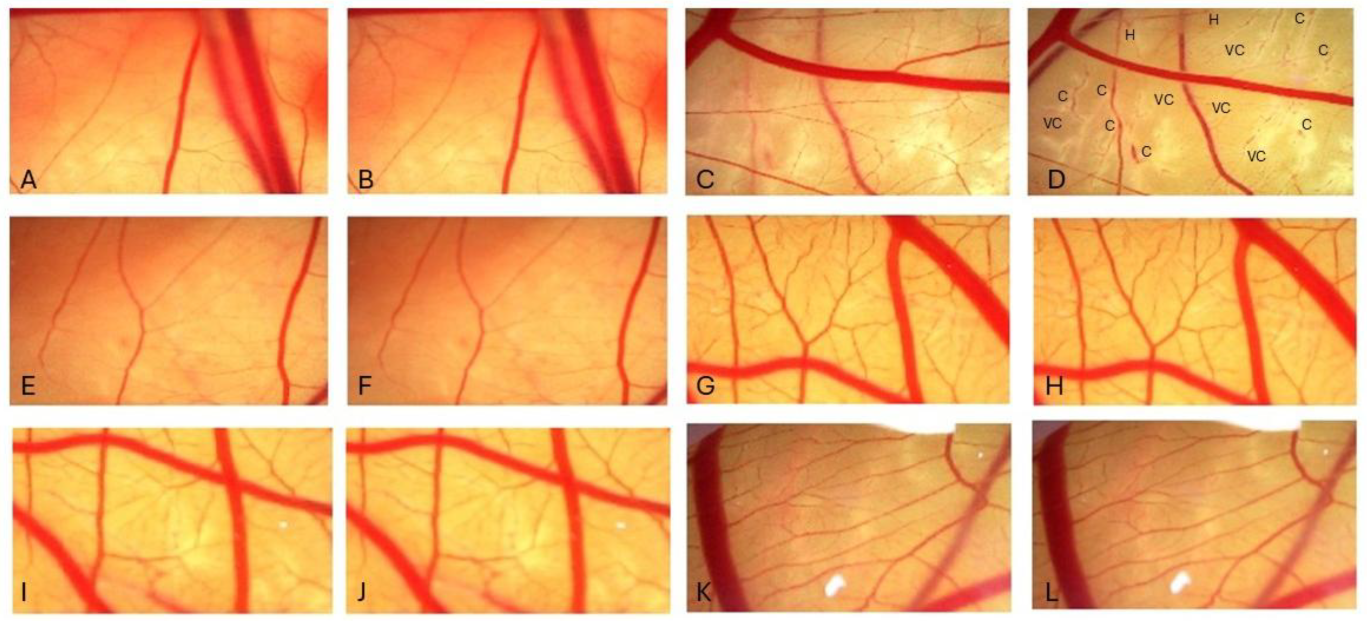
As can be seen in Figure 2, 0.9% sodium chloride (negative control), chitosan hydrochloride, grape seed oil, P. aeruginosa biosurfactant, and the CCh-PaB-GSO nanoemulsion exhibited an irritation potential (IP) of zero, with no alterations (vasoconstriction, hemorrhage, or clotting) in the vascular network of the chorioallantoic membrane during the 300 seconds observation period. As expected, 1% sodium lauryl sulfate (positive control for irritation) showed an IP of 17.74 ± 0.4, with vasoconstriction (6.0 ± 1.0 s), clot formation (63.0 ± 3.0 s), and hemorrhage (48 ± 3.0 s) observed. It was subsequently classified as a severe irritant.

4. Discussion

The dental and cosmetic industries have employed various strategies to develop effective, eco-sustainable, low-cost, and biocompatible products for maintaining oral health that do not trigger microbial resistance. Nanoemulsions are an emerging material for improving the stability and oral bioavailability of functional dental and cosmetic products. To this end, the present study describes, for the first time, the production and application of a novel nanoemulsion formed by the combination of fungal chitosan hydrochloride (ChC), Pseudomonas aeruginosa biosurfactant (PaB), and grape seed oil (GSO). It also demonstrates, in vitro, the potential application of this nanoemulsion in the control of oral diseases such as dental caries, aiming to enhance the metabolic characteristics of its components.
Infrared spectroscopy was used to evaluate the chemical composition of the nanoemulsion and possible interactions between the compounds. As detailed in the results section, both chitosan hydrochloride and grape seed oil exhibited characteristic peaks consistent with the previous literature [13,35,36]. FTIR analysis confirmed interactions between the compounds, evidenced by a prominent band below 1155 cm−1 attributed to saccharide structures, visible in both the ChC spectrum and the CCh-PaB-GSO nanoemulsion spectrum. Additionally, a characteristic peak in the 1036 cm−1 range, associated with the stretching vibration of the C-O group (common to ChC), became more accentuated in the CCh-PaB-GSO spectrum [35,37]. Further suggesting interaction, the final emulsion presented a cloudy appearance with a slight bluish coloration (Figure 3).
The nanoemulsion remained stable during the stability assessment period, maintaining a PDI below 0.3, which indicates uniform and narrow droplet distribution [23,38]. It also maintained a pH of approximately 7.2, a positive zeta potential above +20 mV, and a small droplet size below 300 nm. In this study, we considered droplet sizes greater than 100 nm and smaller than 300 nm to be stable. Particles smaller than 100 nm can be absorbed by the mucosa, and particles larger than 300 nm may tend to aggregate, which could alter the bioactivity of the nanoemulsion. Because the system’s carrier polymer is a chitosan derivative (polycation), the zeta potential was considered stable from +20 mV. During the stability monitoring period, a positive zeta potential was observed, increasing from +20 mV to +30 mV during the observation period, suggesting greater NH4+ availability, likely due to the gradual release of grape seed oil, which directly influences the polymer’s antimicrobial and adsorptive action.
As shown in Table 1, the nanoemulsion increases in droplet size and surface charge during storage, maintaining minimal pH and Pi changes. Therefore, it would be valuable to conduct long-term stability tests to assess its long-term effects and confirm the stability of the formed system. The lack of such an assessment is one of the weaknesses of this study. Common methods for long-term stability testing include HPLC for potency and purity, dissolution tests, and microbial tests for sterility.
The 96-well plate microdilution method is a well-established and widely accepted quantitative methodology for assessing antimicrobial activity, often performed via direct observation of turbidity, optical density readings, or the use of cell viability indicator dyes like resazurin and tetrazolium bromide (MTT) [6]. Resazurin, a blue oxidation-reduction indicator, is reduced to resorufin (pink-red) by metabolically active cells. Thus, a blue-to-purple color signifies the absence of bacterial growth, while pink-to-red variations indicate viable cells with active metabolism due to resazurin reduction by oxidoreductases [6,25]. A known limitation of such viability dyes is their susceptibility to influence by test substance characteristics (e.g., pH) or potential dye-substance interactions, which can lead to non-standard coloration or delayed/inhibited color changes.
The minimum inhibitory concentration (MIC) of the CCh-PaB-GSO nanoemulsion, as well as its individual components ChC, PaB, and GSO, was determined against cariogenic Streptococcus species using resazurin dye as a cell viability indicator. All tested formulations exhibited MIC for the strains of S. mutans, S. sanguis, S. salivarius, and S. mitis.
Regarding the observed interactions in the nanoemulsion, Resende et al. [10] and Farias et al. [19] report that the combination of chitosan and microbial biosurfactant can occur via intermolecular hydrophobic interactions or electrostatic bonding between chitosan’s NH3+ groups and the sulfate groups of anionic surfactants. Such bonds are generally cooperative, particularly at the critical aggregation concentration of the surfactant. Cooperative binding results in micellar-like structures formed along the polymer chain, often accompanied by turbidity in the complexes. However, Burr, Williams, and Ratcliffe [39] and Chiappisi and Gradzielski [40] report non-cooperative interactions between chitosan and synthetic surfactant at low concentrations, resulting in charge neutralization and a turbidity-free, soluble, and clear system. According to Senra et al. [41], the chitosan-surfactant interaction can generate soluble and insoluble aggregates, as several variables, such as hydrophobicity, charge density, chain length, and polymer backbone rigidity, can affect the stoichiometry of the system formed. The authors also claim that anionic surfactants can self-associate, forming micelles under applicable conditions, and can induce micellization at lower concentrations (of surfactant) due to their strong association with chitosan.
The antimicrobial action of chitosan and its derivatives is well documented, though its exact mechanism of action is not yet fully elucidated. Studies indicate that the electrostatic attraction between the polymer’s positively charged amino groups and negatively charged microbial cell surface compounds increases cell permeability, interfering with metabolism and leading to cell death. These amino groups also act as chelators, capturing trace elements essential for cell growth, inhibiting toxin production, and altering microbial metabolism [5,23,42]. This interaction with anionic charges on the bacterial cell surface leads to cell membrane damage, lysis, and leakage of cytoplasmic material. Additionally, chitosan can interfere with mRNA and protein synthesis [7]. Previous research has shown that mouthwashes containing chitosan exhibit favorable results against S. mutans adhesion, biofilm formation, and biofilm maturation, demonstrating greater antimicrobial potential than commercial mouthwashes [43].
Stamford et al. [42] reported that chitosan hydrochloride diluted in water exhibited a lower MIC (0.06 mg/mL) against S. mutans than chitosan diluted in 1% acetic acid (1.25 mg/mL), irrespective of the polymer’s molecular weight and origin (crustacean or fungal). In contrast, we obtained a higher MIC for chitosan hydrochloride for Streptococcus species (3.0 mg/mL) in the present study. The study by Stamford et al. [42] did not specify the pH at which their ChC solution was adjusted, a crucial parameter influencing the formation of NH3 groups in the polymer. In our study, the ChC solution in water was adjusted to pH 7.0 to match the nanoemulsion’s pH. However, more in-depth studies are needed to investigate other parameters that may influence ChC’s antimicrobial action, such as pH variation, molecular weight, degree of deacetylation, zeta potential, and viscosity.
Furthermore, grape seed oil, a vegetable oil recognized for its nutraceutical properties, contributes as an antimicrobial. Its tiny droplets selectively interact with bacterial cell walls without affecting eukaryotic cells, leading to the degradation of pathogenic microorganisms. GSO has reported antibacterial activity against Gram-positive and -negative bacteria, including S. aureus, E. coli, K. pneumoniae, and P. aeruginosa, as well as antifungal activity. Its antioxidant and antimicrobial actions are attributed to its polyphenolic components, such as catechins, epicatechin, gallic acid, monomeric flavonols, and procyanidins [15,18,44]. Additionally, the antimicrobial action of nanoemulsions with essential oils is considered a nonspecific mechanism, consequently preventing the development of resistant strains [38].
CCh-PaB-GSO nanoemulsion exerts antimicrobial action and alters the hydrophobicity of the bacterial surface through the individual action of each of its components with complementary physicochemical properties. CCh, a water-soluble chitosan derivative, cationic salt, interacts electrostatically with the anionic groups present in the microbial cell wall (phosphates and carboxylates), promoting charge neutralization, membrane disorganization, and increased permeability, which leads to the leakage of essential intracellular constituents and consequent cell death [7,42]. The P. aeruginosa biosurfactant, due to its amphiphilic nature, reduces surface tension and inserts itself into lipid bilayers, intensifying membrane disruption and cell lysis [10,19]. Grape seed oil, rich in unsaturated fatty acids (linoleic and oleic) and phenolic compounds, increases membrane fluidity and promotes lipid oxidation, in addition to enhancing the antimicrobial effect through oxidative mechanisms and disruption of cellular integrity [15].
The simultaneous presence of these components in an emulsion increases the affinity for the microbial surface due to the combination of electrostatic (chitosan), hydrophobic (oil and biosurfactant), and physicochemical interactions, altering the charge balance and surface composition of the cell. This modification reduces bacterial hydrophobicity, which is a crucial characteristic for adhesion and biofilm formation, and can therefore compromise the colonization capacity and virulence of microorganisms. Furthermore, the nanoemulsion structure favors the penetration of the system into the biofilm microlayers and the controlled release of active ingredients, prolonging the antimicrobial effect. Thus, the chitosan–biosurfactant–grape seed oil system acts through multiple complementary mechanisms, promoting destabilization of the cell surface, alteration of hydrophobicity and effective inhibition of bacterial growth.
As previously discussed, biofilms are formed by complex communities of pathogenic microorganisms, such as Streptococcus species, that reside on tooth surfaces, metabolize EPS, and produce acids, leading to strong adhesion and the development of oral diseases [45,46]. In our study, the nanoemulsion decreased the adhesion of the Streptococcus strains while simultaneously inhibiting their EPS production and the hydrophobicity of the bacterial cell wall. Higher concentrations of the nanoemulsion resulted in lower values of adhesion, reduced cell wall hydrophobicity, and decreased EPS production for all Streptococcus strains, thereby contributing to the prevention of biofilm formation and oral diseases. This aligns with previous findings on the potential of similar composites, such as fungal chitosan-microbial biosurfactant (PaB included)-Mentha piperita essential oil, in inhibiting cariogenic bacterial biofilm formation [10,19]. Additionally, a mouthwash composed of chitosan hydrochloride and a biosurfactant from Chenopodium quinoa resulted in over 75% inhibition of S. mutans [6], further supporting the role of biosurfactants in reducing the adhesion and proliferation of pathogenic microorganisms on oral cavity surfaces.
Exopolysaccharides constitute the primary structural component of the extracellular matrix in bacterial biofilms, fundamentally dictating the community’s cohesion, stability, and physicochemical resistance. This highly organized extracellular matrix functions as a diffusion barrier, significantly impeding the penetration of antimicrobial agents and regulating local microenvironments crucial for survival. High EPS production, often mediated by glucosyltransferase and glucan-binding proteins in oral bacteria, directly correlates with increased adhesiveness, maturation, and antimicrobial resistance. Conversely, reduced EPS synthesis compromises the mechanical integrity and three-dimensional cohesion of the extracellular matrix. This structural collapse results in reduced biofilm density, increased permeability, and weakened cell–cell interactions, consequently disrupting quorum-sensing signaling and interspecific communication pathways necessary for community persistence. Therefore, EPS reduction is a pivotal strategy that not only destabilizes biofilm architecture but also disrupts the functional homeostasis governing its formation and resilience [28].
The CCh-PaB-GSO nanoemulsion is engineered to act multifactorially in this process, leveraging the complementary properties of its components to achieve EPS modulation and biofilm control:
  • Inhibition of Initial Adhesion and EPS Synthesis: Chitosan Hydrochloride, a polycation, interferes with colonization by electrostatically neutralizing the anionic bacterial surface, preventing initial adhesion. Low molecular weight chitosan and their derivatives also interfere with extracellular glucan synthesis, analogous to the transcriptional repression of gtf and gbp genes observed with other natural compounds [5,28,42].
  • Extracellular Matrix Disruption and Permeabilization: The P. aeruginosa biosurfactant reduces surface tension and intercalates into the extracellular matrix, disrupting the hydrophobic and ionic bonds of the EPS network. This action enhances the permeability of the biofilm and facilitates the diffusion of antimicrobial agents. PaB further modulates microbial communication, potentially repressing polymer synthesis genes [9,47].
  • Membrane Lysis and Bioavailability Enhancement: Grape Seed Oil facilitates integration with the bacterial cell membranes due to its lipophilic nature, intensifying cell lysis and enhancing the overall antimicrobial effect of the nanoemulsion [18].
While the present study effectively demonstrates the nanoemulsion’s activity against a single Streptococcus species, the biological relevance of these findings must be considered within the context of the multispecies oral consortium. The oral biofilm is a highly complex community governed by intricate synergistic and competitive interspecies signaling (e.g., quorum sensing). Pioneer colonizers, such as S. salivarius, S. mutans and S. sanguinis, are foundational; their initial adhesion and robust production of EPS are critical for the subsequent recruitment and co-adhesion of secondary and tertiary colonizers, ultimately leading to a mature, highly acidogenic, and protected multispecies structure. Therefore, the observed CCh-mediated anti-adhesion and PaB-induced EPS disruption are particularly significant, as they fundamentally undermine the scaffolding and architecture upon which the complex biofilm is built. By successfully inhibiting EPS synthesis in these pioneers, the nanoemulsion is proposed to not only reduce the cariogenic potential but also diminish the overall matrix cohesion of the nascent biofilm, thereby increasing the susceptibility of the entire multispecies community to environmental stresses and clearance mechanisms. This targeted attack on the pioneer and structural phase of colonization highlights a potent strategy for comprehensive oral biofilm control.
Beyond its antimicrobial efficacy, the nanoemulsion’s biocompatibility was confirmed through the HET-CAM assay. The HET-CAM is a qualitative and quantitative test that measures acute effects on the small blood vessels and proteins of the chorioallantoic membrane and is one of the most widely accepted non-animal tests [33]. Its primary advantage lies in the membrane’s similarity to mammalian mucous membranes, offering a viable alternative to in vivo testing, such as the Draize rabbit eye test. This low-cost assay enables quick and effective analyses. However, limitations include the inability to analyze substances with color, viscosity, or texture that hinder visualization of the vascular network, or non-liquid substances that cannot be solubilized [33,48,49]. It is also crucial to consider current legislation regarding animal experimentation, as the HET-CAM is classified as an In Vitro test only when the chicken embryo is up to 10 days post-fertilization; beyond this period, it is considered an in vivo test [50].
The HET-CAM (Hens Egg-Chorioallantoic Membrane) test was chosen for this initial study with CCh-PaB-GSO nanoemulsion because it is an alternative, established, and highly sensitive in vivo method for assessing acute irritation of mucous membranes and potential for ocular corrosion/irritation. Given the proposed application of the nanoemulsion in dental caries control, the primary focus at this stage was to ensure immediate safety and the absence of local irritant potential upon contact with the oral mucosa, an essential initial requirement for developing topical dental formulations.
One limitation of this study is the lack of previously published chronic or subchronic toxicity data for the CCh-PaB-GSO nanoemulsion. Therefore, we acknowledge a data gap, and even searching for chronic/subchronic assays of the individual components of the formulation (chitosan hydrochloride, biosurfactant extracted from P. aeruginosa, and grape seed oil) in toxicological databases did not yield directly applicable results to our nanoemulsion system. Therefore, additional toxicity experiments are necessary, considering the need for a more comprehensive assessment in accordance with ethical guidelines and the need to reduce the use of vertebrate animals. We suggest an experimental design integrating the assessment of chronic and subchronic toxicity in future research using validated alternative models, such as those using the nematode Caenorhabditis elegans. C. elegans is a multicellular organism widely recognized in the scientific literature for screening subchronic/chronic toxicity, as it has a short life cycle that allows for the assessment of chronicity endpoints (such as reproduction, growth, and longevity) in days. Its genetic conservation with mammals makes it a useful predictive model for toxicity classification. Another model for toxicity studies is Allium cepa, which allows for the evaluation of mitotic aberrations and subsequent effects on chromosomes through microscopic observations.

5. Conclusions

The pharmaceutical sector has increasingly prioritized the development of sustainable technologies aimed at minimizing adverse effects on both human health and the environment. This shift has been supported by the adoption of green chemistry principles. Within this context, the cosmetics industry shows particular interest in natural alternatives, as cosmetic products are used daily and a considerable fraction ends up directly in the environment. Traditional formulations often rely on hundreds of synthetic ingredients, many of which require substitution to meet sustainability criteria.
In line with consumer demand for natural products, the present study demonstrated that the CCh-PaB-GSO nanoemulsion is stable, non-irritating (based on the HET-CAM test results for acute toxicity), and a promising active material for cosmetic applications. The nanoemulsion is proposed to control biofilm by integrating the distinct mechanisms of action of its constituents: CCh-mediated adhesion interference and gene modulation, PaB-driven extracellular matrix dispersion, and GSO-enhanced cell permeability. These mechanisms collectively contribute to the crucial destabilization of the EPS matrix and subsequent loss of functional homeostasis.
Specifically, the system, grape seed oil loaded with P. aeruginosa biosurfactant and chitosan hydrochloride, displayed antimicrobial properties and significantly inhibited biofilm formation, indicating its potential as a sustainable substitute for conventional chemical agents in oral care products.
Even with the promising results from single-species biofilms, it is necessary to verify the effect of the nanoemulsion in multispecies biofilm systems, which are closer to the complexity of oral biofilms in vivo. As future perspectives, we plan the following stages: (i) Toxicological Safety: Although the HET-CAM test confirms the low acute irritation of the CCh-PaB-GSO nanoemulsion, future chronic and sub-chronic toxicity studies, using alternative in vivo models such as the nematode C. elegans, will be conducted to provide a complete toxicological safety profile. (ii) Multispecies Efficacy: Performance of multispecies biofilm assays. (iii) Clinical Relevance: A preclinical randomized double-blind trial in adult individuals will be carried out to evaluate the use of a mouthwash with CCh-PaB-GSO nanoemulsion for the quantification of microorganisms in dental biofilm and maintenance of oral pH. (iv) Product Development: Sensory and acceptance analysis of oral hygiene products. Such future research will contribute to highlighting the possible application of the CCh-PaB-GSO nanoemulsion in oral hygiene formulations, for example, mouthwash or toothpaste, or even in dental materials for treatment or prevention at the clinical level.

Author Contributions

Conceptualization, T.C.M.S., L.R.R.B., F.H.X.-J., R.D.R., L.A.S. and M.A.D.d.R.; methodology, T.C.M.S., L.R.R.B., F.H.X.-J. and R.D.R.; validation, T.C.M.S., L.R.R.B., R.D.R. and L.A.S.; formal analysis, R.D.R.; investigation, T.C.M.S., A.V.P.S., L.R.R.B. and I.T.d.F.P.M.; resources, T.C.M.S., L.A.S. and M.A.D.d.R.; writing—original draft preparation, T.C.M.S., A.V.P.S., I.T.d.F.P.M. and L.R.R.B.; writing—review and editing, F.H.X.-J., R.D.R., L.A.S. and M.A.D.d.R.; visualization, T.C.M.S. and L.R.R.B.; supervision, T.C.M.S., L.A.S. and M.A.D.d.R.; project administration, L.A.S.; funding acquisition, L.A.S. All authors have read and agreed to the published version of the manuscript.

Funding

This study was funded by the Brazilian fostering agencies: Fundação de Apoio à Ciência e Tecnologia do Estado de Pernambuco (FACEPE) scholarship (process no IBPG-0626-2.12/21) awarded to A.V.P.S., the Conselho Nacional de Desenvolvimento Científico e Tecnológico (CNPq) financial support (grant number 405118/2024-4) and post-doctoral scholarship (process no. 300885/2021-0, and process no 200901/2025-7) awarded to L. R. R. B., and post-doctoral scholarship (process no. 200900/2025-0) awarded to T.C.M.S.) and the Coordenação de Aperfeiçoamento de Pessoal de Nível Superior (CAPES) (Finance Code 001), and PhD scholarship (process no 88887.492524/2020-00) and Sandwich PhD scholarship (PDSE) (process no 88881.846689/2023-01) awarded to I.T.d.F.P.M.

Institutional Review Board Statement

Not applicable.

Informed Consent Statement

Not applicable.

Data Availability Statement

The original contributions presented in the study are included in the article; further inquiries can be directed to the corresponding author.

Acknowledgments

The authors thank the Universidade Federal de Pernambuco—Brasil, the Instituto Keizo Asami (ILIKA), the Analytical Center of the Department of Fundamental Chemistry UFPE, the Laboratories of Universidade Católica de Pernambuco (UNICAP), and the Instituto Avançado de Tecnologia e Inovação (IATI) for technical support and availability of infrastructure; and Coordenação de Aperfeiçoamento de Pessoal de Nível Superior—Brasil (CAPES), Conselho Nacional de Desenvolvimento Científico e Tecnológico- Brasil (CNPq) and Fundação de Amparo à Ciência e Tecnologia do Estado de Pernambuco -FACEPE, Brasil for financial support.

Conflicts of Interest

The authors declare no conflicts of interest.

Abbreviations

The following abbreviations are used in this manuscript:
CChChitosan hydrochloride
PaBPseudomonas aeroginosa biosurfactant
GSOGrape seed oil

References

  1. Al-Marzooq, F.I.; Christidis, N. The interconnection of oral and systemic health. Sci. Rep. 2025, 15, 14931. [Google Scholar] [CrossRef]
  2. Murray, P.E.; Coffman, J.A.; Garcia-Godoy, F. Oral Pathogens Substantial Burden on cancer, Cardiovascular Diseases, Alzheimer’s, Diabetes, and Other Systemic Diseases: A Public Health Crisis—A Comprehensive Review. Pathogens 2024, 13, 1084. [Google Scholar] [CrossRef]
  3. Shen, Z.; Kuang, S.; Zhang, Y.; Chen, J.; Wang, S.; Xu, C.; Huang, Y.; Zhang, M.; Huang, S.; Wang, J.; et al. Restoring periodontal tissue homoeostasis prevents cognitive decline by reducing the number of Serpina3nhigh astrocytes in the hippocampus. Innovation 2024, 5, 100547. [Google Scholar] [CrossRef]
  4. Wang, Z.; Zeng, Y.; Ahmed, Z.; Qin, H.; Bhatti, I.A.; Cao, H. Calcium-dependent antimicrobials: Nature-inspired materials and designs. Exploration 2024, 4, 20230099. [Google Scholar] [CrossRef]
  5. Carvalho, M.M.S.G.; Stamford, T.C.M.; Santos, E.P.; Tenorio, P.; Sampaio, F. Chitosan as oral antimicrobial agent. In Science Against Microbial Pathogens: Communicating Current Research and Technological Advances; Mendez, A., Ed.; Formatex: Badajoz, Spain, 2011; pp. 542–550. [Google Scholar]
  6. Souza, I.R.; Bezerra, K.G.O.; Oliveira, C.L.; Meira, H.M.; Stamford, T.C.M.; Converti, A.; Sarubbo, L.A.; Rufino, R.D. Mouthwash Containing Plant-Derived Biosurfactant and Chitosan Hydrochloride: Assessment of Antimicrobial Activity, Antibiofilm Activity, and Genotoxicity. Appl. Sci. 2024, 14, 6711. [Google Scholar] [CrossRef]
  7. Wassel, M.O.; Khattab, M.A. Antibacterial activity against Streptococcus mutans and inhibition of bacterial induced enamel demineralization of propolis, miswak, and chitosan nanoparticles based dental varnishes. J. Adv. Res. 2017, 8, 387–392. [Google Scholar] [CrossRef] [PubMed]
  8. Chen, C.Y.; Chung, Y.C. Antibacterial effect of water-soluble chitosan on representative dental pathogens Streptococcus mutans and Lactobacilli brevis. J. Appl. Oral. Sci. 2012, 20, 620–627. [Google Scholar] [CrossRef] [PubMed]
  9. Datta, M.; Chattopadhyay, I. Applications of microbial biosurfactants in human health and environmental sustainability: A narrative review. Discov. Med. 2024, 1, 160. [Google Scholar] [CrossRef]
  10. Resende, A.H.M.; Farias, J.M.; Silva, D.D.B.; Rufino, R.D.; Luna, J.M.; Stamford, T.C.M.; Sarubbo, L.A. Application of biosurfactants and chitosan in toothpaste formulation. Colloids Surf. B Biointerfaces. 2019, 181, 77–84. [Google Scholar] [CrossRef]
  11. Maj, M.; Tylkowski, B.; Konopka, P.; Woźniak-Budych, M.; Staszak, K.; Staszak, M.; Kaźmierski, Ł.; Bajek, A.; Jastrzab, R. Advancing oral health: Harnessing the potential of chitosan and polyphenols in innovative mouthwash formulation. Biomed. Pharmacother 2024, 175, 116654. [Google Scholar] [CrossRef]
  12. Yadav, A.A.; Mujawar, S.S.; Shinde, N.A.; Phalake, S.S.; Khot, V.M.; Kashte, S.B. A multifunctional tragacanth gum-chitosan hydrogel loaded with manganese ferrite nanoparticles for dental caries prevention and remineralization. Int. J. Biol. Macromol. 2025, 322, 146886, ISSN 0141-8130. [Google Scholar] [CrossRef] [PubMed]
  13. Li, X.M.; Wu, Z.Z.; Zhang, B.; Pan, Y.; Meng, R.; Chen, H.Q. Fabrication of chitosan hydrochloride and carboxymethyl starch complex nanogels as potential delivery vehicles for curcumin. Food Chem. 2019, 293, 197–203. [Google Scholar] [CrossRef] [PubMed]
  14. Zhang, L.; Zhang, Y.; Li, W.; Zhou, Y.; Guo, R.; Pei, X.; Xie, J. Chitosan and its functional derivatives for nutraceutical delivery: Focus on quaternized, hydrochloride, and carboxymethyl forms. Trends Food Sci. Technol. 2025, 164, 105237. [Google Scholar] [CrossRef]
  15. Joujou, F.M.; Darra, N.E.; Rajha, H.N.; Sokhn, E.S.; Alwan, N. Evaluation of synergistic/antagonistic antibacterial activities of fatty oils from apricot, date, grape, and black seeds. Sci. Rep. 2024, 14, 6532. [Google Scholar] [CrossRef] [PubMed]
  16. Delimont, N.M.; Carlson, B.N. Prevention of dental caries by grape seed extract supplementation: A systematic review. Nutr. Health. 2020, 26, 43–52. [Google Scholar] [CrossRef] [PubMed]
  17. Zhao, W.; Xie, Q.; Bedran-Russo, A.K.; Pan, S.; Ling, J.; Wu, C.D. The preventive effect of grape seed extract on artificial enamel caries progression in a microbial biofilm-induced caries model. J. Dent. 2014, 42, 1010–1018. [Google Scholar] [CrossRef] [PubMed]
  18. Khah, M.D.; Ghanbarzadeh, B.; Nezhad, L.R.; Ostadrahimi, A. Effects of virgin olive oil and grape seed oil on physicochemical and antimicrobial properties of pectin-gelatin blend emulsified films. Int. J. Biol. Macromol. 2021, 171, 262–274. [Google Scholar] [CrossRef] [PubMed]
  19. Farias, J.M.; Stamford, C.M.; Resende, A.H.M.; Aguiar, J.S.; Rufino, R.D.; Luna, J.M.; Sarubbo, L.A. Mouthwash containing biosurfactant and chitosan: An eco-sustainable option for controlling cariogenic microorganisms. Int. J. Biol. Macromol. 2019, 129, 853–860. [Google Scholar] [CrossRef]
  20. Silva, E.J.; Correa, P.F.; Almeida, D.G.; Luna, J.M.; Rufino, R.D.; Sarubbo, L.A. Recovery of contaminated marine environments by biosurfactant-enhanced bioremediation. Colloids Surf. B Biointerfaces 2018, 172, 127–135. [Google Scholar] [CrossRef]
  21. Berger, L.R.R.; Stamford, T.C.M.; De Oliveira, K.A.R.; De Miranda, A.P.P.; Lima, M.A.B.; Pintado, M.M.E.; Câmara, M.P.S.; Franco, L.O.; Magnani, M.; Souza, E.L. Chitosan produced from Mucorales fungi using agroindustrial by-products and its efficacy to inhibit Colletotrichum species. Int. J. Biol. Macromol. 2018, 102, 635–641. [Google Scholar] [CrossRef]
  22. Synowiecki, J.; Al-Khateeb, N.A.A.Q. Mycelia of Mucor rouxii as source of chitin and chitosan. Food Chem. 1997, 60, 605–610. [Google Scholar] [CrossRef]
  23. Araujo, A.S.; de Lima, G.S.; Nunes, I.S.; de Oliveira Farias de Aguiar, J.C.R.; Navarro, D.M.A.F.; Melo, N.F.C.B.; Magalhes, N.S.S.; Franca, R.; Carvalho, R.S.F.; Stamford, T.C.M. Chitosan hydrochloride-gum Arabic-passion fruit seed oil nanoparticle edible coating to control fungal infection and maintain quality parameters of strawberries. Food Control 2024, 161, 110360. [Google Scholar] [CrossRef]
  24. Tsai, M.L.; Chen, R.H.; Bai, S.W.; Chen, W.Y. The storage stability of chitosan/tripolyphosphate nanoparticles in a phosphate buffer. Carbohydr. Polym. 2011, 84, 756–761. [Google Scholar] [CrossRef]
  25. Driessche, F.V.; Rigole, P.; Brackman, G.; Coenye, T. Optimization of resazurin-based viability staining for quantification of microbial biofilms. J. Microbiol. Methods 2014, 98, 31–34. [Google Scholar] [CrossRef] [PubMed]
  26. Doern, C.D. When does 2 plus 2 equal 5? A review of antimicrobial synergy testing. J. Clin. Microbiol. 2014, 52, 4124–4128. [Google Scholar] [CrossRef]
  27. Huston, A.L.; Methe, B.; Deming, J.W. Purification, characterization and sequencing of an extracellular cold-active aminopeptidase produced by marine psychrophile Colwellia psychrerythraea strain 34H. Appl. Environ. Microbiol. 2004, 70, 3321–3328. [Google Scholar] [CrossRef]
  28. AlKanderi, S.; AlFreeh, M.; Bhardwaj, R.G.; Karched, M. Sugar Substitute Stevia Inhibits Biofilm Formation, Exopolysaccharide Production, and Downregulates the Expression of Streptococcal Genes Involved in Exopolysaccharide Synthesis. Dent. J. 2023, 11, 267. [Google Scholar] [CrossRef]
  29. Dubois, M.; Gilles, K.A.; Hamilton, J.K.; Rebers, P.A.; Smith, F. Colorimetric method of determination of sugars and related substances. Anal. Chem. 1956, 18, 350–356. [Google Scholar] [CrossRef]
  30. Sano, H.; Shibasaki, R.; Matsukubo, T.; Takaesu, Y. Effect of molecular mass ans degree of deacetylation of chitosan on adsortption of Streptococcus sobrinus 6715 to saliva treated hydroxyapatite. Bull. Tokyo Dent. Coll. 2002, 48, 75–82. [Google Scholar] [CrossRef]
  31. Luepke, N.P. Hen’s egg chorioallantoic membrane test for irritation potential. Food Chem. Toxicol. 1985, 23, 287–291. [Google Scholar] [CrossRef]
  32. Steiling, W.; Bracher, M.; Coutellemont, P.; Silva, O. The HET-CAM, a useful in vitro assay for assessing the eye irritation properties of cosmetic formulations and ingredients. Toxicol. Vitr. 1999, 13, 375–384. [Google Scholar] [CrossRef]
  33. Freire, P.L.; Stamford, T.C.M.; Albuquerque, A.J.; Sampaio, F.; Cavalcante, H.M.M.; Macedo, R.O.; Galembeck, A.; Flores, M.A.P.; Rosenblat, A. Action of silver nanoparticles toward biological systems: Cytotoxicity evaluation using hen’s egg test and inhibition of Streptococcus mutans biofilm formation. Int. J. Antimicrob. Agents. 2015, 45, 183–187. [Google Scholar] [CrossRef] [PubMed]
  34. Wackerly, D.; Mendenhall, W.; Scheaffer, R. Mathematical Statistics with Applications, 5th ed.; Duxbury Press: Boston, MA, USA, 1995; Volume 1, 798p. [Google Scholar]
  35. Gechev, B.; Zsivanovits, G.; Iliev, A.; Marudova, M. Chitosan/grapeseed oil multicomponent edible films-design and properties. J. Phys. Conf. Ser. 2023, 2436, 012029. [Google Scholar] [CrossRef]
  36. Ge, J.; Yue, P.; Chi, J.; Liang, J.; Gao, X. Formation and stability of anthocyanins-loaded nanocomplexes prepared with chitosan hydrochloride and carboxymethyl chitosan. Food Hydrocoll. 2018, 74, 23–31. [Google Scholar] [CrossRef]
  37. Vladimír, M.; Matwijczuk, A.P.; Niemczynowicz, A.; Kycia, R.A.; Karcz, D.; Gładyszewska, B.; Ślusarczyk, L.; Burg, P. Chemometric approach to characterization of the selected grape seed oils based on their fatty acids composition and FTIR spectroscopy. Sci. Rep. 2021, 11, 19256. [Google Scholar] [CrossRef]
  38. Mutlu, N. Effects of grape seed oil nanoemulsion on physicochemical and antibacterial properties of gelatin-sodium alginate film blends. Int. J. Biol. Macromol. 2023, 237, 124207. [Google Scholar] [CrossRef] [PubMed]
  39. Burr, S.J.; Williams, P.A.; Ratcliffe, I. Synthesis of cationic alkylated chitosans and an investigation of their rheological properties and interaction with anionic surfactant. Carbohydr. Polym. 2018, 201, 615–623. [Google Scholar] [CrossRef] [PubMed]
  40. Chiappisi, L.; Gradzielski, M. Co-assembly in chitosan–surfactant mixtures: Thermodynamics, structures, interfacial properties and applications. Adv. Colloid. Int. Sci. 2015, 220, 92–107. [Google Scholar] [CrossRef]
  41. Senra, T.D.A.; Campana-Filho, S.P.; Desbrières, J. Surfactant-polysaccharide complexes based on quaternized chitosan. Characterization and application to emulsion stability. Eur. Polym. J. 2018, 104, 128–135. [Google Scholar] [CrossRef]
  42. Stamford, T.C.M.; Stamford-Arnaud, T.M.; Cavalcante, H.M.M.; Macedo, R.O.; Campos-Takaki, G.M. Microbiological Chitosan: Potential Application as Anticariogenic Agent. In Practical Applications in Biomedical Engineering; InTech: London, UK, 2013. [Google Scholar] [CrossRef]
  43. Costa, E.M.; Silva, S.; Madureira, A.R.; Cardelle-Cobas, A.; Tavaria, F.K.; Pintado, M.M. A comprehensive study into the impact of a chitosan mouthwash upon oral microorganism’s biofilm formation in vitro. Carbohydr. Polym. 2014, 101, 1081–1086. [Google Scholar] [CrossRef]
  44. Ahmed, S.A.A.; Mahsoub, F.; El Gamal, S.A.; Khamis, T.; Faroh, K.Y.; Abdelwarith, A.A.; Younis, E.M.; Saad, M.F.; Ali, H.S.; Davies, S.J.; et al. Chitosan-grape seed oil nanoemulsion enriched diet promotes performance, antioxidant-immune metrics and modifies immune- gene action and morphological architecture in Nile tilapia against Aeromonas veronii. Aquac. Rep. 2025, 41, 102697. [Google Scholar] [CrossRef]
  45. Govindarajan, D.K.; Mohanarangam, M.; Kadirvelu, L.; Sivaramalingam, S.S.; Jothivel, D.; Ravichandran, A.; Periasamy, S.; Kandaswamy, K. Biofilms and oral health: Nanotechnology for biofilm control. Discov. Nano 2025, 20, 114. [Google Scholar] [CrossRef] [PubMed]
  46. Zheng, T.; Jing, M.; Gong, T.; Yan, J.; Wang, X.; Xu, M.; Zhou, X.; Zeng, J.; Li, Y. Regulatory mechanisms of exopolysaccharide synthesis and biofilm formation in Streptococcus mutans. J. Oral. Microbiol. 2023, 15, 2225257. [Google Scholar] [CrossRef] [PubMed]
  47. Sultan, F.; Maji, D.; Phatake, R.S.; Kumar, K. Pharmaceutical applications of microbial biosurfactants. Int. J. Pharm. 2025, 681, 125887. [Google Scholar] [CrossRef]
  48. Vargas, A.; Zeisser-Labouèbe, M.; Lange, N.; Gurny, R.; Delie, F. The chick embryo and its chorioallantoic membrane (CAM) for the in vivo evaluation of drug delivery systems. Adv. Drug Deliv. Rev. 2007, 59, 1162–1176. [Google Scholar] [CrossRef] [PubMed]
  49. Saw, C.L.L.; Heng, P.W.S.; Liew, C.V. Chick chorioallantoic membrane as an in situ biological membrane for pharmaceutical formulation development: A review. Drug Dev. Ind. Pharm. 2008, 34, 1168–1177. [Google Scholar] [CrossRef]
  50. Bagley, D.M.; Waters, D.; Kong, B.M. Development of a 10-day chorioallantoic membrane vascular assay as an alternative to the Draize rabbit eye irritation test. Food Chem. Toxicol. 1994, 32, 1155–1160. [Google Scholar] [CrossRef]
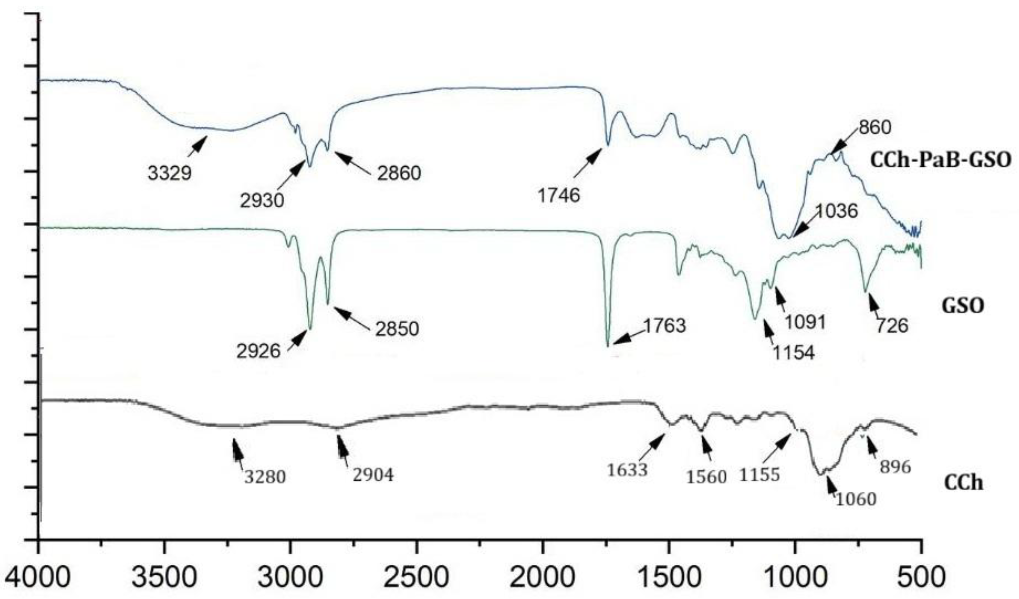
Figure 1. Fourier transform infrared spectroscopy analysis of chitosan hydrochloride (CCh), grape seed oil (GSO) and nanoemulsion chitosan hydrochloride-Pseudomonas aeruginosa biosurfactant-grape seed oil (CCh-PaB-GSO).
Applsci 15 11773 g001
Figure 2. Determination of irritation potential using the HET-CAM assay (Chorioallantoic Membrane assay of the fertilized hen’s egg) with observation up to 5 min (300 s) for signs of vasoconstriction (VC), hemorrhage (H), and coagulation (C). The panels show observations at time zero and 300 s for: (A,B) 0.9% sodium chloride solution (negative irritation control); (C,D) 1% sodium lauryl sulfate (SLS) (positive irritation control); (E,F) Fungal chitosan hydrochloride (ChC); (G,H) Pseudomonas aeruginosa biosurfactant (PaB); (I,J) Grape seed oil (GSO); and (K,L) CCh-PaB-GSO nanoemulsion.
Applsci 15 11773 g002
Figure 3. Sample of the nanoemulsion obtained by high-energy emulsification technic by the combination of chitosan hydrochloride with Pseudomonas aeruginosa biosurfactant and grapeseed oil.
Applsci 15 11773 g003
Table 1. Stability of the fungal chitosan hydrochloride-Pseudomonas aeruginosa biosurfactant-grape seed oil nanoemulsion (ChC-PaB-GSO) over 30 days: Average Size, Polydispersity Index (PDI), Zeta Potential, and pH. Time 0 corresponds to the day the emulsion was prepared.
ParametersTime (Days)
071530
Avereage size (nm)169.5 ± 3.25186.4 ± 1.7188 ± 1.97203.4 ± 0.99
PDI0.2410.2640.2680.271
Zeta potential (mV)+20.25 ± 2.42+25.4 ± 2.05+28.71 ± 1.85+31.94 ± 0.8
pH7.27 ± 0.87.3 ± 0.157.34 ± 0.27.33 ± 0.18
Table 2. Minimum inhibitory concentrations (MICs) of the fungal chitosan hydrochloride (ChC), Pseudomonas aeruginosa biosurfactant (PaB), grape seed oil (GSO), alone, combined in binary pairs or in the final nanoemulsion formulation (ChC-PaB-GSO) against cariogenic Streptococcus species.
MicroorganismTest Substances Alone and in Combination
CChPaBGSOCCh + PaBCCh + GSOPaB + GSOCCh-PaB-GSO
CChPaBCChGSOPaBGSOCChPaBGSO
Streptococcus mutans3.00.154.82.00.12.03.20.13.22.00.13.2
Streptococcus sanguis3.00.154.82.00.12.03.20.13.22.00.13.2
Streptococcus salivarius3.00.154.82.00.12.03.20.13.22.00.13.2
Streptococcus mitis3.00.154.82.00.12.03.20.13.22.00.13.2
Table 3. Adhesion (%) of cariogenic Streptococcus species (S. mutans, S. sanguis, S. salivarius, and S. mitis) to a circular glass coverslip surface in the presence and absence of 5% sucrose at different concentrations of the CCh-PaB-GSO nanoemulsion.
CCh-PaB-GSO
Nanoemulsion (mg/mL)
S. mutansS. sanguisS. salivariusS. mitis
0% Sucrose 5% Sucrose0% Sucrose 5% Sucrose0% Sucrose 5% Sucrose0% Sucrose 5% Sucrose
0100 (±0.00)100 (±0.00)100 (±0.00)100 (±0.00)100 (±0.00)100 (±0.00)100 (±0.00)100 (±0.00)
0.2562.73 (±0.38)91.00 (±1.00)61.10 (±0.17)88.00 (±1.73)51.33 (±1.53)87.33 (±0.58)52.00 (±1.73)86.00 (±1.00)
0.5020.33 (±0.58)48.00 (±1.73)18.57 (±0.40)46.00 (±1.00)18.00 (±1.73)43.00 (±1.00)18.67 (±1.15)44.33 (±0.58)
1.0014.9 (±0.85)41.57 (±0.51)17.17 (±0.76)43.10 (±1.01)11.00 (±1.73)39.33 (±0.58)16.00 (±1.73)39.07 (±0.90)
1.5011.73 (±0.64)36.00 (±1.73)13.00 (±1.00)36.33 (±0.58)11.00 (±1.00)34.60 (±0.53)15.00 (±1.00)35.33 (±0.58)
2.007.57 (±0.51)31.73 (±1.55)8.53 (±0.50)37.00 (±0.00)10.00 (±1.00)28.67 (±1.15)8.00 (±0.00)31.13 (±1.03)
2.505.00 (±0.00)26.33 (±1.15)7.00 (±0.00)22.00 (±2.00)7.00 (±0.00)21.97 (±1.00)7.00 (±0.00)22.37 (±0.55)
3.004.00 (±0.00)17.97 (±0.95)4.33 (±0.58)12.57 (±1.43)7.00 (±0.00)14.23 (±0.68)4.67 (±0.58)11.77 (±0.68)
Table 4. Exopolysaccharide (EPS) production (%) by cariogenic Streptococcus species (S. mutans, S. sanguis, S. salivarius, and S. mitis) at different concentrations of the CCh-PaB-GSO nanoemulsion in the presence of 5% sucrose.
CCh-PaB-GSO Nanoemulsion (mg/mL)S. mutansS. sanguisS. salivariusS. mitis
0100 (±0.00)100 (±0.00)100 (±0.00)100 (±0.00)
0.2558.97 (±1.00)44.90 (±1.01)54.33 (±0.58)41.00 (±0.10)
0.5047.77 (±0.90)31.67 (±0.58)32.33 (±0.58)31.57 (±0.51)
1.0017.77 (±1.10)12.53 (±0.50)17.60 (±0.53)15.70 (±0.30)
1.507.73 (±0.30)5.67 (±0.58)6.17 (±0.29)5.37 (±0.32)
Table 5. Bacterial cell surface hydrophobicity (%) of cariogenic Streptococcus species (S. mutans, S. sanguis, S. salivarius, and S. mitis) at different concentrations of the CCh-PaB-GSO nanoemulsion in the presence of 5% sucrose.
CCh-PaB-GSO Nanoemulsion (mg/mL)S. mutansS. sanguisS. salivariusS. mitis
0100 (±0.00)100 (±0.00)100 (±0.00)100 (±0.00)
0.2597.87 (±0.20)91.90 (±1.01) 78.87 (±1.03)88.33 (±0.58)
0.5059.13 (±1.00)56.20 (±0.80)46.27 (±1.10)51.57 (±0.59)
1.0042.33 (±1.50)36.73 (±0.61)30.90 (±1.01)28.80 (±0.72)
1.5012.43 (±0.50)19.90 (±0.17)15.47 (±061)20.90 (±1.01)
Disclaimer/Publisher’s Note: The statements, opinions and data contained in all publications are solely those of the individual author(s) and contributor(s) and not of MDPI and/or the editor(s). MDPI and/or the editor(s) disclaim responsibility for any injury to people or property resulting from any ideas, methods, instructions or products referred to in the content.