Review

From Pathogens to Partners: The Beginnings of Gut Microbiota Research

Department of Environmental Protection, Faculty of Geology, Geophysics and Environmental Protection, AGH University of Krakow, al. Mickiewicza 30, 30-059 Krakow, Poland
Appl. Sci. 2025, 15(21), 11376; https://doi.org/10.3390/app152111376
Submission received: 3 September 2025 / Revised: 20 October 2025 / Accepted: 21 October 2025 / Published: 23 October 2025

Abstract

The investigation of the human intestinal microbiota has rapidly become one of the most dynamic and interdisciplinary fields in modern biomedical science, and for good reasons. Since the 1990s, when the microbiota was first described as the “neglected organ”, research has expanded exponentially, uncovering its fundamental roles in maintaining immune homeostasis, regulating host metabolism, which gave rise to the term “second liver”, and influencing neural activity through the dense network of enteric neurons, which justifies its characterization as the “second brain”. Furthermore, the remarkable genetic richness of the microbiota, comprising a gene pool vastly exceeding that of the human genome, has earned it the title of the “second genome”. Tracing the origins of this scientific field reveals that the concept of the gut as a complex microbial ecosystem emerged gradually, shaped by pivotal developments in microbiology and medicine throughout the 19th and early 20th centuries. The rise of the germ theory, the advancement of microscopy, and the discovery of key microbial phenomena, including fermentation, decomposition, bacteriophages, probiotics, and antibiotics, collectively transformed our understanding of microorganisms from pathogens to essential symbionts. This review aims to provide a historical perspective on how these landmark discoveries laid the conceptual and methodological foundations for contemporary microbiota research. By highlighting the scientific milestones that shifted perceptions of microbes from “bad germs” to “good germs”, it seeks to offer readers, whether from biomedical, ecological, or philosophical backgrounds, an integrative view of how this paradigm evolved and why it remains central to current human health discussions.

1. Introduction

It is not an overstatement to suggest that human life, and particularly human health, relies on microbial symbiotic organisms [1,2,3,4,5,6]. Consequently, research on the microbiota has flourished in recent years [7], and justifiably so. In 1992, the Italian physiologist Velio Bocci (1928–2019) referred to the human gut microbiota as a “neglected organ” [8]. Since then, additional metaphors such as the “second brain” [9,10], “second liver” [11], and “second genome” [1] have been introduced, reflecting the dynamic progress in this field.
Microorganisms that colonize multicellular organisms, including humans, are collectively referred to as the microbiota. This term encompasses viruses, archaea, bacteria, fungi, and protozoa [5,12], with some researchers also including multicellular parasites [13]. The microbiota colonizes various body sites such as the digestive tract (particularly the large intestine), respiratory system, reproductive organs, and skin, with bacteria markedly predominating–outnumbering other microbial groups by 2–3 orders of magnitude [14]. The abundance and diversity of microorganisms inhabiting the human body vary considerably and depend on numerous factors, including sex, mode of delivery (natural childbirth or cesarean section), infant feeding method (breastfeeding or formula feeding), place of residence (rural versus urban), cohabitation with siblings, partner, family members or pets, professional activity, environmental exposures, dietary habits, lifestyle, and medical history, among many others [3,4,5,15,16,17]. In the 1970s, it was widely accepted that symbiotic microorganisms outnumber host cells by roughly ten to one. More recent estimates, however, suggest a ratio closer to 1:1. The “reference man” (a 20–30-year-old male, 70 kg in weight and 170 cm in height) is estimated to consist of approximately 3.0 × 10¹³ human cells, whereas his microbiota comprise about 3.8 × 10¹³ microbial units, with intestinal microorganisms strongly dominating. It should be noted, however, that these estimates include red blood cells; without them, the ratio of microbial to human cells would be closer to 10:1. Moreover, the 1:1 ratio appears to be relatively stable across variables such as sex, age, height, and body mass [14,15]. While numerical equivalence between host and microbial cells is now recognized, a striking disparity exists at the genetic level, reaching two orders of magnitude [15,16]. The number of human protein-coding genes is currently estimated at approximately 20,000–25,000, with recent analyses suggesting fewer than 20,000 [18], whereas the collective gene repertoire of the human gut microbiota comprises several million genes [19,20].
In microbiota research, the term microbiome is also frequently employed. The introduction of this term is often, though inaccurately, attributed to the Jewish-American geneticist and microbiologist Joshua Lederberg (1925–2008), Nobel laureate in Physiology or Medicine in 1958 “for his discoveries concerning genetic recombination and the organization of the genetic material of bacteria” [21]. This attribution usually dates the term to 2001, and Lederberg is sometimes even credited with defining the concept of microbiota. However, both terms had already been in use long before 2001, applied in different contexts and with varying definitions [22]. The terms microbiota and microbiome are still at times used interchangeably, which is imprecise, and the concept of the microbiome is also frequently conflated with that of the metagenome. To address this ambiguity, in 2019 an expert panel was convened to establish international research standards for microbiota and microbiome studies, resulting in the clarification and refinement of these definitions. According to the current consensus, microbiota refers to the “community of microorganisms” inhabiting a specific niche, whereas microbiome encompasses the “microbiota and their ‘theatre of activity’ (structural elements, metabolites/signal molecules, and the surrounding environmental conditions)” [12], drawing on a definition originally proposed in 1988 [23]. In contrast, metagenome denotes the “collection of genomes and genes from the members of a microbiota” [12].
It is well established that distinct body sites are colonized by characteristic microbial communities. For instance, the oral cavity is dominated by members of the genus Streptococcus, whereas the gastrointestinal tract is primarily inhabited by representatives of the genus Bacteroides. The skin microbiota is enriched in Staphylococcus and Propionibacterium genus members, while Lactobacillus representatives predominate in the vaginal microbiota [1]. Nevertheless, inter-individual variability within these communities is considerable, challenging the notion of a uniform “constitutive” microbiota. Instead, the concept of a “constitutive” microbiome may be more appropriate [3]. Recent studies have further highlighted the profound interdependence between gut microorganisms and their human hosts, shaped by hundreds of thousands of years of co-evolution [24]. Many members of the human microbiota have become so adapted to the intestinal environment that they cannot thrive, or even survive, outside it. Their often-reduced genomes exemplify a modern definition of symbiosis, understood as “the biological and long-term relationship between two partners of different species, which gives rise to new structures or metabolic dependencies and may lead to genetic integration” [25].
Advancements in modern biomedical science have significantly expanded our understanding of the gut microbiota and its integral role in maintaining human metabolic homeostasis. Intestinal microorganisms contribute to energy extraction from food, participate in the detoxification of xenobiotics, and synthesize essential vitamins. They also degrade complex dietary polysaccharides and plant-derived polymers, such as cellulose, lignin, and pectin, otherwise indigestible to humans, converting them into simple sugars and short-chain fatty acids (SCFAs). These metabolites serve as an important energy source for intestinal epithelial cells, exert anti-inflammatory effects, and strengthen the integrity of the intestinal barrier. The gut microbiota also plays a pivotal role in the development and regulation of the host immune system. It provides a controlled environment in which immune tolerance and responsiveness are shaped, while simultaneously reinforcing the physical and immunological barriers of the gut. Resident microbes protect the host against pathogenic colonization not only through competitive exclusion but also by stimulating mucin production, promoting epithelial regeneration, and inducing the secretion of antimicrobial peptides and immunoglobulins. Furthermore, the gut maintains bidirectional communication with the central nervous system via the vagus nerve, forming the gut–brain axis. Through neuroendocrine, immunological, and metabolic pathways, this complex network enables the microbiota to modulate stress responses, emotional states, and cognitive functions, underscoring its profound influence on overall human health and well-being [10,26,27,28,29].
A properly functioning intestine maintains a dynamic state of quasi-equilibrium between the host and its microbial inhabitants. A sustained disruption of this balance is referred to as dysbiosis, a concept reintroduced into the context of gut microbial ecology by the German veterinarian, microbiologist, and dietitian Helmut Haenel (1919–1993), who contrasted it with eubiosis, representing a state of microbial equilibrium and physiological stability [30]. Dysbiosis is increasingly recognized as a key factor underlying a wide spectrum of chronic disorders. It has been implicated in inflammatory bowel diseases (IBD), autoimmune conditions (e.g., rheumatoid arthritis, multiple sclerosis), certain types of cancer, and metabolic disorders such as insulin resistance, type 2 diabetes, obesity, and cardiovascular disease. Moreover, alterations in the gut microbiota have been associated with neuropsychiatric and neurodevelopmental disorders, including depression, anxiety, bipolar disorder, and autism spectrum disorders [3,5,6,10,28,29].
The continuous and rapid expansion of knowledge regarding the gut microbiota and its intricate relationship with host health and disease has been driven by the development of modern research methodologies, particularly those that emerged in the latter half of the twentieth century. The refinement of anaerobic culturing techniques enabled the investigation of a much broader spectrum of intestinal microorganisms [31], while the wide introduction of germ-free animal models, predominantly mice and rats, provided new opportunities to study host–microbe interactions under controlled conditions [32]. The molecular revolution marked a turning point, with the advent of DNA sequencing methods that allowed for the characterization of complex microbial communities without the need for laborious and often inefficient cultivation. The subsequent development of next-generation sequencing (NGS) technologies has made it possible not only to identify microbial taxa but also to infer the metabolic potential and functional capacities of entire microbial ecosystems. Today, researchers can integrate a wide range of powerful high-throughput approaches, including genomics and metagenomics (genome-level analyses) [33], transcriptomics (transcriptome profiling) [34], proteomics (protein-level analyses) [35,36], and metabolomics (metabolite profiling) [37], to obtain a comprehensive and multidimensional view of the gut microbiota and its role in human health.
The journey toward a deeper understanding of the complex symbiotic relationships between humans and their microbiota is far from complete and will undoubtedly continue to yield groundbreaking discoveries. To fully appreciate current advances, however, it is necessary to revisit the historical roots of this discipline and examine how its conceptual foundations were established. The primary objective of the present review is to trace the evolution of microbiota research from its early origins in the mid-16th century through the mid-20th century, highlighting the pivotal scientific breakthroughs, theoretical developments, and technological innovations that collectively shaped the field. Particular attention is given to the interplay between emerging microbiological concepts and broader scientific paradigms, including the development of microscopy, the formulation of the germ theory, and the growing recognition of microorganisms as key agents in health, disease, and biogeochemical processes. This historical overview concludes in the mid-20th century, a period marked by a temporary stagnation in microbiota research due to several converging scientific and methodological factors discussed below. The subsequent resurgence of interest in later decades has been so profound and multifaceted that it merits a separate, dedicated analysis beyond the scope of this manuscript.

2. From the Spontaneous Generation to the Germ Theory

Throughout most of human history, the existence of microscopic organisms, imperceptible to the naked eye, remained entirely unknown. Nevertheless, ancient scholars occasionally speculated about the presence of invisible pathogenic agents [38]. One early example is the Roman polymath Marcus Terentius Varro (116–27 BC), who cautioned against living near swamps, suggesting that they might harbor minute creatures capable of entering the body through the mouth or nose and causing disease [39]. Substantive progress in the understanding of microbial life, however, only became possible with the advent of microscopy. At the same time, the concept of spontaneous generation, originating with the Greek philosopher and polymath Aristotle (384–322 BC), exerted a profound influence on scientific thought for centuries. According to this theory, living organisms could arise from decaying organic matter under the influence of certain forms of energy, such as sunlight. In this framework, spontaneous generation was regarded as one of several mechanisms of life’s origin, alongside oviparity and viviparity [40].

2.1. Microscopy

The use of magnifying lenses has been documented since ancient times. However, it was not until the 1590s (Figure 1, Table S1) that the first rudimentary microscopes, capable of magnifications of approximately tenfold, were developed. These pioneering instruments are generally attributed to the Dutch optician Zacharias Janssen (1580–1638) and either his brother or father, Hans, although historical details regarding their identities remain uncertain [41]. A major milestone in microscopy followed soon after, driven by another Dutchman, Antonie Philips van Leeuwenhoek (1632–1723), a public official, entrepreneur, and passionate naturalist. While magnifying lenses were already an integral part of his trade as a draper, Leeuwenhoek’s intellectual pursuits took a transformative turn upon encountering the book by the English naturalist, biologist, physicist, and astronomer Robert Hooke (1635–1703), titled “Micrographia: or Some Physiological Descriptions of Minute Bodies Made by Magnifying Glasses, with Observations and Inquiries Thereupon” [42]. Fascinated by Hooke’s detailed microscopic illustrations, Leeuwenhoek resolved to explore the microscopic world himself. Lacking access to professional instruments, he constructed his own simple microscope–essentially a small metal plate fitted with a lens and a specimen holder. Through persistent experimentation, he refined his lens-making techniques to achieve magnifications approaching 300×, surpassing those of contemporary professional devices. His meticulous investigations extended to a vast range of materials, like dust particles, mineral crystals, soil, water, and various bodily secretions, through which he first observed red blood cells, spermatozoa, and, most notably, a remarkable diversity of microscopic life forms, which he termed animalcules. Although the biological framework of his time precluded their classification into fungi, protozoa, algae, or bacteria, Leeuwenhoek is now recognized as the first person to observe these microorganisms, earning him the title of the father of microbiology [43,44]. His observations of animalcules in saliva, dental plaque, and fecal samples (where he was also the first to describe parasitic protozoa [45]) position him also as the pioneer of human microbiota research. Nevertheless, Leeuwenhoek’s groundbreaking findings faded into relative obscurity for nearly 150 years, likely due to the technical difficulties of reproducing his results and operating his delicate instruments. The true emergence of microbiology as an independent discipline occurred only in the 19th century, during the period of intense scientific debate surrounding the theory of spontaneous generation.

2.2. The Theory of Spontaneous Generation

Despite the long-standing and deeply rooted acceptance of the theory of spontaneous generation, several pioneering figures throughout the history of science challenged this prevailing paradigm. One of them was William Harvey (1578–1657), an English physician and biologist with a particular interest in physiology and reproductive biology. Through a series of experiments, Harvey formulated the principle that omne vivum ex ovo–all living beings originate from eggs, in the broad sense [46]. Subsequently, Francesco Redi (1626–1697), a distinguished Tuscan physician, poet, and naturalist renowned for his contributions to parasitology and toxicology, conducted his famous experiment demonstrating that fly larvae did not appear on meat sealed within glass containers, as adult flies could not access it to deposit eggs [47,48,49]. This work provided the first direct experimental refutation of spontaneous generation. Further insights into the biology of insect reproduction were provided by Maria Sibylla Merian (1647–1717), a German-Dutch painter, naturalist, and pioneering entomologist. Through meticulous observation and exquisite illustration, she documented the developmental stages of numerous insect species, revealing the intricate and often astonishing complexity of their life cycles [50,51,52].
Another important contributor, Louis Joblot (1645–1723), a French naturalist and contemporary of Leeuwenhoek, conducted methodologically rigorous experiments to test the validity of spontaneous generation. He prepared decoctions of plant material, sterilized them by boiling, and compared samples sealed from air exposure with those left open. Microbial growth was observed only in the uncovered samples, leading Joblot to reject the idea of spontaneous generation. His findings were presented in a detailed treatise [53], which also included practical instructions for microscopy. Unfortunately, his contributions received limited recognition at the time due to unfavorable historical circumstances [54]. The Italian naturalist Lazzaro Spallanzani (1729–1799) further advanced this line of inquiry by demonstrating that hermetically sealed flasks containing boiled meat broth remained free of microbial growth [55]. Later, in 1836, the German chemist and microbiologist Franz Ferdinand Schulze (1815–1873) conducted experiments in which air was passed through concentrated sulfuric acid before entering sterile media. The absence of microbial contamination in these conditions provided compelling evidence against the spontaneous generation hypothesis [56]. These pioneering studies laid the foundation for subsequent experimental work by researchers such as Theodor Schwann (1810–1882), a German physiologist best known for his role in the development of cell theory [57], and John Tyndall (1820–1893), an Irish physicist, chemist, and microbiologist [58]. Collectively, these investigations were instrumental in shaping modern understanding of the origins of life and in establishing the experimental framework for microbiology as a scientific discipline.

2.3. Infectious Diseases and the Rise of the Germ Theory

The second major thread in the history of microbiology concerns the gradual evolution of ideas about the origins of infectious diseases. For centuries, their causes were attributed to the cosmos influences, sinful living, divine punishment or trial [59], miasma (bad air or night air, infected by rotting organic material) [60], imbalance of the body’s four humors (blood, phlegm, yellow bile and black bile) [61,62], and various scapegoats such as heathens, Jews, foreigners, beggars, prostitutes, or lepers [63].
The recognition of the microbiological origin of infectious diseases emerged only in the 19th century and was closely intertwined with the eventual rejection of the theory of spontaneous generation. At the forefront of this scientific transformation stood one of the most influential figures in modern science, the French chemist and microbiologist Louis Pasteur (1822–1895). Renowned for his pioneering contributions to fermentation research, food preservation, and vaccine development, Pasteur initially trained as a chemist, and his entry into microbiology was largely serendipitous. He was first consulted to address the so-called “disease of fermentation”, which was causing significant economic losses in the wine and brewing industries. Through systematic experimentation, Pasteur demonstrated that different fermentation processes were driven by distinct groups of microorganisms, thereby linking biological activity to chemical transformation. In doing so, he developed the process that would later bear his name, i.e., pasteurization. Subsequently, Pasteur turned his attention to a devastating epidemic among silkworms that threatened the silk industry. He identified two distinct infectious diseases of microbial origin and devised methods to prevent their spread by recognizing and removing contaminated eggs, effectively rescuing the industry. Pasteur also played a pivotal role in the final refutation of the theory of spontaneous generation. Building upon the earlier work of Lazzaro Spallanzani, he designed a refined version of the boiled broth experiment using flasks with long, S-shaped necks. These vessels allowed air exchange but prevented the entry of airborne particles and microorganisms. This ingenious modification addressed one of the main criticisms of Spallanzani’s work—that life failed to appear simply due to oxygen deprivation in sealed containers. Pasteur’s broths remained sterile for months, but once the flasks were tilted and dust from the curved necks entered the liquid, microbial growth promptly occurred. These and related investigations culminated in 1878, when Pasteur formally articulated the germ theory (Figure 1, Table S1), proposing that processes such as fermentation, putrefaction, and infectious diseases have a microbiological etiology [64,65,66,67,68]. This paradigm shift laid the conceptual foundation for modern microbiology, epidemiology, and infectious disease control.
Although the germ theory is commonly attributed to Louis Pasteur, it is important to acknowledge that the concept was not entirely novel. Similar ideas had appeared much earlier, even over two millennia ago, for instance, in the writings of Marcus Terentius Varro, already mentioned in this narrative [39]. The Italian physician, teacher, and poet Girolamo Fracastoro (1478 or 1483–1553) postulated that infectious diseases could be transmitted through the air via imperceptible particles, which he termed spores or materies morbi [69]. The German Jesuit, theologian, inventor, and physician Athanasius Kircher (1601 or 1602–1680) attributed the cause of plague to “small worms” observed in blood samples, which are now suspected to have been red blood cells, yet his intuition regarding the transmission of disease by living agents (contagium animatum) was remarkably prescient [70]. The Slovenian physician Marko Anton Plenčič (1705–1786), also known as Marcus Antonius von Plenciz and sometimes referred to as the “Slovenian Pasteur”, argued that infectious diseases arise from microscopic organisms that multiply within the body and can disseminate through the air [71]. Comparable views were expressed by the English physician and writer Henry Holland (1788–1873) [72]. To these early proponents, one must also add already mentioned Robert Hooke, Antonie Philips van Leeuwenhoek, and others whose observations laid the empirical groundwork for modern microbiology. By the latter half of the 19th century, the scientific community had begun to accept the existence and ubiquity of microorganisms; however, it was Pasteur who ultimately gathered enough scientific evidence that firmly established the germ theory.
For the sake of historical accuracy, it should be acknowledged that the pioneer of food preservation was the French inventor and entrepreneur Nicolas Appert (1749–1841) [73,74]. Although pasteurization was initially applied to wine and beer, its subsequent adaptation to milk was introduced and refined by later researchers [75]. Furthermore, diseases affecting silkworms were systematically studied and correctly identified by the French chemist Pierre Jacques Antoine Béchamp (1816–1908) prior to Pasteur’s work. Béchamp also investigated fermentation processes and other related phenomena, and his findings contributed to the conceptual development of the germ theory [68].
The second major figure in the development of germ theory was the distinguished German physician and bacteriologist Heinrich Hermann Robert Koch (1843–1910), who perfected laboratory techniques for cultivating microorganisms, applying staining methods, and conducting microscopic observations. He is best known for elucidating the life cycles of Bacillus anthracis (anthrax), Vibrio cholerae (cholera), and Mycobacterium tuberculosis (tuberculosis), work that led to the formulation of the now-famous Koch’s postulates (Figure 1, Table S1) [76,77]. These postulates, cited below according to [78], were developed in collaboration with the German bacteriologist Friedrich August Johannes Loeffler (1852–1915) and were grounded in the earlier conceptual framework proposed by the German physician, pathologist, and anatomist Friedrich Gustav Jakob Henle (1809–1885):
  • The organism must be shown to be invariably present in characteristic form and arrangement in the diseased tissue.
  • The organism, which from its relationship to the diseased tissue appears to be responsible for the disease, must be isolated and grown in pure culture.
  • The pure culture must be shown to induce the disease experimentally.
  • The organism should be re-isolated from the experimentally infected subject.
In 1905, Robert Koch was awarded the Nobel Prize in Physiology or Medicine “for his investigations and discoveries in relation to tuberculosis” [21]. It is noteworthy, however, that the discovery of Vibrio cholerae had been first made three decades earlier by the Italian physician and anatomist Filippo Pacini (1812–1883), though his contribution was formally recognized only posthumously [79]. Similarly, the causative agent of anthrax had been observed prior to Koch’s work, albeit unnamed, in 1850 by the French physician and pathologist Pierre François Olive Rayer (1793–1867) [80], in collaboration with the French physician and microbiologist Casimir-Joseph Davaine (1812–1882). In 1863, Davaine further demonstrated that anthrax could be transmitted between animals [81], thereby becoming the first to establish the pathogenic role of bacteria in infectious disease [82].
Early investigations into the gut microbiota (as detailed further) emerged concurrently with the development of the germ theory and were initially dominated by the perception of intestinal microbes as pathogenic rather than commensal. This view gradually evolved with subsequent discoveries that revealed the existence and functional significance of “good germs”.

3. “Bad Germs” and “Good Germs”

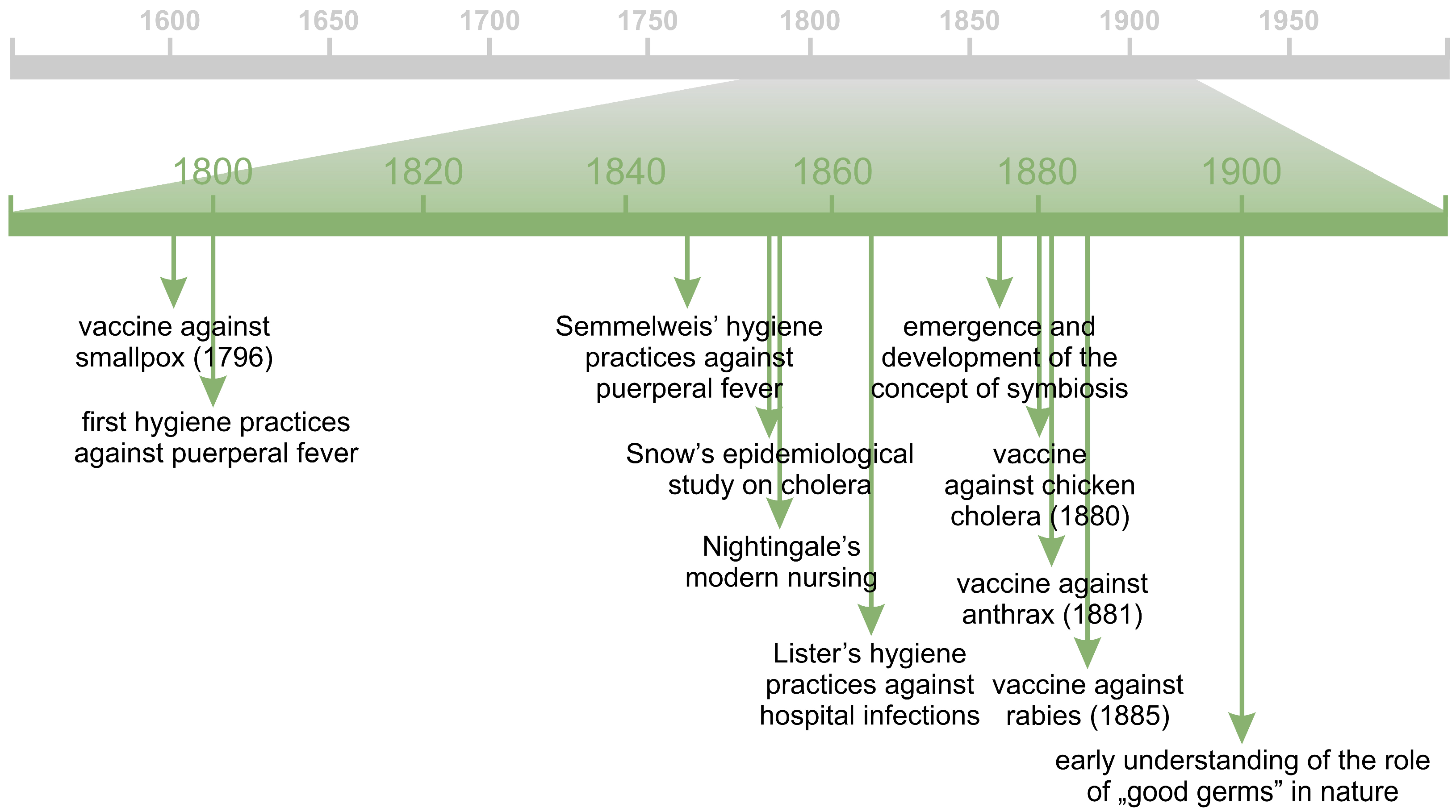
The germ theory sparked a veritable microbiological “gold rush”, initiating an intensive search for pathogenic microorganisms. The identification of the etiological agents of numerous infectious diseases subsequently facilitated the development of some of the earliest vaccines, including those against chicken cholera (1880), anthrax (1881), and rabies (1885), pioneered by Pasteur and his contemporaries. Nonetheless, it is important to recognize that the first effective vaccine predated the formal establishment of the germ theory. In 1796, Edward Jenner (1749–1823), an English physician, introduced the smallpox vaccine (Figure 2, Table S2), marking a seminal advancement in immunology. This achievement was built upon earlier practices of variolation, a precursor technique for inducing immunity. Notably, smallpox remains the first, and, to date, the only human disease to have been eradicated [65,83,84,85].
The germ theory also provided a crucial framework for understanding the necessity and significance of hygiene. Nineteenth-century hospitals, much like contemporary urban environments, were often overcrowded, lacked running water and adequate sanitation, and were generally unsanitary, with medical personnel routinely exposed to blood and bodily secretions. Under such conditions, the efficacy of medical interventions was severely compromised. Hospital-acquired infections, including putrid fever, hospital gangrene, puerperal fever, wet gangrene, and hospital rot [86,87], spread rapidly, and mortality rates reflected the remark of Scottish obstetrician James Young Simpson (1811–1870) in 1869: “a soldier had more chance of survival on the field of Waterloo than a man who goes to the hospital” [88]. It is noteworthy, however, that awareness of hygiene predated these formal developments. The eminent medieval Persian physician, alchemist, and philosopher Abu Bakr Muhammad Ibn Zakariya Al Razi (865–925), more commonly known as Rhazes, provided accurate and objective descriptions of various ailments and associated the occurrence of infectious diseases with poor sanitary conditions [89,90]. In the 18th century, British physician John Pringle (1707–1782) formulated hygiene principles for military hospitals and prisons, advocating reduced overcrowding, proper ventilation, and systematic laundering of patients’ clothing. Pringle is also credited with the first use of the term antiseptic and is regarded as the father of modern military medicine [91].
By the late 18th and early 19th centuries, a growing understanding of hygiene emerged, particularly regarding puerperal fever, a major cause of maternal mortality. Medical practitioners such as Scottish obstetrician Alexander Gordon (1752–1799) [92] and American physician Oliver Wendell Holmes (1809–1894) [93] emphasized the critical importance of handwashing and overall hygiene. Hungarian obstetrician Ignaz Philipp Semmelweis (1818–1865), through the implementation of stringent hygiene practices in his obstetrics ward, drastically reduced mortality rates among his patients. However, he encountered such an astonishing disdain and lack of comprehension among his colleagues, who thought it impertinent to suggest that gentlemen’s hands were dirty and regarded handwashing as too cumbersome, that it drove him to madness [94,95]. A significant contemporary figure was Florence Nightingale (1820–1910), an English nurse, statistician, social reformer, and writer. She recognized the central role of hygiene in patient care, emphasizing proper diet, environmental conditions, and comprehensive care. Despite facing resistance similar to her peers, her intelligence, dedication, and empathy established her as a pioneer of modern nursing [96]. Finally, Joseph Lister (1827–1912), inspired by Pasteur’s discoveries, successfully implemented antiseptic surgical techniques. By combining scientific rigor, meticulous attention to detail, and compassionate patient care, Lister developed effective methods for treating and dressing postoperative wounds to prevent infection. His innovations included routine disinfection of surgical sites and sterilization of instruments, leading to a dramatic reduction in patient mortality. Although initially met with resistance, Lister’s work ultimately cemented the practical application of the germ theory in clinical settings [87,97].
Simultaneously, the fields of epidemiology and sanitary microbiology were beginning to take shape. In 1854, the British physician John Snow (1813–1858) conducted one of the earliest epidemiological investigations during a cholera outbreak in London, demonstrating that the source of the disease was a contaminated water pump affected by sewage. Following the closure of the pump, the incidence of cholera cases declined markedly [98]. This observation corroborated the earlier hypothesis of another British physician, William Budd (1811–1880), who proposed that cholera and certain other gastrointestinal diseases were transmitted via water and sewage containing living microorganisms capable of proliferating in the human intestine upon ingestion [99].
Applied microbiology arose therefore under the prominence of “bad germs”. Moreover, the theory of evolution, introduced in 1859 by the British naturalist and geologist Charles Robert Darwin (1809–1882) [100], which gradually gained widespread acceptance, appeared to underscore a view of the relationship between humans and microorganisms as a life-and-death struggle for survival, seemingly at odds with notions of non-antagonistic symbiosis. Nevertheless, the ecological and beneficial roles of microorganisms began to receive recognition in other domains. In agriculture, for example, the dominance of the humus theory gradually gave way to an emphasis on organic matter remineralization and the critical role of mineral elements in plant nutrition. Consequently, interest in agricultural practices, environmental microbiology, agricultural chemistry, and plant pathology intensified. Among plant diseases that drew significant attention was tobacco mosaic, now known to be caused by a virus. Key advances in its study were made by the Dutch botanist and microbiologist Martinus Willem Beijerinck (1851–1931) [101], who is also recognized for his isolation of rhizobial symbionts capable of nitrogen fixation, as well as bacteria involved in sulfur cycling [102,103]. Another foundational figure in environmental microbiology was the Russian microbiologist Sergei Nikolaevich Winogradsky (1856–1953). His extensive investigations in soil microbiology elucidated the biogeochemical cycles of elements in natural ecosystems and highlighted the pivotal roles of bacteria therein. Winogradsky’s research encompassed diverse microbial processes, including photosynthesis, nitrification, sulfur oxidation, iron reduction, and the degradation of complex organic compounds within the soil matrix [104].
By the late 19th and early 20th centuries, scientific discourse began to recognize the significance of “good germs”, particularly bacterial decomposers essential for sustaining life (Figure 2, Table S2). It became increasingly apparent that pathogenic microorganisms represented only a minor subset within the broader framework of microbial ecology. In 1877, the German botanist, plant pathologist, and mycologist Albert Bernhard Frank (1839–1900) introduced the term symbiosis into scientific discourse [105] and subsequently coined the term mycorrhiza [106,107]. The following year, the concept was further developed and popularized by the German physician, botanist, mycologist, and lichenologist Heinrich Anton de Bary (1831–1888) [108,109,110], who defined symbiotic partners as organisms that must coexist in close physical association and belong to distinct species [111]. His seminal lecture on the subject, published in 1879 in both French [112] and German [113], helped establish the modern scientific understanding of ecological interrelationships. Collectively, these contributions marked a critical turning point in the recognition of the diverse roles of microorganisms beyond pathogenicity.

4. Beginnings of the Human Gut Microbiota Studies

The beginning of research on the microbiota of multicellular organisms is dated back to the mid-19th century [114] (Figure 3, Table S3), but it is noteworthy, that the pioneering investigations into the human microbiota, albeit somewhat inadvertently, were conducted by the aforementioned, Antonie Philips van Leeuwenhoek, at the turn of the 17th and 18th centuries (Figure 1, Table S1).
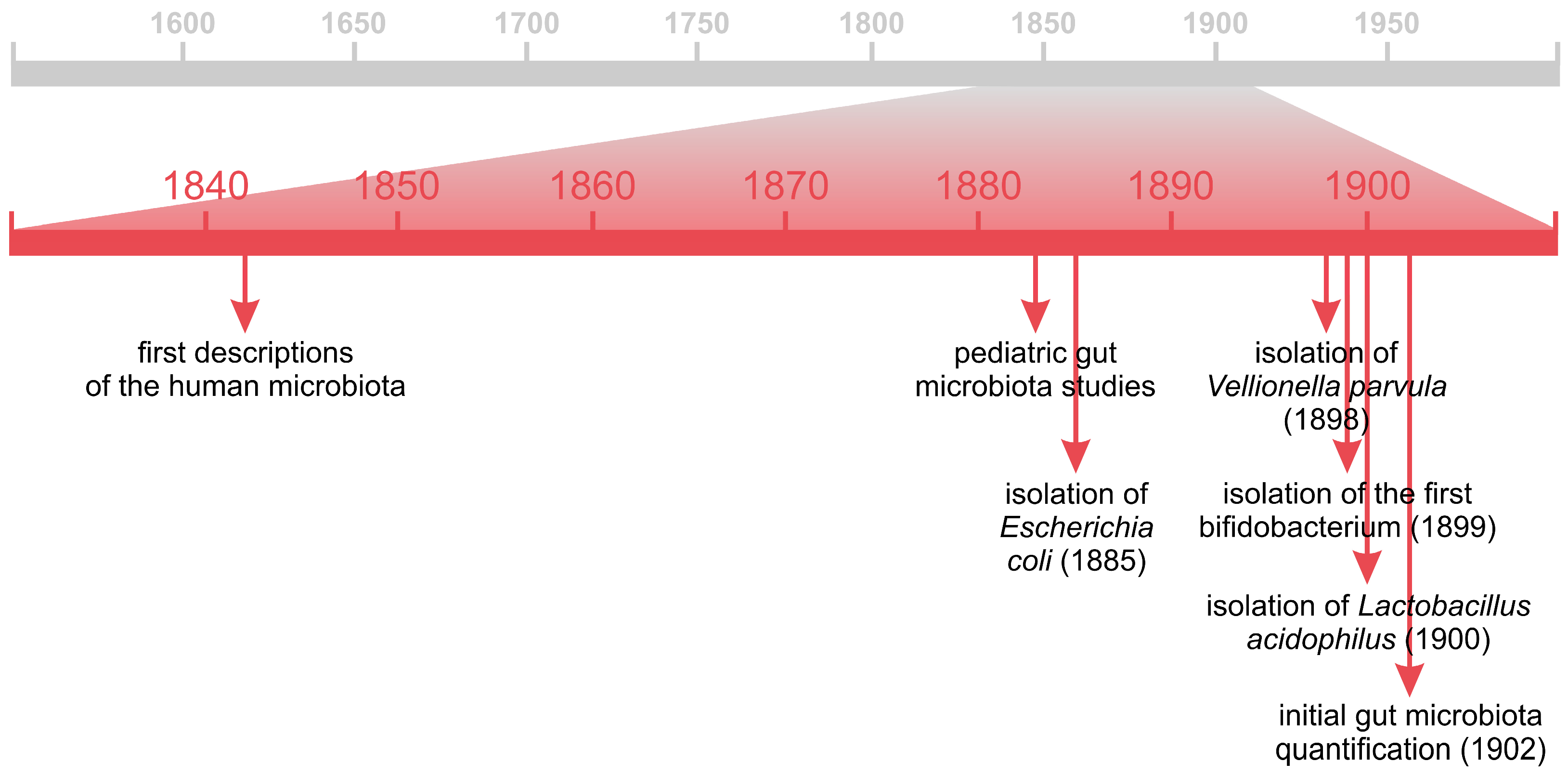
In 1842, the Scottish anatomist John Goodsir (1814–1867) reported the presence of “vegetable organisms of an undescribed form” in the gastric contents of a young patient [115]. Among these, he identified a microorganism that he named Sarcina ventriculi, owing to its distinctive cubic arrangements that evoked the orderly formations of Roman legions. Although the nomenclature Sarcina persists to this day, the taxonomic classification of this bacterium has since been revised to reflect modern microbiological standards. Goodsir’s observations were made prior to the formal establishment of the germ theory, and he interpreted these organisms as belonging to the plant kingdom, hence his designation of them as “vegetable organisms” [116]. In 1853, an American paleontologist, parasitologist and anatomist, Joseph Leidy (1823–1891) published a book titled “A Flora and Fauna within Living Animals”, describing the species of “plants” and “animals” (flora and fauna) inhabiting host organisms [117]. Subsequently, the presence of numerous microorganisms in the gastrointestinal tract, particularly in feces, as well as their diet-related variability, was delineated, for instance, by the British physician and microscopist Lionel Smith Beale (1828–1906) in 1867 [118], by the German botanist and mycologist Ernst Hans Hallier (1831–1904) in 1869 [119], by the American surgeon and microscopist Joseph Janvier Woodward (1833–1884) in 1879 [120], by the German physician and hygienist Julius August Christian Uffelmann (1837–1894) in 1881 [121], or by the Scottish bacteriologist Allan Macfadyen (1860–1907) and his collaborators in 1891 [122].
Significant advances in human microbiota research were pioneered by three eminent pediatricians: the German-Austrian Theodor Escherich (1857–1911), the French Henry Tissier (1866–1926), and the Austrian Ernst Moro (1874–1951). Escherich’s investigations focused primarily on the gut microbiota of infants and children, as well as the microbial composition of maternal milk. He sought to define the “normal” bacterial composition of the intestinal tract, i.e., the typical assemblage of microbial species present in healthy individuals, and to characterize its dynamic changes immediately following birth. For instance, he demonstrated that meconium was sterile at birth. Additionally, Escherich aimed to elucidate the role of intestinal bacteria in digestion under both physiological and pathological conditions. Through his studies, he described several fecal-associated bacterial species and is best known for his discovery in 1885 of a rod-shaped, facultatively anaerobic, Gram-negative bacterium, initially named Bacterium coli commune and later renamed in 1919 as Escherichia coli [123]. Today, this microorganism is one of the best studied gut inhabitant [124] and arguably one of the most scientifically influential bacterial species [125]. Initially, Escherichia coli was thought to dominate the infant gut microbiota [114]; however, this assumption was rapidly challenged by Tissier, who in 1899 isolated the first bifidobacterial species from infant feces. This Gram-positive, anaerobic, rod-shaped bacterium was originally designated Bacillus bifidus communis [66,126], with the genus name Bifidobacterium being formally proposed in 1924 [127]. Tissier suggested that the high prevalence of bifidobacteria in the feces of healthy, breastfed infants could contribute to their lower incidence of diarrhea. In his pediatric practice, he administered bifidobacteria therapeutically to manage this condition, representing one of the earliest documented examples of the intentional oral use of live microorganisms for health benefits [66].
Ernst Moro, a student of Theodor Escherich, is best known for describing the umklammerungsreflex (embracing reflex) in newborns, today widely recognized as the Moro reflex [128]. However, his contributions to early microbiota research are equally noteworthy. Focusing on the physiology of infant digestion [129], Moro demonstrated that breastfed infants exhibited superior immune development compared to those nourished with cow’s milk. In 1900, he discovered a rod-shaped, Gram-positive, microaerophilic to anaerobic bacterium, which he named Bacillus acidophilus [130]; later reclassified as Lactobacillus acidophilus. It is worth emphasizing the taxonomic challenges associated with the classification of this and similar microorganisms. During Moro’s time, bacterial taxonomy relied primarily on morphological characteristics, with rod-shaped forms commonly designated as Bacillus. Only with the advent of molecular techniques, particularly DNA sequencing, did more accurate and phylogenetically informed classification systems emerge. Consequently, many microorganisms have undergone reclassifications, in some cases multiple times, often accompanied by changes in nomenclature. The genus Lactobacillus serves as a prime example of this complexity. Recent genomic studies [131,132] have revealed that Lactobacillus represents an exceptionally diverse group, both genetically and ecologically, encompassing species adapted to a wide range of environments. Accordingly, a major taxonomic revision led to the division of this genus into 25 new genera, reflecting its extensive heterogeneity [131]. In 1898, another gut-associated bacterium, strictly anaerobic, Gram-negative, and coccus-shaped, was isolated (Figure 3, Table S3). Initially named Staphylococcus parvulus [133], it was reclassified in 1933 as Veillonella parvula [134]. The first quantitative estimation of intestinal microorganisms was conducted in 1902 by the German physician Julius Strasburger (1871–1934) [135]. Remarkably, comparable assessments of intestinal microbial abundance did not emerge again until several decades later, underscoring the pioneering character of his work.
It is important to recognize that these discoveries were deeply rooted in the methodological foundations established by other microbiologists, most notably already mentioned Heinrich Hermann Robert Koch, whose innovations in bacterial isolation and in vitro cultivation profoundly transformed microbiological research. Two additional figures also warrant mention in this context: the Danish bacteriologist Hans Christian Joachim Gram (1853–1938) and the German physician, bacteriologist, and hygienist Hans Ernst August Buchner (1850–1902). Gram developed the differential staining method, now universally known as Gram staining, which enables the classification of bacteria based on cell wall structure [136], whereas Buchner pioneered anaerobic culture techniques that significantly advanced the study of oxygen-sensitive microorganisms [123].

5. “Good Germs” in the Service of Human Health

5.1. Probiotics

5.1.1. From the Theory of Autointoxication to Probiotic Bacteria

The theory of autointoxication has deep historical roots, proposing that metabolic by-products generated within the intestines could penetrate the bloodstream, resulting in systemic poisoning and contributing to the onset of numerous, if not all, diseases. This concept was closely intertwined with the above-mentioned humoral theory of medicine, which held that illness arose from imbalances among the four bodily humors (blood, phlegm, yellow bile, and black bile), with enemas commonly prescribed as a means to restore internal equilibrium [137]. The revival of autointoxication theory in the 19th century (Figure 4, Table S4) was initiated by the German clinician and medical writer Herman Senator (1834–1911) [138] and was later strongly advocated by figures such as the English physician and medical author Robert Bell (1846–1926) [139] and the French physician and pathologist Charles Jacques Bouchard (1837–1915) [140]. The gastrointestinal tract was believed to play a crucial role in a wide range of nervous system disorders, including emotional disturbances, irritability, nervous tension, anxiety, and mood fluctuations, which were attributed to inadequate and overly processed diets, as well as excessive consumption of alcohol and even tea [141,142]. It is noteworthy that the American surgeon William Beaumont (1785–1853) had experimentally recognized the existence of a gut–brain connection as early as 1838 [143]. Within the framework of autointoxication theory, the colon, in particular, acquired a distinctly negative reputation, often described as a “septic reservoir” or “sewage system”. This perception stemmed from the contemporary understanding of the vast microbial populations inhabiting decomposing organic matter. The colon was thus conceptualized as a receptacle accumulating waste and putrefying digestive residues–an ideal breeding ground for harmful microorganisms [141,142,144,145]. Consequently, a wide array of questionable remedies and elaborate colonic cleansing devices emerged, including the well-known J.B.L. Cascade, designed by the controversial British entrepreneur Charles Alfred Tyrrell (1843–1918) [137]. Some proponents, such as the eminent yet contentious British surgeon William Arbuthnot Lane (1856–1943) [146,147], went so far as to advocate for partial or even total colectomy.
The theory of autointoxication also exerted a profound influence on the Russian zoologist and immunologist Ilya Ilyich Mechnikov (1845–1916), internationally known by the French spelling of his name, Élie Metchnikoff. He is best known for discovering phagocytes, for which he was co-awarded the Nobel Prize in Physiology or Medicine in 1908, “in recognition of their work on immunity” [21]. As a distinguished scholar and mentor, Metchnikoff made seminal contributions to the development of immunology, embryology, physiology, pathology, and zoology [148]. His scientific curiosity eventually led him to investigate the intestinal microbiota (both human and animal) through a series of pioneering studies conducted independently [149,150,151], collaboratively [152], and by his close associates [153,154]. These works examined the composition of the intestinal microbiota, its variation with age, diet, and environmental conditions, and its potential involvement in the processes of aging and degeneration. Comparative analyses were also undertaken on the intestinal microbiota of children from diverse geographical regions. Among the bacterial species identified were numerous putrefactive microorganisms, such as Bacillus putrificus, Bacillus sporogenes, and Bacillus welchii (now classified as Clostridium perfringens), which were implicated in intestinal putrefaction leading to the generation of toxic metabolites. In addition, these investigations stimulated the development of germ-free animal models, which provided crucial insights into the physiological roles of intestinal microorganisms [155,156,157]. It is noteworthy, however, that the first germ-free animal, a guinea pig, had already been produced between 1885 and 1896 by the American-British bacteriologist George Henry Falkiner Nuttall (1862–1937) and the German biochemist Hans Thierfelder (1858–1930) [158].
Although Metchnikoff regarded the colon as an evolutionary vestige and an organ whose microbial inhabitants potentially accelerate aging and shorten lifespan, he did not advocate for radical surgical interventions. Instead, he proposed a more constructive strategy: the replacement of harmful intestinal microorganisms with benign or beneficial counterparts [149,150,151]. During his in vitro investigations on Vibrio cholerae, Metchnikoff observed that certain bacterial species exhibited antagonistic activity against the pathogen, leading him to hypothesize that interindividual variability in disease resistance could be attributed to differences in intestinal microbial composition. Concurrently, research conducted across various European laboratories demonstrated that lactic acid-producing bacteria exert inhibitory effects on putrefactive microorganisms, which were then believed to play a central role in autointoxication. Metchnikoff further noted the exceptional health and longevity of Bulgarian peasants whose diet was rich in fermented dairy products, particularly yogurt (sour milk, bulg. кисело мляко, kiselo mlyako). He attributed these beneficial effects to the presence of Lactobacillus bulgaricus in their diet [66,114,144,159]. This bacterium had been discovered in 1905 by the Bulgarian physician and bacteriologist Stamen Gigow Grigorow (1878–1945), who initially named it Bacillus bulgaricus [66], while it is currently classified as Lactobacillus delbrueckii subsp. bulgaricus [160]. These observations, particularly Grigorow’s findings, inspired Metchnikoff to formulate his celebrated “Mechnikov hypothesis”, often summarized as “yogurt brings long life” [4,66]. Lactobacillus bulgaricus is therefore considered the first scientifically recognized probiotic microorganism, and soon after, the first commercial probiotic preparation (lactobacilline tablets) was produced by the French company Le Ferment [142]. The growing popularity of fermented milk products subsequently spurred the establishment of new enterprises devoted to their production. Among them, a company that began operations in Europe and later expanded to the United States, now globally known as Danone, was established [66].
Although it soon became evident that “Metchnikoff’s bacteria” did not persist within the colon [161], the concept of probiotics as antagonists to putrefactive intestinal microbes remained influential. Researchers subsequently sought lactic acid-producing bacteria capable of surviving transit through the gastrointestinal tract, particularly within the large intestine. This pursuit culminated in 1930 with the discovery of Lactobacillus casei by the Japanese microbiologist Minoru Shirota (1899–1982) (Figure 4, Table S4). In 1935, he introduced a fermented milk beverage containing this bacterium, marketed under the name Yakult. The strain originally isolated by Shirota was initially identified as Lactobacillus acidophilus, but later reclassified as Lactobacillus paracasei. Nevertheless, it continues to be widely referred to as Lactobacillus casei Shirota (LcS) for historical and practical reasons [66,162]. Today, LcS remains one of the most extensively studied and commercially utilized probiotic strains. However, the European Food Safety Authority (EFSA) has not conclusively confirmed its specific health-promoting or immunomodulatory effects [163].
It is noteworthy that health-promoting and health-supporting foods such as yogurt and other fermented products are by no means a modern invention. These dietary items have been integral to human nutrition for millennia, dating back nearly to 10,000 BC, with their beneficial effects on health widely recognized. However, for obvious reasons, these effects were not attributed to the presence or activity of microorganisms until much later [66,159,164]. Although Metchnikoff is often regarded as the pioneer of probiotic research, it should be recalled that Henry Tissier, mentioned earlier, administered bifidobacteria orally for the treatment of infant diarrhea around the same time. Moreover, at least a decade earlier, the Polish physician, pediatrician, neurologist, and social activist Józef Polikarp Brudziński (1874–1917) conducted a study in which he used a suspension of Bacillus lactis aerogenes to treat infants suffering from acute infectious diarrhea. For this reason, he is sometimes referred to as the true father of probiotics [165].
In the broader context of microbiota research and its relationship with nutrition, two American figures also deserve recognition: the microbiologist Arthur Isaac Kendall (1877–1959) and the physician, inventor, and nutritionist John Harvey Kellogg (1852–1943). Kendall was among the first to propose that the gastrointestinal microbiota could exert a protective effect against pathogenic bacteria. He emphasized the importance of bacterial metabolism and metabolites rather than taxonomic identity, highlighting the impact of diet on shaping the intestinal microbiota [166]—a concept that gained experimental validation nearly a century later. Today, Kendall is recognized as a trailblazer in modern microbiota research and a visionary of contemporary methodological approaches [167]. Kellogg, for his part, directed a well-known health resort where he promoted his distinctive dietary and lifestyle philosophy. A staunch advocate of vegetarianism, he developed numerous innovative food products, such as cornflakes, granola, peanut butter, and soy milk, that transformed American breakfast culture. He championed physical exercise, outdoor activity, and abstinence from alcohol, tobacco, and even tea and coffee, while emphasizing adequate hydration [168]. These lifestyle recommendations, remarkably prescient for his time, align closely with current principles for maintaining a healthy gut microbiota and overall well-being. Nonetheless, Kellogg’s pioneering contributions to gut microbiota research and health promotion have been somewhat overshadowed by his controversial involvement in the eugenics movement [169].
In the pre-antibiotic era, intestinal infections represented a major public health challenge, particularly among soldiers during World War I, who were already severely affected by the harsh conditions of trench warfare. In response, numerous physicians and researchers sought innovative strategies to combat gastrointestinal infections. Among them was the German physician and scientist Alfred Nissle (1874–1965), whose work focused on identifying bacterial strains with antagonistic activity against Salmonella spp., Shigella spp., and other enteropathogens. In 1917, during a military campaign in the Balkans, Nissle observed that one soldier remained strikingly resistant to dysentery, despite widespread infection among his comrades. He hypothesized that this resistance might stem from protective components of the soldier’s intestinal microbiota. From the soldier’s stool, Nissle subsequently isolated a bacterial strain later designated Escherichia coli Nissle 1917 (EcN) (Figure 4, Table S4). Both Nissle’s original investigations and subsequent research have demonstrated the strain’s pronounced antagonistic activity against a range of pathogenic microorganisms, along with distinctive physiological properties and an excellent safety profile. Building upon these findings, Nissle developed the probiotic formulation Mutaflor, designed to deliver viable bacteria specifically to the distal regions of the gastrointestinal tract. Remarkably, more than a century after its introduction, Mutaflor remains in clinical use, with substantial evidence supporting the beneficial health effects of Escherichia coli Nissle 1917 [66,114,170].

5.1.2. Current Understanding of Probiotics, Prebiotics, Synbiotics and Postbiotics

The term probiotic derives from the Latin pro (for) and the Greek βίος (life), literally meaning “for life”. Its earliest conceptualization referred broadly to “active substances that are essential for a healthy development of life”. The term is believed to have been introduced into the scientific lexicon in 1953 (Figure 4, Table S4) by the German bacteriologist, hygienist, and food scientist Werner Georg Kollath (1892–1970) [159,170], whose legacy remains somewhat controversial due to his eugenic sympathies. The following year, the term was employed to contrast the detrimental effects of antibiotics on the gut microbiota with the beneficial influence of specific bacterial species on intestinal homeostasis [171]. Over subsequent decades, the definition of probiotics underwent substantial conceptual evolution. In 1965, it was described as substances secreted by one organism that stimulate the growth of another [172], while by 1974 its scope had been expanded to encompass both microbial organisms and non-living agents capable of promoting microbial balance [173]. A more operational definition emerged in 1989, identifying probiotics as live microbial feed supplements that beneficially affect the host animal by improving its intestinal microbial balance [174]. In 1994, the World Health Organization (WHO) acknowledged probiotics as a potential adjunct in immune defense, particularly relevant in the context of rising antibiotic resistance. In 2001 WHO, alongside with the Food and Agriculture Organization (FAO) redefined probiotics as “live microorganisms which when administered in adequate amounts confer health benefits on the host” [175]. It broadened the preceding comprehension of probiotics, as outlined by the Lactic Acid Bacteria Industrial Platform (LABIP), to encompass products administered via diverse routes, beyond solely oral delivery, such as vaginally [170]. Finally, in 2014, an expert consensus document was released regarding the extent and proper application of the term probiotic. It was the result of the panel work of the International Scientific Association for Probiotics and Prebiotics (ISAPP) held in October 2013 [176]. Probiotics are subject to several requirements that must be met: (1) need to be safe for use in human (for instance, they ought not to facilitate the spreading of antibiotic resistance); (2) need to demonstrate activity and viability within the gastrointestinal tract, existing in adequate quantities to substantiate any positive impacts (it is worth noting that their longevity is restricted and typically ceases within a few days after discontinuation of their intake); (3) need to exhibit metabolic activity within the human intestine, with certain strains capable of persisting and proliferating; (4) need to provide a proven physiological advantage [177].
The conceptual evolution of probiotics reflects a gradual shift from broadly defined growth-promoting substances toward living microorganisms with rigorously documented, strain-specific health effects. Early definitions were largely intuitive and lacked precision, often encompassing non-microbial products or vaguely described bioactive substances. In contrast, contemporary understanding is grounded in evidence-based research, emphasizing that only microorganisms with reproducible, strain-dependent health benefits and well-established safety profiles can be regarded as probiotics. Within this framework, the status of so-called probiotic foods (e.g., yogurts, fermented vegetables, and other traditionally fermented products) remains ambiguous. Although these products may contain microorganisms with potential probiotic properties and are generally considered health-promoting, their microbial composition is seldom standardized or comprehensively characterized, thereby precluding their formal classification as probiotic products under the current definition. At present, a growing body of literature highlights the need to revisit and refine the definition of probiotics to reflect advances in microbiome science, although the 2014 consensus definition remains the prevailing reference in both scientific and regulatory contexts [178,179].
The concept of probiotics is inherently intertwined with that of prebiotics. Prebiotics are defined as non-digestible dietary components, predominantly carbohydrates such as fructooligosaccharides (FOS), galactooligosaccharides (GOS), xylooligosaccharides (XOS), inulin (IN), or dietary fiber, that selectively stimulate the growth and metabolic activity of beneficial microorganisms within the gastrointestinal tract. Owing to their chemical structure, these compounds resist digestion and absorption in the small intestine and undergo fermentation primarily in the large intestine, particularly in the colon, resulting in the production of physiologically relevant metabolites, including short-chain fatty acids (SCFAs) [164,180]. Human milk represents the quintessential natural prebiotic source. In the early 20th century, pediatricians, most notably mentioned earlier Henry Tissier, observed that bifidobacteria were significantly more prevalent in the stools of breastfed infants compared to those fed with cow milk. They hypothesized the presence of a specific human milk-derived substrate that favored the growth of these bacteria, which they termed the “bifidus factor” or “bifidogenic factor”. It is now known that this factor comprises a complex mixture of approximately 200 structurally distinct human milk oligosaccharides (HMOs), unique to humans. These oligosaccharides range from short-chain trisaccharides (e.g., sialyllactose, fucosyllactose) to high-molecular-weight glycans, such as N-acetyllactosamine polymers. Only a minor fraction of HMOs is directly utilized by the infant; instead, they primarily serve as selective substrates for Bifidobacterium longum subsp. infantis, a bacterial strain that predominates in the gut microbiota of healthy, naturally born, breastfed infants. In return, Bifidobacterium infantis produces SCFAs that nourish the intestinal epithelium, contribute to mucosal barrier integrity, and modulate immune system maturation. Moreover, this species synthesizes sialic acid, an essential compound involved in neurodevelopment, which may partially account for observed differences in cognitive outcomes between breastfed and formula-fed infants. Structurally, HMOs also resemble glycans present on host epithelial surfaces to which pathogenic microorganisms typically adhere. Consequently, they act as molecular decoys, preventing pathogen attachment and infection. Notably, certain HMOs have demonstrated the ability to inhibit HIV adhesion, which may explain the relatively low rate of mother-to-child HIV transmission through breastfeeding despite the presence of the virus in milk. Given their multifaceted biological roles, Bifidobacterium infantis (as a probiotic) and HMOs (as prebiotics) have been incorporated into therapeutic strategies for extremely premature neonates at risk of necrotizing enterocolitis. Furthermore, HMO-based prebiotics are currently being developed for use in adult populations, highlighting their expanding biomedical potential [126,181,182,183,184,185]. In recent years, the concept of prebiotics has evolved beyond traditional oligosaccharides to encompass a broader range of substrates, including polyphenols, organic acids, and even bioactive peptides, provided they selectively modulate the composition or activity of the host microbiota in ways that confer health benefits [186].
The integration of the beneficial properties of probiotics and prebiotics led to the emergence of the synbiotic concept. According to the definition proposed in 2019 by the ISAPP, a synbiotic is defined as “a mixture comprising live microorganisms and substrate(s) selectively utilized by host microorganisms that confers a health benefit on the host”. Importantly, the combined activity of both components must demonstrate an enhanced effect compared to their individual actions. Prebiotics facilitate the survival and colonization of beneficial microorganisms in the gastrointestinal tract by modulating physicochemical parameters such as pH and oxygen availability. This effect is indirect, resulting from microbial fermentation processes that alter the local microenvironment, thereby creating favorable conditions for the persistence and metabolic activity of probiotic strains. A well-designed synbiotic aims to optimize the functionality of both components, leading to additive or synergistic effects. In an additive relationship, the probiotic and prebiotic act independently to confer health benefits. These formulations are referred to as complementary synbiotics. In contrast, in a synergistic relationship, the prebiotic is specifically selected to support the growth, viability, or metabolic activity of the co-administered probiotic strain, thereby enhancing its health-promoting properties. These are known as synergistic synbiotics. The most common combinations used in synbiotic formulations include Lactobacillus spp. and/or Bifidobacterium spp. together with fructooligosaccharides (FOS), galactooligosaccharides (GOS), or inulin [187,188,189,190].
It is also pertinent to highlight two additional categories of biogenic products that contribute to maintaining gut health and exert broader systemic benefits. The first are postbiotics, which consist of inactivated (e.g., pasteurized) microbial cells, their structural components, or metabolites, particularly molecules exhibiting anti-inflammatory or immunomodulatory activity [190]. ISAPP defines postbiotics as a “preparation of inanimate microorganisms and/or their components that confers a health benefit on the host” [191]. A related category encompasses parabiotics, which refer specifically to purified bacterial metabolites or bioactive compounds derived from microorganisms that similarly promote host health [190].
Currently approved prebiotics, probiotics, synbiotics and postbiotics are typically used following antibiotic therapy, but are increasingly also employed to support the treatment of conditions such as diarrhea, irritable bowel syndrome (IBS), inflammatory bowel disease (IBD), cardiovascular diseases, hypertension, hypercholesterolemia, lactose intolerance, obesity, and cancer. They are also utilized in regulating lipid metabolism, modulating the immune system, supporting uptake and bioavailability of minerals, or laxation [180,192]. Currently, probiotics mainly utilize strains from the genera Lactobacillus and Bifidobacterium, which can be administered either individually or in various combinations [164,180,192], as well as non-pathogenic strain Escherichia coli Nissle 1917 [170]. The use of a species of yeast, Saccharomyces cerevisiae var. boulardii, has also been approved for similar purposes [193,194]. More recent formulations employ Akkermansia muciniphila for the management of obesity, type 2 diabetes, and cardiovascular diseases [195], as well as for mitigating aging-related processes [196]. Additionally, Bacteroides fragilis [197] and Faecalibacterium prausnitzii [198] have been investigated for their anti-inflammatory potential, alongside selected strains belonging to the genera Pediococcus, Lactococcus, Enterococcus, Streptococcus, Propionibacterium, and Bacillus [192]. Probiotics are also gaining increasing attention as psychobiotics, supporting a treatment of psycho-related disorders such as depression, anxiety, chronic stress or low mood, through the gut–brain axis, with Lactobacillus spp. and Bifodobacterium spp. mainly used for this purpose [177,199]. It is worth noting that the idea of using probiotic bacteria to treat mental disorders was first proposed as early as 1910 [200], though it is only now gaining widespread recognition.
Into the realm of probiotics, genetic engineering is also increasingly venturing. Engineered probiotics are being developed in areas such as: diagnosis and treatment of inflammatory bowel disease (Escherichia coli Nissle 1917 (EcN); yeast), detection of bacterial infections (lactic-acid bacteria against Staphylococcus aureus; Lactobacillus lactis against Vibrio cholerae), treatment of bacterial infections (Lactobacillus casei against listeriosis; EcN against enterococci and Pseudomonas aeruginosa; Saccharomyces cerevisiae var. boulardii against Clostridium difficile), treatment of tumors (Escherichia coli SLIC; EcN SYNB1891), treatment of metabolic diseases (Lactobacillus reuteri and EcN SYNB1618 in phenylketonuria; Escherichia coli SYNB8802 in hyperoxaluria; EcN SYNB1020 in hyperammonemia; Lactobacillus gasseri and Lactococcus lactis AG019 in diabetes) [201].

5.2. Phages

Towards the end of the 19th century and throughout the early decades of the 20th century, scientific interest increasingly focused on microbial antagonism and its potential applications in the prevention and treatment of various diseases, particularly infectious ones. This growing attention, driven in part by advances in gut microbiota research, laid the groundwork for numerous seminal discoveries; many of which, however, have since faded into relative obscurity.
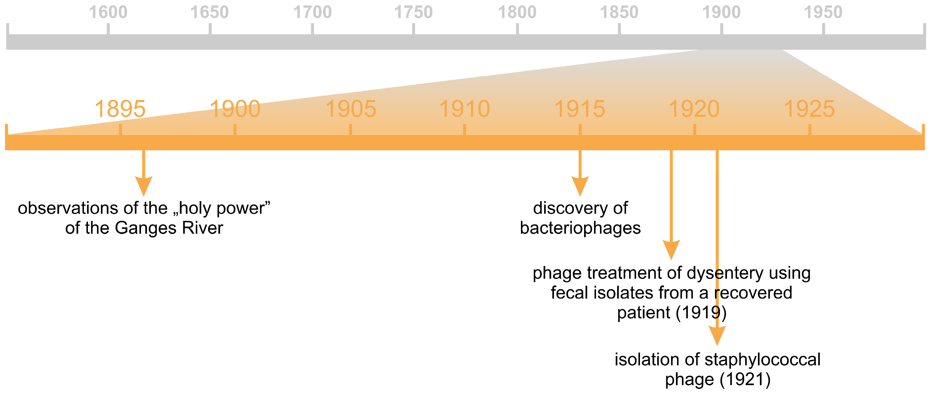
In the late 19th century, during his investigations into infectious diseases in India, the English bacteriologist Ernest Hanbury Hankin (1865–1939) made a remarkable observation. Despite the pronounced microbial contamination of the Ganges River, individuals bathing in its waters appeared to exhibit reduced susceptibility to certain illnesses, particularly cholera, an effect that local populations attributed to the river’s “holy power” (Figure 5, Table S5). Scientists, however, sought a more rational explanation. Although this interpretation remains a subject of debate, the antibacterial properties of the river water were later ascribed to bacteriophages [202,203], first described in 1915 by the English bacteriologist Frederick William Twort (1877–1950) [204]. Some researchers, however, consider the true pioneer of phage research to be the Franco-Canadian microbiologist Félix d’Hérelle (1873–1949) [205,206], who not only independently discovered bacteriophages but also correctly elucidated their biological nature in 1917 [207]. Merely two years later, he employed phages isolated from the stool samples of convalescent patients to treat individuals suffering from dysentery. He subsequently extended their application to the management of various other infectious diseases, including skin, bone, and ocular infections, as well as cholera, plague, typhoid, paratyphoid, and septicemia, in multiple countries around the world (e.g., Brazil, Egypt, China, Germany, Great Britain, Greece, India, Italy, Japan, Senegal, the Soviet Union, and the United States), achieving substantial reductions in mortality rates. In 1933, d’Hérelle established the Laboratoire du Bactériophage in France, further advancing the therapeutic exploration of phages. Nevertheless, the clinical use of phage therapy gradually declined, primarily due to difficulties in large-scale production, the limited understanding of phage biology, and additional sociopolitical and scientific factors, including skepticism toward d’Hérelle’s unconventional ideas. Ultimately, phage therapy was discontinued in most Western institutions, persisting mainly in certain Eastern European countries [202,205], notably Poland. The subsequent revival of interest in phage therapy culminated in the establishment of the Phage Therapy Unit in 2005, as part of the Hirszfeld Institute of Immunology and Experimental Therapy of the Polish Academy of Sciences in Wrocław, Poland [208]. At the time, this facility was unique in Europe and has since achieved significant clinical and scientific success [209].
Currently, it is well established that bacteriophages constitute an integral component of the gut microbiota, displaying markedly greater diversity in healthy individuals compared to those with various pathological conditions. Their primary function involves the protection of the intestinal epithelial barrier and mucosal tissues against pathobiont colonization and infection [164]. This protective capacity is associated with the pronounced tropism of phages for mucosal surfaces, a feature believed to have deep evolutionary origins and to contribute significantly to the maintenance of intestinal homeostasis [210].
The multifaceted aspects of “good germs” are, to some extent, interconnected through the work of a somewhat overlooked yet highly influential Belgian physician and microbiologist, André Gratia (1893–1950) [211]. In his investigations, Gratia demonstrated that the discoveries made by Twort [204] and d’Hérelle [207] in fact represented two manifestations of the same phenomenon, i.e., the activity of bacteriophages. In 1921 (Figure 5, Table S5), he achieved a major milestone by isolating the first staphylococcal bacteriophage [212,213]. While studying antagonistic interactions among co-cultured strains of Escherichia coli, Gratia identified colicins—proteinaceous antibacterial substances produced by certain bacterial strains, characterized by a narrow spectrum of activity [214]. Moreover, he was among the first to observe the inhibitory effects of Penicillium molds on bacterial growth, thus indirectly anticipating the discovery of penicillin [215]. Unfortunately, a severe illness prevented him from pursuing this promising line of research further, allowing others to make the subsequent breakthroughs. Although he never returned to this topic, Gratia continued to conduct pioneering studies in virology, genetics, and molecular biology, which further underscored his contribution to the early development of modern microbiological sciences [211].

5.3. Antibiotics and Other Antimicrobial Compounds

5.3.1. Antibiotics

Although antibiotics and other antimicrobial agents were not discovered and clinically applied until the first half of the 20th century (Figure 6, Table S6), evidence suggesting their existence had surfaced several decades earlier. As early as 1874, the British physician William Roberts (1830–1899) observed that bacterial growth was inhibited on culture media overgrown with molds of the genus Penicillium [216]. A year later, John Tyndall—already mentioned in this narrative—reported similar findings while investigating airborne particulates, noting that media colonized by molds were devoid of bacterial contamination [217]. Despite being published, these observations received little attention at the time, as the germ theory was still in its infancy and not yet widely accepted [38]. Approximately two decades later, in 1897, the French physician Ernest Duchesne (1874–1912) provided a detailed account in his doctoral dissertation of the antagonistic interactions between bacteria and filamentous fungi, proposing their potential therapeutic application. However, Duchesne’s pioneering insights went largely unrecognized, and his work was soon forgotten, only to be rediscovered decades later, following the landmark discovery of the first true antibiotic [218].
It is worth emphasizing that microbial antagonism was concurrently observed by several other researchers who arrived at similar conclusions. Among the figures previously mentioned, Louis Pasteur and André Gratia deserve particular acknowledgment for their pioneering contributions in this regard. Nevertheless, the decisive breakthrough in this field was achieved by the Scottish physician and bacteriologist Alexander Fleming (1881–1955). In 1928, while conducting experiments with Staphylococcus cultures, Fleming noticed that bacterial growth was inhibited on a plate accidentally contaminated with a mold colony. He correctly inferred that the mold produced a substance with potent antibacterial activity and named it penicillin, after the genus Penicillium from which it was derived [219]. Crucially, Fleming demonstrated that this substance, in contrast to conventional disinfectants, exhibited selective toxicity, lethal to prokaryotic cells while harmless to eukaryotic ones, marking a transformative advance in antimicrobial therapy. However, Fleming was unable to purify or stabilize the compound in sufficient quantities for therapeutic use. This challenge was later overcome by a research team at Oxford University, led by the Australian pharmacologist Howard Walter Florey (1898–1968) and the British biochemist Ernst Boris Chain (1906–1979). Their efforts resulted in the successful isolation, chemical characterization, and large-scale production of penicillin, enabling its application as the first true antibiotic of natural origin [216,220]. The results of their research, published in 1940 [221], revolutionized modern medicine, and in 1945, Fleming, Florey, and Chain were jointly awarded the Nobel Prize in Physiology or Medicine “for the discovery of penicillin and its curative effect in various infectious diseases” [21].
The second major breakthrough in antibiotic discovery was the isolation of streptomycin by a research team led by the Jewish-American microbiologist Selman Abraham Waksman (1888–1973). In contrast to the serendipitous discovery of penicillin, the identification of streptomycin was the outcome of a systematic and methodical investigation. As a soil microbiologist, Waksman recognized that the soil harbors a vast diversity of metabolically active microorganisms, particularly actinomycetes, capable of producing bioactive secondary metabolites. His team screened several thousand microbial isolates for antimicrobial potential, confirming inhibitory activity in approximately 10% of the tested strains. Among these, a compound isolated from Streptomyces griseus, later named streptomycin, demonstrated a broad antimicrobial spectrum and, most importantly, proved highly effective against Mycobacterium tuberculosis, which at the time remained one of a leading cause of global mortality. The decisive discovery of streptomycin’s anti-tuberculosis activity was made by Waksman’s colleague, the American microbiologist Albert Israel Schatz (1920–2005) [222]. Within less than a decade, the production of streptomycin was successfully scaled to an industrial level, marking the beginning of a new era in antimicrobial therapy. In 1952, Waksman was awarded the Nobel Prize in Physiology or Medicine “for his discovery of streptomycin, the first antibiotic effective against tuberculosis” [21]. Waksman is also credited with coining the term antibiotic, which he defined as “a chemical substance, produced by microorganisms, which has the capacity to inhibit the growth of and even to destroy bacteria and other microorganisms”. The term derives from the prefix anti (against) and the Greek word βίος (life), thus literally meaning “against life”. Waksman’s definition explicitly excluded antimicrobial agents of plant or animal origin, as well as synthetic compounds. Although in contemporary discourse the term antibiotic is sometimes used in a broader sense, encompassing synthetic agents and even certain anticancer drugs, most microbiologists and clinicians continue to adhere to Waksman’s original, microbe-derived definition [223,224]. Interestingly, it is worth recalling that in its early conceptualization, the term probiotic was introduced as the semantic and functional antithesis of antibiotic [171].
The history of antibiotics features numerous distinguished contributors; however, one particularly noteworthy figure is the French-American microbiologist, experimental pathologist, environmentalist, and humanist René Jules Dubos (1901–1982). His pioneering research on soil microorganisms led to the isolation of several bioactive metabolites, thereby laying essential groundwork for the subsequent development of the antibiotic era. Nevertheless, Dubos did not support the indiscriminate use of antimicrobial agents or the wholesale eradication of microorganisms, which he regarded as integral and beneficial components of natural ecosystems. Together with his collaborators, Dubos demonstrated that antibiotics are naturally produced by microorganisms as ecological tools that enable competition and coexistence within complex microbial communities. For this reason, he objected to the term antibiotic, literally meaning against life, and advocated instead for the use of the more precise designation—antibacterial. Dubos also issued early and prescient warnings regarding the overuse and misuse of antibiotics, emphasizing the extraordinary adaptive potential of microorganisms and predicting the inevitable emergence of antimicrobial resistance, a concern that remains profoundly relevant to contemporary medicine [225,226].

5.3.2. Chemotherapeutics

The second major category of antimicrobial agents comprises synthetic chemical compounds, developed independently of natural sources. The pioneering discovery in this field is credited to the eminent German chemist and bacteriologist Paul Ehrlich (1854–1915). In recognition of his fundamental contributions to immunology, particularly his research on the mechanisms of the immune system’s defense, Ehrlich was awarded the Nobel Prize in Physiology or Medicine in 1908, which he shared with the aforementioned Ilya Metchnikoff, “in recognition of their work on immunity” [21]. In collaboration with the Japanese bacteriologist Sahachirō Hata (1873–1938), Ehrlich initiated a groundbreaking search for a chemotherapeutic agent effective against the spirochete Treponema pallidum, the causative pathogen of syphilis. It had already been established that Treponema pallidum exhibited sensitivity to arsenic; thus, the researchers sought an arsenic-based compound capable of selectively targeting the microorganism while minimizing host toxicity. Through an exceptionally systematic screening process, testing over 600 distinct chemical compounds, Ehrlich and Hata pioneered the modern approach to pharmaceutical chemical screening, thereby establishing themselves as true forebears of contemporary drug discovery. Their meticulous efforts culminated in 1909 with the identification of compound 606, subsequently named arsphenamine, and later commercialized as Salvarsan (Figure 6, Table S6). This agent is widely recognized as the first “magic bullet” in medical history, selectively attacking pathogenic microorganisms while sparing the host. A few years later, Salvarsan was superseded by Neosalvarsan, an improved derivative characterized by enhanced water solubility and a reduced toxicity profile. Ehrlich is also credited with formulating the concept of chemotherapy, initially defined as the use of synthetic chemical compounds to combat infections caused by microorganisms and parasites—a concept later extended to encompass anticancer therapy. Moreover, he introduced the term chemotherapeutics to denote such compounds, thereby laying the conceptual and methodological foundations for modern pharmacology [227,228,229].
The second major milestone in the development of chemotherapeutic agents was reached in 1932, with the discovery of the antimicrobial activity of prontosil red—an azo dye later referred to simply as Prontosil—against streptococci and staphylococci. This landmark achievement is attributed to the pioneering work of the German pathologist and microbiologist Gerhard Domagk (1895–1964), who was awarded the 1939 Nobel Prize in Physiology or Medicine “for the discovery of the antibacterial effects of prontosil” [21]. Initial experiments conducted in mice demonstrated that prontosil red exhibited pronounced in vivo efficacy, yet lacked activity in vitro, a paradox that soon prompted further investigation. Subsequent studies revealed that the compound itself was a prodrug, exerting antibacterial effects only after biotransformation by intestinal bacteria into its active metabolite, sulfanilamide, a representative of the sulfonamide class of compounds. Because sulfanilamide possesses a relatively simple chemical structure and can be synthesized at low cost, it rapidly became the prototype for a vast family of sulfonamide derivatives. In the following years, thousands of such analogs were developed and systematically evaluated, leading to the widespread adoption of sulfonamide-based chemotherapeutics in clinical medicine. Although their prominence waned with the advent of more potent antibiotics, sulfonamides later regained scientific and clinical interest, fueled by renewed research and novel applications in both antimicrobial and non-infectious disease contexts [230,231].

6. Summary and Conclusions

When the germ theory was formulated in the 19th century and the etiology of numerous infectious diseases—affecting plants, animals, and humans alike—was elucidated, it became evident that microorganisms play a fundamental role in the circulation of elements in nature and that they are ubiquitous, engaging in a broad spectrum of interactions with other living organisms. Consequently, the microbiota of various animal species also began to attract scientific attention. Although the remarkable diversity of these symbiotic microorganisms was soon recognized, they were initially regarded as passive inhabitants of the host, with no specific physiological function attributed to them. However, the pioneering investigations of early gut microbiota researchers, together with concurrent advances in biology, ecology, microbiology, human physiology, and medicine, laid the foundation for more profound explorations of the host–microbe relationship.
In the first half of the 20th century, microbiology entered a period of remarkable expansion. Alongside the continuing refinement of medical microbiology, technical and industrial microbiology began to flourish. Microorganisms became valuable tools for large-scale production of alcohols and organic acids, paving the way for biotechnological applications that bridged industry and agriculture. This era demonstrated the enormous economic and practical potential of microbial processes and firmly established microbiology as one of the most dynamic branches of applied science [232]. At the same time, the discovery of antibiotics and chemotherapeutic agents provided humanity with an unprecedented weapon against pathogenic microorganisms, fueling justified optimism in the battle against infectious diseases. In the turbulent context of two world wars, when infections claimed the lives of countless soldiers not only through wounds but also through epidemics, scientific and medical attention naturally gravitated toward combating “bad germs”. Consequently, interest in commensal and symbiotic microorganisms, including the gut microbiota, temporarily waned.
It is generally accepted that the field experienced a major revival following the publication of “Microorganisms Indigenous to Man” [233] (1962) by the American physician and microbiologist Theodor Rosebury (1904–1976) [234]. This seminal monograph offered the first comprehensive synthesis of previously fragmented research on human-associated microorganisms. Rosebury approached the human microbiota as an integrated and dynamic ecosystem, emphasizing the interdependence of microbial and host physiology. He meticulously organized dispersed historical data, described the anatomical distribution and colonization mechanisms of microorganisms, and proposed that they play essential physiological roles, such as vitamin synthesis, facilitation of digestion, and protection against pathogens. He also presciently warned that the overuse of antibiotics could destabilize the delicate equilibrium of the intestinal environment, anticipating concerns that would only gain prominence decades later [235]. Since then, research on the human microbiota has undergone a renaissance. Advances in analytical and experimental methodologies, such as the use of germ-free animal models, anaerobic culturing techniques, fecal microbiota transplantation, high-throughput sequencing, and multi-omics approaches [236], have fundamentally reshaped our understanding of host–microbe interactions. These developments have inaugurated a new era in which the microbiota is no longer viewed merely as an accompaniment to human life but as an essential component of health and disease. The continuing evolution of this field promises to further refine our comprehension of the complex symbiosis that underpins all multicellular life.
This manuscript presents a comprehensive and integrative approach, which brings together diverse historical, biological, and medical perspectives to provide a coherent overview of the origins and early development of microbiota research. By organizing the information into clearly structured sections, supported by illustrative tables and figures, this work aims to facilitate an understanding of the key conceptual and methodological milestones that shaped the field. Nevertheless, certain limitations must be acknowledged. The review is focused primarily on the historical period up to the mid-20th century and therefore does not address the numerous breakthroughs that occurred thereafter. Future analyses could build on this foundation by tracing subsequent advances, including the emergence of molecular and systems biology, to complete the picture of modern microbiome research.

Supplementary Materials

The following supporting information can be downloaded at: https://www.mdpi.com/article/10.3390/app152111376/s1, Table S1: Timeline of the most significant discoveries related to the theory of spontaneous generation and germ theory; Table S2: Timeline of the most significant discoveries shaping our understanding of the roles of “bad germs” and “good germs” in nature; Table S3: Timeline of the most significant discoveries from the early period of the human gut microbiota research; Table S4: Timeline of the most significant discoveries related to the theory of autointoxication and the probiotic properties of microorganisms; Table S5: Timeline of the most significant discoveries from the “era of phages”; Table S6: Timeline of the most significant discoveries from the “era of antibiotics”.

Funding

The study was financially supported by the Faculty of Geology, Geophysics and Environmental Protection at the AGH University of Krakow, Poland (No. 16.16.140.315).

Conflicts of Interest

The author declares no conflicts of interest.

References

  1. Grice, E.A.; Segre, J.A. The Human Microbiome: Our Second Genome. Annu. Rev. Genom. Hum. Genet. 2012, 13, 151–170. [Google Scholar] [CrossRef] [PubMed]
  2. Verma, D.; Garg, P.K.; Dubey, A.K. Insights into the human oral microbiome. Arch. Microbiol. 2018, 200, 525–540. [Google Scholar] [CrossRef] [PubMed]
  3. Rinninella, E.; Raoul, P.; Cintoni, M.; Franceschi, F.; Miggiano, G.A.D.; Gasbarrini, A.; Mele, M.C. What is the Healthy Gut Microbiota Composition? A Changing Ecosystem across Age, Environment, Diet, and Diseases. Microorganisms 2019, 7, 14. [Google Scholar] [CrossRef]
  4. Lu, Z. Microbiota Research: From History to Advances. E3S Web Conf. 2020, 145, 01014. [Google Scholar] [CrossRef]
  5. Hou, K.; Wu, Z.-X.; Chen, X.-Y.; Wang, J.-Q.; Zhang, D.; Xiao, C.; Zhu, D.; Koya, J.B.; Wei, L.; Li, J.; et al. Microbiota in health and diseases. Signal Transduct. Target. Ther. 2022, 7, 135. [Google Scholar] [CrossRef] [PubMed]
  6. Shi, Q.; Dai, L.; Zhao, Q.; Zhang, X. A review on the effect of gut microbiota on metabolic diseases. Arch. Microbiol. 2022, 204, 192. [Google Scholar] [CrossRef]
  7. Hooks, K.B.; O’Malley, M.A. Contrasting Strategies: Human Eukaryotic Versus Bacterial Microbiome Research. J. Eukaryot. Microbiol. 2020, 67, 279–295. [Google Scholar] [CrossRef]
  8. Bocci, V. The neglected organ: Bacterial flora has a crucial immunostimulatory role. Perspect. Biol. Med. 1992, 35, 251–260. [Google Scholar] [CrossRef]
  9. Gershon, M. The Second Brain: A Groundbreaking New Understanding of Nervous Disorders of the Stomach and Intestine; Harper Collins: New York, NY, USA, 1998. [Google Scholar]
  10. Cryan, J.F.; O’Riordan, K.J.; Cowan, C.S.M.; Sandhu, K.V.; Bastiaanssen, T.F.S.; Boehme, M.; Codagnone, M.G.; Cussotto, S.; Fulling, C.; Golubeva, A.V.; et al. The Microbiota-Gut-Brain Axis. Physiol. Rev. 2019, 99, 1877–2013. [Google Scholar] [CrossRef]
  11. Lindell, A.E.; Zimmermann-Kogadeeva, M.; Patil, K.R. Multimodal interactions of drugs, natural compounds and pollutants with the gut microbiota. Nat. Rev. Microbiol. 2022, 20, 431–443. [Google Scholar] [CrossRef]
  12. Berg, G.; Rybakova, D.; Fischer, D.; Cernava, T.; Champomier Vergès, M.-C.; Charles, T.; Chen, X.; Cocolin, L.; Eversole, K.; Corral, G.H.; et al. Microbiome definition re-visited: Old concepts and new challenges. Microbiome 2020, 8, 103. [Google Scholar] [CrossRef]
  13. Vemuri, R.; Shankar, E.M.; Chieppa, M.; Eri, R.; Kavanagh, K. Beyond Just Bacteria: Functional Biomes in the Gut Ecosystem Including Virome, Mycobiome, Archaeome and Helminths. Microorganisms 2020, 8, 483. [Google Scholar] [CrossRef]
  14. Sender, R.; Fuchs, S.; Milo, R. Revised Estimates for the Number of Human and Bacteria Cells in the Body. PLoS Biol. 2016, 14, e1002533. [Google Scholar] [CrossRef]
  15. Thursby, E.; Juge, N. Introduction to the human gut microbiota. Biochem. J. 2017, 474, 1823–1836. [Google Scholar] [CrossRef]
  16. Gilbert, J.; Blaser, M.J.; Caporaso, J.G.; Jansson, J.; Lynch, S.V.; Knight, R. Current understanding of the human microbiome. Nat. Med. 2018, 24, 392–400. [Google Scholar] [CrossRef]
  17. Weyrich, L.S. The evolutionary history of the human oral microbiota and its implications for modern health. Periodontology 2020, 85, 90–100. [Google Scholar] [CrossRef] [PubMed]
  18. Amaral, P.; Carbonell-Sala, S.; De La Vega, F.M.; Faial, T.; Frankish, A.; Gingeras, T.; Guigo, R.; Harrow, J.L.; Hatzigeorgiou, A.G.; Johnson, R.; et al. The status of the human gene catalogue. Nature 2023, 622, 41–47. [Google Scholar] [CrossRef]
  19. Yang, X.; Xie, L.; Li, Y.; Wei, C. More than 9,000,000 Unique Genes in Human Gut Bacterial Community: Estimating Gene Numbers Inside a Human Body. PLoS ONE 2009, 4, e6074. [Google Scholar] [CrossRef] [PubMed]
  20. Zhu, B.; Wang, X.; Li, L. Human gut microbiome: The second genome of human body. Protein Cell 2010, 1, 718–725. [Google Scholar] [CrossRef] [PubMed]
  21. The Nobel Prize. Available online: https://www.nobelprize.org (accessed on 1 September 2025).
  22. Prescott, S.L. History of medicine: Origin of the term microbiome and why it matters. Hum. Microbiome J. 2017, 4, 24–25. [Google Scholar] [CrossRef]
  23. Whipps, J.; Lewis, K.; Cooke, R. Mycoparasitism and plant disease control. In Fungi in Biological Control Systems; Burge, M., Ed.; Manchester University Press: Manchester, UK, 1988; pp. 161–187. [Google Scholar]
  24. Suzuki, T.A.; Fitzstevens, J.L.; Schmidt, V.T.; Enav, H.; Huus, K.E.; Ngwese, M.M.; Grießhammer, A.; Pfleiderer, A.; Adegbite, B.R.; Zinsou, J.F.; et al. Codiversification of gut microbiota with humans. Science 2022, 377, 1328–1332. [Google Scholar] [CrossRef] [PubMed]
  25. Zakharov, I.A.; Goryacheva, I.I. Hereditary Symbionts: Genomic Integration. Russ. J. Genet. 2020, 56, 639–654. [Google Scholar] [CrossRef]
  26. Beaumont, M.; Roura, E.; Lambert, W.; Turni, C.; Michiels, J.; Chalvon-Demersay, T. Selective nourishing of gut microbiota with amino acids: A novel prebiotic approach? Front. Nutr. 2022, 9, 1066898. [Google Scholar] [CrossRef]
  27. Abeltino, A.; Hatem, D.; Serantoni, C.; Riente, A.; De Giulio, M.M.; De Spirito, M.; De Maio, F.; Maulucci, G. Unraveling the Gut Microbiota: Implications for Precision Nutrition and Personalized Medicine. Nutrients 2024, 16, 3806. [Google Scholar] [CrossRef]
  28. Dawson, S.L.; Todd, E.; Ward, A.C. The Interplay of Nutrition, the Gut Microbiota and Immunity and Its Contribution to Human Disease. Biomedicines 2025, 13, 329. [Google Scholar] [CrossRef] [PubMed]
  29. Sanz, Y.; Cryan, J.F.; Deschasaux-Tanguy, M.; Elinav, E.; Lambrecht, R.; Veiga, P. The gut microbiome connects nutrition and human health. Nat. Rev. Gastroenterol. Hepatol. 2025, 22, 534–555. [Google Scholar] [CrossRef]
  30. Hooks, K.B.; O’Malley, M.A. Dysbiosis and Its Discontents. mBio 2017, 8, 1492. [Google Scholar] [CrossRef]
  31. Hanišáková, N.; Vítězová, M.; Rittmann, S.K.R. The Historical Development of Cultivation Techniques for Methanogens and Other Strict Anaerobes and Their Application in Modern Microbiology. Microorganisms 2022, 10, 412. [Google Scholar] [CrossRef]
  32. Fiebiger, U.; Bereswill, S.; Heimesaat, M.M. Dissecting the Interplay Between Intestinal Microbiota and Host Immunity in Health and Disease: Lessons Learned from Germfree and Gnotobiotic Animal Models. Eur. J. Microbiol. Immunol. 2016, 6, 253–271. [Google Scholar] [CrossRef]
  33. Wang, W.L.; Xu, S.Y.; Ren, Z.G.; Tao, L.; Jiang, J.W.; Zheng, S.S. Application of metagenomics in the human gut microbiome. World J. Gastroenterol. 2015, 21, 803–814. [Google Scholar] [CrossRef] [PubMed]
  34. Dong, Z.; Chen, Y. Transcriptomics: Advances and approaches. Sci. China Life Sci. 2013, 56, 960–967. [Google Scholar] [CrossRef]
  35. Lai, L.A.; Tong, Z.; Chen, R.; Pan, S. Metaproteomics Study of the Gut Microbiome. Methods Mol. Biol. 2019, 1871, 123–132. [Google Scholar] [CrossRef]
  36. Al-Amrani, S.; Al-Jabri, Z.; Al-Zaabi, A.; Alshekaili, J.; Al-Khabori, M. Proteomics: Concepts and applications in human medicine. World J. Biol. Chem. 2021, 12, 57–69. [Google Scholar] [CrossRef]
  37. Clish, C.B. Metabolomics: An emerging but powerful tool for precision medicine. Cold Spring Harb. Mol. Case Stud. 2015, 1, a000588. [Google Scholar] [CrossRef] [PubMed]
  38. Straus, E.W.; Straus, A. Medical Marvels: The 100 Greatest Advances in Medicine; Prometheus Books: Amherst, NY, USA, 2006. [Google Scholar]
  39. Hempelmann, E.; Krafts, K. Bad air, amulets and mosquitoes: 2000 years of changing perspectives on malaria. Malar. J. 2013, 12, 232. [Google Scholar] [CrossRef]
  40. Deichmann, U. Origin of life. The role of experiments, basic beliefs, and social authorities in the controversies about the spontaneous generation of life and the subsequent debates about synthesizing life in the laboratory. Hist. Philos. Life Sci. 2012, 34, 341–359. [Google Scholar]
  41. Davidson, M.W. Pioneers in Optics: Zacharias Janssen and Johannes Kepler. Microsc. Today 2009, 17, 44–47. [Google Scholar] [CrossRef]
  42. Chapman, A. England’s Leonardo—Robert Hooke (1635–1703) and the art of experiment in Restoration England. Proc. R. Inst. Great Br. 1996, 67, 239–275. [Google Scholar]
  43. Gest, H. The discovery of microorganisms by Robert Hooke and Antoni van Leeuwenhoek, Fellows of the Royal Society. Notes Rec. R. Soc. 2004, 58, 187–201. [Google Scholar] [CrossRef]
  44. Lane, N. The unseen world: Reflections on Leeuwenhoek (1677) ‘Concerning little animals’. Philos. Trans. R. Soc. B 2015, 370, 20140344. [Google Scholar] [CrossRef] [PubMed]
  45. Dobell, C. The Discovery of the Intestinal Protozoa of Man. Proc. R. Soc. Med. (Sect. Hist Med.) 1920, 13, 1–15. [Google Scholar] [CrossRef]
  46. Foote, E.T. Harvey: Spontaneous generation and the egg. Ann. Sci. 1969, 25, 139–163. [Google Scholar] [CrossRef]
  47. Redi, F. Esperienze Intorno Alla Generazione Degl’ Insetti; Accademia della Crusca: Firenze, Italy, 1668. [Google Scholar]
  48. Bardell, D. Francesco Redi’s Description of the Spontaneous Generation of Gall Flies. Am. Biol. Teach. 1985, 47, 237–238. [Google Scholar] [CrossRef]
  49. Hawgood, B.J. Francesco Redi (1626–1697): Tuscan philosopher, physician and poet. J. Med. Biogr. 2003, 11, 28–34. [Google Scholar] [CrossRef]
  50. Van Lenteren, J.C.; Godfray, H.C.J. European science in the Enlightenment and the discovery of the insect parasitoid life cycle in The Netherlands and Great Britain. Biol. Control 2005, 32, 12–24. [Google Scholar] [CrossRef]
  51. Vidal, S. The history of Hymenopteran parasitoid research in Germany. Biol. Control 2005, 32, 25–33. [Google Scholar] [CrossRef]
  52. Todd, K. Maria Sibylla Merian (1647–1717): An early investigator of parasitoids and phenotypic plasticity. Terr. Arthropod Rev. 2011, 4, 131–144. [Google Scholar] [CrossRef]
  53. Joblot, L. Descriptions et Usages de Plusieurs Nouveaux Microscopes, Tant Simples Que Composez: Avec de Nouvelles Observations Faites Sur une Multitude Innombrable D’insectes, et D’autres Animaux de Diverses Especes, Qui Naissent Dans des Liqueurs Préparées, & Dans Celles Qui ne le Sont Point; Chez Jacques Collombat: Paris, France, 1718. [Google Scholar]
  54. Dolan, J.R. Re-visiting the ridiculed rival of Leeuwenhoek: Louis Joblot (1645–1723). Protist 2022, 173, 125882. [Google Scholar] [CrossRef]
  55. Ariatti, A.; Mandrioli, P. Lazzaro Spallanzani. A blow against spontaneous generation. Aerobiologia 1993, 9, 101–107. [Google Scholar] [CrossRef]
  56. Schulze, F. Vorläufige Mittheilung der Resultate einer experimentellen Beobachtung über Generatio aequivoca. Poggendorff’s Annal. D Phys. U Chem. 1836, 115, 487–489. [Google Scholar] [CrossRef]
  57. Müller-Wille, S. Cell Theory, Specificity, and Reproduction, 1837–1870. Stud. Hist. Philos. Biol. Biomed. Sci. 2010, 41, 225–231. [Google Scholar] [CrossRef]
  58. Weed, L.A. John Tyndall and His Contribution to the Theory of Spontaneous Generation. Ann. Med. Hist. 1942, 4, 55–62. [Google Scholar] [PubMed]
  59. Krzysztofik, M. The Image of Disease in Religious, Medical-Astrological and Social Discourses: Old Polish Literature as an Example of Early Modern European Mentality. J. Relig. Health 2022, 61, 3340–3349. [Google Scholar] [CrossRef]
  60. Halliday, S. Death and miasma in Victorian London: An obstinate belief. BMJ 2001, 323, 22–29. [Google Scholar] [CrossRef]
  61. Jouanna, J. The Legacy of the Hippocratic Treatise The Nature of Man: The Theory of the Four Humours. In Greek Medicine from Hippocrates to Galen; van der Eijk, P., Ed.; Brill: Leiden-Boston, MA, USA, 2012; pp. 335–359. [Google Scholar]
  62. Sumrall, L.; O’Malley, M.A. Conceptual parallels: Microbiome research and ancient medicine. Perspect. Biol. Med. 2024, 67, 406–423. [Google Scholar] [CrossRef] [PubMed]
  63. Huremović, D. Brief History of Pandemics (Pandemics Throughout History). In Psychiatry of Pandemics; Huremović, D., Ed.; Springer: Cham, Switzerland, 2019; pp. 7–35. [Google Scholar] [CrossRef]
  64. Smith, T. Pasteur and insect pathogens. Nat. Struct. Biol. 1999, 6, 720. [Google Scholar] [CrossRef]
  65. Berche, P. Louis Pasteur, from crystals of life to vaccination. Clin. Microbiol. Infect. 2012, 18, 1–6. [Google Scholar] [CrossRef]
  66. Ozen, M.; Dinleyici, E.C. The history of probiotics: The untold story. Benef. Microbes 2014, 6, 159–165. [Google Scholar] [CrossRef]
  67. Roll-Hansen, N. Revisiting the Pouchet-Pasteur controversy over spontaneous generation: Understanding experimental method. Hist. Philos. Life Sci. 2018, 40, 68. [Google Scholar] [CrossRef]
  68. Cavaillon, J.-M.; Legout, S. Louis Pasteur: Between Myth and Reality. Biomolecules 2022, 12, 596. [Google Scholar] [CrossRef]
  69. Pesapane, F.; Marcelli, S.; Nazzaro, G. Hieronymi Fracastorii: The Italian scientist who described the “French disease”. An. Bras. Dermatol. 2015, 90, 684–686. [Google Scholar] [CrossRef] [PubMed]
  70. Torrey, H.B. Athanasius Kircher and the Progress of Medicine. Osiris 1938, 5, 246–275. [Google Scholar] [CrossRef]
  71. Galea, J. Doctors during the 1837 Cholera Epidemic in Malta: Unearthing the Truth. Malta Med. J. 2018, 30, 32–41. [Google Scholar]
  72. Holland, H. On the hypothesis of insect life as a cause of disease? In Medical Notes and Reflections; Holland, H., Ed.; Longman, Orme, Brown, Green, and Longmans: London, UK, 1839; pp. 560–589. [Google Scholar] [CrossRef]
  73. Appert, N. L’art de Conserver Pendant Plusieurs Années Toutes les Substances Animales et Végétales; Patris: Paris, France, 1810. [Google Scholar]
  74. Garcia, R.; Adrian, J. Nicolas Appert: Inventor and Manufacturer. Food Rev. Int. 2009, 25, 115–125. [Google Scholar] [CrossRef]
  75. Currier, R.W.; Widness, J.A. A Brief History of Milk Hygiene and Its Impact on Infant Mortality from 1875 to 1925 and Implications for Today: A Review. J. Food Prot. 2018, 81, 1713–1722. [Google Scholar] [CrossRef]
  76. Kaufmann, S.H.E. Robert Koch, the Nobel Prize, and the Ongoing Threat of Tuberculosis. N. Engl. J. Med. 2005, 353, 2423–2426. [Google Scholar] [CrossRef]
  77. Blevins, S.M.; Bronze, M.S. Robert Koch and the ‘golden age’ of bacteriology. Int. J. Infect. Dis. 2010, 14, 744–751. [Google Scholar] [CrossRef] [PubMed]
  78. Tabrah, F.L. Koch’s Postulates, Carnivorous Cows, and Tuberculosis Today. Hawaii Med. J. 2011, 70, 144–148. [Google Scholar]
  79. Bentivoglio, M.; Pacini, P. Filippo Pacini: A determined observer. Brain Res. Bull. 1995, 38, 161–165. [Google Scholar] [CrossRef] [PubMed]
  80. Riedel, S. Anthrax: A continuing concern in the era of bioterrorism. Bayl. Univ. Med. Cent. Proc. 2005, 18, 234–243. [Google Scholar] [CrossRef]
  81. Davaine, C.J. Recherche sur les infusoires du sang dans la maladie connue sous le nom de sang de rate. Comptes Rendus De L’académie Des Sci. 1863, 57, 220–223. [Google Scholar]
  82. Estey, R.H. A Note on Casimir-Joseph Davaine 1812-1882. Agric. Hist. 1975, 49, 549–552. [Google Scholar]
  83. Belongia, E.A.; Naleway, A.L. Smallpox Vaccine: The Good, the Bad, and the Ugly. Clin. Medi Res. 2003, 1, 87–92. [Google Scholar] [CrossRef] [PubMed]
  84. Plotkin, S. History of vaccination. Proc. Natl. Acad. Sci. USA 2014, 111, 12283–12287. [Google Scholar] [CrossRef]
  85. Saleh, A.; Qamar, S.; Tekin, A.; Singh, R.; Kashyap, R. Vaccine Development Throughout History. Cureus 2021, 13, 16635. [Google Scholar] [CrossRef]
  86. Neuhauser, D. Surgical experience, hospital size and severity adjusted mortality: James Y Simpson, 1869. Qual. Saf. Health Care 2005, 14, 67–68. [Google Scholar] [CrossRef]
  87. Fitzharris, L. The Butchering Art: Joseph Lister’s Quest to Transform the Grisly World of Victorian Medicine; Farrar, Straus and Giroux: New York, NY, USA, 2017. [Google Scholar]
  88. Roberts, W.C. Facts and ideas from anywhere. Baylor Univ. Med. Cent. Proc. 2018, 31, 257–267. [Google Scholar] [CrossRef]
  89. Amr, S.S.; Tbakhi, A.T. Abu Bakr Muhammad Ibn Zakariya Al Razi (Rhazes): Philosopher, Physician and Alchemist. Ann. Saudi Med. 2007, 27, 305–307. [Google Scholar] [CrossRef]
  90. Hajar, R. The Air of History (Part IV): Great Muslim Physicians Al Rhazes. Hist. Med. 2013, 14, 93–95. [Google Scholar] [CrossRef] [PubMed]
  91. Selwyn, S. Sir John Pringle: Hospital reformer, moral philosopher and pioneer of antiseptics. Med. Hist. 1966, 10, 266–274. [Google Scholar] [CrossRef]
  92. Bennet, P.N. Alexander Gordon (1752–1799) and his writing: Insights into medical thinking in the late eighteenth century. J. R. Coll. Physicians Edinb. 2012, 42, 165–171. [Google Scholar] [CrossRef]
  93. Dunn, P.M. Oliver Wendell Holmes (1809–1894) and his essay on puerperal fever. Arch. Dis. Child. Fetal Neonatal Ed. 2007, 92, 325–327. [Google Scholar] [CrossRef] [PubMed]
  94. Best, M.; Neuhauser, D. Ignaz Semmelweis and the birth of infection control. Qual. Saf. Health Care 2004, 13, 233–234. [Google Scholar] [CrossRef]
  95. Pittet, D.; Allegranzi, B. Preventing sepsis in healthcare—200 years after the birth of Ignaz Semmelweis. Euro Surveill. 2018, 23, 18-00222. [Google Scholar] [CrossRef] [PubMed]
  96. Karimi, H.; Alavi, N.M. Florence Nightingale: The Mother of Nursing. Nurs. Midwifery Stud. 2015, 4, e29475. [Google Scholar] [CrossRef] [PubMed]
  97. Pitt, D.; Aubin, J.M. Joseph Lister: Father of modern surgery. Can. J. Surg. 2012, 55, 8–9. [Google Scholar] [CrossRef]
  98. Ramsey, M.A.E. John Snow, MD: Anaesthetist to the Queen of England and pioneer epidemiologist. Baylor Univ. Med. Cent. Proc. 2006, 19, 24–28. [Google Scholar] [CrossRef]
  99. Moorhead, R. William Budd and typhoid fever. J. R. Soc. Med. 2002, 95, 561–564. [Google Scholar] [CrossRef]
  100. Darwin, C. On the Origin of Species by Means of Natural Selection, or the Preservation of Favoured Races in the Struggle for Life; John Murray: London, UK, 1859. [Google Scholar]
  101. Bos, L. Beijerinck’s work on tobacco mosaic virus: Historical context and legacy. Philos. Trans. R. Soc. B-J. 1999, 354, 675–685. [Google Scholar] [CrossRef]
  102. Burton, A. It’s life, Martinus, but not as we know it. Front. Ecol. Environ. 2011, 9, 84. [Google Scholar] [CrossRef]
  103. Soumare, A.; Diedhiou, A.G.; Thuita, M.; Hafidi, M.; Ouhdouch, Y.; Gopalakrishnan, S.; Kouisni, L. Exploiting Biological Nitrogen Fixation: A Route Towards a Sustainable Agriculture. Plants 2020, 9, 1011. [Google Scholar] [CrossRef]
  104. Dworkin, M.; Gutnick, D. Sergei Winogradsky: A founder of modern microbiology and the first microbial ecologist. FEMS Microbiol. Rev. 2012, 36, 364–379. [Google Scholar] [CrossRef]
  105. Frank, A.B. Über die biologischen Verhältnisse des Thallus einiger Krustflechten. Cohn Beitr. Biol. Pflanz. 1877, 2, 123–200. [Google Scholar]
  106. Frank, A.B. Ueber die auf Wurzelsymbiose beruhende Ernährung gewisser Bäume durch unterirdische Pilze. Ber. Deutsch. Bot. Ges. 1885, 3, 128–145. [Google Scholar]
  107. Trappe, J.M. A.B. Frank and mycorrhizae: The challenge to evolutionary and ecologic theory. Mycorrhiza 2005, 15, 277–281. [Google Scholar] [CrossRef]
  108. Sapp, J. Saltational symbiosis. Theory Biosci. 2010, 129, 125–133. [Google Scholar] [CrossRef]
  109. Kutschera, U.; Hossfeld, U. Physiological phytopathology: Origin and evolution of a scientific discipline. J. Appl. Bot. Food Qual. 2012, 85, 1–5. [Google Scholar]
  110. Oulhen, N.; Schulz, B.J.; Carrier, T.J. English translation of Heinrich Anton de Bary’s 1878 speech, ‘Die Erscheinung der Symbiose’ (‘De la symbiose’). Symbiosis 2016, 69, 131–139. [Google Scholar] [CrossRef]
  111. In this issue. Nat. Rev. Microbiol. 2008, 6, 709. [CrossRef]
  112. Bary, A. De la symbiose. Rev. Int. Sci. 1879, 3, 301–309. [Google Scholar]
  113. Bary, A. Die Erscheinung der Symbiose: Vortrag; Verlag von Karl, J. Trübner: Strassburg, Germany, 1879. [Google Scholar]
  114. Farré-Maduell, E.; Casals-Pascual, C. The origins of gut microbiome research in Europe: From Escherich to Nissle. Hum. Microbiome J. 2019, 14, 100065. [Google Scholar] [CrossRef]
  115. Goodsir, J.; Wilson, G. History of a Case in Which a Fluid Periodically Ejected from the Stomach Contained Vegetable Organisms of an Undescribed Form. Edinb. Med. Surg. J. 1842, 57, 430–443. [Google Scholar] [CrossRef]
  116. Donaldson, K.; Henry, C. John Goodsir: Discovering Sarcina ventriculi and diagnosing Darwin’s dyspepsia. Scott. Med. J. 2020, 65, 40–45. [Google Scholar] [CrossRef] [PubMed]
  117. Leidy, J. A Flora and Fauna Within Living Animals; Smithsonian Institution: Washington, DC, USA, 1853. [Google Scholar]
  118. Smith Beale, L. The Microscope in Its Application to Practical Medicine; Lindsay and Blakiston: Philadelphia, PA, USA, 1867. [Google Scholar]
  119. Hallier, E. Die Parasiten der Infectionskrankheiten. In Zeitschrift für Parasitenkunde; Hallier, E., Zürn, F.A., Eds.; Mauke’s Verlag: Jena, Germany, 1869; pp. 117–184. [Google Scholar]
  120. Barnes, J.K.; Woodward, J.J. The Medical and Surgical History of the War of the Rebellion. Part II. Volume 1. Medical History; Government Printing Office: Washington, DC, USA, 1879. [Google Scholar]
  121. Uffelman, J. Untersuchungen über das mikroskopische und chemische Verhaltender Fäces natürlicher nährter Säuglinge und über die Verdauung der einzelnen Nahrungsbestandtheile Seitens derselben. Dtsch. Arch. Klin. Med. 1881, 28, 437–475. [Google Scholar]
  122. Macfadyen, A.; Nencki, M.; Sieber, N. Research into the Chemical Processes in the Small Intestine of Man. J. Anat. Physiol. 1891, 25, 390–427. [Google Scholar]
  123. Shulman, S.T. Theodor Escherich: The First Pediatric Infectious Disease Physician? Clin. Infect. Dis. 2007, 45, 1025–1029. [Google Scholar] [CrossRef]
  124. Blount, Z.D. The Natural History of Model Organisms: The unexhausted potential of E. coli. eLife 2015, 4, e05826. [Google Scholar] [CrossRef] [PubMed]
  125. Ruiz, N.; Silhavy, T.J. How Escherichia coli Became the Flagship Bacterium of Molecular Biology. J. Bacteriol. 2022, 204, e00230-22. [Google Scholar] [CrossRef]
  126. Lee, J.-H.; O’Sullivan, D.J. Genomic Insight into Bifidobacteria. Microbiol. Mol. Biol. Rev. 2010, 74, 378–416. [Google Scholar] [CrossRef]
  127. Orla-Jensen, S. La classification des bacteries lactiques. LAIT 1924, 4, 468–474. [Google Scholar] [CrossRef]
  128. Weirich, A.; Hoffmann, G.F. Ernst Moro (1874–1951)—A great pediatric career started at the rise of university-based pediatric research but was curtailed in the shadows of Nazi laws. Eur. J. Pediatr. 2005, 164, 599–606. [Google Scholar] [CrossRef]
  129. Moro, E. Untersuchungen über diastatisches Enzym in den Stühlen von Säuglingen und in der Muttermilch. Jahrb. Kinderheilkd. 1898, 47, 342–361. [Google Scholar]
  130. Moro, E. Über den Bacillus acidophilus n. spec.: Ein Beitrag zur Kenntnis der normalen Darmbakterien des Säuglings. Jahrb. Kinderheilkd. 1900, 52, 38–55. [Google Scholar]
  131. Zheng, J.; Wittouck, S.; Salvetti, E.; Franz, C.M.A.P.; Harris, H.M.B.; Mattarelli, P.; O’Toole, P.W.; Pot, B.; Vandamme, P.; Walter, J.; et al. A taxonomic note on the genus Lactobacillus: Description of 23 novel genera, emended description of the genus Lactobacillus Beijerinck 1901, and union of Lactobacillaceae and Leuconostocaceae. Int. J. Syst. Evol. Microbiol. 2020, 70, 2782–2858. [Google Scholar] [CrossRef] [PubMed]
  132. Oberg, T.S.; McMahon, D.J.; Culumber, M.D.; McAuliffe, O.; Oberg, C.J. Invited review: Review of taxonomic changes in dairy-related lactobacilli. J. Dairy. Sci. 2022, 105, 2750–2770. [Google Scholar] [CrossRef] [PubMed]
  133. Veillon, A.; Zuber, A. Recherches sur quelques microbes strictement anaérobies et leur rôle en pathologie. Arch. Med. Exp. 1898, 10, 517–545. [Google Scholar]
  134. Prévot, A.R. Études de systématique bactérienne. I: Lois générale. II: Cocci anaérobies. Ann. Sci. Nat. Bot. 1933, 15, 23–260. [Google Scholar]
  135. Strasburger, J. Untersuchungen über die Bakterienmenge in menschlichen Fäces. Z. Klin. Med. 1902, 48, 413–444. [Google Scholar]
  136. Jacobson, W. Gram’s discovery of his staining technique. J. Infect. 1983, 7, 97–98. [Google Scholar] [CrossRef]
  137. Ernst, E. Colonic irrigation and the theory of autointoxication: A triumph of ignorance over science. J. Clin. Gastroenterol. 1997, 24, 196–198. [Google Scholar] [CrossRef]
  138. Senator, H. Über einen Fall von Hydrothionämie und über Selbstinfection durch abnorme Verdauungs vorgänge. Berl. Klin. Wochenschr. 1868, 24, 254–256. [Google Scholar]
  139. Bell, R. Constipation viewed as a disease ‘per se’ and as an exciting cause of disease. Lancet 1880, 115, 243–244. [Google Scholar] [CrossRef]
  140. Bouchard, C. Leçons Sur les Auto-Intoxications Dans les Maladies; Librairie, F. Savy: Paris, France, 1887. [Google Scholar]
  141. Mathias, M. Autointoxication and historical precursors of the microbiome–gut–brain axis. Microb. Ecol. Health Dis. 2018, 29, 1548249. [Google Scholar] [CrossRef]
  142. Lewandowska-Pietruszka, Z.; Figlerowicz, M.; Mazur-Melewska, K. The History of the Intestinal Microbiota and the Gut-Brain Axis. Pathogens 2022, 11, 1540. [Google Scholar] [CrossRef]
  143. Beaumont, W. Experiments and Observations on the Gastric Juice and the Physiology of Digestion; Maclachlan and Stewart: Edinburgh, UK, 1838. [Google Scholar]
  144. Podolsky, S.H. Cultural Divergence: Elie Metchnikoff’s Bacillus bulgaricus Therapy and His Underlying Concept of Health. Bull. Hist. Med. 1998, 72, 1–27. [Google Scholar] [CrossRef]
  145. Whorton, J. Civilisation and the colon: Constipation as the “disease of diseases”. BMJ 2000, 321, 1586–1589. [Google Scholar] [CrossRef]
  146. Lane, W.A. Remarks on The Results of the Operative Treatment of Chronic Constipation. BMJ 1908, 1, 126–130. [Google Scholar] [CrossRef]
  147. Morris, M.; Price, T.; Cowan, S.W.; Yeo, C.J.; Phillips, B. William Arbuthnot Lane (1856–1943): Surgical Innovator and His Theory of Autointoxication. Am. Surg. 2017, 83, 1–2. [Google Scholar] [CrossRef]
  148. Cavaillon, J.-M.; Legout, S. Centenary of the death of Elie Metchnikoff: A visionary and an outstanding team leader. Microbes Infect. 2016, 18, 577–594. [Google Scholar] [CrossRef]
  149. Metchnikoff, E. Essais Optimistes; A. Maloine: Paris, France, 1907. [Google Scholar]
  150. Metchnikoff, E. Études sur la flore intestinale. Putréfaction intestinale. Ann. Inst. Pasteur 1908, 22, 929–955. [Google Scholar]
  151. Metchnikoff, E. Études sur la flore intestinale. Poisons intestinaux et scléroses. Ann. Inst. Pasteur 1910, 24, 755–770. [Google Scholar]
  152. Metchnikoff, E.; Wollman, E. Sur quelques essais de désintoxication intestinale. Ann. Inst. Pasteur 1912, 26, 825–849. [Google Scholar]
  153. Tsiklinsky, P.V. Sur la flore microbienne termophile du canal intestinal de l’homme. Ann. Inst. Pasteur 1903, 17, 217–240. [Google Scholar]
  154. Choukévitch, J. Etude de la flore bactérienne du gros intestin du cheval. Ann. Inst. Pasteur 1911, 25, 345–367. [Google Scholar]
  155. Metchnikoff, O. Note sur l’influence des microbes dans le développement des têtards. Ann. Inst. Pasteur 1901, 15, 631–634. [Google Scholar]
  156. Wollman, E. Sur l’élevage de mouches stériles. Ann. Inst. Pasteur 1911, 25, 79. [Google Scholar]
  157. Cohendy, M. Expériences sur la vie sans microbes. Ann. Inst. Pasteur 1912, 26, 106. [Google Scholar]
  158. Basic, M.; Bleich, A. Gnotobiotics: Past, present and future. Lab. Anim. 2019, 53, 232–243. [Google Scholar] [CrossRef]
  159. Gasbarrini, G.; Bonvicini, F.; Gramenzi, A. Probiotics History. J. Clin. Gastroenterol. 2016, 50, 116–119. [Google Scholar] [CrossRef]
  160. Teixeira, P. LACTOBACILLUS|Lactobacillus delbrueckii ssp. bulgaricus. In Encyclopedia of Food Microbiology, 2nd ed.; Batt, C.A., Tortorello, M.L., Eds.; Academic Press: Cambridge, MA, USA, 2014; pp. 425–431. [Google Scholar] [CrossRef]
  161. Herter, C.A.; Kendall, A.I. An Observation on the Fate of B. bulgaricus (In Bacillac) in the Digestive Tract of a Monkey. J. Biol. Chem. 1908, 5, 293–302. [Google Scholar] [CrossRef]
  162. Sutula, J.; Coulthwaite, L.; Verran, J. Culture media for differential isolation of Lactobacillus casei Shirota from oral samples. J. Microbiol. Methods 2012, 90, 65–71. [Google Scholar] [CrossRef]
  163. NDA (EFSA Panel on Dietetic Products, Nutrition and Allergies). Scientific Opinion on the substantiation of a health claim related to Lactobacillus casei strain Shirota and maintenance of the upper respiratory tract defence against pathogens by maintaining immune defences pursuant to Article 13(5) of Regulation (EC) No 1924/2006. EFSA J. 2010, 8, 1860. [Google Scholar] [CrossRef]
  164. Vitetta, L.; Vitetta, G.; Hall, S. Immunological Tolerance and Function: Associations Between Intestinal Bacteria, Probiotics, Prebiotics, and Phages. Front. Immunol. 2018, 9, 2240. [Google Scholar] [CrossRef]
  165. Krawczyk, R.T.; Banaszkiewicz, A. Dr. Józef Brudziński—The true ‘Father of probiotics’. Benef. Microbes 2021, 12, 211–213. [Google Scholar] [CrossRef]
  166. Kendall, A.I. Some Observations on the Study of the Intestinal Bacteria. J. Biol. Chem. 1909, 6, 499–507. [Google Scholar] [CrossRef]
  167. Aziz, R.K. A hundred-year-old insight into the gut microbiome! Gut Pathog. 2009, 1, 21. [Google Scholar] [CrossRef]
  168. Kellog, J.H. Autointoxication or Intestinal Toxemia; The Modern Medicine Publishing Co.: Battle Creek, MI, USA, 1919. [Google Scholar]
  169. Fee, E.; Brown, T.M. John Harvey Kellogg, MD: Health Reformer and Antismoking Crusader. Am. J. Public. Health 2002, 92, 935. [Google Scholar] [CrossRef]
  170. Sonnenborn, U.; Schulze, J. The non-pathogenic Escherichia coli strain Nissle 1917—Features of a versatile probiotic. Microb. Ecol. Health Dis. 2009, 21, 122–158. [Google Scholar] [CrossRef]
  171. Vergin, F. Anti- und Probiotica. Hippokrates 1954, 25, 116–119. [Google Scholar]
  172. Lilly, D.M.; Stillwell, R.H. Probiotics: Growth Promoting Factors Produced by Microorganisms. Science 1965, 147, 747–748. [Google Scholar] [CrossRef]
  173. Parker, R.B. Probiotics, the Other Half of Antibiotic Story. Anim. Nutr. Health 1974, 29, 4–8. [Google Scholar]
  174. Fuller, R. Probiotics in man and animals. J. Appl. Bacteriol. 1989, 66, 365–378. [Google Scholar] [CrossRef]
  175. Alok, A.; Singh, I.D.; Singh, S.; Kishore, M.; Jha, P.C.; Iqubal, A. Probiotics: A New Era of Biotherapy. Adv. Biomed. Res. 2017, 6, 31. [Google Scholar] [CrossRef]
  176. Hill, C.; Guarner, F.; Reid, G.; Gibson, G.R.; Merenstein, D.J.; Pot, B.; Morelli, L.; Canani, R.B.; Flint, H.J.; Salminen, S.; et al. The International Scientific Association for Probiotics and Prebiotics consensus statement on the scope and appropriate use of the term probiotic. Nat. Rev. Gastroenterol. Hepatol. 2014, 11, 506–514. [Google Scholar] [CrossRef]
  177. Accettulli, A.; Corbo, M.R.; Sinigaglia, M.; Speranza, B.; Campaniello, D.; Racioppo, A.; Altieri, C.; Bevilacqua, A. Psycho-Microbiology, a New Frontier for Probiotics: An Exploratory Overview. Microorganisms 2023, 10, 2141. [Google Scholar] [CrossRef]
  178. Martin, R.; Langella, P. Emerging Health Concepts in the Probiotics Field: Streamlining the Definitions. Front. Microbiol. 2019, 10, 1047. [Google Scholar] [CrossRef]
  179. Das, T.K.; Pradhan, S.; Chakrabarti, S.; Mondal, K.C.; Ghosh, K. Current status of probiotic and related health benefits. Appl. Food Res. 2022, 2, 100185. [Google Scholar] [CrossRef]
  180. Pandey, K.R.; Naik, S.R.; Vakil, B.V. Probiotics, prebiotics and synbiotics—A review. J. Food Sci. Technol. 2015, 52, 7577–7587. [Google Scholar] [CrossRef]
  181. Van Niekerk, E.; Autran, C.A.; Nel, D.G.; Kirsten, G.F.; Blaauw, R.; Bode, L. Human milk oligosaccharides differ between HIV-infected and HIV-uninfected mothers and are related to necrotizing enterocolitis incidence in their preterm very-low-birth-weight infants. J. Nutr. 2014, 144, 1227–1233. [Google Scholar] [CrossRef]
  182. Underwood, M.A.; Gaerlan, S.; De Leoz, M.L.A.; Dimapasoc, L.; Kalanetra, K.M.; Lemay, D.G.; German, J.B.; Mills, D.A.; Lebrilla, C.B. Human milk oligosaccharides in premature infants: Absorption, excretion, and influence on the intestinal microbiota. Pediatr. Res. 2015, 78, 670–677. [Google Scholar] [CrossRef]
  183. Šuligoj, T.; Vigsnæs, L.K.; Van den Abbeele, P.; Apostolou, A.; Karalis, K.; Savva, G.M.; McConnell, B.; Juge, N. Effects of Human Milk Oligosaccharides on the Adult Gut Microbiota and Barrier Function. Nutrients 2020, 12, 2808. [Google Scholar] [CrossRef]
  184. Carr, L.E.; Virmani, M.D.; Rosa, F.; Munblit, D.; Matazel, K.S.; Elolimy, A.A.; Yeruva, L. Role of Human Milk Bioactives on Infants’ Gut and Immune Health. Front. Immunol. 2021, 12, 604080. [Google Scholar] [CrossRef]
  185. Mills, D.A.; German, J.B.; Lebrilla, C.B.; Underwood, M.A. Translating neonatal microbiome science into commercial innovation: Metabolism of human milk oligosaccharides as a basis for probiotic efficacy in breast-fed infants. Gut Microbes 2023, 15, 2192458. [Google Scholar] [CrossRef]
  186. Ali, S.; Hamayun, M.; Siraj, M.; Khan, S.A.; Kim, H.-Y.; Lee, B. Recent advances in prebiotics: Classification, mechanisms, and health applications. Future Foods 2025, 12, 100680. [Google Scholar] [CrossRef]
  187. Markowiak, P.; Śliżewska, K. Effects of Probiotics, Prebiotics, and Synbiotics on Human Health. Nutrients 2017, 9, 1021. [Google Scholar] [CrossRef]
  188. Swanson, K.S.; Gibson, G.R.; Hutkins, R.; Reimer, R.A.; Reid, G.; Verbeke, K.; Scott, K.P.; Holscher, H.D.; Azad, M.B.; Delzenne, N.M.; et al. The International Scientific Association for Probiotics and Prebiotics (ISAPP) consensus statement on the definition and scope of synbiotics. Nat. Rev. Gastroenterol. Hepatol. 2020, 17, 687–701. [Google Scholar] [CrossRef]
  189. Gomez Quintero, D.F.; Kok, C.R.; Hutkins, R. The Future of Synbiotics: Rational Formulation and Design. Front. Microbiol. 2022, 13, 919725. [Google Scholar] [CrossRef]
  190. Smolinska, S.; Popescu, F.-D.; Zemelka-Wiacek, M. A Review of the Influence of Prebiotics, Probiotics, Synbiotics, and Postbiotics on the Human Gut Microbiome and Intestinal Integrity. J. Clin. Med. 2025, 14, 3673. [Google Scholar] [CrossRef]
  191. Salminen, S.; Collado, M.C.; Endo, A.; Hill, C.; Lebeer, S.; Quigley, E.M.M.; Sanders, M.E.; Shamir, R.; Swann, J.R.; Szajewska, H.; et al. The International Scientific Association of Probiotics and Prebiotics (ISAPP) consensus statement on the definition and scope of postbiotics. Nat. Rev. Gastroenterol. Hepatol. 2021, 18, 649–667. [Google Scholar] [CrossRef]
  192. Latif, A.; Shehzad, A.; Niazi, S.; Zahid, A.; Ashraf, W.; Iqbal, M.W.; Rehman, A.; Riaz, T.; Aadil, R.M.; Khan, I.M.; et al. Probiotics: Mechanism of action, health benefits and their application in food industries. Front. Microbiol. 2023, 14, 1216674. [Google Scholar] [CrossRef]
  193. Hatoum, R.; Labrie, S.; Fliss, I. Antimicrobial and probiotic properties of yeasts: From fundamental to novel applications. Front. Microbiol. 2012, 3, 421. [Google Scholar] [CrossRef]
  194. Van Tilburg Bernardes, E.; Kuchařová Pettersen, V.; Gutierrez, M.W.; Laforest-Lapointe, I.; Jendzjowsky, N.G.; Cavin, J.-B.; Vicentini, F.A.; Keenan, C.M.; Ramay, H.R.; Samara, J.; et al. Intestinal fungi are causally implicated in microbiome assembly and immune development in mice. Nat. Commun. 2020, 11, 2577. [Google Scholar] [CrossRef]
  195. Chiantera, V.; Laganà, A.S.; Basciani, S.; Nordio, M.; Bizzarri, M. A Critical Perspective on the Supplementation of Akkermansia muciniphila: Benefits and Harms. Life 2023, 13, 1247. [Google Scholar] [CrossRef]
  196. Zeng, S.-Y.; Liu, Y.-F.; Liu, J.-H.; Zeng, Z.-L.; Xie, H.; Liu, J.-H. Potential effects of Akkermansia muciniphila in aging and aging-related diseases: Current evidence and perspectives. Aging Dis. 2023, 14, 2015–2027. [Google Scholar] [CrossRef]
  197. He, Q.; Niu, M.; Bi, J.; Du, N.; Liu, S.; Yang, K.; Li, H.; Yao, J.; Du, Y.; Duan, Y. Protective effects of a new generation of probiotic Bacteroides fragilis against colitis in vivo and in vitro. Sci. Rep. 2023, 13, 15842. [Google Scholar] [CrossRef]
  198. Maioli, T.U.; Borras-Nogues, E.; Torres, L.; Barbosa, S.C.; Martins, V.D.; Langella, P.; Azevedo, V.A.; Chatel, J.-M. Possible Benefits of Faecalibacterium prausnitzii for Obesity-Associated Gut Disorders. Front. Pharmacol. 2021, 12, 740636. [Google Scholar] [CrossRef] [PubMed]
  199. Bravo, J.A.; Forsythe, P.; Chew, M.V.; Escaravage, E.; Savignac, H.M.; Dinan, T.G.; Bienenstock, J.; Cryan, J.F. Ingestion of Lactobacillus strain regulates emotional behavior and central GABA receptor expression in a mouse via the vagus nerve. PNAS 2011, 108, 16050–16055. [Google Scholar] [CrossRef] [PubMed]
  200. Phillips, J.G.P. The Treatment of Melancholia by the Lactic Acid Bacillus. J. Mental Sci. 1910, 56, 422–430. [Google Scholar] [CrossRef]
  201. Ma, J.; Lyu, Y.; Liu, X.; Jia, X.; Cui, F.; Wu, X.; Deng, S.; Yue, C. Engineered probiotics. Microb. Cell Factor. 2022, 21, 72. [Google Scholar] [CrossRef]
  202. Summers, W.C. On the Origins of the Science in Arrowsmith: Paul de Kruif, Felix d’Herelle, and Phage. J. Hist. Med. Allied Sci. 1991, 46, 315–332. [Google Scholar] [CrossRef]
  203. Abedon, S.T.; Thomas-Abedon, C.; Thomas, A.; Mazure, H. Bacteriophage prehistory. Bacteriophage 2011, 1, 174–178. [Google Scholar] [CrossRef]
  204. Twort, F.W. An Investigation on the Nature of Ultra-Microscopic Viruses. Lancet 1915, 186, 1241–1243. [Google Scholar] [CrossRef]
  205. Fruciano, D.E.; Bourne, S. Phage as an antimicrobial agent: d’Herelle’s heretical theories and their role in the decline of phage prophylaxis in the West. Can. J. Infect. Dis. Med. Microbiol. 2017, 18, 19–26. [Google Scholar] [CrossRef]
  206. Summers, W.C. Félix Hubert d’Herelle (1873–1949): History of a scientific mind. Bacteriophage 2016, 6, 1270090. [Google Scholar] [CrossRef] [PubMed]
  207. d’Hérelle, F. Sur un microbe invisible antagoniste des bacilles dysentériques. Comptes Rendus De L’académie Des Sci. 1917, 165, 373–375. [Google Scholar]
  208. Phage Therapy Unit (Hirszfeld Institute of Immunology and Experimental Therapy of the Polish Academy of Sciences). Available online: https://hirszfeld.pl/en/structure/iitd-pan-medical-center/phage-therapy-unit (accessed on 1 September 2025).
  209. Żaczek, M.; Weber-Dąbrowska, B.; Międzybrodzki, R.; Łusiak-Szelachowska, M.; Górski, A. Phage Therapy in Poland—A Centennial Journey to the First Ethically Approved Treatment Facility in Europe. Front. Microbiol. 2020, 11, 1056. [Google Scholar] [CrossRef]
  210. Barr, J.J.; Auro, R.; Furlan, M.; Whiteson, K.L.; Erb, M.L.; Pogliano, J.; Stotland, A.; Wolkowicz, R.; Cutting, A.S.; Doran, K.S.; et al. Bacteriophage adhering to mucus provide a non–host-derived immunity. Proc. Natl. Acad. Sci. USA 2013, 110, 10771–10776. [Google Scholar] [CrossRef] [PubMed]
  211. Gratia, J.-P. André Gratia: A forerunner in microbial and viral genetics. Genetics 2000, 156, 471–476. [Google Scholar] [CrossRef] [PubMed]
  212. Gratia, A. Preliminary report on a Staphylococcus bacteriophage. Proc. Soc. Exp. Biol. Med. 1921, 18, 192–193. [Google Scholar] [CrossRef]
  213. Gratia, A. Studies on the d’Hérelle phenomenon. J. Exp. Med. 1921, 34, 115–126. [Google Scholar] [CrossRef]
  214. Gratia, A. Sur un remarquable exemple d’antagonisme entre deux souches de colibacille. Comptes Rendus Biol. 1925, 93, 1040–1042. [Google Scholar]
  215. Gratia, A.; Dath, S. Propriétés bacteriolytiques de certaines moisissures. Comptes Rendus Biol. 1925, 91, 1442–1443. [Google Scholar]
  216. Foster, W.; Raoult, A. Early descriptions of antibiosis. J. R. Coll. Gen. Pract. 1974, 24, 889–894. [Google Scholar] [PubMed]
  217. Tyndall, J. The optical deportment of the atmosphere in relation to the phenomena of putrefaction and infection. Philos. Trans. R. Soc. 1876, 166, 27–74. [Google Scholar] [CrossRef]
  218. Duckett, S. Ernest Duchesne and the concept of fungal antibiotic therapy. Lancet 1999, 354, 2068–2071. [Google Scholar] [CrossRef]
  219. Fleming, A. On the antibacterial action of cultures of a Penicillium with special reference to their use in the isolation of B. influenza. Br. J. Exp. Pathol. 1929, 10, 226–236. [Google Scholar] [CrossRef]
  220. Gaynes, R. The Discovery of Penicillin—New Insights After More Than 75 Years of Clinical Use. Emerg. Infect. Dis. 2017, 23, 849–853. [Google Scholar] [CrossRef]
  221. Chain, E.; Florey, H.W.; Gardner, A.G.; Heatley, N.G.; Jennings, M.A.; Orr-Ewing, J.; Sanders, A.G. Penicillin as a chemotherapeutic agent. Lancet 1940, 236, 226–228. [Google Scholar] [CrossRef]
  222. Woodruff, H.B.; Selman, A. Waksman, Winner of the 1952 Nobel Prize for Physiology or Medicine. Appl. Environ. Microbiol. 2014, 80, 2–8. [Google Scholar] [CrossRef]
  223. Clardy, J.; Fischbach, M.; Currie, C. The natural history of antibiotics. Curr. Biol. 2009, 19, R437–R441. [Google Scholar] [CrossRef]
  224. Sánchez, S.; Demain, A.L. (Eds.) Antibiotics: Current Innovations and Future Trends; Caister Academic Press: Poole, UK, 2015. [Google Scholar] [CrossRef]
  225. Honigsbaum, M. Antibiotic antagonist: The curious career of René Dubos. Lancet 2015, 387, 118–119. [Google Scholar] [CrossRef] [PubMed]
  226. Rasmussen, N. René Dubos, the Autochthonous Flora, and the Discovery of the Microbiome. J. Hist. Biol. 2022, 55, 537–558. [Google Scholar] [CrossRef] [PubMed]
  227. Lennox Thorburn, A. Paul Ehrlich: Pioneer of chemotherapy and cure by arsenic (1854–1915). Br. J. Vener. Dis. 1983, 59, 404–405. [Google Scholar] [CrossRef]
  228. Tampa, M.; Sarbu, I.; Matei, C.; Benea, V.; Georgescu, S.R. Brief History of Syphilis. J. Med. Life 2014, 7, 4–10. [Google Scholar] [PubMed]
  229. Gelpi, A.; Gilbertson, A.; Tucker, J.D. Magic bullet: Paul Ehrlich, Salvarsan and the birth of venereology. Sex. Transm. Infect. 2015, 91, 68–69. [Google Scholar] [CrossRef]
  230. Van Miert, A.S. The sulfonamide-diaminopyrimidine story. J. Vet. Pharmacol. Ther. 1994, 17, 309–316. [Google Scholar] [CrossRef]
  231. Watkinson-Deane, A. From dyes to D-Day: The first antibacterial drugs. Bull. R. Coll. Surg. Engl. 2024, 106, 42. [Google Scholar] [CrossRef]
  232. Vitorino, L.C.; Bessa, L.A. Technological Microbiology: Development and Applications. Front. Microbiol. 2017, 8, 827. [Google Scholar] [CrossRef]
  233. Rosebury, T. Microorganisms Indigenous to Man; McGraw-Hill Book Company: New York, NY, USA, 1962. [Google Scholar]
  234. Fine, D.H. Dr. Theodor Rosebury: Grandfather of modern oral microbiology. J. Dent. Res. 2006, 85, 990–995. [Google Scholar] [CrossRef]
  235. Yong, E. I Contain Multitudes: The Microbes Within Us and a Grander View of Life; HarperCollins—Ecco: New York, NY, USA, 2016. [Google Scholar]
  236. Milestones in Human Microbiota Research. Nature 2019. Available online: https://www.nature.com/immersive/d42859-019-00041-z/index.html (accessed on 1 September 2025).
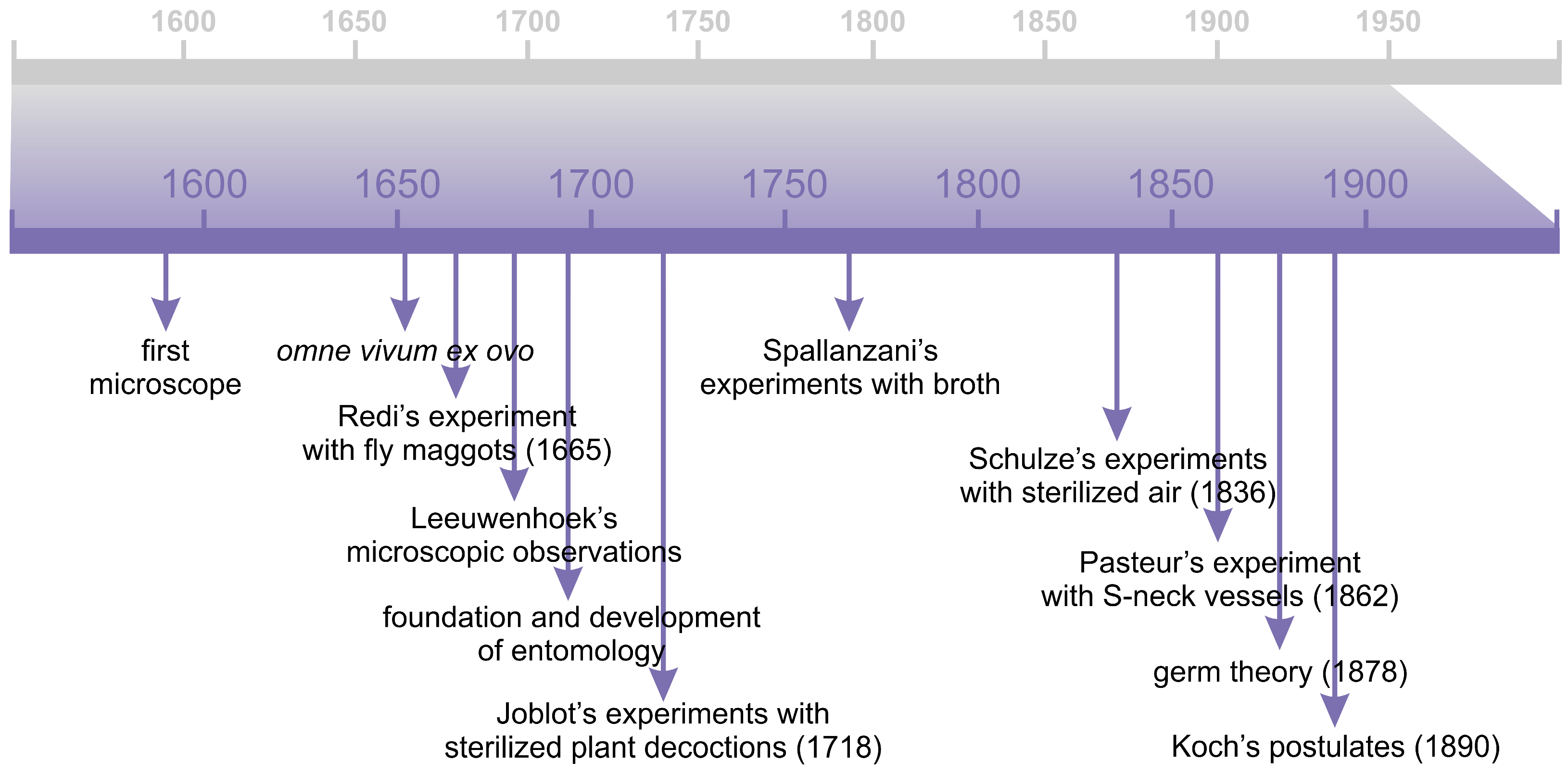
Figure 1. Timeline of the most significant discoveries related to the theory of spontaneous generation and germ theory.
Applsci 15 11376 g001
Figure 2. Timeline of the most significant discoveries shaping our understanding of the roles of “bad germs” and “good germs” in nature.
Applsci 15 11376 g002
Figure 3. Timeline of the most significant discoveries from the early period of the human gut microbiota research.
Applsci 15 11376 g003
Figure 4. Timeline of the most significant discoveries related to the theory of autointoxication and the probiotic properties of microorganisms.
Applsci 15 11376 g004
Figure 5. Timeline of the most significant discoveries from the “era of phages”.
Applsci 15 11376 g005
Figure 6. Timeline of the most significant discoveries from the “era of antibiotics”.
Applsci 15 11376 g006
Disclaimer/Publisher’s Note: The statements, opinions and data contained in all publications are solely those of the individual author(s) and contributor(s) and not of MDPI and/or the editor(s). MDPI and/or the editor(s) disclaim responsibility for any injury to people or property resulting from any ideas, methods, instructions or products referred to in the content.