Article

Antifungal and Cytocompatible Properties of Juglans regia Extract for Dental Applications: A Novel Approach Against Oral Candida Infections

by
Diego Garcia Miranda
1,2,3,4,†,
Lucas de Paula Ramos
1,5,†,
Flavia Villaça Morais
6,7,
Letícia Silva Nascimento
5,
Isadora Abdalla Ferreira
5,
Bruno Martini Guimarães
5,
Fernanda Malagutti Tomé
7,
Flavia Pires Rodrigues
8 and
Florence Carrouel
1,*
1
Laboratory “Health Systemic Process” (P2S), UR4129, Faculty of Medicine Laennec, University Claude Bernard Lyon 1, University of Lyon, 7 rue Guillaume Paradin, 69008 Lyon, France
2
Multimaterials and Interfaces Laboratory (LMI), CNRS UMR 5615, University Claude Bernard Lyon 1, University of Lyon, 6 rue Victor Grignard, 69622 Villeurbanne, France
3
Department of Biosciences and Oral Diagnosis, Institute of Science and Technology, São Paulo State University, Avenida Francisco José Longo 777, São José dos Campos 12245-000, SP, Brazil
4
School of Medicine, Anhembi Morumbi University, Avenida Deputado Benedito Matarazzo 6070, São José dos Campos 12230-002, SP, Brazil
5
School of Dentistry, Federal University of Alfenas—UNIFAL, R. Gabriel Monteiro da Silva, 700—Centro, Alfenas 37130-001, MG, Brazil
6
Cellular and Molecular Fungal Biology Laboratory, Research and Development Institute, University of Vale do Paraíba, São José dos Campos 12244-000, SP, Brazil
7
Department of Health Sciences, Paulista University UNIP, Rod. Pres. Dutra, km 157.5—Limoeiro, São José dos Campos 12240-420, SP, Brazil
8
Faculty of Medicine and Health, School of Dentistry, Oral Biology, University of Leeds, Worsley Building, Woodhouse, Leeds LS2 9JT, UK
*
Author to whom correspondence should be addressed.
These authors contributed equally to this work.
Appl. Sci. 2025, 15(19), 10531; https://doi.org/10.3390/app151910531
Submission received: 3 September 2025 / Revised: 23 September 2025 / Accepted: 28 September 2025 / Published: 29 September 2025
(This article belongs to the Special Issue Advanced Dental Materials and Its Applications)

Abstract

Featured Application

This study addresses the preclinical analysis of the use of Juglans regia glycolic extract as a possible pharmacological agent in dental materials to combat oral candidiasis.

Abstract

Oral Candida infections result from the overgrowth of this opportunistic fungus in the oral mucosa. Risk factors include immunosuppression, antibiotic or corticosteroid use, xerostomia, and conditions such as diabetes mellitus. Fungal resistance in Candida spp. has become a significant challenge, especially due to the excessive use of conventional antifungals such as azoles, echinocandins, and polyenes. Therefore, this study aims to determine the spectrum of antifungal activity of Juglans regia and assess its cytotoxicity on hepatocytes. Thus, a broth microdilution test was conducted according to the CLSI (M27-A3) guidelines. After initial screening, biofilm tests were conducted using the crystal violet (CV) and metabolic activity assays (MTT). Cytotoxicity was evaluated on human hepatocytes (HEPG2). The J. regia extract showed dose-dependent antifungal activity. At a concentration of 200 mg/mL, inhibition was greater according to the CV test in Candida albicans (31%) and Candida tropicalis (30.4%), while the MTT assay indicated a greater reduction in viability in C. albicans (61%) and C. glabrata (53.5%). At 100 mg/mL, C. albicans remained sensitive (37.7% CV; 71.6% MTT), while C. krusei and C. dubliniensis showed low viability by MTT (18.4% and 11.8%, respectively). At 50 mg/mL, C. albicans remained affected (74.3% MTT), but C. krusei, C. dubliniensis, and C. guilliermondii showed the lowest viability values (≤19.4% MTT), suggesting greater sensitivity to lower concentrations. These results indicate variation in susceptibility between species, with C. albicans being consistently inhibited, while C. krusei and C. dubliniensis responded better to lower doses. The extract showed cytocompatibility when applied to human hepatoma cells (HEPG2) and therefore holds significant potential for developing a new therapeutic approach.

1. Introduction

Antifungal resistance has emerged as a major global public health challenge, with increasing prevalence and diverse underlying mechanisms. The multifactorial problem includes genetic mutations in drug targets (such as ERG11, a gene encoding lanosterol 14α-demethylase, an enzyme involved in ergosterol biosynthesis in Candida yeasts), overexpression of drug efflux via ABC and MFS transporters, and alterations in the composition and integrity of the fungal cell membrane [1,2].
The problem is further aggravated by the indiscriminate use of antifungals in both clinical and agricultural settings, while the therapeutic arsenal remains limited and the development of new drug classes progresses slowly [1,2,3]. Oral candidiasis is more than a local discomfort; it may impair feeding, reduce quality of life, and serve as an early indicator of systemic disease. In severe cases, it increases the risk of invasive candidiasis, a life-threatening infection associated with high morbidity and mortality [4]. Current therapies rely heavily on azoles and echinocandins, but resistance is increasingly reported, especially in Candida glabrata and emerging multidrug-resistant pathogens such as Candida auris [5,6,7,8]. These trends highlight the urgent need for alternative, safe, and effective antifungal strategies tailored to oral health.
In this context, phytotherapy offers promising opportunities. Natural products are known to provide diverse bioactive compounds with antimicrobial properties, often with fewer side effects and lower risks of resistance development compared with synthetic drugs [1,9]. Juglans regia (Walnut), belonging to the Juglandaceae family and traditionally used in folk medicine, represents a particularly interesting candidate. Extracts from its leaves, bark, and fruits are rich in polyphenols and flavonoids such as quercetin, myricetin, and avicularin, compounds with documented antimicrobial and antioxidant activities [10,11,12,13,14,15,16,17,18,19,20]. In traditional dental hygiene practices, especially in South Asia, walnut bark and leaves have been used for oral cleaning and to alleviate gum disease [14,17].
Recent studies have confirmed the antimicrobial potential of J. regia against various bacterial and fungal species, including drug-resistant strains. For example, Sytykiewicz et al. [18] demonstrated that methanolic and aqueous extracts prepared from J. regia leaves exhibited significant antifungal activity against Candida albicans isolates. Raja et al. [19] further reinforced the anti-Candida activity of the ethanolic root extract, which caused marked morphological alterations in yeast cells and suppressed key virulence factors essential for pathogenicity. In addition, Jafer and Naser [20] reported that both aqueous and methanolic extracts displayed antimicrobial activity against pathogenic yeasts and bacteria, with the methanolic extract consistently more potent across all tested microorganisms. These examples highlight the broad antifungal potential of J. regia and support further exploration of its pharmacological properties.
Thus, the objective of this study was to evaluate the antifungal activity of a glycolic extract of J. regia against six clinically relevant Candida species, as well as to evaluate its cytocompatibility on human hepatoma cells (HEPG2). The overarching objective was to explore its potential as a novel antifungal agent for dental applications, particularly in preventive and therapeutic formulations targeting oral candidiasis.

2. Materials and Methods

2.1. Chemical Reagents

Phosphate buffer saline (PBS) (code: P2272, Sigma-Aldrich®, St. Louis, MO, USA), Methanol (CAS nº: 67-56-1, purity: 99.8%, Synth®, Diadema, Brazil), RPMI 1640 medium with glutamine, without bicarbonate and phenol red indicator (Himedia, Mumbai, India), MOPS [3-(N-morpholino) propane sulfonic acid] pH 7.0 ± 0.1 (Sigma-Aldrich, St. Louis, MO, USA), Sabouraud Dextrose agar (Difco Laboratories, Detroit, MI, USA), Violet Crystal (CV) (1% v/v) (Synth, Diadema, Brazil), acetic acid (Synth, Diadema, Brazil), Eagle’s medium modified by Dulbecco (DMEM; LGC Biotechnology®, Cotia, Brazil), Fetal Bovine Serum (FBS; Invitrogen®, New York, NY, USA), 7-Hidróxi-3H-fenoxazin-3-ona-10-óxido (Resazurin) (CAS nº: 62758-13-8, code: R1017, Sigma-Aldrich®, St. Louis, MO, USA), and ethanol (CAS nº: 64-17-5, purity:99.5%, Synth®, Diadema, Brazil) were used in this study.

2.2. Equipment

A Class II biological safety cabinet (Veco®, biosseg-06, Sumaré, São Paulo, Brazil), analytical balance (Mettler Toledo®, Balance XPR106DUH/A, Columbus, OH, USA), type I ultrapure water purification system (Allcron®, direct-Pure® Genie, São Paulo, Brazil), autoclave (Cristofoli Biossegurança®, Vitale21, Campo Mourão, Parana, Brazil), Phmeter (Digimed®, DM-20, São Paulo, Brazil), CO2 incubator (Sanyo®, MCO-19AIC(UV), Osaka, Japan), water bath precision (TermoFisher Scientific®, TSGP02, Waltham, MA, USA), refrigerated centrifuge (Labnet®, HEREMLE Z300®, Madrid, Spain), inverted microscope (Ziess®, Axiovert 40C, Jena, Thuringia, Germany), and spectrophotometer (Lonza Biotek®, ELX808LBS, Winooski, VT, USA) were used in this study.

2.3. Plant Extract

Glycolic extract of J. regia (Walnut) (CAS n°: 84012-43-1; lot: PRODO18746, Mapric Greentech Company®, São Paulo, Brazil) was created by percolation using a mixture of 20% (w/w) plant leaf material. All physical−chemical characterizations (HPLC and MALDI-TOF) were reported in Miranda et al. [15], revealing the presence of three major compounds: Gallic acid, Regiolona, and Miquelianina, detected at m/z 170.12, 231.25, and 440.1, respectively.

2.4. Fungal Strains

Antifungal activity was evaluated with the following reference strains (ATCC—American Type Culture Collection): Candida albicans (ATCC 18804), Candida dubliniensis (ATCC MYA 646), Candida glabrata (ATCC 9030), Candida guilhermondii (ATCC 6260), Candida krusei (ATCC 6258), and Candida tropicalis (ATCC 13803). The strains were seeded on Sabouraud dextrose agar for reactivation in a bacteriological incubator at 37 °C and kept for 24 h.

2.5. Antimicrobial Activity in Planktonic Cultures

The minimum inhibitory concentration (MIC) and minimum microbicidal concentration (MMC) of the extract were determined by the broth microdilution method, according to the Clinical and Laboratory Standards Institute guidelines (CLSI—M27-A3 for yeast). This inoculum was prepared from the cultures incubated at 37 °C for 24 h in a sterile physiological solution (0.9% NaCl) and standardized to 106 CFU/mL. Subsequently, 10 dilutions of the extract were prepared in 96-well microplates using RPMI 1640 medium buffered with MOPS for yeasts, generating values of 100, 50, 25, 12.5, 6.25, 3.12, 1.56, 0.78, 0.39, and 0.19 mg/mL of the extract. Then, 100 µL of the standardized inoculum was added in each well, followed by 24 h of incubation. MIC was determined at the last well showing no turbidity. MMC was determined by inoculating 10 μL of wells aliquots on Sabouraud Dextrose agar for yeast.

2.6. Synthesis of Biofilm Matrix

Biofilms were performed in 96-well microplates from standard suspensions containing 107 CFU/mL incubated at 37 °C under agitation at 75 rpm for 48 h, with medium replacement after 24 h. After incubation, J. regia extract was placed in contact with the biofilms. Nystatin 100,000 IU/mL was used as the positive control, and sterile saline (0.9% NaCl) was used as the negative control. For all treatments, biofilms were exposed to the extract for 5 min, and the final volume in each well was 200 µL. Subsequently, solutions were discarded, and biofilm was washed twice with sterile saline (0.9% NaCl) [21,22].

2.7. Crystal Violet (CV) Staining

Biofilms were fixed with 200 µL of methanol per 20 min. Then, the solution was removed, and the plate was incubated at 37 °C for 24 h for drying. Crystal violet (1% v/v) was added for 5 min, and then the dye was removed. The wells were washed with sterile physiological solution (NaCl 0.9%) and 33% acetic acid. The plate was read in a microplate reader at a wavelength of 570 nm, and optical densities (ODs) were converted to biofilm biomass [21].

2.8. Metabolic Activity of Microorganisms by MTT Assay

MTT solution (0.5 mg mL, 100 µL/well) was added to biofilms and incubated for 1 h at 37 °C (Sigma Aldrich, Saint Louis, MI, USA). After incubation, the solution was removed, and 200 µL of dimethyl sulfoxide (DMSO; Sigma Aldrich, Saint Louis, MI, USA) was added into the wells. The plates were incubated again at 37 °C for 10 min and placed on the stirrer (Micro plate shaker MIX-1500, Miulab, Hangzhou, China), under constant agitation for 10 min. The plates were read on a microplate reader (Lonza Biotek ELX808LBS, Winooski, VT, USA) at a wavelength of 570 nm, and ODs were converted to the metabolic activity of the microorganism. Cell viability was expressed as a percentage of the untreated control, which was set to 100% [22].

2.9. Cell Viability by Resazurin Assay

Human hepatoma cells (HEPG2; obtained from the Bank of Cells of Rio de Janeiro, Association Scientific Technical Paul Ehrlich (APABCAM—RJ)) were cultured in Dulbecco’s Modified Eagle’s Medium supplemented with 10% FBS, incubated at 37 °C and 5% CO2. The cytotoxicity assay was performed with 4 × 10⁴ viable cells, plated on a 96-well microplate, and incubated (37 °C and 5% CO2) for 24 h. After cell adhesion, the supernatant was removed to apply J. regia extract at concentrations of 50, 100, and 200 mg/mL for 5 min. The control group received only DMEM + 10% FBS treatment for the same period. Each experimental group was performed with n = 10 and two independent repetitions. Afterwards, the wells were washed twice with sterile saline (0.9% NaCl). Then, the plates were used for the cell viability test.
Metabolic activity was assessed using resazurin at 440 µM. For this, 50 µL of the resazurin suspension was added to each well of the microplate, followed by the addition of 450 µL of DMEM + 10% FBS. Incubation in the dark was carried out for 16 h, after which the plate was read in a spectrophotometer at a wavelength of 570 nm. The optical densities (ODs) obtained were converted into a percentage of cell viability using the following formula (Equation (1)):
% Metabolic activity = (OD treated group × 100)/average OD control group

2.10. Statistical Analysis

The obtained data were initially analyzed for normality using D’Agostino, Shapiro−Wilk, and Kolmogorov−Smirnov tests. Data showing normality were evaluated by ANOVA complemented by Turkey’s test. Data that did not show normality were evaluated by Kruskal−Wallis complemented by Dunn’s test. All analyses utilized a p-value of 0.0001.

3. Results

3.1. Antifungal Activity in Planktonic Cultures

The antifungal activity of J. regia extract against planktonic cultures of Candida species is summarized in Table 1. The MIC was 12.5 mg/mL for C. dubliniensis, C. guilhermondii, and C. tropicalis, indicating higher susceptibility of these species. In contrast, C. albicans, C. glabrata, and C. krusei presented lower sensitivity, with a MIC of 25 mg/mL.
The MMC was 25 mg/mL for most species, but higher values were required for C. albicans and C. dubliniensis (50 mg/mL).

3.2. Anti-Biofilm Action—Biomass

J. regia treatments at 50, 100, and 200 mg/mL reduced the biomass of C. albicans biofilms by 31.9%, 32.7%, and 31%, respectively (Figure 1). For C. tropicalis and C. guilliermondii, biomass reduction increased with concentration, reaching 30.4% and 27%, respectively, at 200 mg/mL, with values comparable or superior to nystatin. C. krusei and C. dubliniensis showed moderate but consistent reductions (≈21–28%), with the extract outperforming nystatin in the case of C. dubliniensis, as shown in Figure 1.

3.3. Anti-Biofilm Action—MTT (Metabolic Activity)

The metabolic activity of Candida biofilms was markedly reduced following treatment with J. regia extract (Figure 2). The strongest effect was observed against C. albicans, with reductions ranging from 61% to 74.3%, which were comparable or superior to nystatin (63.3%). C. glabrata also showed a consistent inhibition (≈53–57%), although slightly lower than the reference drug.
For C. tropicalis and C. guilliermondii, inhibition increased with concentration, reaching 44.9% and 51.3%, respectively, at 200 mg/mL, with the latter surpassing nystatin. In contrast, C. krusei and C. dubliniensis exhibited only modest responses (≤20% reduction), highlighting species-dependent variability.

3.4. Cell Viability by Resazurin Essay

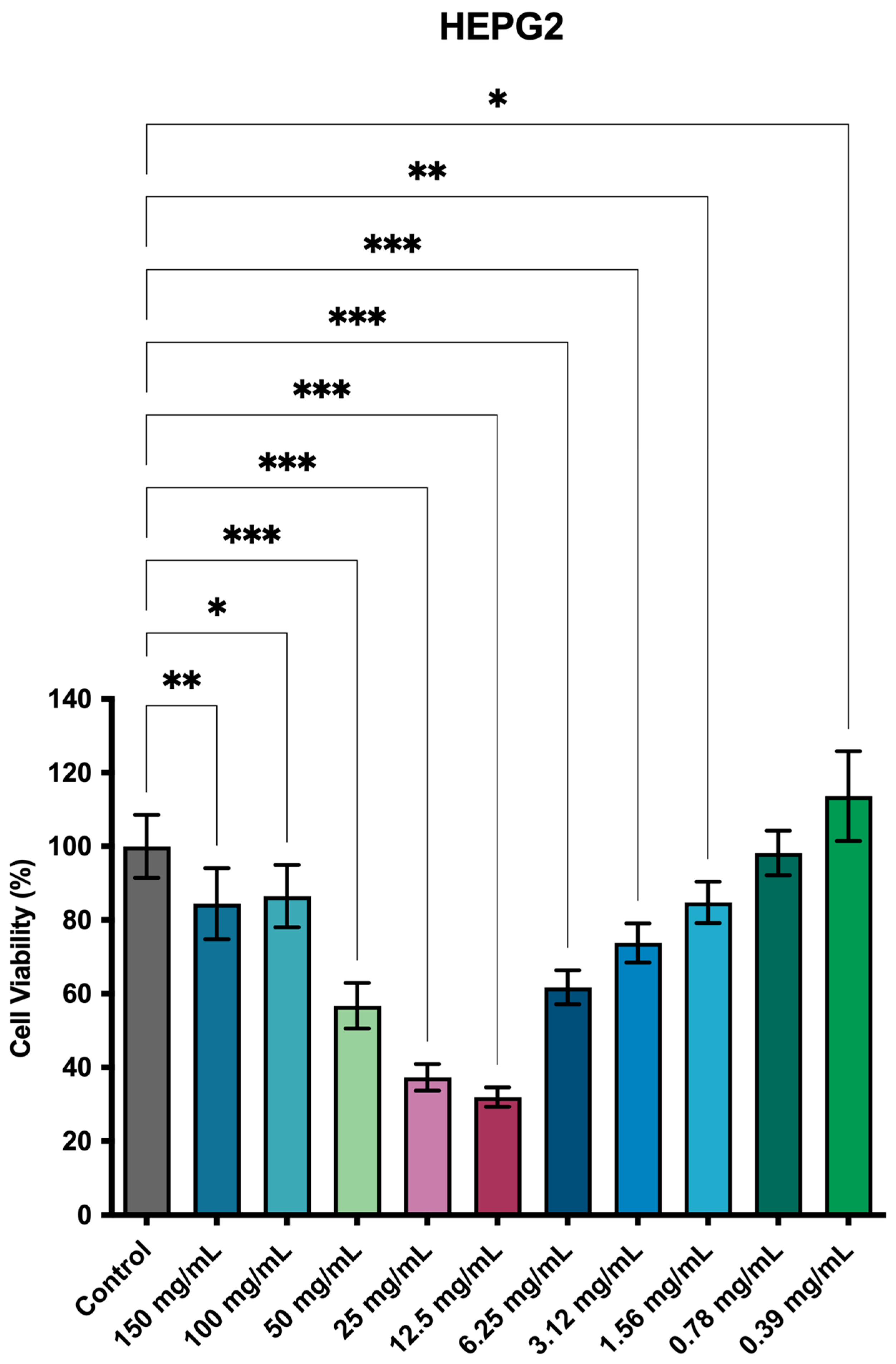
The cytocompatibility of Juglans regia extract was assessed on HEPG2 cells using the resazurin assay (Figure 3). The results revealed a non-linear, biphasic dose–response pattern. At intermediate concentrations (25–50 mg/mL), cell viability decreased substantially (≈39–54%), suggesting cytotoxic effects. At higher concentrations (100–200 mg/mL), however, viability partially recovered (≈80–85%), indicating possible adaptive cellular responses.
At lower concentrations (0.39–3.12 mg/mL), cell viability was maintained or even enhanced (≥100% at 0.39 mg/mL), consistent with a hormetic effect frequently described for plant-derived compounds. This profile suggests that the extract may exert stimulatory effects at very low doses, toxicity at intermediate doses, and relative tolerance at higher exposures.

4. Discussion

The present study investigated the antifungal and cytocompatible properties of a glycolic extract of Juglans regia against six clinically relevant Candida species. Our findings highlight both the potential and the complexity of using natural extracts in dental applications.
The antifungal activity of J. regia in planktonic cultures was species dependent. The MIC and MMC results showed that the antifungal effect of J. regia was species-dependent, with C. dubliniensis, C. guilliermondii, and C. tropicalis exhibiting higher susceptibility, while C. albicans, C. glabrata, and C. krusei required higher concentrations. These results are consistent with prior studies showing variable activity of J. regia extracts depending on the solvent used, plant part studied, and fungal strain tested [16,17,18,19,20]. The higher concentrations needed to eradicate C. albicans are noteworthy, given its prevalence in oral candidiasis [4]. This suggests that J. regia may act in a primarily fungistatic manner against certain species, a finding consistent with those reported by D’Angeli et al. [16] and Noumi et al. [17].
In this study, J. regia extract promoted modest reductions in biofilm biomass (≈21–32%), but a much stronger inhibition of metabolic activity, particularly in C. albicans (61–74%) and C. glabrata (≈54%). This indicates that the extract primarily affects cellular metabolism rather than disrupting the biofilm matrix. For instance, C. glabrata biofilms exhibited strong metabolic inhibition despite relatively modest biomass reductions, while C. dubliniensis showed the opposite profile, with notable biomass reduction but minimal metabolic inhibition. These results suggest that the extract targets distinct aspects of biofilm physiology, depending on the species. Previous reports support this interpretation, as J. regia may interfere with key metabolic pathways [23,24], and the sensitivity of each species appears linked to its intrinsic biofilm phenotype [25]. Taken together, these data highlight that J. regia extract impacts biofilm viability more strongly than structural mass, an important distinction for interpreting its antifungal potential. These findings reinforce the idea that J. regia does not act through a single mechanism, but rather exerts multifactorial effects depending on the fungal species and biofilm structure. This complexity may be advantageous, as multi-targeted agents are less likely to induce resistance compared to single-molecule drugs [1,9].
The cytocompatibility assays, based on OECD guideline 129, revealed a biphasic, hormetic response to J. regia extract, which represents one of the most intriguing findings of this study. At low concentrations (0.39–3.12 mg/mL), the extract stimulated cell viability, while intermediate concentrations (25–50 mg/mL) showed cytotoxic effects, and higher concentrations (100–200 mg/mL) partially restored viability. Such U-shaped dose–response curves are well-documented in toxicology and pharmacology, and are particularly common for plant-derived compounds [26]. Hormesis may reflect adaptive responses of mammalian cells, including the activation of antioxidant defense pathways or modulation of stress-response genes. In the case of J. regia, this phenomenon could be attributed to the chemical complexity of the extract, which contains diverse metabolites with potentially synergistic or antagonistic interactions. Although preliminary, these findings highlight the importance of conducting a thorough characterization of the bioactive compounds within the extract and elucidating their underlying mechanisms of action [27].
From a translational perspective, this non-linear profile highlights both opportunities and risks. On the one hand, the stimulatory effects at low doses may support the safe use of diluted formulations in preventive products such as mouth rinses. On the other hand, the cytotoxicity observed at intermediate concentrations underscores the importance of dose optimization and careful safety testing before clinical application.
When compared with conventional antifungals, J. regia extract showed efficacy comparable to nystatin against C. albicans and, in some cases, even superior activity against C. dubliniensis and C. guilliermondii. However, the extract was less effective against C. krusei, a species known for intrinsic resistance to azoles and reduced susceptibility to polyenes [5,6,7]. This variability emphasizes that J. regia is unlikely to replace conventional antifungals, but could serve as a complementary agent. Its potential use in preventive strategies, where complete eradication is not required but suppression of pathogenic overgrowth is beneficial, deserves further investigation [4].
This study presents several limitations. First, the study was conducted exclusively in vitro, and results may not translate directly to the oral cavity, where saliva, host immunity, and microbial interactions influence antifungal efficacy. Second, the short exposure time (5 min) was chosen to simulate a mouth rinse, but longer or repeated exposures may be necessary in clinical settings [4]. Third, the extract is chemically complex, and its active components were not directly identified in this study. Previous research suggests that flavonoids such as quercetin, myricetin, rutin, and avicularin contribute to antifungal effects [16,23], but their relative roles remain unclear. Fourth, the cytotoxicity results must be interpreted with caution due to the use of HEPG2 cells, which are immortalized hepatocytes in 2D monolayer configuration. Future studies should include multiple primary cell types relevant to oral application. Finally, a major limitation is that the antifungal concentrations required to inhibit Candida spp. (12.5–200 mg/mL) overlap with the concentration range showing cytotoxic effects in HepG2 cells (25–50 mg/mL), which restricts the direct translational potential of the extract.
Despite these limitations, our findings open promising perspectives. The extract could be incorporated into dental materials, varnishes, or mouth rinses designed for patients at risk of oral candidiasis, such as denture wearers, elderly individuals, or immunocompromised patients. Its multifactorial mode of action may reduce the likelihood of resistance development. Future studies should focus on the isolation and characterization of the active compounds, mechanistic studies exploring mitochondrial disruption, oxidative stress induction, or inhibition of efflux pumps, in vivo models of oral candidiasis to assess efficacy, bioavailability, and safety and formulation research to optimize delivery and stability in dental products.

5. Conclusions

Thus, the antifungal activity observed not only validates the traditional use of the plant in oral hygiene and health practices, but also suggested its potential as a source of bioactive secondary metabolites for novel antifungal agents. However, the effects were species-dose-dependent, with strong activity against C. albicans and C. glabrata, but limited action against C. krusei and C. dubliniensis biofilms. Moreover, given that the concentrations required for antifungal effects are relatively high compared to standard drugs (e.g., nystatin), further work is needed to isolate and characterize the active constituents, optimize formulations, and improve selectivity. Importantly, these results are limited to in vitro assays. Further research on the mechanisms of action, toxicity, and in vivo efficacy will be essential before considering any translational application. The biphasic profile observed also indicates that caution is required when extrapolating doses, as toxicity may vary depending on the concentration and cell type. While this non-linear response could represent both a challenge and an opportunity for the development of antifungal agents derived from J. regia, careful attention to the overlap between antifungal and cytotoxic concentrations will be essential to ensure translational feasibility.

Author Contributions

Conceptualization: D.G.M., L.d.P.R., F.V.M., F.M.T., F.P.R., and F.C.; methodology: D.G.M., L.d.P.R., F.V.M., L.S.N., I.A.F., B.M.G., F.M.T., F.P.R., and F.C.; validation: D.G.M., L.d.P.R., F.V.M., F.M.T., F.P.R., and F.C.; formal analysis: D.G.M., L.d.P.R., F.V.M., F.M.T., F.P.R., and F.C.; investigation: D.G.M., L.d.P.R., F.V.M., L.S.N., I.A.F., B.M.G., F.M.T., F.P.R., and F.C.; resources: D.G.M., L.d.P.R., F.V.M., F.M.T., F.P.R., and F.C.; data curation: D.G.M., L.d.P.R., F.V.M., L.S.N., I.A.F., B.M.G., F.M.T., F.P.R., and F.C.; writing—original draft preparation: D.G.M., L.d.P.R., F.V.M., L.S.N., I.A.F., B.M.G., F.M.T., F.P.R., and F.C.; writing—review and editing: D.G.M., L.d.P.R., F.V.M., F.M.T., F.P.R., and F.C.; visualization: D.G.M., L.d.P.R., F.V.M., F.M.T., F.P.R., and F.C.; supervision: D.G.M., L.d.P.R., F.M.T., and F.C.; project administration: D.G.M., L.d.P.R., F.V.M., F.M.T., F.P.R., and F.C.; funding acquisition: D.G.M., L.d.P.R., F.V.M., F.M.T., F.P.R., and F.C. All authors have read and agreed to the published version of the manuscript.

Funding

This research received no external funding.

Institutional Review Board Statement

Not applicable.

Informed Consent Statement

Not applicable.

Data Availability Statement

The raw data supporting the conclusions of this article will be made available by the authors upon request.

Conflicts of Interest

The authors declare no conflicts of interest.

References

  1. Perlin, D.S.; Rautemaa-Richardson, R.; Alastruey-Izquierdo, A. The global problem of antifungal resistance: Prevalence, mechanisms, and management. Lancet Infect. Dis. 2017, 17, e383–e392. [Google Scholar] [CrossRef] [PubMed]
  2. Gupta, A.K.; Venkataraman, M. Antifungal resistance in superficial mycoses. J. Dermatol. Treat. 2022, 33, 1888–1895. [Google Scholar] [CrossRef] [PubMed]
  3. van Rhijn, N.; Rhodes, J. Evolution of antifungal resistance in the environment. Nat. Microbiol. 2025, 10, 1804–1815. [Google Scholar] [CrossRef] [PubMed]
  4. Contaldo, M.; Di Stasio, D.; Romano, A.; Fiori, F.; Della Vella, F.; Rupe, C.; Lajolo, C.; Petruzzi, M.; Serpico, R.; Lucchese, A. Oral Candidiasis and Novel Therapeutic Strategies: Antifungals, Phytotherapy, Probiotics, and Photodynamic Therapy. Curr. Drug Deliv. 2023, 20, 441–456. [Google Scholar] [CrossRef] [PubMed]
  5. Espinel-Ingroff, A.; Pfaller, M.A.; Bustamante, B.; Canton, E.; Fothergill, A.; Fuller, J.; Gonzalez, G.M.; Lass-Flörl, C.; Lockhart, S.R.; Martin-Mazuelos, E.; et al. Multilaboratory study of epidemiological cutoff values for detection of resistance in eight Candida species to fluconazole, posaconazole, and voriconazole. Antimicrob. Agents Chemother. 2014, 58, 2006–2012. [Google Scholar] [CrossRef]
  6. Lima, S.M.F.; Sousa, M.G.A.C.; Freire, M.S.; Almeida, J.A.; Cantuaria, A.P.C.; Silva, T.A.M. Immune Response Profile against Persistent Endodontic Pathogens Candida albicans and Enterococcus faecalis In Vitro. J. Endod. 2015, 41, 1061–1065. [Google Scholar] [CrossRef]
  7. Rodríguez-Leguizamón, G.; Fiori, A.; Lagrou, K.; Gaona, M.A.; Ibáñez, M.; Patarroyo, M.A.; Van Dijck, P.; Gómez-López, A. New echinocandin susceptibility patterns for nosocomial Candida albicans in Bogotá, Colombia, in ten tertiary care centres: An observational study. BMC Infect. Dis. 2015, 15, 108. [Google Scholar] [CrossRef] [PubMed]
  8. Castanheira, M.; Messer, S.A.; Rhomberg, P.R.; Pfaller, M.A. Antifungal susceptibility patterns of a global collection of fungal isolates: Results of the SENTRY Antifungal Surveillance Program (2013). Diagn. Microbiol. Infect. Dis. 2016, 85, 200–204. [Google Scholar] [CrossRef] [PubMed]
  9. Jafari, E.; Andalib, S.; Abed, A.; Rafieian-Kopaei, M.; Vaseghi, G. Neuroprotective, antimicrobial, antioxidant, chemotherapeutic, and antidiabetic properties of Salvia Reuterana: A mini review. Avicenna J. Phytomed. 2015, 5, 10–16. [Google Scholar] [PubMed]
  10. Almeida, I.F.; Fernandes, E.; Lima, J.L.F.C.; Costa, P.C.; Bahia, M.F. Walnut (Juglans regia) leaf extracts are strong scavengers of pro-oxidant reactive species. Food Chem. 2008, 106, 1014–1020. [Google Scholar] [CrossRef]
  11. Fernández-Agullóa, A.; Pereira, E.; Freire, M.S.; Valentão, P.; Andradec, P.B.; González-Álvareza, J. Influence of solvent on the antioxidant and antimicrobial properties of walnut (Juglans regia L.) green husk extracts. Ind. Crops Prod. 2013, 42, 126–132. [Google Scholar] [CrossRef]
  12. Joshan, D.S.; Singhb, S.K. Investigational study of Juglans regia extract and quercetin against photoaging. Biomed. Aging Pathol. 2013, 3, 193–200. [Google Scholar] [CrossRef]
  13. Rather, M.A.; Dar, B.A.; Dar, M.Y.; Wani, B.A.; Shah, W.A.; Bhat, B.A.; Ganai, B.A.; Bhat, K.A.; Anand, R.; Qurishi, M.A. Chemical composition, antioxidant and antibacterial activities of the leaf essential oil of Juglans regia L. and its constituents. Phytomedicine 2012, 19, 1185–1190. [Google Scholar] [CrossRef] [PubMed]
  14. Farooqui, A.; Khan, A.; Borghetto, I.; Kazmi, S.U.; Rubino, S.; Paglietti, B. Synergistic Antimicrobial Activity of Camellia sinensis and Juglans regia against Multidrug-Resistant Bacteria. PLoS ONE 2015, 10, e0118431. [Google Scholar] [CrossRef] [PubMed]
  15. Miranda, D.G.; Carrouel, F.; Attik, N.; Araujo, G.F.; Dos Santos Lopes, N.F.; Marcucci, M.C.; Rodrigues, F.P.; Caires, G.A.; Vigerelli, H.; Godoi, B.H.; et al. Juglans regia and Pfaffia paniculata extracts: Implications for periodontal disease treatment and correlation with Alzheimer’s risk. Front. Cell Infect. Microbiol. 2025, 15, 1585438. [Google Scholar] [CrossRef] [PubMed] [PubMed Central]
  16. D’Angeli, F.; Malfa, G.A.; Garozzo, A.; Li Volti, G.; Genovese, C.; Stivala, A.; Nicolosi, D.; Attanasio, F.; Bellia, F.; Ronsisvalle, S.; et al. Antimicrobial, Antioxidant, and Cytotoxic Activities of Juglans regia L. Pellicle Extract. Antibiotics 2021, 10, 159. [Google Scholar] [CrossRef] [PubMed] [PubMed Central]
  17. Noumi, E.; Snoussi, M.; Hajlaoui, H.; Valentin, E.; Bakhrouf, A. Antifungal properties of Salvadora persica and Juglans regia L. extracts against oral Candida strains. Eur. J. Clin. Microbiol. Infect. Dis. 2009, 29, 81–88. [Google Scholar] [CrossRef]
  18. Sytykiewicz, H.; Chrzanowski, G.; Czerniewicz, P.; Leszczyński, B.; Sprawka, I.; Krzyżanowski, R.; Matok, H. Antifungal Activity of Juglans regia (L.) Leaf Extracts Against Candida albicans Isolates. Pol. J. Environ. Stud. 2015, 24, 1339–1348. [Google Scholar] [CrossRef]
  19. Raja, V.; Ahmad, S.; Irshad, M.; Wani, W.; Siddiqi, W.; Shreaz, S. Anticandidal activity of ethanolic root extract of Juglans regia (L.): Effect on growth, cell morphology, and key virulence factors. J. Med. Mycol. 2017, 27, 476–486. [Google Scholar] [CrossRef]
  20. Jafer, F.N.; A Naser, L. The Biological Activity of Aqueous and Methanolic Extracts of Juglans regia on Yeasts and Pathologic Bacteria. Arch. Clin. Microbiol. 2020, 11, 2405–2410. [Google Scholar] [CrossRef]
  21. de Sá Assis, M.A.; de Paula Ramos, L.; Abu Hasna, A.; de Queiroz, T.S.; Pereira, T.C.; Nagai de Lima, P.M.; Berretta, A.A.; Marcucci, M.C.; Talge Carvalho, C.A.; de Oliveira, L.D. Antimicrobial and Antibiofilm Effect of Brazilian Green Propolis Aqueous Extract against Dental Anaerobic Bacteria. Molecules 2022, 27, 8128. [Google Scholar] [CrossRef] [PubMed] [PubMed Central]
  22. Miranda, D.G.; Carrouel, F.; Silva, T.C.A.; Rozzatto, M.C.; Hasna, A.A.; Santos, C.E.R.; Morais, F.V.; de Oliveira, L.D.; de Paula Ramos, L. New Insights into Cutaneous Asepsis: Synergism between Pfaffia and Rosemary Extracts. Antibiotics 2024, 13, 226. [Google Scholar] [CrossRef] [PubMed] [PubMed Central]
  23. Al-Aboody, M.S.; Mickymaray, S. Anti-Fungal Efficacy and Mechanisms of Flavonoids. Antibiotics 2020, 9, 45. [Google Scholar] [CrossRef]
  24. Ibrahim Bhardwaj, N.; Puri, A.; Wadhwan, V.; Nangia, R.; Jahan, I. Antimicrobial effect of Juglans regia bark with commonly used antibiotics against initial colonizers of Plaque and Caries: A comparative study. J. Oral Maxillofac. Pathol. 2023, 27, 443–454. [Google Scholar] [CrossRef] [PubMed] [PubMed Central]
  25. Vitális, E.; Nagy, F.; Tóth, Z.; Forgács, L.; Bozó, A.; Kardos, G.; Majoros, L.; Kovács, R. Candida biofilm production is associated with higher mortality in patients with candidaemia. Mycoses 2020, 63, 352–360. [Google Scholar] [CrossRef] [PubMed]
  26. Calabrese, E.J. Hormesis mediates acquired resilience: Using plant-derived chemicals to enhance health. Annu. Rev. Food Sci. Technol. 2021, 12, 355–381. [Google Scholar] [CrossRef]
  27. Murray, K.O.; Clanton, T.L.; Horowitz, M. Epigenetic responses to heat: From adaptation to maladaptation. Exp. Physiol. 2022, 107, 1144–1158. [Google Scholar] [CrossRef] [PubMed] [PubMed Central]
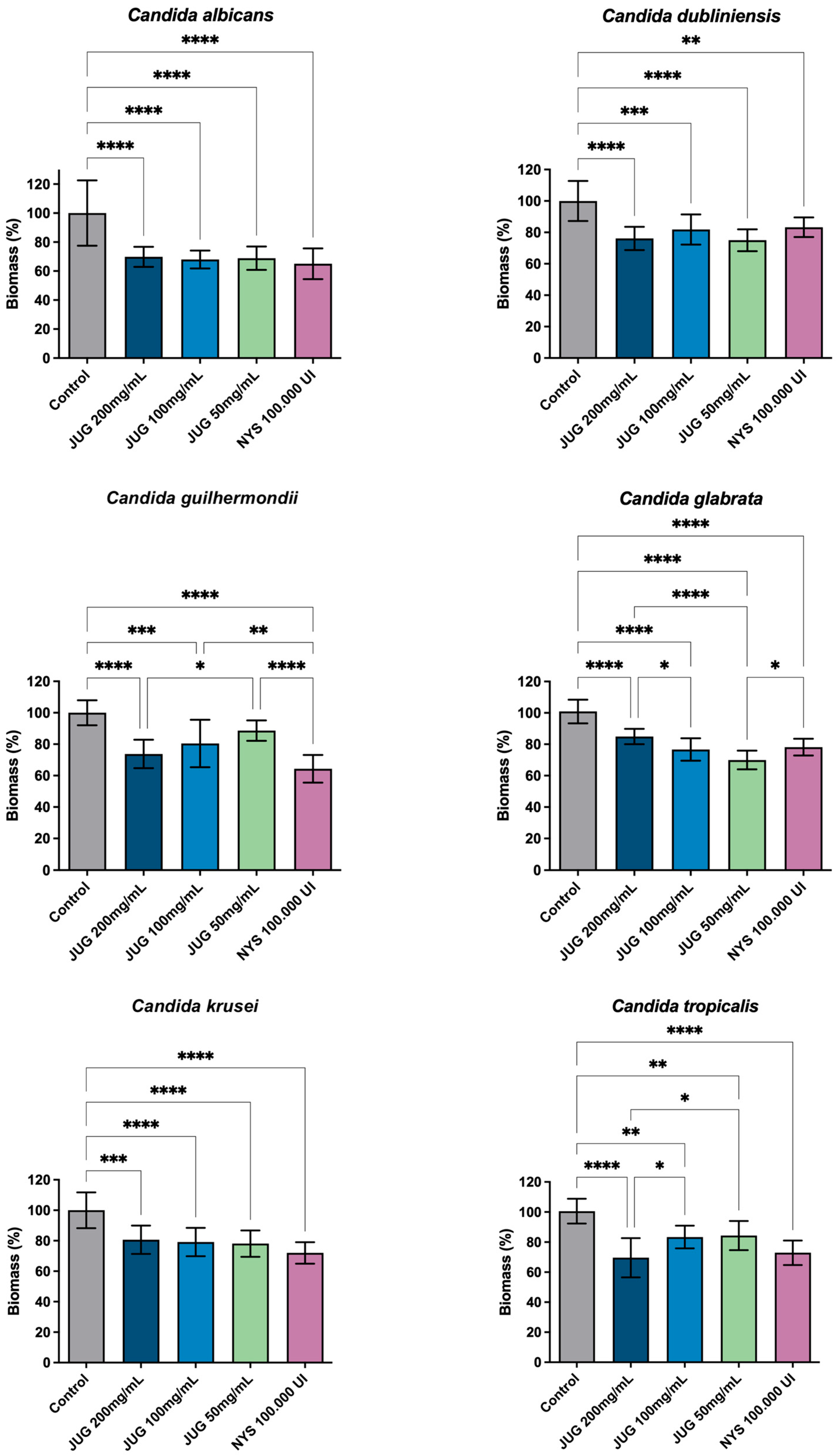
Figure 1. Reduction in the biomass of Candida spp. biofilms after treatment for 5 min with J. regia extract. Legend: (*) 0.033; (**) 0.002; (***) < 0.0002; (****) < 0.0001; JUG—Juglans regia extract; NYS—nystatine.
Applsci 15 10531 g001
Figure 2. Reduction in the metabolic activity of Candida spp. biofilms after treatment for 5 min with J. regia extract. Legend: (*) 0.033; (**) 0.002; (***) < 0.0002; (****) < 0.0001; JUG—Juglans regia extract; NYS—nystatine.
Applsci 15 10531 g002
Figure 3. Cytocompatibility of J. regia extract applied to human hepatocytes (HEPG2). Legend: (*) 0.033; (**) 0.002; (***) < 0.0002.
Applsci 15 10531 g003
Table 1. Antifungal activity of Juglans regia extract in planktonic cultures of Candida species.
Candida spp.MICMMC
C. albicans25 mg/mL50 mg/mL
C. dubliniensis12.5 mg/mL50 mg/mL
C. guilhermondii12.5 mg/mL25 mg/mL
C. glabrata25 mg/mL25 mg/mL
C. krusei25 mg/mL25 mg/mL
C. tropicalis12.5 mg/mL25 mg/mL
Legend: MIC—minimum inhibitory concentration; MMC—minimum microbicidal concentration.
Disclaimer/Publisher’s Note: The statements, opinions and data contained in all publications are solely those of the individual author(s) and contributor(s) and not of MDPI and/or the editor(s). MDPI and/or the editor(s) disclaim responsibility for any injury to people or property resulting from any ideas, methods, instructions or products referred to in the content.