Article

Compressive, Dimensional, and Antimicrobial Characteristics of 3D-Printed Acrylonitrile Butadiene Styrene (ABS) Following Dental Disinfection

1
School of Dentistry, IMU University, Bukit Jalil, Kuala Lumpur 57000, Malaysia
2
School of Dental Sciences, Universiti Sains Malaysia, Kota Bharu 16150, Malaysia
3
Adelaide Dental School, University of Adelaide, Adelaide, SA 5000, Australia
4
College of Dentistry and Dental Clinics, University of Iowa, Iowa City, IA 52242, USA
*
Author to whom correspondence should be addressed.
Appl. Sci. 2025, 15(19), 10428; https://doi.org/10.3390/app151910428
Submission received: 1 September 2025 / Revised: 23 September 2025 / Accepted: 24 September 2025 / Published: 25 September 2025

Abstract

Objective: To investigate the compressive, dimensional, and antimicrobial properties of thermoplastic Acrylonitrile Butadiene Styrene (ABS) 3D printed hollow blocks following chemical disinfection. Methods: Forty-two 3D printed ABS cubes were immersed in tap water, 0.12% chlorhexidine gluconate, 3% hydrogen peroxide, 5% sodium bicarbonate, 0.5% sodium hypochlorite, and commercial Potassium Caroate (Polident) for 28 days (4 cycles). Linear Outer (OM) and Inner (IM) dimensions, Root Mean Square (RMS), and mass were recorded before and after each immersion cycle. An additional set of seven cubes was untreated and served as a control. Fourier transform infrared spectroscopy (FTIR) was executed on one randomly selected sample from each group before and after immersion. Following the completion of the immersion cycles, an evaluation of compressive strength was performed using a universal testing machine. Subsequently, from each group, a single ABS cube was randomly selected for the introduction of Streptococcus mutans and Candida albicans, followed by a 14-day incubation period with Scanning Electron Microscope (SEM) evaluation. Results: There were no significant differences (p > 0.05) between OM, IM, and compressive strength measurements (F = 1.036, p = 0.443) across all groups. RMS values increased for OM and decreased for IM. Notably, cubes that underwent immersion in a 0.12% chlorhexidine gluconate solution displayed considerable changes in mass (p < 0.05), exhibiting a low positive correlation (ρ = 0.339). The 0.12% chlorhexidine gluconate group exhibited the emergence of a new OH peak (3000–3500) in FTIR, whereas the 3% hydrogen peroxide group experienced the disappearance of the styrene peak (1300–1500). Exposure of ABS to C. albicans and S. mutans demonstrated clear surfaces under SEM with 0.12% chlorhexidine gluconate, 0.5% sodium hypochlorite, and Polident. Conclusions: Disinfection with 0.5% sodium hypochlorite and Potassium Caroate produced minimal mechanical changes and resisted growth of C. albicans and S. mutans. 0.12% chlorhexidine gluconate and 3% hydrogen peroxide altered dimensional and chemical compositions of 3D printed ABS following disinfection.

1. Introduction

Commercially available 3D printing filaments used in fused deposition modelling (FDM)-based printing encompass a variety of materials, including acrylonitrile butadiene styrene (ABS), polycarbonates (PC), polylactic acid (PLA), and polyethylene terephthalate glycol (PETG) [1,2,3]. ABS has found extensive application in the realm of health sciences due to its exceptional mechanical properties and resistance to temperature. This includes the fabrication of diagnostic models, organ printing, and tissue scaffolding [4,5,6,7].
One of the key advantages of 3D printing dental prostheses is the ability to make hollow prostheses, thereby dramatically reducing the weight and improving comfort for the patient [8]. However, the effectiveness of such a hollow prosthesis hinges significantly on its ability to demonstrate robust dimensional stability and compressive characteristics during repeated masticatory forces and to effectively prevent the proliferation of microbes after undergoing disinfection processes.
The oral environment, due to its conducive conditions including temperature, nutrient availability, moisture, and surface suitability, hosts a diverse microflora, notably including Streptococcus and Candida species, capable of colonising intraoral prostheses [6]. Consequently, practising proper prosthetic hygiene and disinfection is pivotal to ensure the well-being of oral tissues and to extend the shelf life of the prosthetics [6]. Insufficient oral hygiene maintenance can lead to the accumulation of microorganisms on denture surfaces, contributing to the development of denture stomatitis [6]. Among various disinfection techniques, chemical disinfection stands out for its convenience, cost-effectiveness, and safety [6]. However, previous research has indicated that exposure to specific disinfectant solutions can compromise physical properties such as dimensions, compressive strength, and chemical composition of dentures [9,10,11]. Importantly, 3D printed hollow ABS does not possess the same attributes as conventional denture base materials like condensed acrylic resin. Therefore, an independent evaluation is necessary to determine the mechanical stability after disinfection and the antimicrobial effectiveness of ABS.
The incorporation of 3D printed ABS in prosthesis fabrication can be justified only if it displays satisfactory mechanical stability and the ability to hinder microbial growth post-disinfection. The present research endeavour aims to investigate the mechanical and antimicrobial properties of ABS after exposure to a variety of disinfecting agents.

2. Materials and Methods

This in vitro study did not involve any human or animal subjects and was therefore exempt from ethical considerations across all involved bodies. Consequently, informed consent was not applicable. The study methods are described in the following steps.
  • Fabrication of ABS cubes for in vitro assessment
  • Pre-Immersion Assessments
  • The Immersion Procedure
  • Post Immersion Assessments
  • Statistical Analyses

2.1. Fabrication of ABS Cubes for In Vitro Assessment

A sample size of 42 was determined in accordance with the effect size of 0.64, alpha of 0.05 and power of 0.80 [12]. The samples were divided into six groups (A–F) with seven specimens in each group and were immersed in different disinfectants. An additional group of 7 specimens labelled “no immersion” was fabricated to act as a control for compressive strength and microbial accumulation analysis, and was stored dry within an airtight container. An open-source CAD software (FreeCAD v0.18, Jurgen Riegel; Germany) was used to design the samples into 20mm hollow cubes (Figure 1A) as suggested by Sharma et al. [12] and Marichelyam et al. [13]. To prevent human error, identification codes were 3D printed onto one of the cube surfaces for each cube (Figure 1B). Based on print settings of 0.1mm layer height, 1.6mm wall thickness and 100% infill density, an open-source software Cura 4.6.1 (Ultimaker Inc., Geldermalsen, The Netherlands) (Figure 1C) was utilised for 3D printing preparation of the cubes. The cubes were 3D printed using Fused Deposition Modelling (FDM)-based printer (Ender 3, Creality; Shenzhen, China) (Figure 1D) with generic 1.75mm ABS filament (Fabbxile Technologies, Pulau Pinang, Malaysia). Based on the manufacturer’s instructions, a temperature of 230 °C was used for the nozzle and 105 °C for the printer bed. Excess material on the printed cubes was then trimmed [12].

2.2. Pre-Immersion Assessments

2.2.1. Linear Measurements for Dimensional Stability

Twelve outer measurements (OM) and eight inner measurements (IM) of the 3D printed ABS cubes were made for each sample (1–7) from each group (A–F) using a digital slide calliper (Mitutoyo Absolute 500, Mitutoyo, Kanagawa, Japan). Each group resulted in 84 OM and 56 IM [12,13]. The accuracy and precision were analysed using RMS in accordance with the International Organisation for Standardisation, ISO 5725-1, where the value 0 was considered perfect accuracy [12,14].
RMS = 1 n .   i = 0 n ( r i   - o i ) 2
  • ri: ideal value derived from CAD software
  • oi: obtained physical value

2.2.2. Mass Analysis

The mass of each ABS cube from Groups A to F was measured using an electronic balance (BSA423S, Sartorius AG; Göttingen, Germany). The mass was re-measured every seven days of immersion for evaluating changes in structural compositions, otherwise noticeable by a change in mass [13].

2.2.3. Fourier Transform Infrared Spectroscopy (FTIR)

Prior to immersion, infrared spectroscopy was performed on a random sample from each group using FTIR (Tensor 27, Bruker Corporation, Ettlingen, Germany) to record the pre-immersion transmittance results. Spectroscopic analysis was carried out using single channel transmittance with a resolution of 4/cm with 32 sample scans. Data were recorded from 4000/cm to 600/cm wavelength on an interferogram of 14,220 points.

2.3. The Immersion Procedure

Curylofo et al. [15] suggested a 25-day controlled immersion to simulate daily denture, whereas two other studies [16,17] suggested 4 weeks for changes to be observed. Thus, a 28-day controlled submission was adapted on the ABS cubes. Dental disinfectants and their respective concentration used were based on past studies and mixed according to the manufacturer’s instructions as follows:
  • Control: No immersion
  • Group A: Tap water [18]
  • Group B: 0.12% Chlorhexidine gluconate (Listerine; Pfizer, Manhattan, USA) [19]
  • Group C: 3% Hydrogen Peroxide (Polylab, Kuala Lumpur, Malaysia) [19]
  • Group D: 5% Sodium bicarbonate solution (Sweet Berry Nhd, Kota Bharu, Malaysia) [20]
  • Group E: 0.5% Sodium hypochlorite (Depex, Sun Jiang Sdn Bhd, Selangor, Malaysia) [21,22]
  • Group F: Commercial antibacterial denture cleanser consisting of Potassium Caroate (Potassium Monopersulfate) (Polident, GlaxoSmithKline, Brentford, UK) [23,24]
ABS cubes in all the sets were immersed in 250 mL of respective disinfectants within air-tight containers placed on a stationary bench at room temperature, 26 °C to 32 °C [21,22]. Every 7 days, the solutions were replaced with fresh solutions to minimise unwanted microbial growth and optimise chemical efficacy [25]. Before re-immersion, the cubes and containers were cleaned under running water and dried for 15 min [18], after which the new OM, IM, RMS values and mass were measured. The procedure was repeated four times over 28 days. After 28 days, FTIR was repeated on the same samples used previously for pre-immersion transmittance, and results were interpreted according to published reference values [26,27]. Compressive strength analysis and microbial accumulation tests were performed with “no immersion” as the positive control.

2.4. Post Immersion Assessments

All OM and IM of all samples from groups A to F were re-measured, and RMS was calculated to evaluate the material’s dimensional stability following every seven days of immersion. In order to record the post-immersion transmittance results, infrared spectroscopy was repeated after 28 days.

2.4.1. Compressive Strength Analysis

Compressive force was measured in MPa using a universal testing machine (Shimadzu; Kyoto, Japan) with a loading rate set at 1.0 mm/min. Three random points along the total length for both height and width of the ABS cubes were measured. The average value of each parameter was documented.

2.4.2. Microbial Accumulation Test

One sample was randomly chosen using the Python v3.x function “randint (0, 6)” from each experimental group (A–F, no immersion as negative control) for microbial accumulation of ATCC 10,231 strain [28] of C. albicans (KwikStik; Microbiologics, St Cloud, MN, USA) and ATCC 25,175 strain of S. mutans (KwikStik; Microbiologics, USA). Microbes were streaked onto a trypticase soy agar plate (Isolac, Shah Alam, Malaysia) according to manufacturer instructions and incubated at 37 °C for 48 h. The inoculation procedure was as follows. Tryptic soy broth (TSB) (Biolife, Monza, Italy) was prepared by mixing 18 g of TSB powder in 600 mL of distilled water. The broth was autoclaved at 121 °C for 15 min. The cultured microbes were suspended in 30 mL of the prepared tryptic soy broth and adjusted to match the turbidity of 0.5 McFarland unit (equivalent to 1.5 × 10⁸ CFUs/mL). The cubes from each experimental group were placed within individual sterile laboratory wells and inoculated with 5 mL of microbial broth. They were then incubated at 37 °C for 14 days.

2.4.3. Scanning Electron Microscopy (SEM)

After 14 days, the ABS cubes were prepared for SEM according to the protocol proposed by Fischer et al. [29]. The microbial broth was removed, and the ABS cubes were washed with Phosphate-Buffered Saline (PBS) to remove non-adherent fungi before being fixed in 10% formalin by slow dispersion of the fixative agent. The cubes were then dehydrated in ascending grades of ethanol by subsequent exchanges of the following dilutions: once in 25% ETOH for five minutes, once in 50% ETOH for five minutes, once in 75% ETOH for five minutes, once in 95% ETOH for five minutes, and 100% ETOH for 10 min three times. The samples were further dried in hexamethyldisilane (HMDS) (Sigma–Aldrich; Merck KGaA, Darmstadt, Germany) as follows: once in 1:1 HMDS:ETOH for 10 min and twice in 100% HMDS for 10 min at room temperature, after which excess liquid was wicked with filter paper and allowed for four hours of drying at room temperature. The cubes were then observed using a scanning electron microscope (TM3000; Hitachi, Tokyo, Japan) set at magnification of ×200, ×500, ×1000 and ×2500.

2.5. Statistical Analyses

Statistical software (SPSS v24.0, IBM Corp., Chicago, IL, USA) was used to perform all statistical analyses. Normality was evaluated with the Shapiro–Wilk and Kolmogorov–Smirnov tests. Analysis of OM and IM was conducted using the Kruskal–Wallis one-way test, whereas the mass change per group over the course of 28 days of immersion was analysed using Pearson’s Correlation Coefficient.

3. Results

The assessment of inter-rater reliability involved two reviewers who compared 20 randomly selected linear measurements (12 for OM and 8 for IM). The outcome yielded a K value of 0.99 (Cronbach’s α = 0.99), indicating a high level of agreement between the raters.

3.1. Linear Measurements (OM, IM, and RMS)

Over the course of the 28-day immersion period, there were no significant alterations observed in either OM (Table 1) or IM (Table 2), with p > 0.05. Nonetheless, there were indications of minor expansion and contraction events, as evidenced by irregular mean and standard deviation (SD) values across five measurement intervals during the immersion cycle.
The analysis of accuracy using the RMS values revealed consistent findings across all groups. Specifically, there was an observed rise in OM accuracy, shifting from 7.92 ± 0.13 to 9.08 ± 0.37. Conversely, for IM, a decline in accuracy was noted, transitioning from 5.65 ± 0.50 to 5.15 ± 0.39. In the context of RMS interpretation, a value of 0 signifies perfect accuracy, and values approaching 0 are indicative of higher overall accuracy [12]. A comprehensive overview of the detailed RMS values is provided in Supplementary File.

3.2. Mass Analysis

Throughout the 28 days of immersion, the mass analysis showed a negligible Pearson correlation value (<0.30) in all sets, except Set B, which revealed significant changes (p < 0.05) with a low positive correlation (=0.34) (Table 3).

3.3. Infrared Spectroscopy

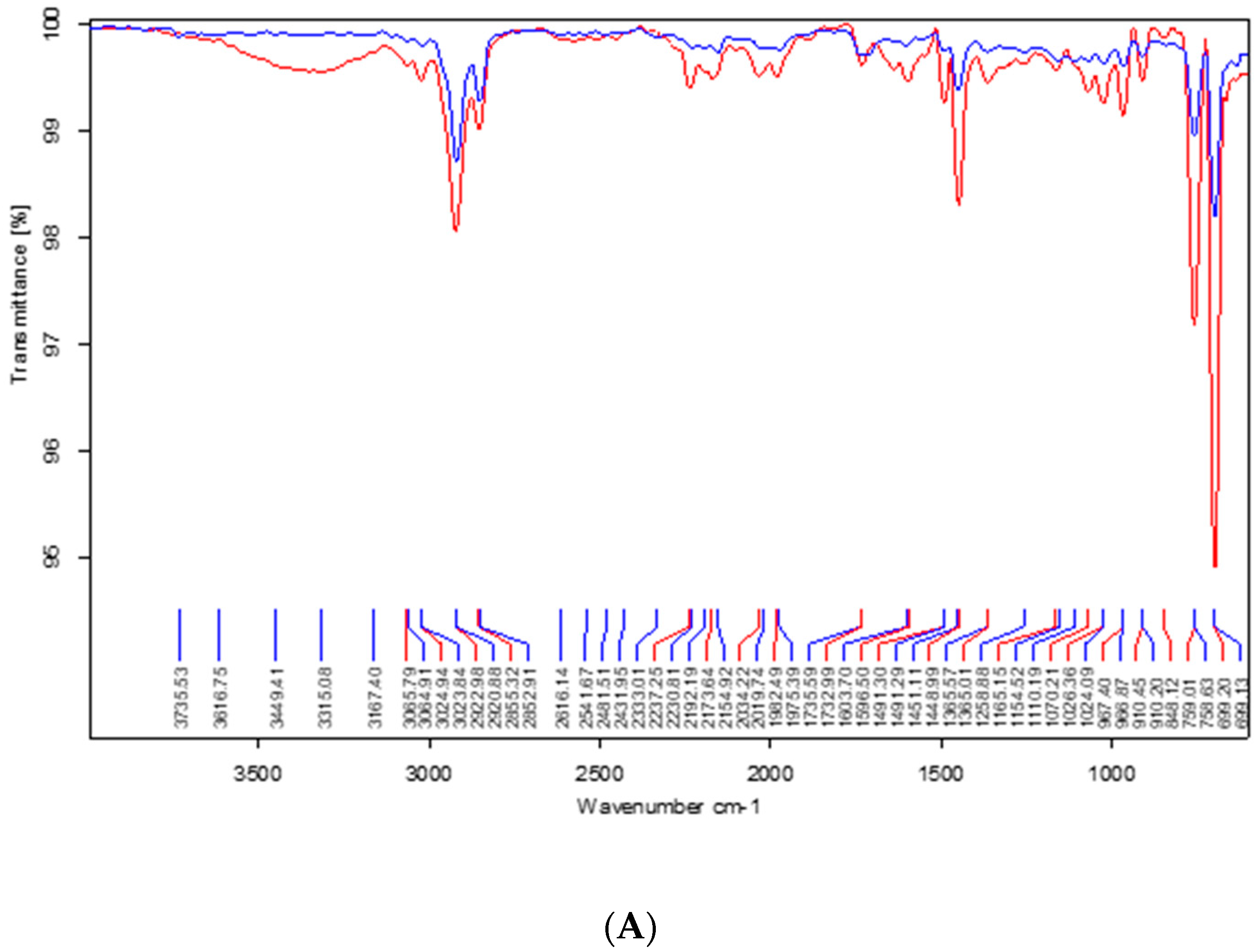
ABS, a composite comprised of Acrylonitrile, Butadiene, and Styrene, exhibited distinct bands in its baseline spectra across all pre-immersion samples. Notably, the presence of aromatic combination bands potentially indicated styrene double bonds, as was highlighted by bands within the 1300–1500 cm−1 range (Figure 2A). Additional peaks within the 2000–2500 cm−1 range corresponded to the nitrile group (C ≡ N), while an absorption band at 2500–3000 cm−1 suggested the existence of aromatic C-H bonds, aliphatic bond vibrations, and CH2 groups.
Aside from groups B and C, no substantial shifts were discerned in post-immersion peaks across the remaining groups, in contrast to their pre-immersion counterparts. However, in group B, new peaks emerged post-immersion at 3000–3500 and 1000–1100 (Figure 2B), whereas in group C, the disappearance of the peak at 1300–1500 (Figure 2C) suggested a loss of styrene subsequent to immersion in a hydrogen peroxide solution. All the spectroscopic charts are provided in Supplementary File.

3.4. Compressive Strength Analysis

Group F demonstrated the highest mean compressive strength (26.469 ± 1.435). The one-way ANOVA for compressive strength suggested a non-significant difference (F = 1.036, p = 0.443) among the groups. A detailed compressive strength report is presented in Table 4.

3.5. Scanning Electron Microscopy (SEM)

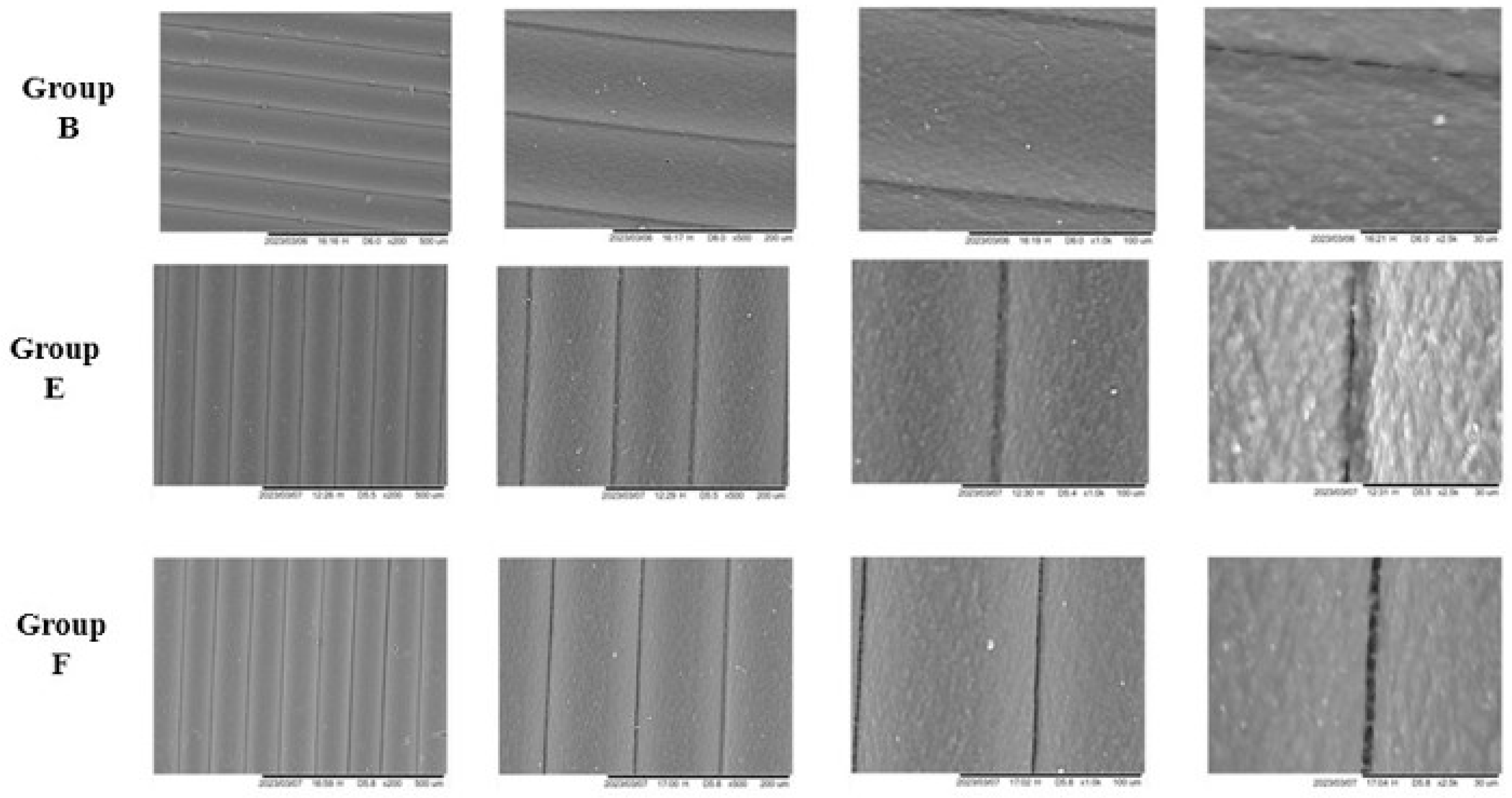
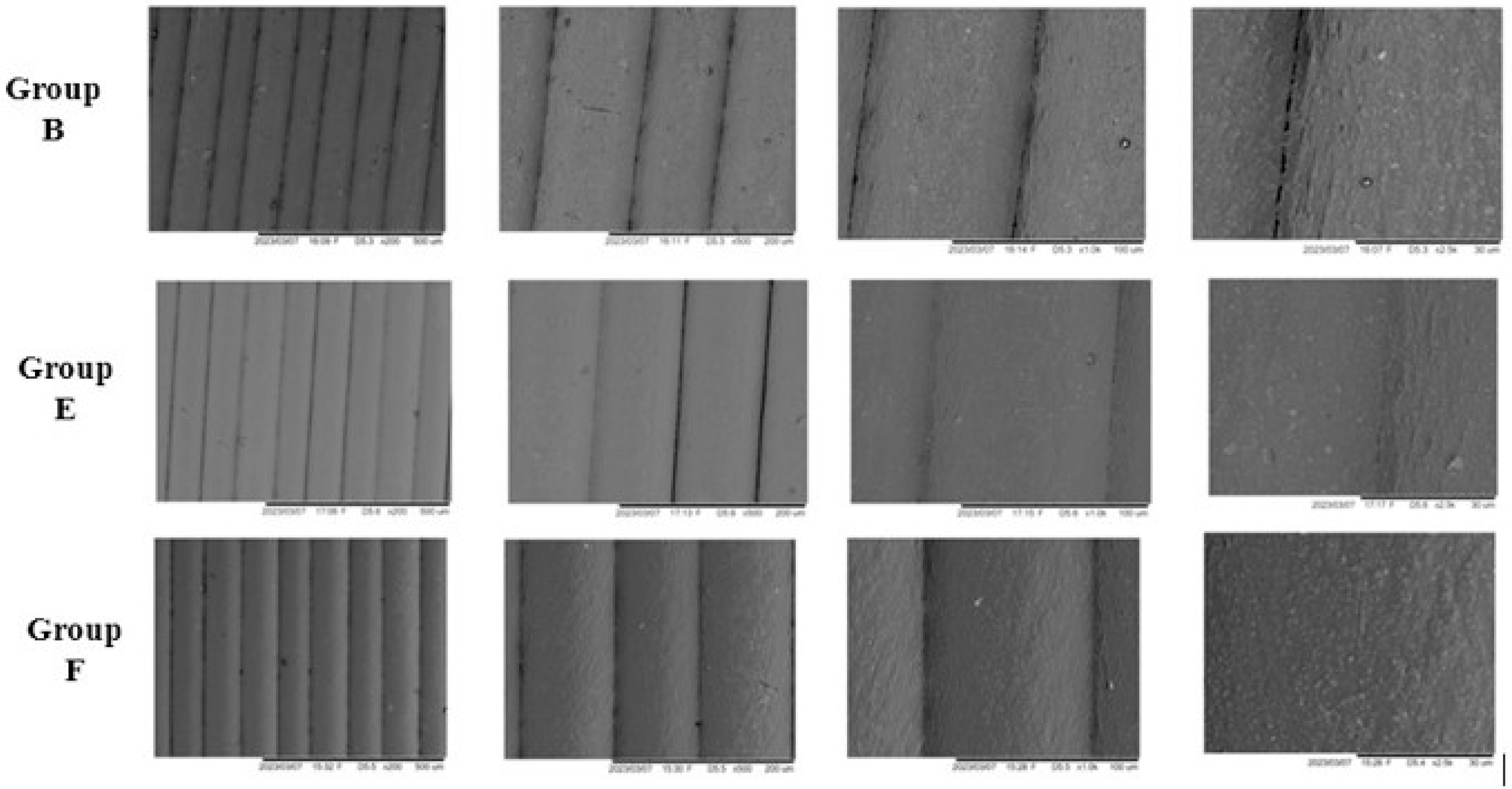
SEM analyses revealed clean surfaces of ABS cubes in group B (0.12% chlorhexidine gluconate), group E (0.5% sodium hypochlorite) and group F (Polident) when inoculated in C. albicans (Figure 3) and S. mutans (Figure 4).
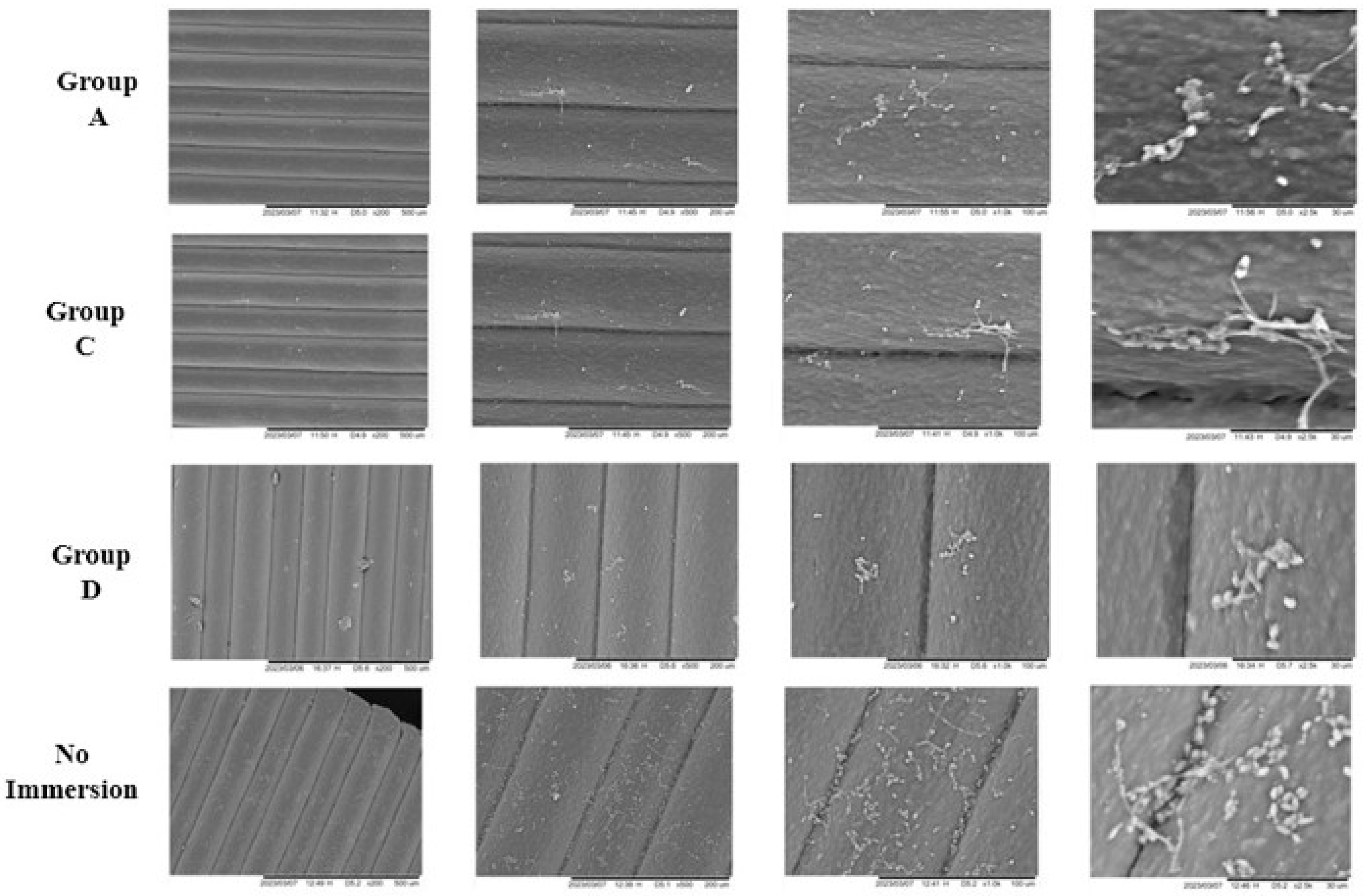
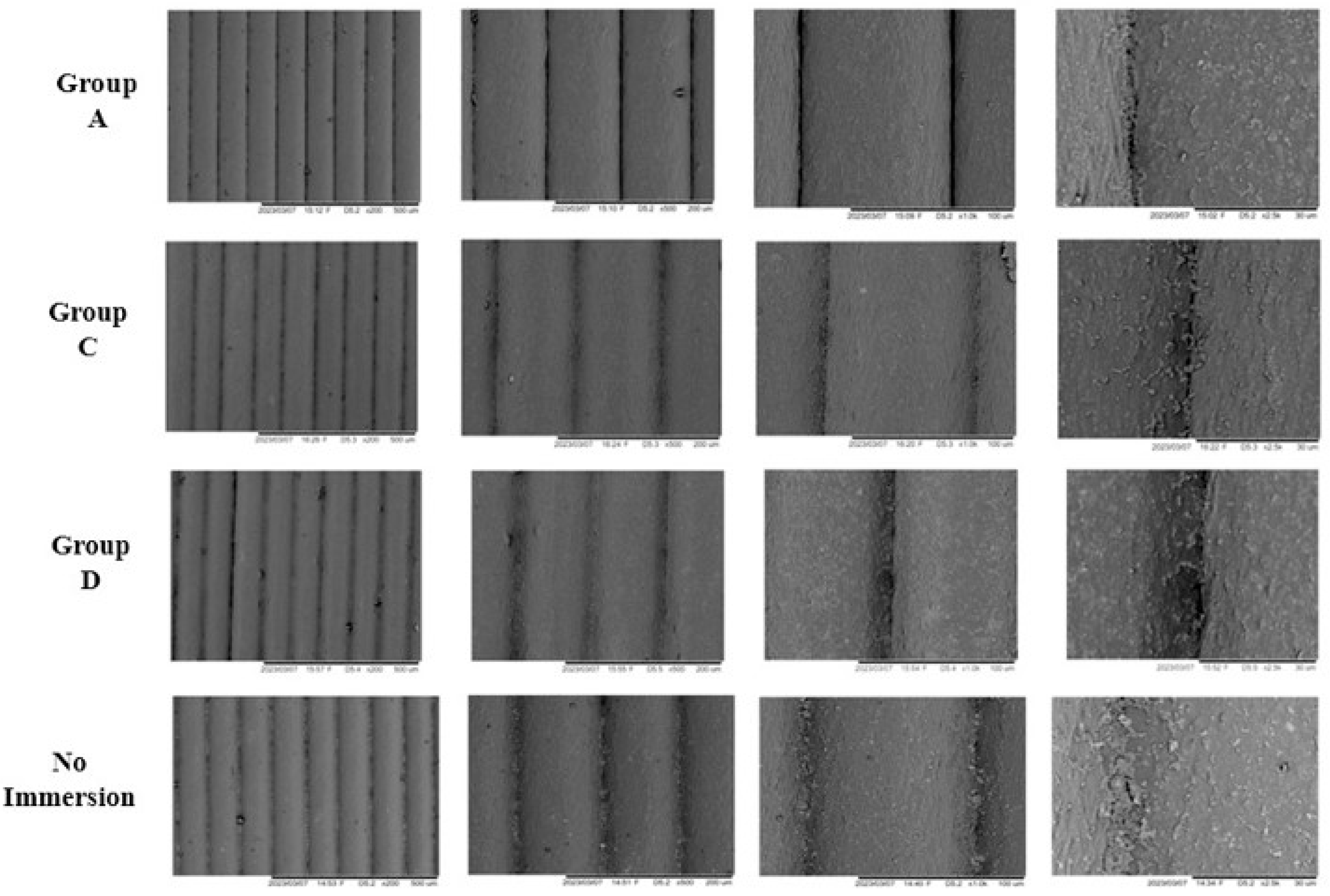
SEM examinations demonstrated distinct indications of colonisation by C. albicans and S. mutans in multiple experimental groups, including Group A (tap water), Group C (3% Hydrogen Peroxide), Group D (5% sodium bicarbonate), and the control group devoid of immersion. Concerning C. albicans (Figure 5), the “no immersion” group displayed the most even colonisation across all dimensions of the cube’s surface. The prevalent forms of C. albicans were pseudo-hyphae and yeast cells, frequently arranging into branched chains of elongated structures with constrictions at septal junctions with round to oval morphologies. In the context of S. mutans (Figure 6), Groups A, C, and D showcased a relatively more dispersed arrangement, whereas the “no immersion” set exhibited the highest degree of colonisation, revealing aggregated and partially detaching cocci embedded within amorphous extracellular matrix-like structures.

4. Discussion

Conventional denture base materials such as acrylic resin and polymethyl methacrylate have limitations with ongoing endeavours looking at finding accessible substitutes that are both lighter, require less material and can be effectively disinfected using dental products. Acrylonitrile butadiene styrene is an amorphous thermoplastic terpolymer that is predominantly composed of polybutadiene grafted with acrylonitrile and styrene. It can offer enhanced impact strength derived from polybutadiene. It integrates the mechanical robustness, stiffness, and dimensional stability of acrylonitrile and styrene components, thus increasing its durability as a denture base material [30]. Nonetheless, it is widely recognised that thermoplastic materials used to make intraoral prostheses can harbour microbial growth in tight contacts, crevices, and hollow areas, necessitating regular disinfection during their service life.
While prior literature indicated prosthetic deterioration due to chemical disinfection [11], the assessment of the mechanical strength and antimicrobial attributes of 3D printed ABS directed toward hollow prosthesis fabrication subsequent to chemical disinfection remained unexplored. Hence, this current in vitro study was carried out to appraise the effects of disinfectants on the mechanical and antimicrobial efficacy of 3D printed ABS filament.

4.1. Linear Measurements (OM, IM, and RMS)

The dimensional stability of the 3D-printed ABS cubes was assessed using both linear outer and inner measurements to evaluate how well the material maintained its shape following immersion in different disinfectant solutions. The results showed gradual, but not statistically significant, dimensional changes after exposure. This is likely due to ABS having good resistance to disinfectants containing mild alkalis, as well as its hydrophobic properties, which may limit oxidative agents such as hydrogen peroxide and sodium hypochlorite from penetrating and causing bulk degradation. However, to place these findings in context, long-term changes were better captured and explained through Root Mean Square (RMS) analyses.
Discrepancies in RMS values were observed within the ABS cubes when compared to the original CAD model, even before the immersion phase. These findings align with prior research conducted by Sharma et al. [12] and Lee et al. [31], who pointed out that 3D printed samples tend to exhibit a degree of inaccuracy due to variations in printer and material characteristics. Over the course of 28 days, the RMS values consistently increased in terms of outer measurements (OM), suggesting a gradual decline in dimensional precision concerning the outer linear aspects. Throughout the 28-day immersion period in dental disinfectants, the box plots displaying mean and standard deviation illustrated fluctuating variations in both outer measurements and inner measurements. These fluctuations indicated minor instances of expansion and contraction, which were deemed insignificant.

4.2. Mass Analysis

Most groups showed trivial and inconsequential alterations, except for Set B (0.12% chlorhexidine gluconate). In Set B, a noteworthy increase in mass was observed in the ABS cubes following the immersion cycle. This could be attributed to the absorption of hydroxyl molecules from the 0.12% chlorhexidine gluconate, which is corroborated by spectroscopic findings. Conversely, the mass changes in the other sets hint at the possibility that any mass lost due to disinfectant infiltration might have been counterbalanced by mass gain through fluid absorption.

4.3. Infrared Spectroscopy

The use of FTIR spectroscopy analysed interactions between polymers and gauged the miscibility of the ABS polymer through hydrogen bonding [27]. Among the consistent findings, the sodium hypochlorite solution and commercial Potassium Caroate product exhibited minimal alteration in post-immersion peaks, reinforcing the notion that these substances are suitable, as recommended by other authors [21,22], for conventional prosthodontic practices. Noticeable spectroscopic changes emerged between the initial baseline spectra and the subsequent post-immersion spectra, particularly evident in the cases of 0.12% chlorhexidine and hydrogen peroxide.
The spectroscopy outcomes for ABS submerged in 0.12% chlorhexidine revealed additional peaks within the 3000–3500 cm−1 and 1000–1100 cm−1 ranges, suggesting the absorption of alcohol compounds (hydroxyl groups) and cyclohexane rings from the chlorhexidine gluconate solution into the ABS cubes. This absorption likely accounts for the significant increase in mass observed post-immersion in the 0.12% chlorhexidine group. The findings may be explained by surface adsorption and diffusion of chlorhexidine molecules into the amorphous ABS micro-voids. Furthermore, the hydroxyl groups in chlorhexidine could have formed weak hydrogen bonds with polar segments of ABS, while the hydrophobic cyclohexane rings may have aligned with the styrene components. These dual interactions can potentially explain the presence of alcohol and cyclohexane ring signatures with significant mass changes but without evidence of significant bulk dimensional changes.
As for hydrogen peroxide, the disappearance of peaks in the 1300–1500 range was noticeable, indicating the absence of the styrene peak following immersion. This implies that chemical bonds were disrupted during immersion, potentially rendering the material more toxic [32].

4.4. Compressive Strength Analysis

The decision to use a hollow cube-like design was strategic. Dentures have multiple uneven vertices and rounded corners that, over time, can wear down, become sharp, or even crack under occlusal load. These irregularities are difficult to standardise and replicate in a laboratory setting. In contrast, a 3D-printed hollow cube provides a controlled and repeatable model: it contains 8 corners and 12 edges that act as potential weak points, along with 8 internal points to simulate standardised crevices. The 6 flat external faces also allow for uniform compressive force application during bench-press testing, avoiding the uneven distribution that occurs with dentition-based dentures. While this model does not fully reproduce clinical conditions, it enabled the current investigation to systematically evaluate a single generic material against multiple disinfectants under a single protocol.
Furthermore, a prior investigation involving the immersion of 3D printed resin unveiled noteworthy declines in mechanical performance upon exposure to various substances like distilled water, peroxide-based effervescent tablets, and sodium hypochlorite [33]. However, in the context of this current study focusing on compressive strength, it was observed that the strength displayed by the 3D printed cubes after immersion did not degrade significantly. According to the findings reported by Dundar et al., the resilience of thermoplastics in withstanding substantial plastic deformation can be attributed to the relative movements of their molecular chains [34]. For ABS, the incorporation of randomly distributed rubber particles within its molecular structure further enhances its toughness and capacity for enduring greater plastic deformation [34]. Nevertheless, it is important to acknowledge that the material behaviour of thermoplastics under load is notably influenced by hydrostatic pressure, potentially leading to varying responses in tension and compression scenarios [34].

4.5. Scanning Electron Microscopy (SEM)

The SEM analysis conducted in this study revealed that the surfaces of ABS cubes remained clean following immersion in 0.5% sodium hypochlorite, 0.12% chlorhexidine gluconate, and Potassium Caroate. These results align with prior research that showcased the efficacy of 0.5% sodium hypochlorite in disinfection and biofilm removal [34,35], along with the effectiveness of 0.12% chlorhexidine gluconate [36]. This finding is also consistent with the work of Drake et al., who emphasised that Polident (Potassium Carbonate) exhibited a significant reduction in the colonisation of S. mutans [37]. Polident is characterised as an enzymatic peroxide-based denture cleanser and has been noted for its ability to prevent biofilm formation on denture surfaces [37,38]. Its alkaline nature and effervescent properties when mixed with water aid in mechanically dispersing microbial biofilms. Additionally, the inclusion of hydrogen peroxide in the mixture leads to oxygen release, causing oxidative damage to microbial cells, while proteolytic enzymes assist in breaking down biofilm proteins [39]. In contrast, the bactericidal effect of 0.12% chlorhexidine is attributed to the release of cations that bind to negatively charged sites on bacterial cells. This disrupts cell integrity, interfering with osmosis and eventually resulting in cell death. Moreover, chlorhexidine possesses a unique quality of binding to proteins in mucous membranes, extending its antimicrobial action against fungi and bacteria and subsequently impeding microbial adherence [40]. This effect of chlorhexidine likely contributes to the clean surfaces observed in the ABS cubes immersed in 0.12% chlorhexidine gluconate, indicating potential resistance to S. mutans growth post-disinfection.

4.6. Research Limitations and Future Recommendations

The current investigation had several limitations. This in vitro investigation evaluated disinfectants using simulation-based techniques; however, relying solely on a simulated five-year immersion period may not accurately reflect real-world intraoral conditions. Variables such as saliva, temperature fluctuations, functional stresses, and pH changes play a significant role and require further exploration through in vivo studies. In addition, artificial UV weathering was not included, as it may cause unintended drying or chemical alterations of disinfectant solutions and plastics, as shown in a recent investigation [41]. Any attempt to incorporate UV weathering must be approached with caution, since dental prostheses are not typically exposed to prolonged UV radiation. A more appropriate methodology for assessing 3D-printed hollow materials in the context of dental prosthesis disinfection is therefore needed.
The hollow cube design, while efficient for enabling multiple assessments within a single model, presented limitations in evaluating certain mechanical characteristics such as tensile strength, flexural resistance, and fatigue properties. Previous studies have examined these mechanical properties in other 3D-printed materials used in dentistry and food applications [42]; however, a dedicated investigation of ABS in this context remains warranted and could form the basis for future research.
The study’s antimicrobial property assessment has certain constraints. For instance, the evaluation of less common fungi and bacteria’s microbial growth was not feasible due to financial limitations. The absence of a quantitative analysis of microbial growth, such as measuring colony-forming units (CFU), is a noteworthy limitation that should be addressed in future research endeavours. Furthermore, it is important to note that this study used generic ABS filaments from a specific distributor in South Asia. While the results might not differ substantially, variations could arise if filaments from other regions were employed and should be a topic of subsequent investigations.
While the current study provided insights into the effectiveness of disinfectants on 3D-printed ABS, the results should be interpreted within the context of its limitations, including a limited sample size due to logistical constraints. This may have slightly reduced the study’s statistical power and increased the risk of type II errors. However, future research may benefit more from investigations that closely simulate real-world intraoral conditions, allowing for a broader and more clinically relevant assessment of disinfectant efficacy.

5. Conclusions

The current in vitro study investigated the efficacy of different disinfectants for maintaining the integrity and antimicrobial resistance of 3D printed ABS material. While certain solutions, such as 0.12% chlorhexidine gluconate, exhibited promising resistance against S. mutans and C. albicans growth, the associated mass changes raise concerns. Conversely, 0.5% sodium hypochlorite and Potassium Caroate demonstrated both antimicrobial resistance and minimal dimensional alterations for long-term disinfection.

Supplementary Materials

The following supporting information can be downloaded at: https://www.mdpi.com/article/10.3390/app151910428/s1.

Author Contributions

Conceptualization, N.B.J., A.B., A.P., J.D. and T.H.F.; Methodology, N.B.J., A.B., C.T.Y., K.Y.X., F.R., J.D. and T.H.F.; Software, N.B.J., A.B., F.R. and T.H.F.; Validation, A.B., C.T.Y., K.Y.X., F.R., J.D. and T.H.F.; Formal analysis, N.B.J., A.B. and T.H.F.; Investigation, N.B.J., A.B., C.T.Y., K.Y.X., F.R., A.P. and T.H.F.; Resources, N.B.J., A.B., F.R. and A.P.; Data curation, A.B., C.T.Y., K.Y.X., F.R., A.P. and T.H.F.; Writing—original draft, N.B.J., A.B. and T.H.F.; Writing—review & editing, N.B.J., A.P. and T.H.F.; Visualization, A.B., C.T.Y., K.Y.X. and A.P.; Supervision, A.P., J.D. and T.H.F.; Project administration, T.H.F.; Funding arrangement, T.H.F. All authors have read and agreed to the published version of the manuscript.

Funding

This study received no external funding.

Institutional Review Board Statement

Not applicable.

Informed Consent Statement

Not applicable.

Data Availability Statement

The original contributions presented in this study are included in the article/Supplementary File. Further inquiries can be directed to the corresponding author.

Conflicts of Interest

The authors declare no conflicts of interest.

References

  1. Babu, R.D.; Devaprakasam, D. A Review on Additive Manufacturing Techniques in Medical Applications. Int. J. Adv. Sci. Eng. 2019, 5, 988–997. [Google Scholar] [CrossRef]
  2. Dodziuk, H. Applications of 3D printing in healthcare. Kardiochir. Torakochirurgia Pol. 2016, 13, 283. [Google Scholar] [CrossRef] [PubMed]
  3. Lin, L.; Fang, Y.; Liao, Y.; Chen, G.; Gao, C.; Zhu, P. 3D printing and digital processing techniques in dentistry: A review of literature. Adv. Eng. Mater. 2019, 21, 1801013. [Google Scholar] [CrossRef]
  4. Van Noort, R. The future of dental devices is digital. Dent. Mater. 2012, 28, 3–12. [Google Scholar] [CrossRef] [PubMed]
  5. Fiorenza, L.; Yong, R.; Ranjitkar, S.; Hughes, T.; Quayle, M.; McMenamin, P.G.; Kaidonis, J.; Townsend, G.C.; Adams, J.W. The use of 3D printing in dental anthropology collections. Am. J. Phys. Anthr. 2018, 167, 400–406. [Google Scholar] [CrossRef]
  6. He, Y.; Xue, G.; Fu, J. Fabrication of low cost soft tissue prostheses with the desktop 3D printer. Sci. Rep. 2014, 4, 6973. [Google Scholar] [CrossRef]
  7. Samykano, M.; Selvamani, S.K.; Kadirgama, K.; Ngui, W.K.; Kanagaraj, G.; Sudhakar, K. Mechanical property of FDM printed ABS: Influence of printing parameters. Int. J. Adv. Manuf. Technol. 2019, 102, 2779–2796. [Google Scholar] [CrossRef]
  8. Jamayet, N.B.; Farook, T.H.; Ayman, A.O.; Johari, Y.; Patil, P.G. Digital workflow and virtual validation of a 3D-printed definitive hollow obturator for a large palatal defect. J. Prosthet. Dent. 2023, 129, 798–804. [Google Scholar] [CrossRef]
  9. Sequeira, A.L.; Narayan, A.I.; George, V.T. Effects of nonaldehyde immersion disinfection on the mechanical properties of flexible denture materials. J. Prosthet. Dent. 2019, 121, 843–847. [Google Scholar] [CrossRef]
  10. Raut, A.; Kar, A.K.; Mohanty, A.K.; Hota, S.; Bhushan, P.; Priyadarshini, S. Influence of disinfectants Use on removable oral prosthesis: An in Vitro Study. World 2022, 13, 166–171. [Google Scholar]
  11. Zhang, K.; Zhang, S.; Shi, Y.; Zhang, L.; Fu, B. Effects of disinfectants on physical properties of denture base resins: A systematic review and meta-analysis. J. Prosthet. Dent. 2024, 131, 841–858. [Google Scholar] [CrossRef] [PubMed]
  12. Sharma, N.; Cao, S.; Msallem, B.; Kunz, C.; Brantner, P.; Honigmann, P.; Thieringer, F.M. Effects of Steam Sterilization on 3D Printed Biocompatible Resin Materials for Surgical Guides—An Accuracy Assessment Study. J. Clin. Med. 2020, 9, 1506. [Google Scholar] [CrossRef] [PubMed]
  13. Marichelvam, M.K.; Jawaid, M.; Asim, M. Corn and rice starch-based bio-plastics as alternative packaging materials. Fibers 2019, 7, 32. [Google Scholar] [CrossRef]
  14. ISO (1994) 5725-1; Accuracy (Trueness and Precision) of Measurement Methods and Results-Part 1: General Principles and Definitions. International Organization for Standardization: Geneva, Switzerland, 1994.
  15. Curylofo, P.A.; Raile, P.N.; Vasconcellos, G.L.L.; Macedo, A.P.; Pagnano, V.O. Effect of Denture Cleansers on Cobalt-Chromium Alloy Surface: A Simulated Period of 5 Years’ Use. J. Prosthodont. 2020, 29, 142–150. [Google Scholar] [CrossRef]
  16. Sutula, J.; Coulthwaite, L.; Thomas, L.; Verran, J. The effect of a commercial probiotic drink on oral microbiota in healthy complete denture wearers. Microb. Ecol. Health Dis. 2012, 23, 18404. [Google Scholar] [CrossRef]
  17. Hipólito, A.C.; Barão, V.A.; Faverani, L.P.; Ferreira, M.B.; Assunção, W.G. Color degradation of acrylic resin denture teeth as a function of liquid diet: Ultraviolet-visible reflection analysis. J. Biomed. Opt. 2013, 18, 105005. [Google Scholar] [CrossRef]
  18. Kürkcüoğlu, I.; Özkir, S.E.; Köroğlu, A.; Sahin, O.; Yilmaz, B. Effect of denture cleansing solutions on different retentive attachments. J. Prosthet. Dent. 2016, 115, 606–610. [Google Scholar] [CrossRef]
  19. Buergers, R.; Rosentritt, M.; Schneider-Brachert, W.; Behr, M.; Handel, G.; Hahnel, S. Efficacy of denture disinfection methods in controlling Candida albicans colonization in vitro. Acta Odontol. Scand. 2008, 66, 174–180. [Google Scholar] [CrossRef]
  20. de Sousa, F.A.C.G.; Paradella, T.C.; Koga-Ito, C.Y.; Jorge, A.O.C. Effect of sodium bicarbonate on Candida albicans adherence to thermally activated acrylic resin. Braz. Oral. Res. 2009, 23, 381–385. [Google Scholar] [CrossRef]
  21. Porwal, A.; Khandelwal, M.; Punia, V.; Sharma, V. Effect of denture cleansers on color stability, surface roughness, and hardness of different denture base resins. J. Indian Prosthodont. Soc. 2017, 17, 61. [Google Scholar] [CrossRef]
  22. Stanford, B.D.; Pisarenko, A.N.; Snyder, S.A.; Gordon, G. Perchlorate, bromate, and chlorate in hypochlorite solutions: Guidelines for utilities. J.-Am. Water Work. Assoc. 2011, 103, 71–83. [Google Scholar] [CrossRef]
  23. Lee, H.-E.; Li, C.-Y.; Chang, H.-W.; Yang, Y.-H.; Wu, J.-H. Effects of different denture cleaning methods to remove Candida albicans from acrylic resin denture based material. J. Dent. Sci. 2011, 6, 216–220. [Google Scholar] [CrossRef]
  24. Felipucci, D.N.B.; Davi, L.R.; Paranhos, H.F.O.; Bezzon, O.L.; Silva, R.F.; Pagnano, V.O. Effect of different cleansers on the surface of removable partial denture. Braz. Dent. J. 2011, 22, 392–397. [Google Scholar] [CrossRef] [PubMed]
  25. Valentini-Mioso, F.; Maske, T.T.; Cenci, M.S.; Boscato, N.; Pereira-Cenci, T. Chemical hygiene protocols for complete dentures: A crossover randomized clinical trial. J. Prosthet. Dent. 2019, 121, 83–89. [Google Scholar] [CrossRef] [PubMed]
  26. Nandiyanto, A.B.D.; Oktiani, R.; Ragadhita, R. How to read and interpret FTIR spectroscope of organic material. Indones. J. Sci. Technol. 2019, 4, 97–118. [Google Scholar] [CrossRef]
  27. Fahmy, T.; Sarhan, A.; Elsayed, I.A.; Abdelwahed, H.G. Optical properties of poly (vinyl chloride-co-vinyl acetate-co-2-hydroxypropyl acrylate)/(acrylonitrile-butadiene-styrene) blends. Int. J. Eng. Res. 2018, 11, 1405–1415. [Google Scholar]
  28. Oliveira Paranhos, H.F.; Silva-Lovato, C.H.; De Souza, R.F.; Cruz, P.C.; De Freitas-Pontes, K.M.; Watanabe, E.; Ito, I.Y. Effect of three methods for cleaning dentures on biofilms formed in vitro on acrylic resin. J. Prosthodont. Implant. Esthet. Reconstr. Dent. 2009, 18, 427–431. [Google Scholar] [CrossRef]
  29. Fischer, E.R.; Hansen, B.T.; Nair, V.; Hoyt, F.H.; Dorward, D.W. Scanning electron microscopy. Curr. Protoc. Microbiol. 2012. Chapter 2: Unit2B. 2-2B. 2. [Google Scholar] [CrossRef]
  30. Mangore, R.V.; Ahankari, S.S.; Verma, K.D.; Kar, K.K. Fly ash-reinforced acrylonitrile butadiene styrene composites. In Handbook of Fly Ash; Butterworth-Heinemann: Oxford, UK, 2022; p. 301. [Google Scholar]
  31. Lee, D.; Lee, S.Y.; Kim, H.; Park, C. A Hybrid Dental Model Concept Utilizing Fused Deposition Modeling and Digital Light Processing 3D Printing. Int. J. Prosthodont. 2020, 33, 229–231. [Google Scholar] [CrossRef]
  32. Tshentu, Z.R.; Togo, C.; Walmsley, R.S. Polymer-anchored oxovanadium (IV) complex for the oxidation of thioanisole, styrene and ethylbenzene. J. Mol. Catal. A Chem. 2010, 318, 30–35. [Google Scholar] [CrossRef]
  33. Alkaltham, N.S.; Aldhafiri, R.A.; Al-Thobity, A.M.; Alramadan, H.; Aljubran, H.; Ateeq, I.S.; Khan, S.Q.; Akhtar, S.; Gad, M.M. Effect of denture disinfectants on the mechanical performance of 3D-printed denture base materials. Polymers 2023, 15, 1175. [Google Scholar] [CrossRef] [PubMed]
  34. Dundar, M.A.; Dhaliwal, G.S.; Ayorinde, E.; Al-Zubi, M. Tensile, compression, and flexural characteristics of acrylonitrile–butadiene–styrene at low strain rates: Experimental and numerical investigation. Polym. Polym. Compos. 2021, 29, 331–342. [Google Scholar] [CrossRef]
  35. de Sousa Porta, S.R.; de Lucena-Ferreira, S.C.; da Silva, W.J.; Del Bel Cury, A.A. Evaluation of sodium hypochlorite as a denture cleanser: A clinical study. Gerodontology 2015, 32, 260–266. [Google Scholar] [CrossRef] [PubMed]
  36. Machado de Andrade, I.; Cruz, P.C.; Silva-Lovato, C.H.; de Souza, R.F.; Souza-Gugelmin, M.C.M.; Paranhos, H.d.F.O. Effect of chlorhexidine on denture biofilm accumulation. Journal of Prosthodontics: Implant, Esthetic and Reconstructive Dentistry. J. Prosthodont. 2012, 21, 2–6. [Google Scholar] [CrossRef]
  37. Drake, D.; Wells, J.; Ettinger, R. Efficacy of denture cleansing agents in an in vitro bacteria-yeast colonization model. Int. J. Prosthodont. 1992, 5, 214–220. [Google Scholar]
  38. Dills, S.S.; Olshan, A.M.; Goldner, S.; Brogdon, C. Comparison of the antimicrobial capability of an abrasive paste and chemical-soak denture cleaners. J. Prosthet. Dent. 1988, 60, 467–470. [Google Scholar] [CrossRef]
  39. Han, Y.; Liu, X.; Cai, Y. Effects of two peroxide enzymatic denture cleaners on Candida albicans biofilms and denture surface. BMC Oral Health 2020, 20, 193. [Google Scholar] [CrossRef]
  40. Mohammadi, Z.; Abbott, P.V. The properties and applications of chlorhexidine in endodontics. Int. Endod. J. 2009, 42, 288–302. [Google Scholar] [CrossRef]
  41. Nabi, D.; Carmona, E.; Menger, F.; Römerscheid, M.; Lips, S.; Beck, A.J.; Böhme, A.; Joerss, H.; Jahnke, A.; Tasdemir, D.; et al. Uv Weathering Alters Toxicity and Chemical Composition of Consumer Plastic Leachates. SSRN Electron. J. 2025, 29. [Google Scholar] [CrossRef]
  42. Bin Jamayet, N.; Barman, A.; Rashid, F.; Eusufzai, S.Z.; Özcan, M.; Dudley, J.; Farook, T.H. In Vitro Characterisation of 3D Printable Filaments Subjected to Edible Liquids: An Analysis of Fused Deposition Modelling for Intraoral Applicability. Int. J. Dent. 2024, 2024, 2118412. [Google Scholar] [CrossRef]
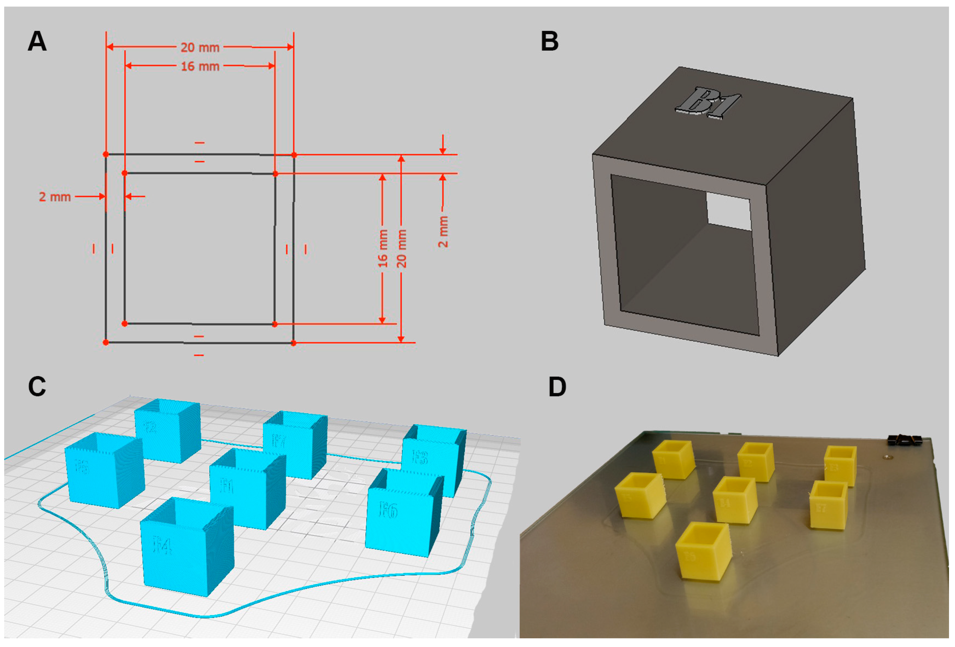
Figure 1. Summary of research workflow. (A) design in CAD, (B) conversion into 3D models, (C) preparation for 3D printing, (D) the final 3D printed products.
Applsci 15 10428 g001
Figure 2. (A) Group A; Blue: Pre-immersion, Red: Post-immersion; (B) Group B (0.12% chlorhexidine gluconate); Blue: Pre-immersion, Red: Post-immersion; (C) Group C (3% hydrogen peroxide); Blue: Pre-immersion, Red: Post-immersion.
Applsci 15 10428 g002aApplsci 15 10428 g002b
Figure 3. SEM images of ABS cube surfaces without adherence (Clean) of C. albicans for Group B, Group E, and Group F at magnification (left to right) of ×200, ×500, ×1000, and ×2500.
Applsci 15 10428 g003
Figure 4. SEM images of ABS cube surfaces without adherence (Clean) of C. albicans for Group B, Group E, and Group F at magnification (left to right) of ×200, ×500, ×1000, and ×2500.
Applsci 15 10428 g004
Figure 5. SEM images of ABS cube surfaces with adherence of C. albicans for Group A, Group C, Group D and no immersion at magnification (left to right) of ×200, ×500, ×1000, and ×2500.
Applsci 15 10428 g005
Figure 6. SEM images of ABS cube surfaces with adherence of S. mutans in Group A, Group C, Group G and No immersion (left to right) at ×200, ×500, ×1000, and ×2500 magnifications.
Applsci 15 10428 g006
Table 1. Analysis of changes in OM in 28 days.
Pre-Immersion
GroupMedian (IQR)2 (df)p-Value *
A232.05 (0.96)1.232 (5)0.942
B232.72 (2.14)
C231.90 (1.35)
D232.33 (1.75)
E231.73 (1.71)
F232.09 (1.50)
7 days post immersion
GroupMedian (IQR)2 (df)p-value *
A231.01 (1.23)8.607 (5)0.126
B231.85 (1.00)
C231.15 (1.72)
D232.04 (2.17)
E231.54 (1.39)
F232.29 (1.12)
14 days post immersion
GroupMedian (IQR)2 (df)p-value *
A231.16 (1.09)4.613 (5)0.465
B231.59 (1.38)
C231.06 (1.71)
D230.78 (1.32)
E231.18 (2.17)
F231.54 (1.16)
21 days post immersion
GroupMedian (IQR)2 (df)p-value *
A230.98 (0.61)7.740 (5)0.171
B232.01 (1.96)
C230.83 (1.16)
D231.27 (1.48)
E231.64 (1.28)
F232.03 (0.81)
28 days post immersion
GroupMedian (IQR)2 (df)p-value *
A230.93 (1.21)5.466 (5)0.362
B231.75 (1.69)
C230.61 (1.48)
D230.88 (1.69)
E230.78 (1.52)
F231.02 (0.45)
* Significant at p < 0.05. Kruskal–Wallis test: Parametric assumptions for all values not met. The Kolmogorov–Smirnov test and the Shapiro–Wilk test are significant (p < 0.05). Data not normally distributed. IQR: Interquartile range. df: Degree of freedom.
Table 2. Analysis of changes in IM in 28 days of immersion.
Pre-Immersion
GroupMedian (IQR)2 (df)p-Value *
A122.51 (1.74)7.389 (5)0.193
B122.70 (1.80)
C121.74 (2.25)
D121.86 (2.26)
E122.98 (1.68)
F123.19 (1.66)
7 days post immersion
GroupMedian (IQR)2 (df)p-value *
A122.96 (1.80)3.675 (5)0.597
B123.34 (1.61)
C122.94 (2.18)
D121.82 (2.11)
E123.07 (2.16)
F123.52 (2.09)
14 days post immersion
GroupMedian (IQR)2 (df)p-value *
A123.38 (1.68)4.816 (5)0.439
B123.84 (1.55)
C122.77 (2.42)
D121.82 (2.54)
E122.77 (2.00)
F123.81 (1.92)
21 days post immersion
GroupMedian (IQR)2 (df)p-value *
A123.12 (1.94)3.977 (5)0.553
B123.63 (2.05)
C122.54 (2.23)
D121.59 (2.48)
E122.60 (1.82)
F123.27 (1.64)
28 days post immersion
GroupMedian (IQR)2 (df)p-value *
A122.87 (1.85)3.448 (5)0.631
B123.28 (1.76)
C122.58 (1.73)
D121.80 (2.84)
E123.04 (2.19)
F123.71 (1.76)
* Significant at p < 0.05. Kruskal–Wallis test: Parametric assumptions for all values not met. The Kolmogorov–Smirnov test and the Shapiro–Wilk test are significant (p < 0.05). Data not normally distributed. IQR: Interquartile range. df: Degree of freedom.
Table 3. Analysis of changes in mass.
GroupMean ± SDCorrelation Coefficientp Value *
A2.76 ± 0.040.1650.342
B2.83 ± 0.040.3390.047 *
C2.82 ± 0.030.1760.311
D2.84 ± 0.050.1780.306
E2.79 ± 0.050.1060.544
F2.76 ± 0.040.1170.504
* Significant at p < 0.05. Pearson’s correlation coefficient. All data normally distributed. SD = standard deviation.
Table 4. One-way ANOVA analysis for compressive strength (MPa).
Immersion MediaMean (SD)F Stat (df)p Value *
No Immersion23.617 (0.175)1.036 (6)0.443
Set A26.110 (0.866)
Set B26.147 (2.894)
Set C26.152 (1.836)
Set D26.206 (1.074)
Set E24.991 (2.319)
Set F26.469 (1.435)
* Significant at p < 0.05.
Disclaimer/Publisher’s Note: The statements, opinions and data contained in all publications are solely those of the individual author(s) and contributor(s) and not of MDPI and/or the editor(s). MDPI and/or the editor(s) disclaim responsibility for any injury to people or property resulting from any ideas, methods, instructions or products referred to in the content.