Article

Computed Tomography Evaluation of the Renal Blood Vessels in the Omani Population

1
Department of Surgery, Sultan Qaboos University Hospital, Muscat 123, Oman
2
Department of Human & Clinical Anatomy, College of Medicine & Health Sciences, Sultan Qaboos University, Muscat 123, Oman
3
Radiology Residency Program, Oman Medical Specialty Board, Muscat 132, Oman
4
Department of Medicine, Royal College of Surgeons, D02 YN77 Dublin, Ireland
5
Department of Radiology & Molecular Imaging, Sultan Qaboos University Hospital, Muscat 123, Oman
*
Author to whom correspondence should be addressed.
Appl. Sci. 2025, 15(18), 9967; https://doi.org/10.3390/app15189967
Submission received: 6 August 2025 / Revised: 30 August 2025 / Accepted: 9 September 2025 / Published: 11 September 2025
(This article belongs to the Special Issue Research Progress in Medical Image Analysis)

Abstract

The renal vessels are known to exhibit variations in different populations. The present retrospective cross-sectional study aimed to evaluate the radiological anatomy of renal arteries and veins in the Omani population. Computed tomography angiography scans were used to assess diameter, laterality, and vascular branching patterns in adults (aged ≥ 18 years) who underwent contrast-enhanced CT angiography of the abdomen and pelvis between 1 January 2023, and 31 December 2024. Normal CT angiograms of cases performed for vascular pathology screening, renal transplant workup, or trauma evaluation with normal findings were included. Measurements included diameters, anatomical course, and vascular variations in the renal arteries and veins. Accessory renal arteries were defined as any additional arteries arising from the aorta supplying the kidney, regardless of the entry point. The mean diameters of the right and left renal arteries were significantly higher in males (p = 0.020 and 0.026, respectively). The right renal vein was significantly larger in females (p = 0.020). Accessory renal arteries were identified in 24.22% (n = 31 patients), including two cases with unilateral double accessory arteries. The right and left RA diameters were 4.51 ± 0.91 mm and 4.95 ± 0.98 mm, respectively, both significantly larger in males (p = 0.020 and 0.026). Supernumerary renal veins were observed in 21 patients; retroaortic and circumaortic left RVs were found in seven and one case(s), respectively. Venous variations were present in 17.2% of the Omani subjects. The findings may enhance preoperative planning, especially in renal transplantation and urologic surgery, by increasing awareness of anatomical variants. This region-specific dataset supports the development of optimized imaging protocols and surgical strategies for better patient care.

1. Introduction

Although the renal vasculature is inherently complex and variable, it is critically important for clinical practice, particularly in renal transplantation, urological surgeries, and vascular interventions. The kidneys receive their main blood supply from the renal arteries, which arise from the abdominal aorta slightly below the superior mesenteric artery, typically at the level of the L1–L2 intervertebral disk [1]. These arteries measure approximately 4–6 mm in length and 5–6 mm in diameter, run posterior to the renal veins, and cross anterior to the renal pelvis before reaching the hilum. In addition to supplying the kidneys, the renal arteries also perfuse adjacent structures, including the adrenal glands and ipsilateral ureters [1]. The right renal artery typically arises from the anterolateral aspect of the abdominal aorta. It passes beneath the inferior vena cava, while the left renal artery originates more laterally and courses horizontally to the left kidney. Before entering the hilum, both renal arteries divide into anterior (75% of blood flow) and posterior (25% of blood flow) divisions [1]. The anterior division further branches into upper, middle, lower, and apical segmental arteries. These segmental arteries subsequently branch into lobar, interlobar, arcuate, and lobular arteries, ultimately forming a network of afferent arterioles that supply blood to the glomeruli [1].
Venous drainage occurs via a single renal vein on each side, originating from the vertebral levels of L1–L2/L3 vertebrae. The renal veins traverse the abdominal cavity medially before ultimately joining the inferior vena cava. Notably, the left renal vein exhibits a longer course compared to its right counterpart [2]. The renal veins exhibit asymmetry, each following a distinct path to drain other organs, such as the gonads, adrenal gland, and diaphragm. Beginning as stellate veins, they progress into arcuate veins, which, in turn, drain into the interlobar veins. These interlobar veins coalesce to form the segmental veins before draining into the renal veins [2]. Variations in the renal vasculature are common, with more than 30% of individuals having more than one renal artery supplying a kidney [1]. Accessory arteries have also been noted to supply the lower poles of the kidneys. In this study, the term accessory renal artery refers to any artery arising from the abdominal aorta in addition to the main renal artery, regardless of whether it enters through the hilum or directly into the renal poles.
Globally, in the African region, a study reported that the incidence of first and second additional renal arteries was 23.2% and 4.5%, respectively, with accessory arteries more frequently observed on the left side (32.0%) in a sample of 130 renal angiograms from transplant donors and 106 cadaveric kidneys [3]. Another study conducted in the same region reported the presence of a renal collar (0.3%), retroaortic veins (0.5%), and additional veins (0.4%) from a sample size of 1008 kidneys evaluated through cadavers and renal venograms [4]. While some international studies, such as one conducted in Turkey, have comprehensively evaluated both renal arteries and veins concurrently in living kidney donors using CT angiography, the existing literature remains limited, as most of the studies have focused primarily on either arteries or veins in isolation [5].
This study aims to address this gap by evaluating renal vascular anatomy among patients through the following objectives: (i) investigate renal vascular structures, including arteries and veins, assessing their diameter, location, and variations using computed tomography (CT) scans and multidetector computed tomography (MDCT) angiography; (ii) determine the prevalence of renal vascular variations in Oman; and (iii) provide the regional dataset on radiological renal vasculature, thereby filling a significant research gap in the field. Ultimately, addressing this regional gap in renal vascular anatomy research is anticipated to improve surgical planning and enhance outcomes in urological surgery and renal transplantation, and minimize surgical complications such as inadvertent rupture of anomalous vessels during vascular pedicle manipulation, which can lead to fatal hemorrhage [3,4].
To the best of our knowledge, this is the first study to concurrently assess renal arterial and venous morphometry and anatomical variations in the Omani population using MDCT, offering critical insights for region-specific surgical and radiological protocols. Alarmingly, according to the Ministry of Health in Oman, the country is facing a challenge with the increase in the number of non-communicable diseases, including chronic kidney disease. The increase in the number of patients progressing to end-stage kidney disease (ESKD) requiring renal replacement therapy is a cause of concern [6]. The situation is a serious issue, as seen from the reported figures in 2014, during which 1339 ESKD patients were reported to undergo dialysis and approximately 1400 patients were recipients of kidney transplants [6]. Considering the estimated annual incidence of ESKD to be approximately 120 patients per million population [6], we performed the present study to examine the normal and abnormal anatomy of the renal vessels, which may be important for clinicians and surgeons. The scientific novelty of this work is the concurrent quantification of both renal arterial and venous diameters and variation frequencies in an Omani cohort using a uniform MDCT protocol, thereby generating population-specific reference values, which have not been reported earlier. The practical significance, in contrast, is the immediate applicability of these data to donor evaluation, preoperative planning, and local radiology workflows in Oman.

2. Materials and Methods

The retrospective cross-sectional study was performed after obtaining ethical approval from the Institutional Medical Research Ethical Committee, College of Medicine, Sultan Qaboos University (REF. NO. SQU-EC/032/2024 MREC # 3388—30 September 2024). The study included contrast-enhanced CT scans of 128 patients (n = 92 males and 36 females) who attended the Radiology Department at Sultan Qaboos University Hospital (SQUH) from 1 January 2023 to 31 December 2024.

2.1. Inclusion Criteria

All patients aged 18 years or more who underwent a CT angiogram with contrast for a suspicion of a vascular pathology (abdominal aortic aneurysm, high arterial blood pressure, a lump in the lower limb) or renal transplantation were included in the study.

2.2. Exclusion Criteria

Patients who were less than 18 years old were excluded from the study. Additionally, patients who underwent CT scans without contrast, patients with significant trauma-related injuries, and patients with inadequate, suboptimal scan quality were also excluded from the study. After applying the inclusion/exclusion criteria, 128 patients (n = 92 males and 36 females) were selected.

2.3. Radiological Study

The radiographic images were obtained from two CT machines available at the Radiology Department at SQUH: a Siemens Force CT machine (Siemens Healthineers, Erlangen, Germany) with 192 × 2 slices and a GE CT machine (GE HealthCare, Chicago, IL, USA) with 256 slices. The CT images were interpreted by a single radiologist working at the hospital who collected the data into a spreadsheet. The diameter of the renal arteries was measured at the midpoint between the origin from the abdominal aorta and the point of bifurcation. The diameter of the vein was measured at the midpoint between the origin of the vein and the point of drainage into the inferior vena cava. For this study, an accessory renal artery was defined as any artery other than the main renal artery that originates from the abdominal aorta and supplies the kidney, either via the hilum or by entering directly into the renal poles. As noted in the prior literature, these arteries are essential for segmental perfusion; their injury may result in ischemic necrosis of the supplied segment [7,8,9]. Terminological inconsistencies exist in the literature, as terms such as supernumerary, multiple, aberrant, and additional are often used interchangeably, creating confusion. We adopt the definition suggested by Graves and others, using “accessory” for arteries originating from the aorta in addition to the main renal artery, regardless of their point of entry into the kidney [7,8,9]. The course of the left renal vein, the presence of the supernumerary vein, and the accessory or double renal artery were visualized using thin cuts of axial and coronal planes.

2.4. Statistical Analysis

The analysis was performed by IBM Statistical Package for Social Sciences (SPSS) software (version 26). A one-sample Kolmogorov–Smirnov (K-S) test was performed to determine whether the diameters of the right and left renal arteries and veins follow a normal distribution or not. The diameters of the right and left renal veins and the diameter of the left renal arteries were found to follow normal distribution (p = 0.073, 0.200, and 0.179, respectively). The diameter of the right renal artery was not found to be normally distributed (p = 0.047). To compare the diameter of the renal veins and the left renal artery between males and females, an independent t-test was performed. To compare the diameter of the right renal artery between males and females, the Mann–Whitney U test was performed. A p-value of 0.05 or less (p ≤ 0.05) was considered to be significant. The course of the vessels and the presence of supernumerary vessels were treated as descriptive statistics, with the incidence being reported among males and females.

3. Results

The mean diameter of the right renal vein (RV) in the 128 patients was found to be 9.63 ± 2.54 mm (range 3.20–16.50 mm). The mean diameter of the right RV in males (n = 92 males) was found to be 9.31 ± 2.36 mm, while in females (n = 36 females), the mean diameter of the right RV was found to be 10.46 ± 2.80 mm. The difference in the diameter of the right RV among males and females was found to be statistically significant (p = 0.020).
The mean diameter of the left RV in the study population was found to be 9.32 ± 1.94 mm (range 3.2–13.5 mm). The mean diameter of the left RV in males and females was found to be 9.29 ± 1.87 mm and 9.40 ± 2.14 mm, respectively. The difference in the diameter of the left RV between males and females was found to be insignificant (p > 0.05).
The mean diameter of the right renal artery (RA) in the study population was found to be 4.51 ± 0.91 mm (range 2.2–6.7 mm). The mean diameter of the left RA was 4.95 ± 0.98 mm (range 2.4–7.5 mm). Table 1 compares the diameter of the right and left RA among males and females with the p-value. Figure 1 shows the axial CT measurement of the left renal artery diameter, which in this case was 5.3 mm.
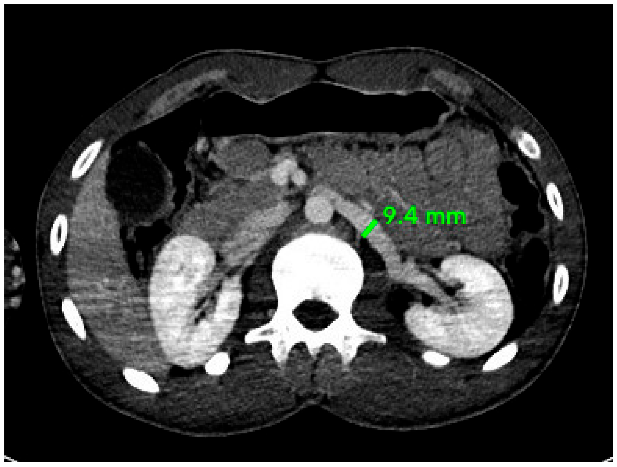
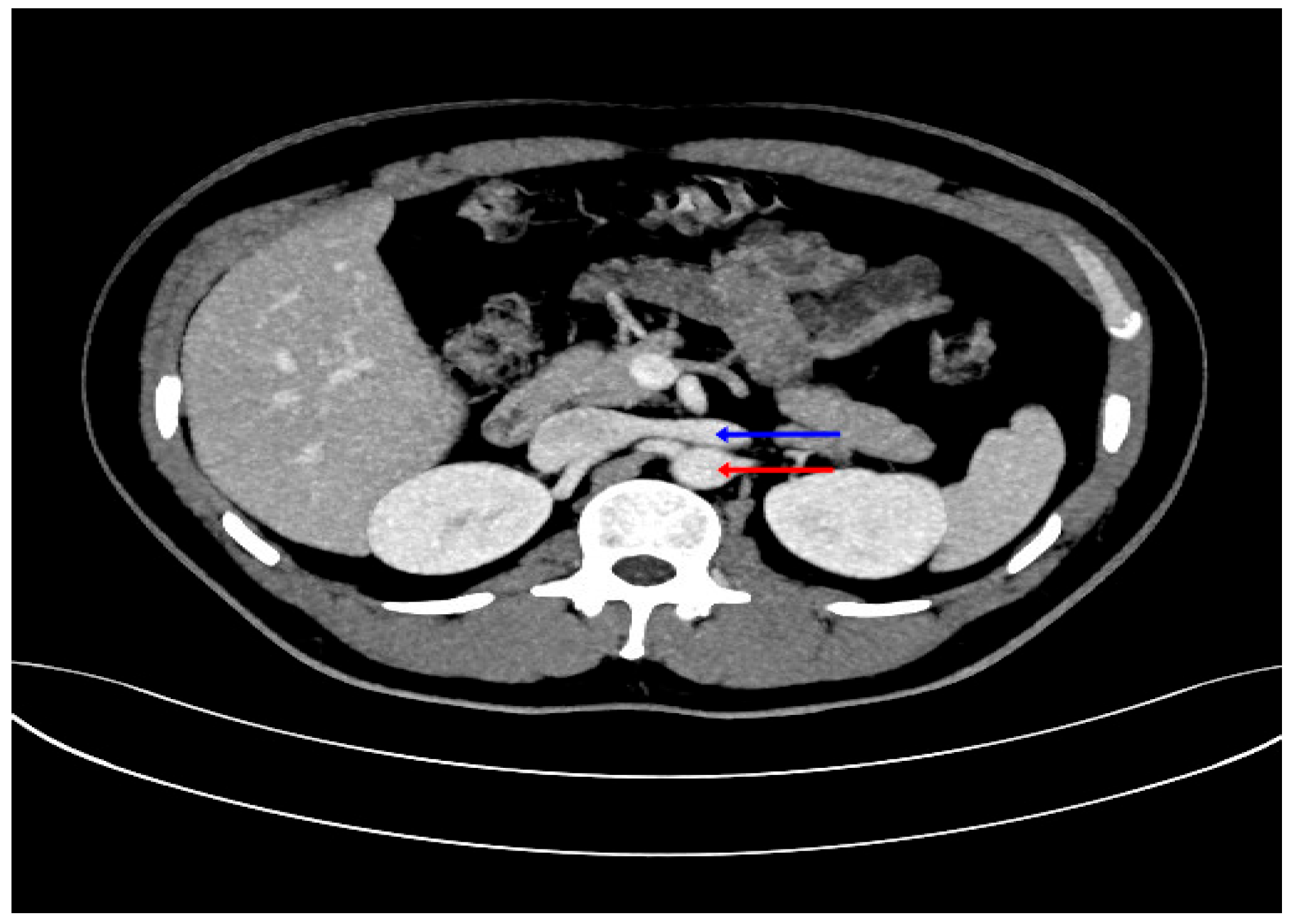
The RV was found at the hilum in all the patients. There was no incidence of RV at the level of the upper or lower pole of the kidney. One supernumerary renal vein was found in 20 (15.63%) patients (11 males and nine females), while in one female patient (0.78%), two supernumerary renal veins were found. In 120 patients (93.75%), the left RV was found as a single RV anterior to the abdominal aorta. Circumaortic and retroaortic left renal veins were also observed as shown in Table 2. Figure 2 illustrates the axial view of the left renal vein, measuring 9.4 mm in diameter. Figure 3 shows a single left RV anterior to the abdominal aorta. Figure 4 shows retroaortic course of the left renal vein, passing posterior to the aorta.
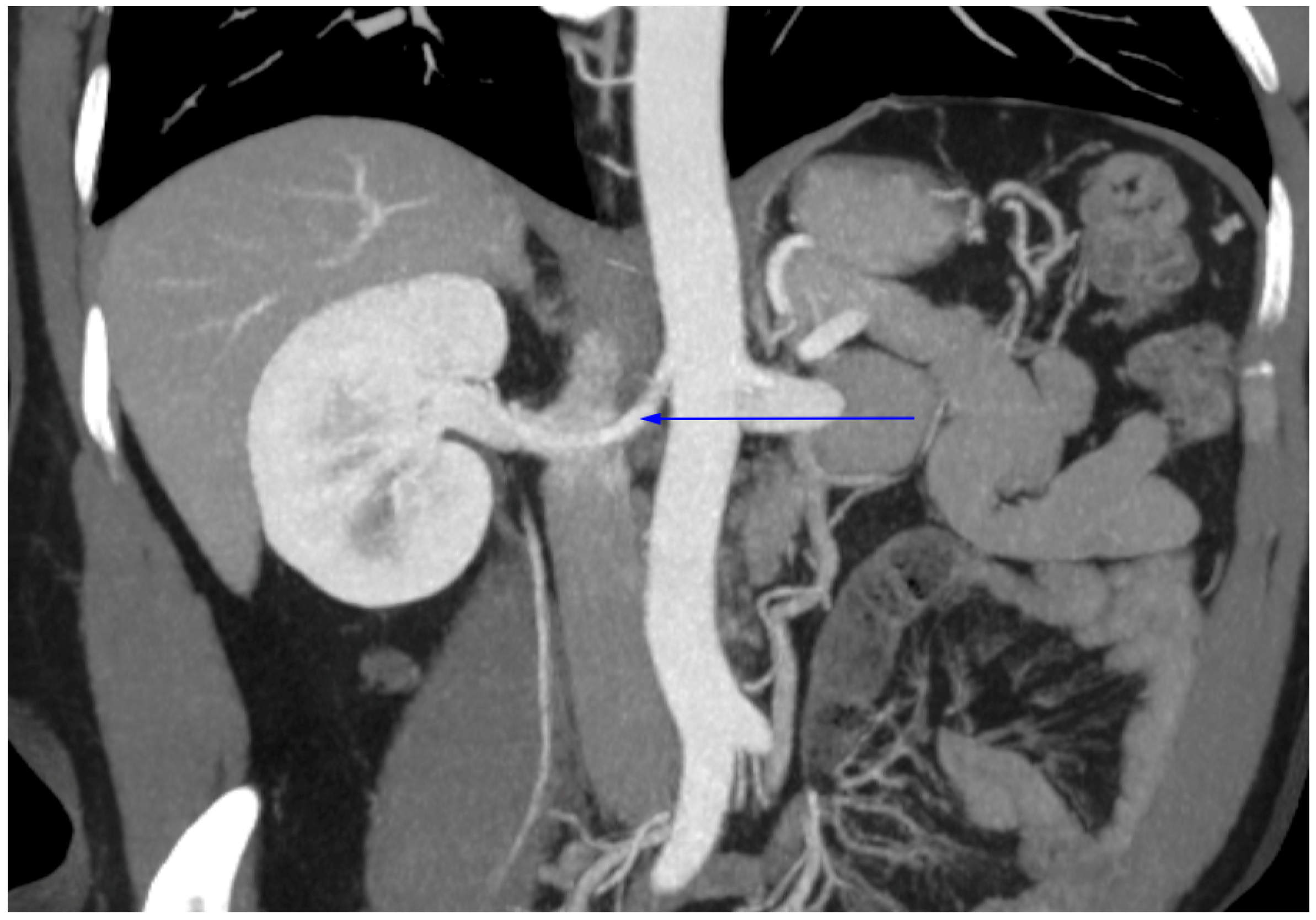
In 97 patients (75.78%), a single renal artery was found bilaterally. Figure 5 shows a single right renal artery. In the other 31 patients (24.22%), accessory renal arteries were observed.
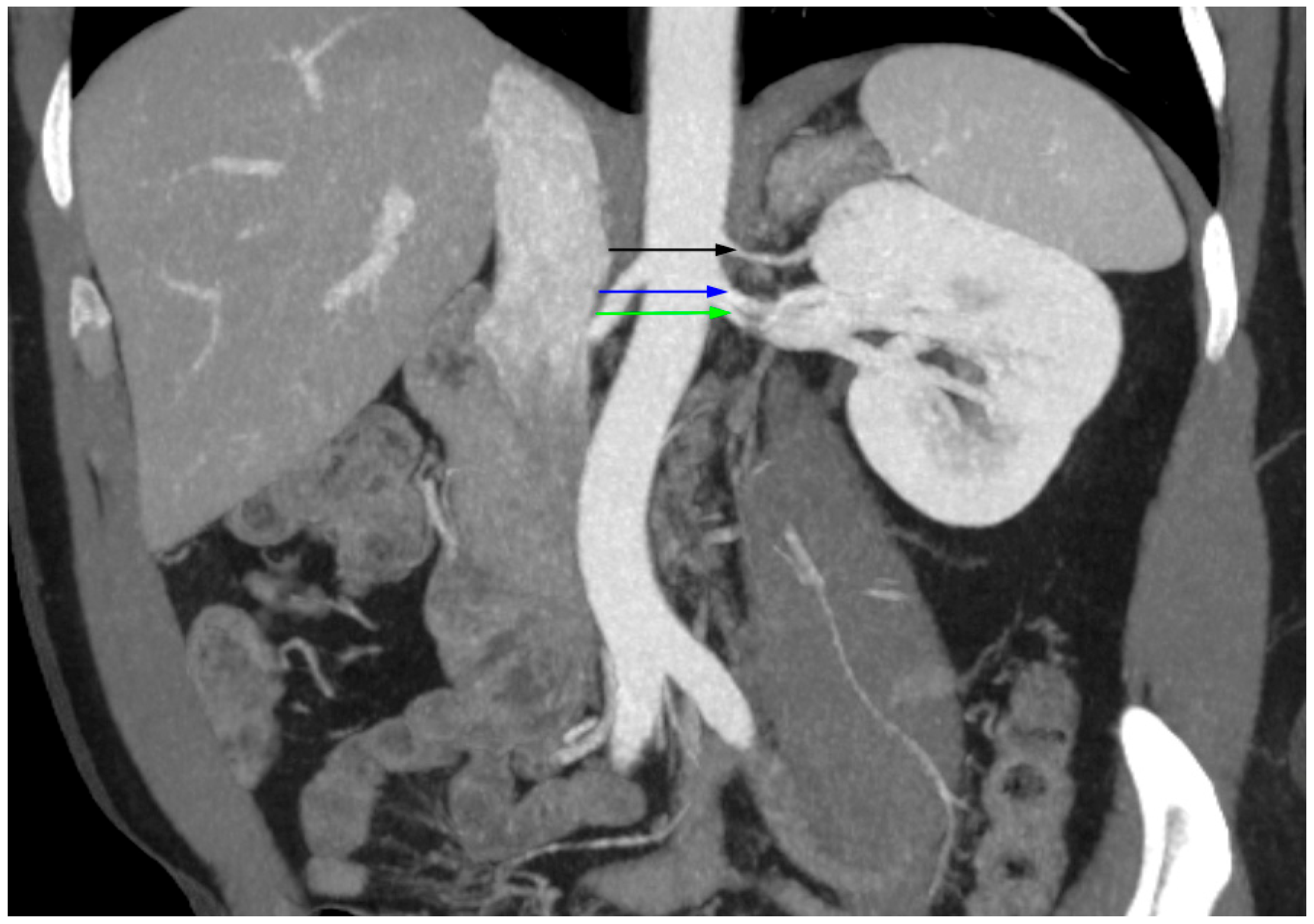
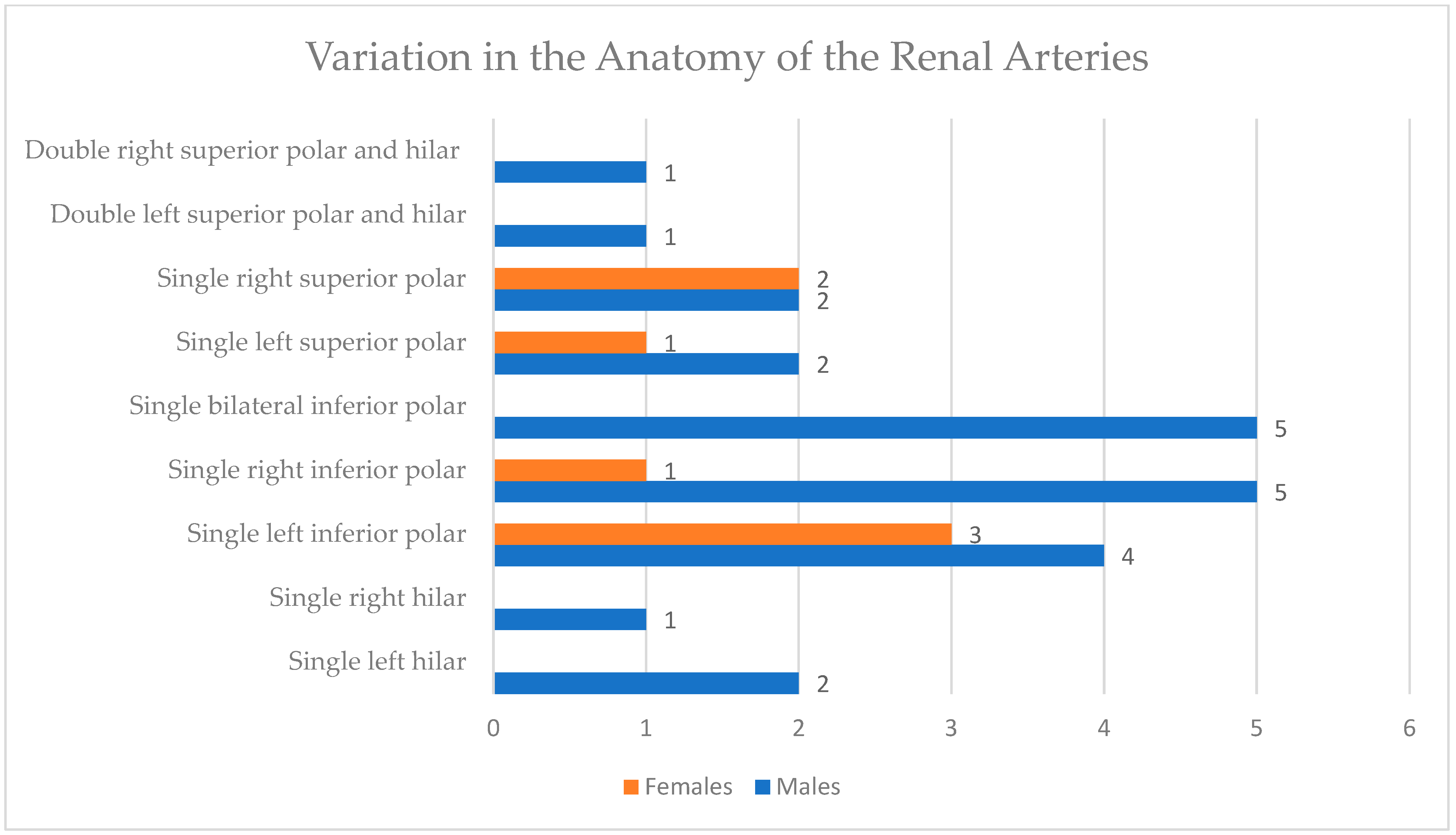
Among patients with accessory RA, one patient showed a left superior polar artery and a right inferior polar artery. In seven patients, superior polar accessory arteries were found, four on the right and three on the left. In 18 patients, inferior polar accessory arteries were observed. Figure 6 illustrates a coronal oblique MIP CT angiogram in which a left accessory renal artery arises from the abdominal aorta and supplies the inferior pole of the left kidney, in addition to the main hilar artery. In three patients, hilar accessory renal arteries were observed, two on the left and one on the right. In the other two patients, two accessory renal arteries were observed. One patient had right superior polar and right hilar accessory arteries, while the other had left superior polar and left hilar accessory arteries. Figure 7 displays a coronal oblique MIP CT angiogram showing two accessory renal arteries on the left: a superior polar artery and a hilar accessory artery, both supplying the left kidney. Figure 8 shows the renal artery variations among males and females.

4. Discussion

Understanding anatomical variation is essential for tailoring surgical approaches and minimizing intraoperative risk. The knowledge of renal vessel anatomy is of great significance, as it directly influences the outcome of procedures such as renal transplantation and urological surgeries. Anatomical variations in renal vasculature may increase the complexity of interventions and may lead to complications if unrecognized. This study examined renal vascular anatomy in 128 patients using contrast-enhanced CT angiography. Key findings included a mean diameter of 9.63 ± 2.54 mm for the right RV, significantly larger in females than males (p = 0.020). At the same time, no significant sex difference was found in the left RV. The right and left RA diameters were 4.51 ± 0.91 mm and 4.95 ± 0.98 mm, respectively, both significantly larger in males (p = 0.020 and 0.026). Venous variations included supernumerary renal veins in 15.63%, circumaortic veins in 0.78%, and retroaortic veins in 5.47%. In terms of arterial anatomy, single RA was seen in 75.78% of patients, while accessory RA was seen in 24.22%.
Our results are consistent with the international literature. In Nepal, a study reported a higher prevalence of accessory RA (32%) compared to 24.22% in our population [10]. Similarly, studies in Sudan and Nigeria reported accessory arteries in 25.6% and 32%, respectively [11,12]. The prevalence of accessory RA in our population is comparable to previous reports. In the same Nepalese study, double right RVs were found in 6.3% and retroaortic veins in 2.1%, lower than our rates of 15.63% and 5.47%, respectively [10]. A meta-analysis by Hostiuc et al. reported multiple RVs in 16.7% of cases, closely matching our findings [13]. Retroaortic and circumaortic veins were reported in 3% and 3.5% in that meta-analysis, whereas in our study, retroaortic veins were slightly more common (5.47%), while circumaortic veins were less frequent (0.78%).
Regarding RA diameter, a study conducted in Karachi, Pakistan, found larger values: 6.66 mm (right) and 6.79 mm (left), with males having larger diameters than females [14]. In our study, both arteries were about 2 mm smaller. A study based in India found mean diameters of 4.83 mm (right) and 5.04 mm (left), which closely align with our findings (4.51 mm and 4.95 mm, respectively) [15]. Both studies, like ours, reported statistically significant differences between males and females. Another regional study from Saudi Arabia reported slightly higher values, i.e., 5.54 mm (right) and 5.48 mm (left) in males, with lower measurements in females. However, it similarly confirmed significant differences in RA diameter between sexes and between the right and left sides [16]. A Polish study, however, found no significant difference between the right and left single RA (p = 0.219) [17]. Table 3 summarizes the current literature on the diameters of the renal arteries.
Regarding RV diameter, a 2018 study conducted in Lahore, Pakistan, on 50 male cadavers was published. The study reported right and left RV diameters of 10.85 ± 2.37 mm and 11.57 ± 1.78 mm, respectively, both larger than our findings (9.63 mm and 9.32 mm) [18]. This may relate to body habitus differences, as the Lahore study involved only males.
As for RV diameter, our study found mean values of 9.63 ± 2.54 mm on the right and 9.32 ± 1.94 mm on the left. These measurements are somewhat lower than those reported in a 2018 cadaveric study from Lahore, Pakistan, which found right and left RV diameters of 10.85 ± 2.37 mm and 11.57 ± 1.78 mm, respectively, in 50 male specimens [18]. The larger diameters observed in the Pakistani study may be attributed to post-mortem vessel distension and the exclusive inclusion of males. In contrast, our cohort included both sexes and utilized in vivo imaging, which may have contributed to the smaller measurements due to physiological variations in vascular tone and volume status. However, a more directly comparable dataset comes from a Turkish study, where the mean right and left RV diameters were 8.8 ± 1.9 mm and 8.9 ± 1.8 mm, respectively, in non-compressed veins [19]. These values are slightly smaller than ours, possibly due to demographic or methodological differences, though both studies used in vivo MDCT techniques. In contrast, a classic cadaveric study from South Africa reported RV diameters averaging 12 ± 2 mm bilaterally [20]. These are substantially larger than our values, possibly due to differences in measurement techniques and the absence of venous tone or blood pressure regulation post-mortem. Notably, their findings showed little difference between the right and left sides, while our study revealed a modest asymmetry, with right veins being slightly larger, especially among females. This asymmetry may reflect the anatomical course and drainage patterns of the renal veins, which differ between sides. Table 4 summarizes the current literature on the diameters of the renal veins.
Normally, the RA is 4–6 cm long and 5–6 mm in diameter; this is only observed in less than 25% of the cases [21]. However, with the presence of an accessory artery, the RA diameter tends to be smaller [22]. In comparison, the RVs are of a larger caliber (1.2 cm in diameter) but are distinguished based on site; the left RV is longer (6–7 cm) compared to the right (3–4 cm) [23]. Factors influencing vessel size in healthy individuals include genetics, intrauterine development, and environmental factors such as body weight or high-salt diets. Age-related vascular rigidity may also affect measurements [24,25]. Furthermore, sex plays a significant role, as shown in our study and the prior literature, with males typically exhibiting larger vascular diameters due to differences in body composition and metabolic demands [26]. Additionally, comorbidities such as hypertension, diabetes, and renal atrophy are known to contribute to vascular morphological changes. These conditions are associated with increased arterial calcification, which may lead to reduced vessel diameter and altered structural integrity through increased stiffness [27]. Moreover, genetic and embryological factors are key determinants of renal vessel morphology. The development of renal vasculature involves tightly coordinated processes, such as vasculogenesis (the de novo formation of vessels) and angiogenesis (the sprouting of vessels from existing ones), which closely coordinate with nephron formation [28]. The ascent of the kidney from the pelvis to its lumbar position during gestation, coupled with the successive changes in its arterial supply, explains the high incidence of anatomical variations in renal vessels, such as accessory renal arteries, which result from the persistence of normally transient embryonic vessels [29]. At the molecular level, signaling pathways such as the VEGF (Vascular Endothelial Growth Factor) pathway and Notch play a fundamental role in regulating the proliferation and differentiation of endothelial cells [28]. Disruptions in these regulatory mechanisms, including genetic mutations, can result in renal and genitourinary anomalies, often presenting with vascular malformations.
From a clinical standpoint, understanding the variations in the renal vessels is crucial to minimizing accidental injuries and hemorrhage in surgeries involving the kidney, such as renal transplantation [30]. The demand for such procedures continues to rise, reflecting the global and local increase in the incidence of ESKD [6,31]. The presence of supernumerary vessels is considered a factor that might complicate renal transplantation surgeries [32]. Previous studies have reported that the presence of accessory renal arteries is associated with an increased rate of renal complications, longer ischemia times, and delayed graft function [33]. Moreover, the loss of an arterial branch other than the main artery can result in delayed graft function, segmental necrosis, and ureteral necrosis [34]. However, variations in the anatomy of the renal arteries should not discourage the utilization of such samples in transplant surgeries, as the demand for such surgeries is rising [35]. However, it is necessary to study the grafts with multiple renal arteries beforehand to minimize the complications and increase the chance of successful renal transplantation [33]. The variation in the anatomy of RVs also has clinical implications. The variation in RV increases the complexity of catheterization, complicating hemodialysis. It is also thought to increase the risk of thrombosis [35]. Unawareness of the presence of circumaortic RV might result in vascular injury, especially to the posterior limb, as the renal veins might deceivingly look normal, and the surgeon might not be aware of the posterior limb, leading to vascular injury and hemorrhage [13]. Retroaortic renal veins can get compressed between the aorta and the vertebra, resulting in urological problems such as hematuria, varicocele, and uteropelvic junction obstruction [36]. Another important complication related to the renal veins is the Nutcracker syndrome (NCS), also known as the left renal vein entrapment syndrome, which has two main forms: anterior NCS and posterior NCS. In the anterior NCS, the left renal vein is compressed between the aorta and the superior mesenteric artery. In posterior NCS, the left renal vein is compressed between the aorta and the vertebral body. The latter is less common but is associated with retroaortic and circumaortic renal veins. NCS usually manifests in pelvic pain, flank pain, hematuria, and varicoceles. It also increases the risk of left renal vein thrombosis and left renal vein hypertension, which might cause chronic kidney disease in the long term [37]. These population-specific findings may support the refinement of CT angiography protocols for renal donor evaluation in Oman and inform surgical training programs to anticipate anatomical variations more commonly encountered in Omani patients.
While several studies have investigated the anatomical variations in RAs and RVs concurrently [38,39], only a limited number have included measurements of their diameters. Therefore, this study offers a more comprehensive characterization of the renal vasculature by incorporating diameter assessment using contrast-enhanced CT scans. Vascular anatomy and dimensions are influenced by a range of factors, including ethnicity, which may contribute to variations in disease susceptibility among different populations [40,41,42]. Ethnicity-related differences in vascular diameter should therefore be taken into account when establishing normative reference values and developing clinical protocols for the diagnosis and management of various conditions. By focusing on the Omani population, this study contributes to the paucity of the literature on ethnic variations in renal vascular anatomy that may be relevant for regional clinical, surgical, and radiological practice. The scientific novelty of this work lies in being the first to provide sex-stratified reference values for both renal arteries and veins in Oman using a standardized MDCT protocol. The practical significance is its direct relevance to donor evaluation, preoperative imaging, and surgical planning in the local setting.
The study had a few limitations. First, the study employed a single-center retrospective analysis, which may limit the generalizability of the findings to the broader Omani population. Second, the sample was male-predominant (72%), which may skew sex-based comparisons. Thus, we admit the potential impact of the male-predominant sample on the overall prevalence rates of variations Third, the study included only patients undergoing CT angiography for suspected pathology or transplantation, which may introduce selection bias. Finally, clinical comorbidities such as hypertension or diabetes, which could influence vascular morphology, were not systematically controlled for. Admittedly, the retrospective nature of the study limits causal inference between anatomical variations and potential contributing factors such as age, comorbidities, or body habitus. Besides renal vessels, the anatomy of the iliac vessels is also important for renal transplant; however, due to constraint of time, we did not focus our study on any other blood vessel other than the renal blood vessels. Future studies could include volume rendering 3D imaging techniques for better visualization.

5. Conclusions

This study presents the first comprehensive evaluation of renal vascular anatomy in Oman, utilizing contrast-enhanced CT imaging. Renal artery and vein variations were frequent, with notable sex-based differences and a high prevalence of supernumerary vessels. The mean diameter of the right and left renal arteries was significantly wider in males compared to females, while the diameter of the renal veins was wider in females. These findings highlight the critical role of preoperative imaging in procedures such as renal transplantation, vascular reconstruction, and urological surgery. The dimensions of the renal vessels are important in determining the compatibility with the recipient vessels. Knowing the exact dimensions of the blood vessels at the preoperative stage may help in the proper stenting of the renal blood vessels. Abnormal vessel diameter may have an effect on the hemodynamics and may result in ischemia.
By establishing region-specific anatomical baselines, this work contributes valuable data to improve surgical planning, reduce intraoperative risk, and inform radiology protocols, donor evaluations, and medical training in Oman. These findings not only align with international reports but also provide a population-specific reference for the Middle East, underscoring the importance of incorporating local data into global transplant practice.
Further research should include larger multicenter studies with balanced representation across sex and age groups. Subgroup analyses based on age, BMI, comorbidities, and ethnicity may help identify determinants of vascular variation. Correlating anatomical variants with clinical outcomes, such as graft ischemia time, blood loss, surgical complexity, or graft function, can provide actionable insights for transplant and laparoscopic surgery. Incorporating genetic and anthropometric data may also advance understanding of population-specific vascular development. Ultimately, translating these anatomical insights into clinical protocols could enhance donor safety, optimize graft survival, and improve surgical outcomes in the Omani and wider regional population.

Author Contributions

Conceptualization, S.D. and S.B.; methodology, S.D., S.B. and A.A.L.; validation, A.A.K. and A.A.L.; formal analysis, A.A.; investigation, A.A.K. and A.A.; data curation, A.A.K. and A.A.; writing—original draft preparation, A.A.L., A.A., A.A.K., A.N.A. and K.A.B.; supervision, S.D. and S.B.; fund acquisition, A.A.L. and S.B.; project administration, S.D. and S.B. All authors have read and agreed to the published version of the manuscript.

Funding

This study was supported by Sultan Qaboos University (grant number: UF/MED/RADI/24/02).

Institutional Review Board Statement

The study was conducted in accordance with the Declaration of Helsinki and was approved by the Medical Research Ethics Committee of the College of Medicine and Health Sciences, Sultan Qaboos University (Approval No. SQU-EC/032/2024, MREC #3388, dated 30 September 2024).

Informed Consent Statement

Patient consent was waived as the study was a retrospective analysis of variations and diameters of renal vessels. The study reported anonymous data, and no identification information of patients was revealed.

Data Availability Statement

As the study involves data from SQUH patients, it cannot be made available online. As such, data is available upon request from the corresponding author.

Acknowledgments

The authors acknowledge the grant received from the university to conduct this study.

Conflicts of Interest

The authors declare no conflicts of interest.

Abbreviations

CTComputed Tomography
ESKDEnd-Stage Kidney Disease
MDCTMultidetector Computed Tomography
NCSNutcracker Syndrome
RARenal Artery
RVRenal Vein
SPSSStatistical Package for Social Sciences
SQUHSultan Qaboos University Hospital
VEGFVascular Endothelial Growth Factor

References

  1. Stephen, W.L. Anatomy, Abdomen and Pelvis, Renal Artery. In StatPearls; StatPearls Publishing: Treasure Island, FL, USA, 2023. Available online: https://www.ncbi.nlm.nih.gov/books/NBK459158/ (accessed on 20 April 2025).
  2. Cole, S.B. Anatomy, Abdomen and Pelvis, Renal Veins. In StatPearls; StatPearls Publishing: Treasure Island, FL, USA, 2023. Available online: https://www.ncbi.nlm.nih.gov/books/NBK538298/ (accessed on 20 April 2025).
  3. Satyapal, K.S.; Haffejee, A.A.; Singh, B.; Ramsaroop, L.; Robbs, J.V.; Kalideen, J.M. Additional renal arteries incidence and morphometry. Surg. Radiol. Anat. 2001, 23, 33–38. [Google Scholar] [CrossRef]
  4. Satyapal, K.S.; Kalideen, J.M.; Haffejee, A.A.; Singh, B.; Robbs, J.V. Left renal vein variations. Surg. Radiol. Anat. 1999, 21, 77–81. [Google Scholar] [CrossRef]
  5. Karayağız, A.H.; Cenal, U.; Ertürk, T.; Özdemir, E.; Polatkan, S.A.V.; Yılmaz, G.; Çakır, Ü.; Berber, İ. Renal arterial and venous system variations in 1073 kidney donors in Turkey. Istanb. Med. J. 2021, 22, 257–260. [Google Scholar] [CrossRef]
  6. Al Alawi, I.H.; Al Salmi, I.; Al Mawali, A.; Sayer, J.A. Kidney disease in Oman: A view of the current and future landscapes. Iran J. Kidney Dis. 2017, 11, 263–270. [Google Scholar]
  7. Ramulu, M.V.; Prasanna, L.C. Accessory renal arteries—Anatomical details with surgical perceptions. J. Anat. Soc. India 2016, 65, 162–167. [Google Scholar] [CrossRef]
  8. Graves, F.T. The aberrant renal artery. J. Anat. 1956, 90, 553–558. [Google Scholar] [PubMed] [PubMed Central]
  9. Gulas, E.; Wysiadecki, G.; Szymański, J.; Majos, A.; Stefańczyk, L.; Topol, M.; Polguj, M. Morphological and clinical aspects of the occurrence of accessory (multiple) renal arteries. Arch. Med. Sci. 2018, 14, 442–453. [Google Scholar] [CrossRef]
  10. Regmi, P.R.; Amatya, I.; Kayastha, P.; Paudel, S.; Suwal, S.; Ghimire, R.K. Normal anatomy and variants of renal vasculature with multidetector computed tomography in a tertiary care hospital: A descriptive cross-sectional study. JNMA J. Nepal Med. Assoc. 2020, 58, 911–914. [Google Scholar] [CrossRef]
  11. Osman, S.; AbdAlla, E.; Ali, L.; MohamedAli, H.; Taha, B.; Elmahdi, T.S.A.; Mohammed, S. Prevalence and patterns of renal vascular variations among potential kidney donors: A computed tomography angiography (CTA) study in Sudan. BMC Nephrol. 2025, 26, 167. [Google Scholar] [CrossRef]
  12. Aremu, A.; Igbokwe, M.; Olatise, O.; Lawal, A.; Maduadi, K. Anatomical variations of the renal artery: A computerized tomographic angiogram study in living kidney donors at a Nigerian Kidney Transplant Center. Afr. Health Sci. 2021, 21, 1155–1162. [Google Scholar] [CrossRef]
  13. Hostiuc, S.; Rusu, M.C.; Negoi, I.; Dorobanțu, B.; Grigoriu, M. Anatomical variants of renal veins: A meta-analysis of prevalence. Sci. Rep. 2019, 9, 10802. [Google Scholar] [CrossRef]
  14. Mohiuddin, M.; Manzoor, A.; Ali, M.; Hassan, N. Analysis of renal artery morphometery in adults: A study conducted by using multidetector computed tomography angiography. Pak. J. Med. Sci. 2017, 33, 943–947. [Google Scholar] [CrossRef]
  15. Hasan, A.A.; Hassan, S.A.; Mubarak, W.A.; Maklad, S.M. A study of evaluation of renal artery measurements using computed tomography angiography in adult Aswanian population. Aswan Univ. Med. J. 2021, 1, 10–18. [Google Scholar] [CrossRef]
  16. Abd Elrahim, E. Computed tomography evaluation of renal artery morphometry in adults: The impact of age and gender. Saudi Med. J. 2020, 41, 34–37. [Google Scholar] [CrossRef]
  17. Majos, M.; Stefańczyk, L.; Szemraj-Rogucka, Z.; Elgalal, M.; De Caro, R.; Macchi, V.; Polguj, M. Does the type of renal artery anatomic variant determine the diameter of the main vessel supplying a kidney? Surg. Radiol. Anat. 2018, 40, 381–388. [Google Scholar] [CrossRef] [PubMed]
  18. Shaheen, R.; Shaukat, A.; Jamil, M.N. Morphometry of renal veins in a cadaver study. Khyber J. Med. Sci. 2018, 11, 208–211. [Google Scholar]
  19. Poyraz, A.K.; Firdolas, F.; Onur, M.R.; Kocakoc, E. Evaluation of left renal vein entrapment using multidetector computed tomography. Acta Radiol. 2013, 54, 144–148. [Google Scholar] [CrossRef]
  20. Satyapal, K.S.; Rambiritch, V.; Pillai, G. Morphometric analysis of the renal veins. Anat. Rec. 1995, 241, 268–272. [Google Scholar] [CrossRef] [PubMed]
  21. Palmieri, B.J.; Petroianu, A.; Silva, L.C.; Andrade, L.M.; Alberti, L.R. Study of arterial pattern of 200 renal pedicle through angiotomography. Rev. Col. Bras. Cir. 2011, 38, 116–121. [Google Scholar] [CrossRef]
  22. Gupta, M.; Kaul, N.V.; Shukla, A.K. Evaluation of diameter of main hilar renal artery to predict the presence of supplementary renal artery by contrast-enhanced MDCT: A retrospective study in Northern India. J. Clin. Diagn. Res. 2022, 16, AC10–AC13. [Google Scholar] [CrossRef]
  23. Radiopaedia.Org. Renal Vein. Available online: https://radiopaedia.org/articles/renal-vein-1 (accessed on 8 June 2025).
  24. Singh, R.P.; Jamal, A. A study of normal renal dimensions at ultrasonography and their influencing factors in an Indian population. Cureus 2023, 15, e40748. [Google Scholar] [CrossRef]
  25. Moorthy, H.K.; Venugopal, P. Measurement of renal dimensions in vivo: A critical appraisal. Indian J. Urol. 2011, 27, 169–175. [Google Scholar] [CrossRef]
  26. Tryggestad, J.B.; Thompson, D.M.; Copeland, K.C.; Short, K.R. Sex differences in vascular compliance in normal-weight but not obese boys and girls: The effect of body composition. Int. J. Pediatr. 2012, 2012, 607895. [Google Scholar] [CrossRef]
  27. Ichii, T.; Morimoto, R.; Okumura, T.; Ishii, H.; Tatami, Y.; Yamamoto, D.; Aoki, S.; Hiraiwa, H.; Furusawa, K.; Kondo, T.; et al. Impact of renal functional/morphological dynamics on the calcification of coronary and abdominal arteries in patients with chronic kidney disease. J. Atheroscler. Thromb. 2017, 24, 1092–1104. [Google Scholar] [CrossRef]
  28. Mohamed, T.; Sequeira-Lopez, M.L.S. Development of the renal vasculature. Semin. Cell Dev. Biol. 2019, 91, 132–146. [Google Scholar] [CrossRef]
  29. García-Barrios, A.; Cisneros-Gimeno, A.I.; Celma-Pitarch, A.; Whyte-Orozco, J. Anatomical study about the variations in renal vasculature. Folia Morphol. 2023, 83, 348–353. [Google Scholar] [CrossRef] [PubMed]
  30. Damaskos, C.; Georgakopoulou, V.E.; Garmpis, N.; Garmpi, A.; Dimitroulis, D. Triple renal arteries in a cadaveric kidney donor: A case report. Cureus 2020, 12, e11639. [Google Scholar] [CrossRef]
  31. Lv, J.-C.; Zhang, L.-X. Prevalence and disease burden of chronic kidney disease. Adv. Exp. Med. Biol. 2019, 1165, 3–15. [Google Scholar] [CrossRef]
  32. Moreno-Alarcón, C.; Server-Pastor, G.; López-González, P.Á.; López-Cubillana, P.; Ruiz-Morcillo, J.C.; Doñate-Iñíguez, G.; Olarte-Barragán, E.H.; Gómez-Gómez, G.A. Must we still be worried about multiple arteries in kidney transplantation? Nephro-Urol. Mon. 2013, 5, 692–696. [Google Scholar] [CrossRef] [PubMed]
  33. Cherchi, V.; Baccarani, U.; Ventin, M.; Pravisani, R.; Puggioni, A.; Zanini, V.; Lorenzin, D.; Vetrugno, L.; Risaliti, A.; Terrosu, G.; et al. Current practice with grafts with multiple renal arteries in kidney transplantation: Role of the methylene blue in the lower pole. Acta Biomed. 2022, 93, e2022006. [Google Scholar] [CrossRef]
  34. Sevmis, M.; Demir, M.E.; Merhametsiz, O.; Aktas, S.; Sevmis, S.; Uyar, M. Grafts with multiple renal arteries in kidney transplantation. Transplant. Proc. 2021, 53, 933–940. [Google Scholar] [CrossRef] [PubMed]
  35. Valenzuela Fuenzalida, J.J.; Vera-Tapia, K.; Urzúa-Márquez, C.; Yáñez-Castillo, J.; Trujillo-Riveros, M.; Koscina, Z.; Orellana-Donoso, M.; Nova-Baeza, P.; Suazo-Santibañez, A.; Sanchis-Gimeno, J.; et al. Anatomical variants of the renal veins and their relationship with morphofunctional alterations of the kidney: A systematic review and meta-analysis of prevalence. J. Clin. Med. 2024, 13, 3689. [Google Scholar] [CrossRef]
  36. Nam, J.K.; Park, S.W.; Lee, S.D.; Chung, M.K. The clinical significance of a retroaortic left renal vein. Korean J. Urol. 2010, 51, 276–280. [Google Scholar] [CrossRef]
  37. Ananthan, K.; Onida, S.; Davies, A.H. Nutcracker syndrome: An update on current diagnostic criteria and management guidelines. Eur. J. Vasc. Endovasc. Surg. 2017, 53, 886–894. [Google Scholar] [CrossRef]
  38. Çınar, C.; Türkvatan, A. Prevalence of Renal Vascular Variations: Evaluation with MDCT Angiography. Diagn. Interv. Imaging 2016, 97, 891–897. [Google Scholar] [CrossRef] [PubMed]
  39. Hekimoglu, A.; Ergun, O. Evaluation of Renal Vascular Variations with Computed Tomography. Afr. J. Urol. 2022, 28, 21. [Google Scholar] [CrossRef]
  40. Sudre, C.H.; Moriconi, S.; Rehwald, R.; Smith, L.; Tillin, T.; Barnes, J.; Atkinson, D.; Ourselin, S.; Chaturvedi, N.; Hughes, A.D.; et al. Accelerated Vascular Aging: Ethnic Differences in Basilar Artery Length and Diameter, and Its Association with Cardiovascular Risk Factors and Cerebral Small Vessel Disease. Front. Cardiovasc. Med. 2022, 9, 939680. [Google Scholar] [CrossRef] [PubMed]
  41. Nonterah, E.A.; Crowther, N.J.; Klipstein-Grobusch, K.; Oduro, A.R.; Kavousi, M.; Agongo, G.; Anderson, T.J.; Asiki, G.; Boua, P.R.; Choma, S.S.R.; et al. Racial and Ethnic Differences in the Association Between Classical Cardiovascular Risk Factors and Common Carotid Intima-Media Thickness: An Individual Participant Data Meta-Analysis. J. Am. Heart Assoc. 2022, 11, e023704. [Google Scholar] [CrossRef]
  42. Masuda, E.M.; Caps, M.T.; Singh, N.; Yorita, K.; Schneider, P.A.; Sato, D.T.; Eklof, B.; Nelken, N.A.; Kistner, R.L. Effect of Ethnicity on Access and Device Complications during Endovascular Aneurysm Repair. J. Vasc. Surg. 2004, 40, 24–29. [Google Scholar] [CrossRef][Green Version]
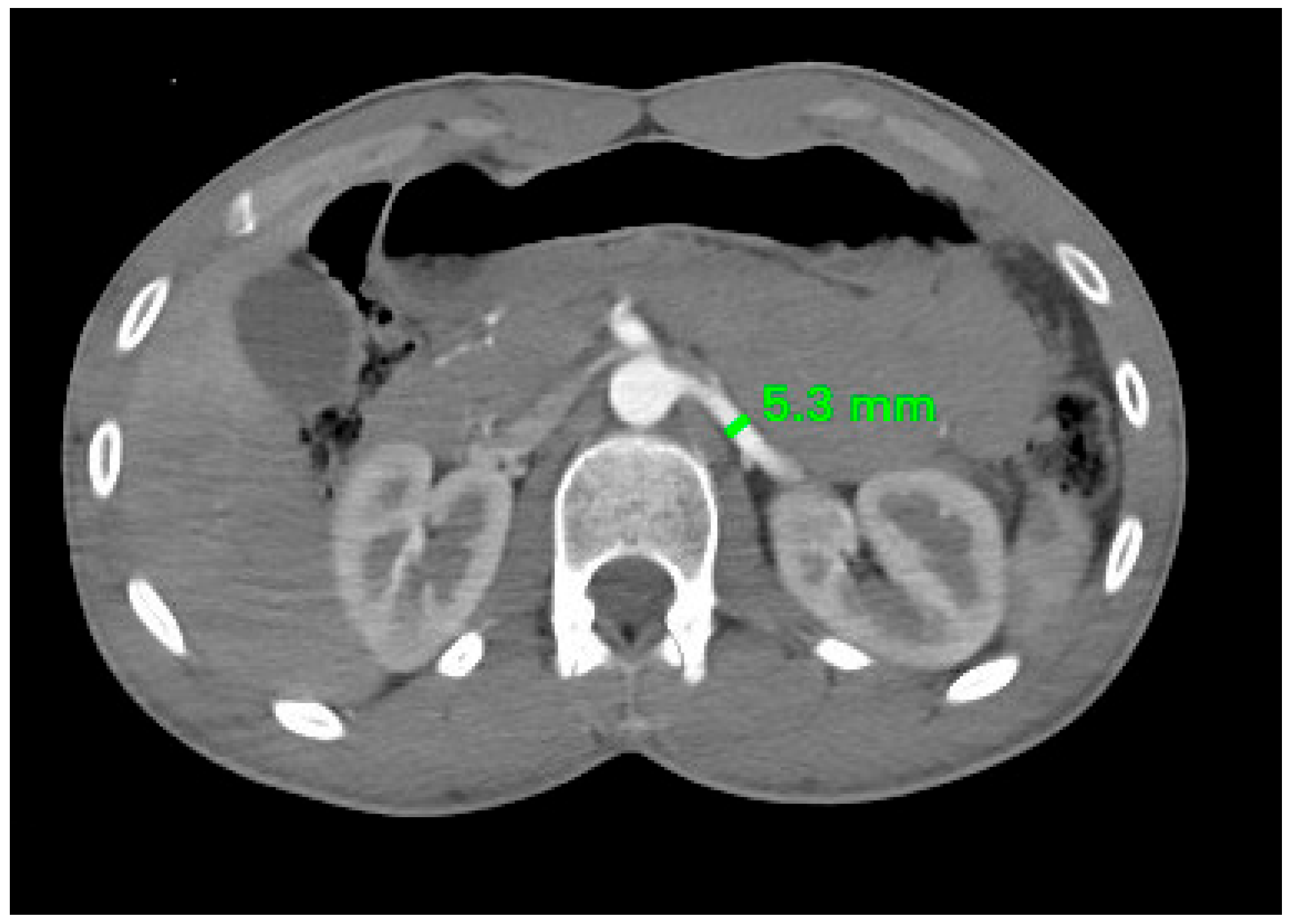
Figure 1. A computed tomography angiogram of an axial plane at the level of the kidneys shows the diameter of the left renal artery to be 5.3 mm.
Applsci 15 09967 g001
Figure 2. Axial CT image (porto-venous phase) showing the diameter of the left renal vein to be 9.4 mm.
Applsci 15 09967 g002
Figure 3. Axial MIP CT image (porto-venous phase) showing a single left renal vein (blue arrow) coursing anterior to the abdominal aorta (red arrow).
Applsci 15 09967 g003
Figure 4. Axial MIP CT image (porto-venous phase) showing a retroaortic left renal vein (blue arrow) coursing posterior to the abdominal aorta (red arrow).
Applsci 15 09967 g004
Figure 5. Coronal oblique MIP CT angiogram showing a single right renal artery supplying the right kidney (blue arrow).
Applsci 15 09967 g005
Figure 6. Coronal oblique MIP CT angiogram showing a left accessory renal artery supplying the inferior pole of the left kidney (red arrow), in addition to the main renal artery (blue arrow).
Applsci 15 09967 g006
Figure 7. Coronal oblique MIP CT angiogram showing two left accessory renal arteries: one superior polar (black arrow) and one accessory hilar artery (green arrow) supplying the left kidney in addition to the main renal artery (blue arrow).
Applsci 15 09967 g007
Figure 8. Variation in the anatomy of the renal arteries in males and females (number of cases).
Applsci 15 09967 g008
Table 1. Mean diameter of right and left renal arteries (RAs) between males and females (mean ± SD).
MalesFemalesp-Value
Right RA4.62 ± 0.93 mm4.22 ± 0.79 mm0.020
Left RA5.07 ± 1.04 mm4.64 ± 0.76 mm 0.026
Table 2. Types of left RV observed in males and females.
MalesFemalesTotal
Single left RV, anterior to aorta8832120
Circumaortic with both trunks1-1
Retroaortic 347
Table 3. Studies assessing the diameters of the renal arteries.
SettingYearPopulation (M/F)Right Renal Artery Diameter (M/F)Left Renal Artery Diameter (M/F)Reference
Karachi, Pakistan2017250
(129M, 121F)
6.66 mm (M), 6.40 mm (F)6.79 mm (M), 6.54 mm (F)[14]
Lodz, Poland2018248
(126M, 122F)
No significant differenceNo significant difference[17]
Taif, Saudi Arabia202050
(34M, 16F)
5.54 mm (M), 5.19 mm (F)5.48 mm (M), 5.29 mm (F)[16]
Aswania, India 2021106
(53M, 53F)
4.83 mm (M), 4.58 mm (F)5.04 mm (M), 4.79 mm (F)[15]
Muscat, Oman (our study) 2025128
(92M, 36F)
4.62 mm (M), 4.22 mm (F)5.07 mm (M), 4.64 mm (F)This study
Table 4. Studies assessing the diameters of the renal veins.
SettingYearPopulationRight Renal Vein Diameter Left Renal Vein DiameterReference
South Africa1995100 cadavers12 ± 2 mm12 ± 2 mm[20]
Turkey20131000 patients8.8 ± 1.9 mm8.9 ± 1.8 mm[19]
Lahore, Pakistan201850 male cadavers10.85 mm11.57 mm[18]
Oman (our study) 2025128 patients
(92M, 36F)
9.31 mm (M), 10.46 mm (F)9.29 mm (M),
9.40 mm (F)
This study
Disclaimer/Publisher’s Note: The statements, opinions and data contained in all publications are solely those of the individual author(s) and contributor(s) and not of MDPI and/or the editor(s). MDPI and/or the editor(s) disclaim responsibility for any injury to people or property resulting from any ideas, methods, instructions or products referred to in the content.