Article

Assessment of the Accumulation of Certain Metals in Human Globus pallidus Using Particle-Induced X-Ray Emission (PIXE), Scanning Electron Microscopy (SEM) and Energy-Dispersive Microanalysis (EDX)

1
Institute of Medical Physics and Biophysics, Faculty of Medicine, Comenius University, 813 72 Bratislava, Slovakia
2
Centre for Nuclear and Accelerator Technologies (CENTA), Faculty of Mathematics, Physics and Informatics, Comenius University, 842 48 Bratislava, Slovakia
3
Institute of Anatomy, Faculty of Medicine, Comenius University, 811 08 Bratislava, Slovakia
*
Authors to whom correspondence should be addressed.
Appl. Sci. 2025, 15(18), 9897; https://doi.org/10.3390/app15189897
Submission received: 2 June 2025 / Revised: 2 September 2025 / Accepted: 8 September 2025 / Published: 10 September 2025

Abstract

Metals are essential for the physiological and biochemical processes in the human brain. However, their accumulation can cause neurotoxic effects, including the generation of reactive oxygen species and structural changes in biomolecules. This study aimed to assess the presence and distribution of metals in the human globus pallidus internus using Particle-Induced X-ray Emission (PIXE) and Scanning Electron Microscopy with Energy-Dispersive X-ray (SEM-EDX). Post-mortem brain tissue samples from six individuals without clinical neuropathological findings were analysed. PIXE analysis revealed the presence of Fe, Cr, Al, Zn, Pb, and Ca. SEM-EDX analysis provided the qualitative elemental composition of an observed aggregate, revealing C, N, O, Na, Ca, Al, Si, S, K, Mg, Cl, Fe, Ni, and Cr. Our findings suggest that metal accumulation in the brain can result from environmental pollution and protein aggregation, as well as biomineralisation processes that sequester metal ions to mitigate their harmful effects. A deeper understanding of these accumulation pathways could contribute to improved therapeutic strategies for neurological diseases associated with metal toxicity.

1. Introduction

Metals in the human brain play a vital role in numerous biochemical reactions critical for its function. However, their accumulation can exert various harmful effects, such as generating reactive oxygen species (ROS) and altering the structure and physicochemical properties of biomolecules. Knowledge of the metallome in health and disease, including the quantitative determination of elemental localisation and concentration, is of great importance for improving our understanding of these harmful effects. This knowledge may lead to more effective methods for eliminating metals from the human brain and improved treatments for diseases associated with their accumulation. Several analytical methods can be used to examine the metal content in biological samples. Particle-Induced X-ray Emission (PIXE) is a technique used for the qualitative and quantitative determination of elemental compositions in various materials. The method relies on the interaction of high-energy protons with the atoms in a sample (Figure 1).
These protons, with sufficient energy, knock inner-shell electrons out of the atoms in the sample. Electrons from higher energy levels fill the vacancies, and X-rays are emitted with energies characteristic of the elements present. PIXE can detect chemical elements at concentrations down to parts per million (ppm). Scanning electron microscopy with energy-dispersive microanalysis (SEM-EDX) is another method used for qualitative determination of elements where electrons interact with the atoms in the sample (Figure 1). Incident electrons with sufficient energy knock inner-shell electrons out of the atoms in the sample. Electrons from higher energy levels fill the vacancies, and X-rays are emitted with energies characteristic of the elements present. The sensitivity of SEM-EDX (100–1000 ppm) is lower than that of PIXE. While the spatial resolution of SEM-EDX can be at the nanometre level, compared to micrometres for micro-PIXE, the effective resolution for our PIXE beam was approximately 1 mm.
Investigating the presence of metals in the human brain is important for developing more accurate diagnostics, targeted therapies, and for assessing the impact of environmental pollution. The presence of metals is important from the perspective of their distribution, concentration, and collocalisation with other metals; thus, a method with high sensitivity and high spatial resolution is needed. In this study, we document the presence of metal accumulations in the human brain, discuss their potential roles, and analyse the possible mechanisms of their formation using PIXE and SEM-EDX.

2. Materials and Methods

2.1. Samples

Post-mortem brain tissue sections from the globus pallidus internus (GPi) were routinely obtained during autopsy to prepare tissue sections for pathological diagnosis at the Department of Pathology of Comenius University, Bratislava, Slovakia. In accordance with § 48, Section 3 of Act No. 581/2004 Coll (as amended), the Health Care Surveillance Authority of the Slovak Republic ordered the performance of autopsies, during which the indicated laboratory examinations were conducted. The analyses presented in this publication were performed as part of these autopsies—related laboratory tests. The Ethics Committee of the Faculty of Medicine, Comenius University, confirmed that no additional ethical approval is required due to the regulatory oversight by the Health Care Surveillance Authority of the Slovak Republic. Furthermore, the Health Care Surveillance Authority of the Slovak Republic agrees to the publication of the anonymised results, which do not contain any personal data that could lead to patient identification. Tissues were collected from six individuals from the Bratislava region who had no clinical history of motor abnormalities, such as involuntary movements involving the limbs, face, or tongue (see Table 1 for details). All procedures were carried out according to the Declaration of Helsinki.
To minimise the risk of metal contamination during the preparation process, special attention was paid to avoiding manipulations with metal instruments. This procedure was used successfully for the investigation of biological tissues using SEM-EDX and PIXE [1,2].
The PIXE and SEM-EDX methods allow for the precise identification and quantification of elemental compositions based on their characteristic X-ray emissions. Table 2 provides a detailed overview of the Kα, Kβ, Lα, and Mα lines corresponding to the characteristic X-ray energies for the elements detected in the samples. The identification of these lines is crucial for determining the presence and concentration of various elements, thus providing valuable information about the samples under study. The data illustrate the wide range of elements detected, from lighter elements such as carbon and oxygen to heavier elements such as lead. This approach combines the high sensitivity and accuracy of PIXE with the detailed analysis capabilities of SEM-EDX, ensuring comprehensive elemental analysis.

2.2. Particle-Induced X-Ray Emission (PIXE)

Samples of approximately 2 cm × 2 cm were sectioned into 5 μm thin sections and placed on the frame holder developed in the CENTA laboratory for the PIXE examination to quantitatively evaluate metal content. X-ray spectra resulting from the interaction of 3 MeV proton beam (with an average diameter of ~1.2 mm) and a beam current of approximately 1 nA were collected using the 70 mm2 Fast SDD detector (AMPTEK, Bedford, MA, USA). Depending on the sample’s surface area, 3 to 18 evenly distributed points were measured on each sample. Several measurements were also performed outside the sample, on the supporting Mylar foil, which served as a blank sample for background correction. The obtained X-ray spectra were processed using GUPIXWIN software (version 3.0.3) [3]. For the current PIXE geometry used in the CENTA laboratory [4,5,6], H values were calibrated using PIXE measurements of several standards. Detector efficiency values were applied according to the manufacturer’s specifications, and detection limits for individual elements were determined using GUPIXWIN software. An example of an X-ray spectrum analysed using GUPIXWIN software is shown in Figure 2. Some peaks were removed from the spectrum (S) due to a low S/N ratio.
The total uncertainty for the PIXE measurements was conservatively estimated to be 20%. This comprehensive estimation accounts for several contributing factors, including charge collection uncertainty due to potential fluctuations in proton beam current and charge integrator instability. Furthermore, statistical fitting errors inherent in the GUPIXWIN software during X-ray spectra processing contribute to the overall uncertainty. The calibration of H values, which relies on measurements of certified standards, introduces additional uncertainties stemming from the reference materials themselves and the calibration methodology. Lastly, despite measuring multiple evenly distributed points, localised inhomogeneities within the biological tissue samples can also influence the precision of the elemental quantification.

2.3. Scanning Electron Microscopy (SEM) and Energy-Dispersive Microanalysis (EDX)

Six samples of the GPi were prepared in two different ways for examination using SEM-EDX for qualitative evaluation:
Samples previously examined by light microscopy (Carl Zeiss, Jena, Germany), mounted without a cover glass, were coated with a 20 nm layer of carbon. The thickness of the samples was ~5 μm.
Samples were embedded into Durcupan ACM (Fluka AG, Busch, Switzerland) as recommended by the manufacturer and cut by an ultramicrotome (C. Reichert, Wien, Austria). The thickness of non-contrasted ultrathin sections on gelatine-coated glass was ~500 nm.
Samples were analysed by SEM EVO LS 15 (Carl Zeiss, Jena, Germany) with an accelerating voltage of 15 kV. Simultaneous EDX was performed with an AMETEK (EDAX, Pleasanton, CA, USA) EDS Element Silicon Drift Detector. Spectra were collected over a 200 s period within an energy range of 0.16–10 keV.

3. Results

PIXE Method

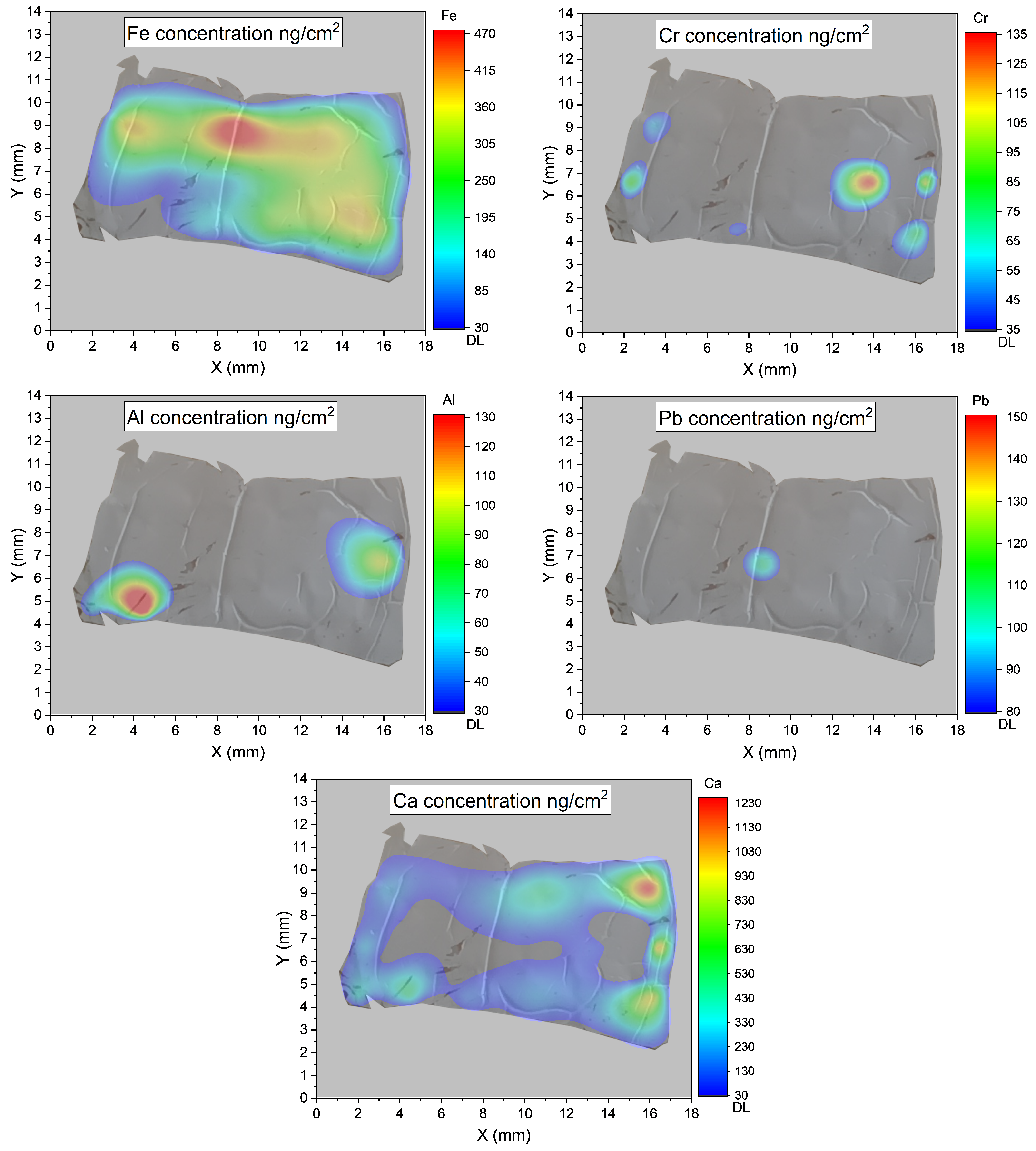
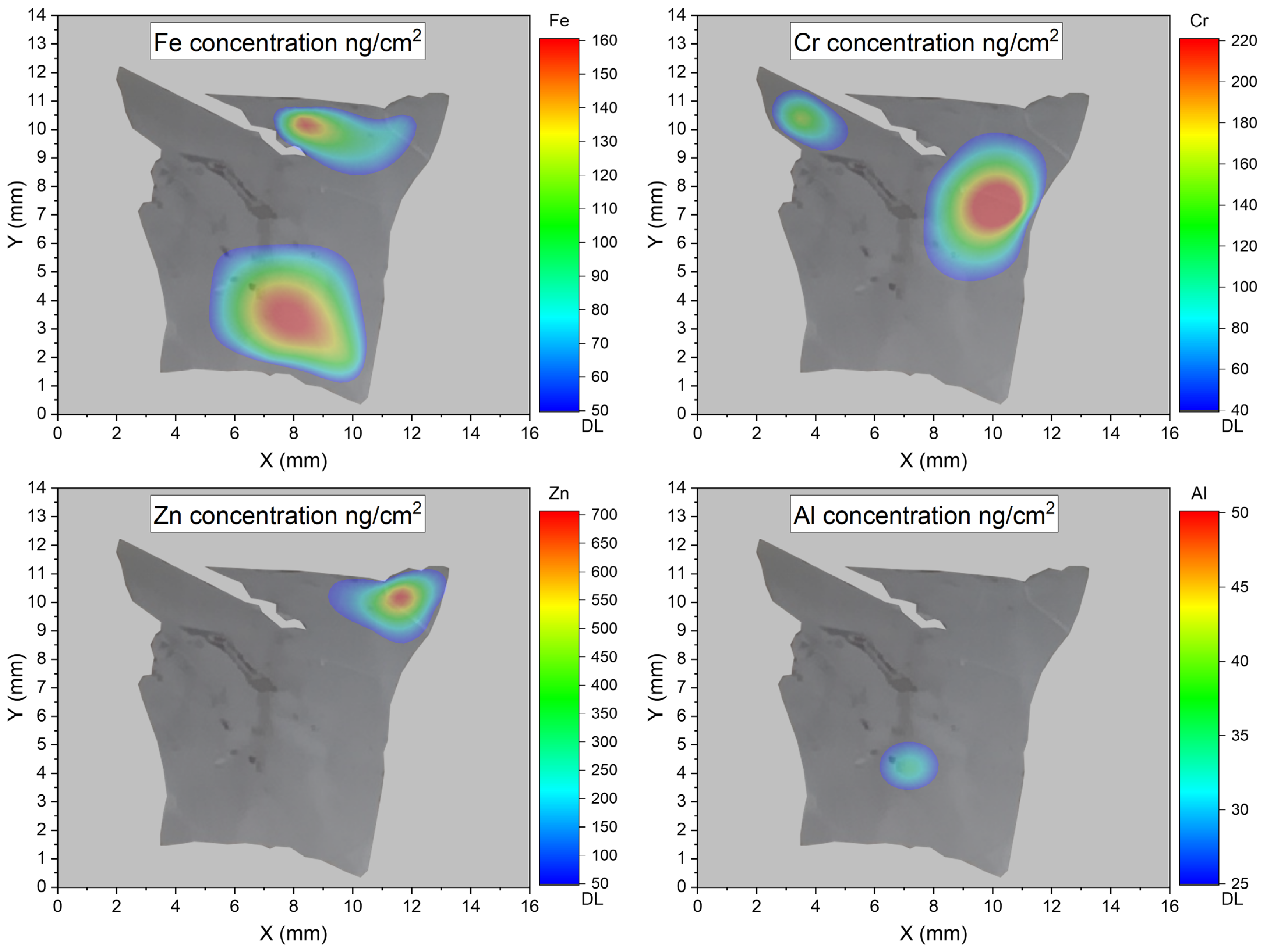
PIXE analysis revealed the presence of Fe, Cr, Al, Zn, Pb, and Ca in four of the six investigated samples. As expected, the PIXE method offers approximately two to three orders of magnitude greater sensitivity than SEM-EDX [4,5,7,8]. The spatial distribution of the detected elements was constructed and is shown in Figure 3 and Figure 4.
Regarding the spatial distribution maps where extensive areas appear below the detection limit, it is important to clarify that the DL values were determined for each individual element using GUPIXWIN software, taking into account the specific background and measurement parameters for each sample. While these grey zones indicate concentrations below this calculated limit, the presence of specific elements, even at trace levels, was confirmed in some regions of interest. For elements like Pb and Al, which are not essential for human metabolism, their detection, even at low concentrations, is significant and indicative of external accumulation. The provided error margin of 20% serves as a conservative estimate for the overall reliability of the quantitative results, particularly for those elements found above the detection limits.

4. SEM-EDX Method

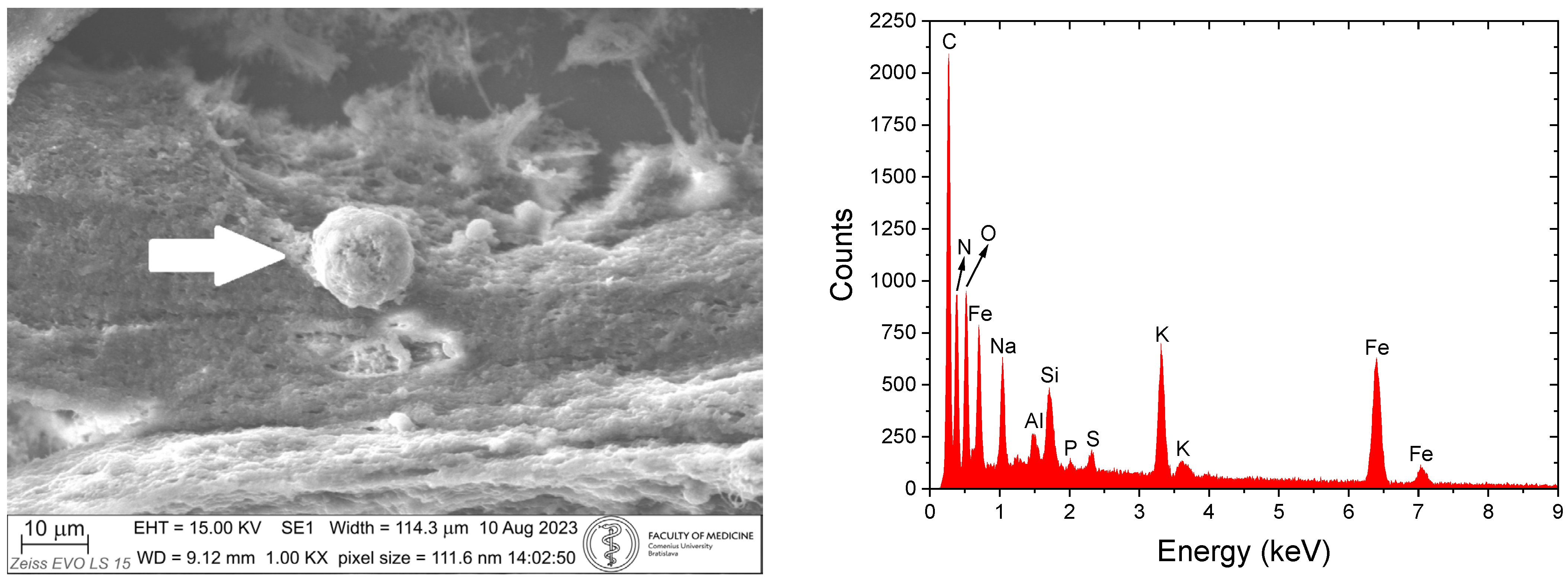
SEM-EDX examination of samples on glass slides revealed micrometre-sized, iron-rich globular structures with a diameter of approximately 15 μm (Figure 5, left). The detection of silicon and sodium was attributed to the glass substrate (Figure 5, right).
Figure 6 shows an SEM image of an irregularly shaped aggregate, approximately 18 µm in size, embedded within the tissue. The aggregate contains C, O, Na, Si, S, Fe, Ni, and Cr. Silicon and sodium likely come from the glass substrate (Figure 6, right).
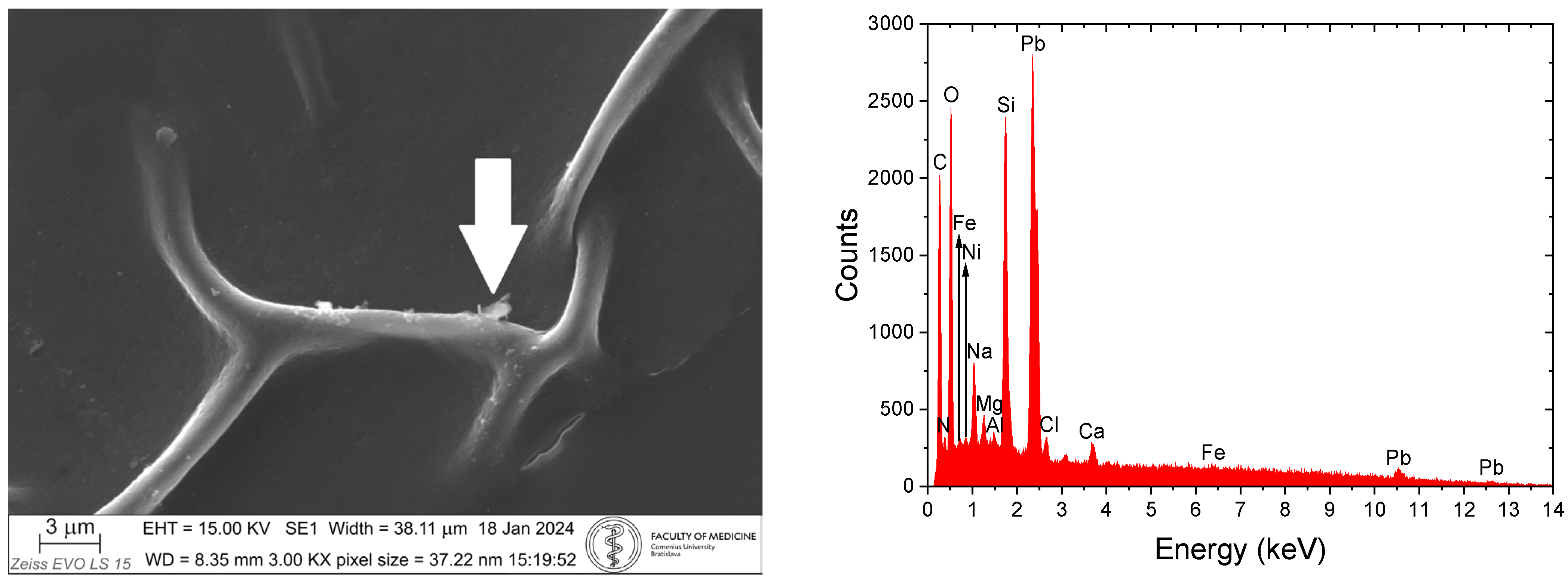
SEM examination of ultrathin sections on gelatine-coated glass shows irregular particles on the surface of the axon. Their size is from ~700 nm (Figure 7, left). SEM-EDX examination reveals the multielemental composition of nanoparticles. Silicon, sodium, magnesium and calcium likely come from the glass substrate (Figure 7, right).
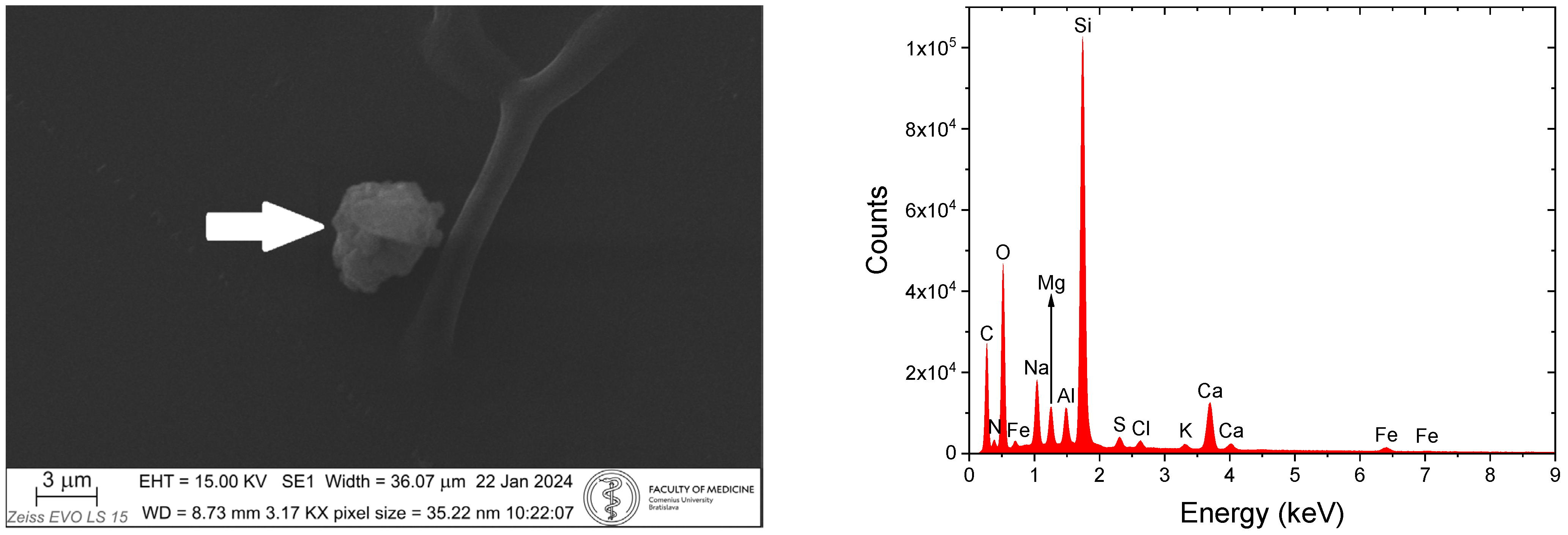
SEM examination of ultrathin sections on gelatine-coated glass shows a globular aggregate composed of smaller irregularly shaped parts (particles) in the vicinity of an axon. The size of particles in the aggregate is from ~500 nm to 3 μm (Figure 8, left). SEM-EDX examination reveals the multielemental composition of the aggregate. Silicon, sodium, magnesium and calcium likely come from the glass substrate (Figure 7, right).
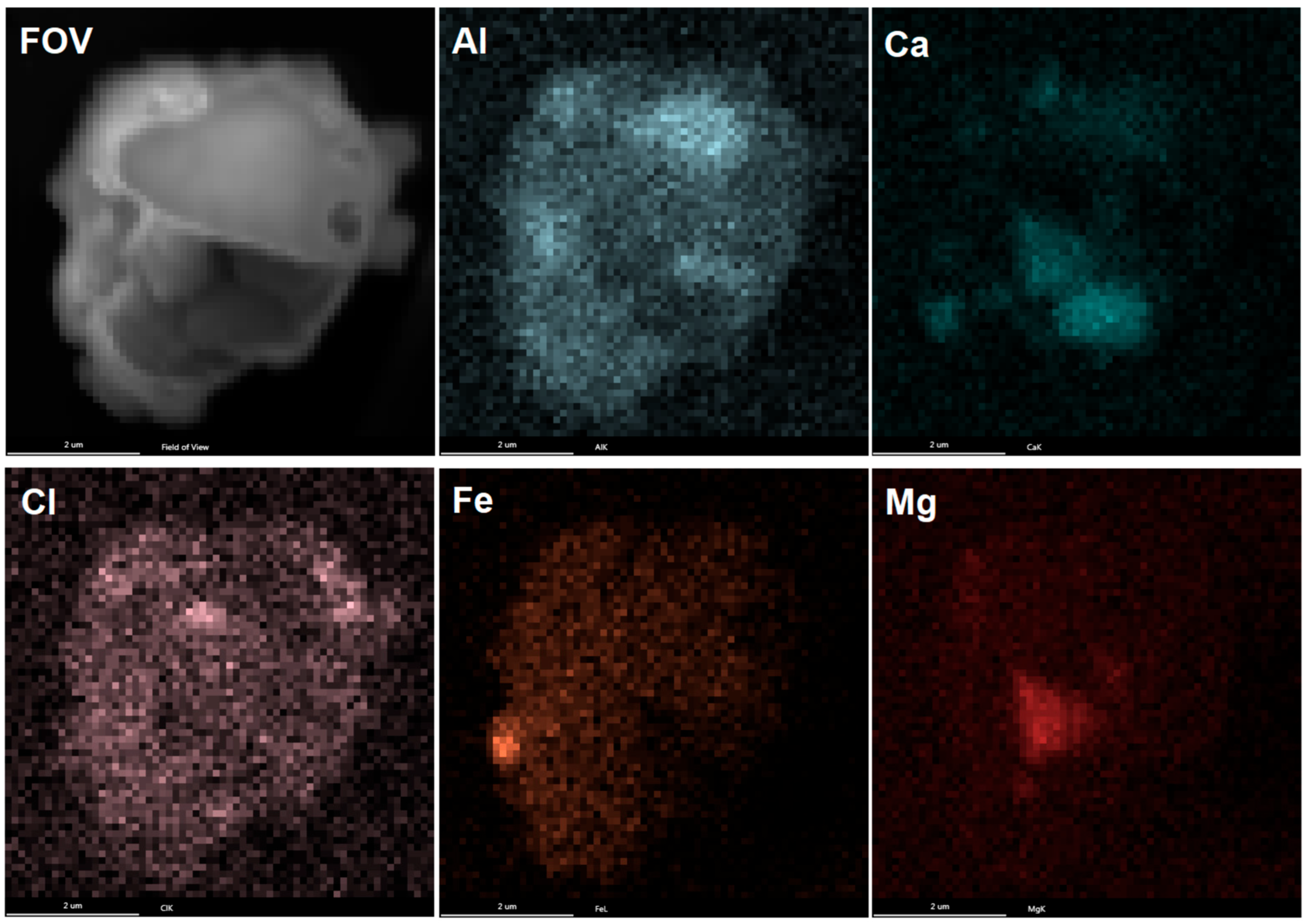
The SEM-EDX maps of the aggregate from Figure 8 are shown in Figure 9. An uneven, focal distribution of Al, Ca, Cl, Fe, and Mg was observed. The aggregate is heterogeneous in terms of mineral compositions with various size distributions and poorly defined morphologies.
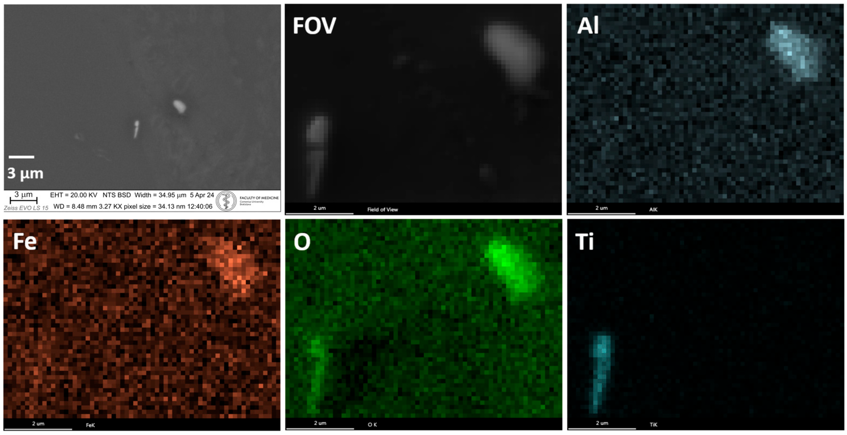
SEM analysis of sample S6 shows two particles with different elemental compositions and shapes (Figure 10). One particle contains Al, O and Fe, the other contains Ti and O.
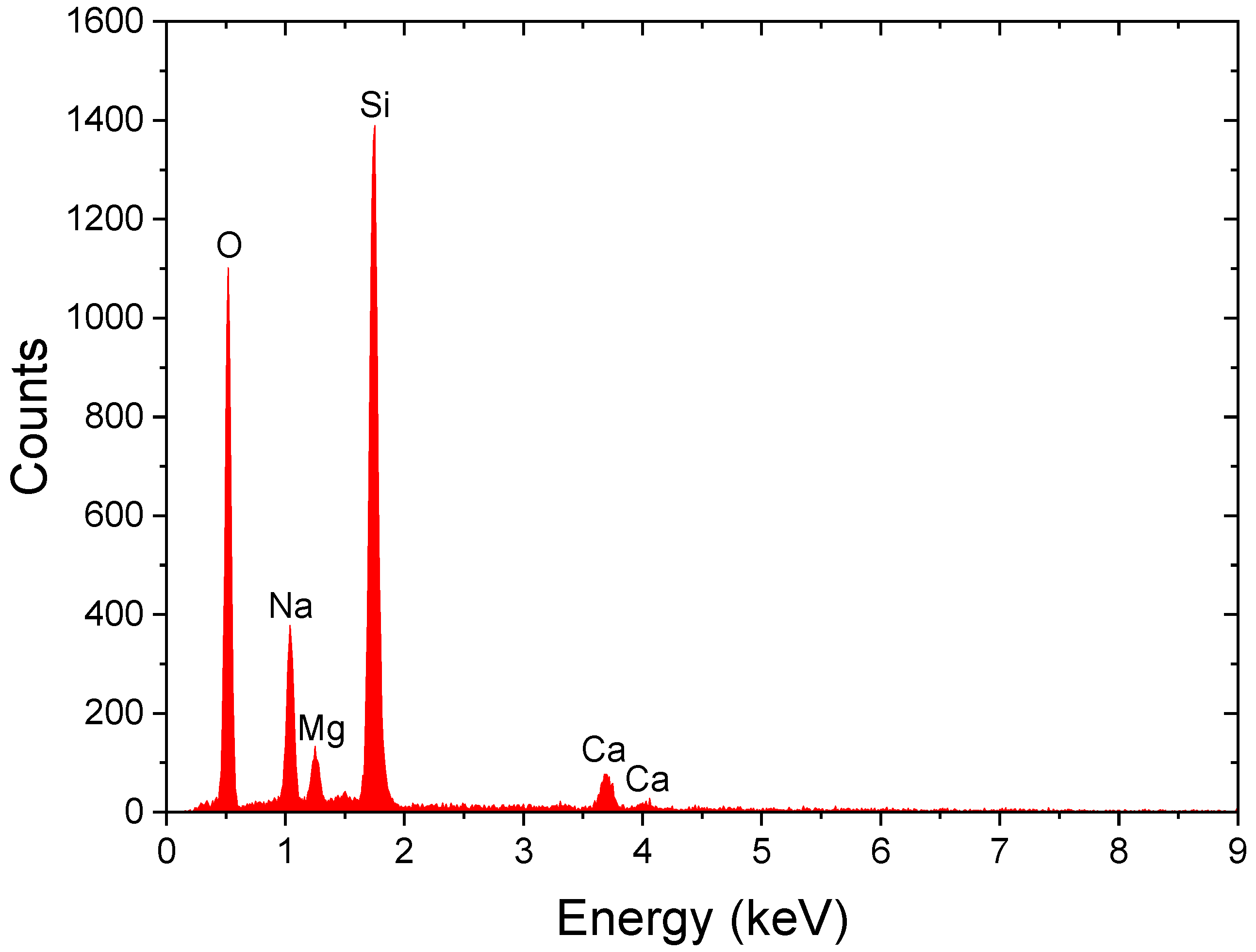
To ascertain potential elemental contribution from the sample substrate, an EDX spectrum of a blank glass slide (without tissue sample) was acquired. This analysis, as shown in Figure 11, revealed the inherent presence of O, Na, Mg, Si, and Ca in the glass material. Consequently, any detection of Na, Mg, Si, and Ca in the tissue sample spectra that directly correlates with the substrate signal was carefully considered and attributed to the supporting glass, as noted in subsequent figures (e.g., Figure 5, Figure 6, Figure 7 and Figure 8). This approach allowed for a more accurate interpretation of elements originating from the biological samples themselves.

5. Discussion

PIXE is a highly sensitive, non-destructive technique suitable for the detection of chemical elements in brain tissue. Our PIXE analysis of samples from individuals without clinical neuropathological findings revealed an accumulation of metals, including Al, Cr, Fe, Zn, and Pb. Many neurological disorders are considered to be due to genetic predispositions, but it is difficult to characterise the metals that trigger them [9]. While these metals can originate from the natural environment, increased industrial activity and urbanisation are thought to enhance their accumulation in the human brain, consequently raising the risk of neuropathologies [10]. The precise mechanisms by which these particles enter the brain remain unclear. Several authors have suggested the nose-to-brain pathway for exogenous particle transport into the brain. Nanoparticle deposits on the olfactory epithelium and subsequent movement to the brain along the olfactory bulb have been observed [11,12,13,14]. Nanoparticles with sizes less than 200 nm are thought to be suitable for the nose-to-brain pathway.
Another possible pathway could be transport from the gastrointestinal tract through the vagus nerves. Direct transport of nanoparticles to the brain may consist of intraneuronal and extraneuronal pathways via intracellular transport across axons and extracellular transport via diffusion and bulk flow through perineuronal channels, perivascular spaces, or lymphatic channels connected to brain tissues [15]. Indirect transport takes place through the vascular system from the respiratory system, leading to the brain by crossing the blood–brain barrier [16]. The accumulation of nanoparticles in the brain may not be limited by one mechanism, but can involve several pathways [17,18].
Aluminium and lead are non-essential metals, and their presence in the human brain likely originates from environmental sources such as air, food, and water pollution. Particulate matter PM 2.5 (the particle size from 1 μm to 2.5 μm) is used as an indicator of pollution. Their presence poses a significant problem for human health because some results indicate a connection between air pollution and neuropathology [19]. Aluminium is a chemical element that is not essential for human metabolism and is not part of proteins. Although its toxicity is well documented, its role in the pathogenesis of several neurological diseases is still unclear. However, its presence in the human brain is associated with Parkinson’s disease and Alzheimer’s disease. From the literature, it can be inferred that aluminium can be used as a marker for some diseases. As the third most abundant metal in the Earth’s crust, its natural sources include minerals such as bauxite, silicates, and cryolite. Other significant sources of this metal are industrial activities, food and water. The sources can vary significantly in aluminium content, and in the way and degree of its uptake.
Lead exhibits severe neurotoxic effects as a result of its ability to mimic or inhibit the action of calcium and zinc ions as regulators of cell function. It alters the release of neurotransmitters from the presynaptic synapse, the developmental processes of synapse formation leading to cognitive deficits, the blockage of voltage-dependent calcium channels in neurons, and the breakdown of the blood–brain barrier [20]. Lead disrupts calcium homeostasis, leading to marked calcium accumulation in lead-exposed cells [21]. Nanoparticles of lead induce calcium release from mitochondria, resulting in apoptosis [22]. The PIXE method can be used for the examination of colocalisation of chemical elements such as Pb and Ca, as has been shown in our results.
Living organisms require varying amounts of metals as they play crucial roles in biochemical reactions critical for brain function. Metals such as selenium, iron, nickel, copper, manganese, molybdenum, chromium, and zinc are required [23]. On the other hand, concentrations higher than physiological level of metals in human cells and tissues are toxic. This toxicity is detected under pathological conditions (iron in Alzheimer’s and Parkinson’s disease, copper in Wilson’s disease) and in tumours [24,25,26]. Romanjuk et al. found calcification processes in cancer tissue with rings containing Fe, Zn, Cu, Cr, and Ni [27]. They discuss that metals can decrease calcium solubility (hydroxyapatite), alter the physical properties of the cell membrane and increase degenerative necrotic changes in breast cancer tissue.
Metal-containing proteins in the brain play vital roles in many neurological functions. Under some pathological conditions, their aggregation may occur. This is a widespread process leading to altered structures and function of cells in the human brain. This process results from altered interaction with ions, altered conditions such as pH, temperature, protein and metal concentration, and biochemical reactions. Protein aggregation can be either accelerated or inhibited in the presence of metal ions [28]. Because ions of chromium, iron, and zinc are an integral part of proteins in the human brain, their aggregation leads to their accumulation, as found in our measurements.
Chromium is an essential trace chemical element distributed in the human body, important for the proper functioning of the hormone insulin. It is involved in the breakdown and absorption of carbohydrates, proteins, and fats. A high concentration of chromium as a protein complex can be found in bone marrow, lungs, lymph nodes, spleen, kidney, and liver. Chromium deposits were detected in vascular siderosis in three cases of neuropathology. The deposits also contained other chemical elements, e.g., phosphorus and calcium [29]. Higher concentrations of Cr, Ca, Se, Co, Ni, and V have been found in malignant and benign brain tumours [30].
Iron is an essential element used for fundamental cell functions and as a catalyst for chemical reactions. It can be found in the human body—mainly in the form of ferritin [31]. It is primarily an iron storage protein located in the cytoplasm of cells and in small amounts in the blood circulation [32,33]. Higher concentrations of iron and ferritin accompany neurological diseases such as Alzheimer’s disease and Parkinson’s disease.
Zinc, like iron, is an important chemical element in the central nervous system. Its physiological function includes the catalysis of biochemical reactions, a contribution to antioxidant function, the regulation of cell growth, and cell proliferation. Zinc is also important for the proper functioning of the immune system [34]. Higher concentrations of zinc cause neuronal death after stroke, epileptic seizures, hypoglycaemia or trauma injury. Nickel is a trace element that plays an important role in biochemical reactions. It is a component of enzymes and proteins with a catalytic function [35,36]. Titanium found in the brain likely comes from environmental pollution or titanium-based implants. It is not known the importance of titanium in the brain or as a part of proteins.
The use of SEM-EDX allows the examination of metal deposition with better resolution at the nanometre level. We found micrometre-sized, round-shaped iron deposits and irregularly shaped nickel deposits. Our previous results revealed that metal particles in the brain are covered with glycoconjugates, which can reduce their toxicity [37]. Deposits can be the result [35,36] of protein/enzyme aggregation and/or pollution of water, air and food.
The accumulation of exogenous nanoparticles in the human brain through different pathways may not explain the presence of several micrometre-sized aggregates. Another mechanism that may be responsible for their presence in the Globus pallidus internus of the human brain is the biomineralisation of nanoparticles. This is the endogenous interaction between the products of metabolic processes and the surrounding cells and tissues. It is a multistep process resulting in the formation of minerals. Biologically induced mineralisation is a process with little control of the organism and cells over mineral formation. Factors such as pH, concentration of organic and inorganic ions, and metabolic activity influence the final products of the process. It results in heterogeneous mineral compositions with large size variations, poor crystallinity, and poorly defined crystal morphologies [38]. Our results from the SEM-EDX examination reveal aggregates with various chemical compositions, large size variations, and morphologies. The SEM-EDX map shows a slight colocalisation of Al with Mg, Fe, and Ca, and colocalisation of Mg and Ca. Diffraction (X-rays, electrons, neutrons) or X-ray spectroscopy provides precise determination of the crystal structure of the aggregate, which is important in the examination of the toxic effect of metals. Calcium, magnesium and chlorine play a crucial role in the nervous system and are important chemical elements in signal transmission, synaptic plasticity, neurotransmitter release, excitability and development. Calcium is a part of calcium-binding proteins and its dysregulation is observed in neurological diseases. Magnesium binds to enzymes and is responsible for their functioning. Chlorine is not part of proteins, but can interact with them and alter their function. Their accumulation can be a response of the organism to the presence of deposits, including sequestering free ions or metals from dysregulated metabolic processes to small areas, reducing their harmful effects, and thus maintaining homeostasis [39]. From our results, we can conclude that the presence of aggregates in the Globus pallidus could be the effect of biologically induced mineralisation. Biologically controlled mineralisation is the process in which minerals are created under the extensive control of cells and tissues. The result of this process is minor size variations in minerals with well-ordered structures, well-defined properties (mechanical, optical, magnetic, electrical) [40]. The function of this process in the human brain is unknown. The biomineralisation of iron in various areas of the human brain is known [41,42] and its products—magnetite and hematite nanoparticles—exhibit enzyme-like catalytic activity [43,44]. Nanoparticles of zinc and chromium also exhibit enzyme-like catalytic activity [45,46]. This raises the question: can the biomineralisation (specifically, biologically controlled biomineralisation) of deposited metals transform them into nanoparticles with enzyme-like catalytic and protective effects on brain tissue?
Biomineralisation of metals in the brain can lead to the formation of metal-based particles with enzyme-like catalytic activity in order to help with the detoxification of ROS, regulate metal homeostasis and protect cells. Even in this case, a synergistic effect of exogenous particle accumulation and endogenous biomineralisation comes into consideration. A high concentration of metals in the human brain can be responsible for alterations in the mechanisms related to cellular metabolism and the excessive production of ROS causes DNA damage. In addition, the toxic effect of metals may not be caused by the presence of a single metal but by a combination of several metals.

6. Conclusions

The presence of metals in the human Globus pallidus appears to be multifactorial. Environmental pollution is arguably a significant contributor to this accumulation. Metals can accumulate through various pathways. Since some metals are parts of proteins that are required for their physiological function, we cannot rule out the possibility of their aggregation and thus their accumulation. Furthermore, biomineralisation processes, particularly biologically induced biomineralisation, may be responsible for producing the micrometre-sized, heterogeneous aggregates observed in this study. It was observed that products of the biologically controlled biomineralisation process exhibit enzyme-like catalytic activity. It is plausible that the organism responds to metal presence through a two-step process: first, biologically induced biomineralisation to sequester toxic ions, followed by biologically controlled biomineralisation to create functional minerals with potentially protective properties.

Author Contributions

Software, D.K.; Validation, J.P., M.J. and J.Z.; Formal analysis, D.K., J.P., M.J. and J.Z.; Investigation, D.K., J.P., M.J. and J.Z.; Writing—original draft, M.K.; Writing—review & editing, J.P., P.P.P. and Š.P. All authors have read and agreed to the published version of the manuscript.

Funding

This research was funded by Cedars-Sinai Medical Center’s International Research and Innovation in Medicine Program and the Association for Regional Cooperation in the Fields of Health, Science and Technology (RECOOP HST Association) and Slovak Research and development Agency project No. APVV-21-0059.

Institutional Review Board Statement

The study was conducted in accordance with the Declaration of Helsinki. Ethical review and approval were waived for this study by the Ethics Committee of the Medical Faculty of Comenius University and University Hospital Bratislava (statement dated 10 July 2023), as the research utilized anonymized post-mortem autopsy data regulated by higher state authority under Slovak Republic legislation (Act No. 581/2004 Coll. on Health Care and amendments), involving no living subjects and excluding personal identifiers. The publication of anonymized results was approved by the Health Care Surveillance Authority of the Slovak Republic (protocol code 6908/2025/960, 39825/2025, dated 9 June 2025), confirming compliance with ethical standards for post-mortem analyses (§ 48 section 3), rendering informed consent inapplicable.

Informed Consent Statement

Patient consent was waived for this study due to its post-mortem nature, involving anonymized biological material from routine autopsies under Slovak Republic legislation (Act No. 581/2004 Coll. on Health Care, amendments, § 48 section 3). Analyses were performed after death for diagnostic/research purposes, making consent inapplicable; all data excluded personal identifiers. Publication was approved by the Health Care Surveillance Authority (protocol 6908/2025/680, dated 9 June 2025), ensuring ethical compliance.

Data Availability Statement

The raw data supporting the conclusions of this article will be made available by the authors on request.

Acknowledgments

We thank Cedars-Sinai Medical Center’s International Research and Innovation in Medicine Program and the Association for Regional Cooperation in the Fields of Health, Science and Technology (RECOOP HST Association) for their support of our study and our organisation as a participating Cedars-Sinai Medical Center—RECOOP Research Center (CRRC). The CENTA group acknowledges partial support provided by the Slovak Research and Development Agency (project No. APVV-21-0059).

Conflicts of Interest

The authors declare no conflict of interest.

References

  1. Pánik, J.; Kopáni, M.; Zeman, J.; Ješkovský, M.; Kaizer, J.; Povinec, P.P. Determination of Metal Elements Concentrations in Human Brain Tissues Using PIXE and EDX Methods. J. Radioanal. Nucl. Chem. 2018, 3, 2313–2319. [Google Scholar] [CrossRef]
  2. Kopáni, M.; Pánik, J.; Filová, B.; Bujdoš, M.; Mišek, J.; KOHAN, M.; Jakuš, J.; Povinec, P.P. PIXE Analysis of Iron in Rabbit Cerebellum after Exposure to Radiofrequency Electromagnetic Fields. Bratisl. Med. J. 2022, 123, 864–871. [Google Scholar] [CrossRef]
  3. University of Guelph. GupixWIN. Available online: https://www.physics.uoguelph.ca/about-gupix-and-gupixwin (accessed on 24 June 2024).
  4. Zeman, J.; Ješkovský, M.; Kaiser, R.; Kaizer, J.; Povinec, P.P.; Staníček, J. PIXE Beam Line at the CENTA Facility of the Comenius University in Bratislava: First Results. J. Radioanal. Nucl. Chem. 2017, 311, 1409–1415. [Google Scholar] [CrossRef]
  5. Kvasniak, J.; Ješkovský, M.; Zeman, J.; Kontuľ, I.; Kaizer, J.; Sučák, K.; Povinec, P.P. Improvements in the PIXE System of the CENTA Laboratory with Application in the Contamination Studies of Tree Rings in Slovakia. Nucl. Instrum. Methods Phys. Res. B 2024, 548, 165254. [Google Scholar] [CrossRef]
  6. Povinec, P.P.; Masarik, J.; Ješkovský, M.; Kaizer, J.; Šivo, A.; Breier, R.; Pánik, J.; Staníček, J.; Richtáriková, M.; Zahoran, M.; et al. Development of the Accelerator Mass Spectrometry Technology at the Comenius University in Bratislava. Nucl. Instrum. Methods Phys. Res. B 2015, 361, 87–94. [Google Scholar] [CrossRef]
  7. Malmqvist, K.G. Accelerator-Based Ion Beam Analysis—An Overview and Future Prospects. Radiat. Phys. Chem. 2004, 71, 817–827. [Google Scholar] [CrossRef]
  8. Kuisma-Kursula, P. Accuracy, Precision and Detection Limits of SEM-WDS, SEM-EDS and PIXE in the Multi-Elemental Analysis of Medieval Glass. X-Ray Spectrom. 2000, 29, 111–118. [Google Scholar] [CrossRef]
  9. Cannon, J.R.; Greenamyre, J.T. The Role of Environmental Exposures in Neurodegeneration and Neurodegenerative Diseases. Toxicol. Sci. 2011, 124, 225–250. [Google Scholar] [CrossRef]
  10. Schottenfeld, D.; Beebe-Dimmer, J.L.; Buffler, P.A.; Omenn, G.S. Current Perspective on the Global and United States Cancer Burden Attributable to Lifestyle and Environmental Risk Factors. Annu. Rev. Public. Health 2013, 34, 97–117. [Google Scholar] [CrossRef] [PubMed]
  11. You, R.; Ho, Y.-S.; Chang, R.C.-C. The Pathogenic Effects of Particulate Matter on Neurodegeneration: A Review. J. Biomed. Sci. 2022, 29, 15. [Google Scholar] [CrossRef] [PubMed]
  12. Moshkin, M.P.; Petrovski, D.V.; Akulov, A.E.; Romashchenko, A.V.; Gerlinskaya, L.A.; Ganimedov, V.L.; Muchnaya, M.I.; Sadovsky, A.S.; Koptyug, I.V.; Savelov, A.A.; et al. Nasal Aerodynamics Protects Brain and Lung from Inhaled Dust in Subterranean Diggers, Ellobius talpinus. Proc. R. Soc. B Biol. Sci. 2014, 281, 20140919. [Google Scholar] [CrossRef]
  13. Kao, Y.-Y.; Cheng, T.-J.; Yang, D.-M.; Wang, C.-T.; Chiung, Y.-M.; Liu, P.-S. Demonstration of an Olfactory Bulb–Brain Translocation Pathway for ZnO Nanoparticles in Rodent Cells In Vitro and In Vivo. J. Mol. Neurosci. 2012, 48, 464–471. [Google Scholar] [CrossRef]
  14. Hopkins, L.E.; Patchin, E.S.; Chiu, P.-L.; Brandenberger, C.; Smiley-Jewell, S.; Pinkerton, K.E. Nose-to-Brain Transport of Aerosolised Quantum Dots Following Acute Exposure. Nanotoxicology 2014, 8, 885–893. [Google Scholar] [CrossRef]
  15. Formica, M.L.; Real, D.A.; Picchio, M.L.; Catlin, E.; Donnelly, R.F.; Paredes, A.J. On a Highway to the Brain: A Review on Nose-to-Brain Drug Delivery Using Nanoparticles. Appl. Mater. Today 2022, 29, 101631. [Google Scholar] [CrossRef]
  16. Li, W.; Lin, G.; Xiao, Z.; Zhang, Y.; Li, B.; Zhou, Y.; Ma, Y.; Chai, E. A Review of Respirable Fine Particulate Matter (PM2.5)-Induced Brain Damage. Front. Mol. Neurosci. 2022, 15, 967174. [Google Scholar] [CrossRef] [PubMed]
  17. Lee, D.; Minko, T. Nanotherapeutics for Nose-to-Brain Drug Delivery: An Approach to Bypass the Blood Brain Barrier. Pharmaceutics 2021, 13, 2049. [Google Scholar] [CrossRef]
  18. Mittal, D.; Ali, A.; Md, S.; Baboota, S.; Sahni, J.K.; Ali, J. Insights into Direct Nose to Brain Delivery: Current Status and Future Perspective. Drug Deliv. 2014, 21, 75–86. [Google Scholar] [CrossRef] [PubMed]
  19. Thiankhaw, K.; Chattipakorn, N.; Chattipakorn, S.C. PM2.5 Exposure in Association with AD-Related Neuropathology and Cognitive Outcomes. Environ. Pollut. 2022, 292, 118320. [Google Scholar] [CrossRef]
  20. Bressler, J.P.; Goldstein, G.W. Mechanisms of Lead Neurotoxicity. Biochem. Pharmacol. 1991, 41, 479–484. [Google Scholar] [CrossRef]
  21. Bressler, J.; Kim, K.; Chakraborti, T.; Goldstein, G. Molecular Mechanisms of Lead Neurotoxicity. Neurochem. Res. 1999, 24, 595–600. [Google Scholar] [CrossRef] [PubMed]
  22. Silbergeld, E.K. Mechanisms of Lead Neurotoxicity, or Looking beyond the Lamppost. FASEB J. 1992, 6, 3201–3206. [Google Scholar] [CrossRef]
  23. Lane, T.W.; Morel, F.M.M. A Biological Function for Cadmium in Marine Diatoms. Proc. Natl. Acad. Sci. USA 2000, 97, 4627–4631. [Google Scholar] [CrossRef]
  24. Caffo, M.; Caruso, G.; La Fata, G.; Barresi, V.; Visalli, M.; Venza, M.; Venza, I. Heavy Metals and Epigenetic Alterations in Brain Tumors. Curr. Genom. 2015, 15, 457–463. [Google Scholar] [CrossRef] [PubMed]
  25. Caruso, G.; Nanni, A.; Curcio, A.; Lombardi, G.; Somma, T.; Minutoli, L.; Caffo, M. Impact of Heavy Metals on Glioma Tumorigenesis. Int. J. Mol. Sci. 2023, 24, 15432. [Google Scholar] [CrossRef]
  26. Pagano, C.; Navarra, G.; Coppola, L.; Savarese, B.; Avilia, G.; Giarra, A.; Pagano, G.; Marano, A.; Trifuoggi, M.; Bifulco, M.; et al. Impacts of Environmental Pollution on Brain Tumorigenesis. Int. J. Mol. Sci. 2023, 24, 5045. [Google Scholar] [CrossRef]
  27. Romanjuk, A.; Lyndin, M.; Moskalenko, R.; Gortinskaya, O.; Lyndina, Y. The Role of Heavy Metal Salts in Pathological Biomineralization of Breast Cancer Tissue. Adv. Clin. Exp. Med. 2016, 25, 907–910. [Google Scholar] [CrossRef]
  28. Poulson, B.G.; Szczepski, K.; Lachowicz, J.I.; Jaremko, L.; Emwas, A.-H.; Jaremko, M. Aggregation of Biologically Important Peptides and Proteins: Inhibition or Acceleration Depending on Protein and Metal Ion Concentrations. RSC Adv. 2020, 10, 215–227. [Google Scholar] [CrossRef]
  29. Duckett, S. Abnormal Deposits of Chromium in the Pathological Human Brain. J. Neurol. Neurosurg. Psychiatry 1986, 49, 296–301. [Google Scholar] [CrossRef]
  30. Hayat, L. Cations in Malignant and Benign Brain Tumors. J. Environ. Sci. Health Part A Environ. Sci. Eng. Toxicol. 1996, 31, 1831–1840. [Google Scholar] [CrossRef]
  31. Plays, M.; Müller, S.; Rodriguez, R. Chemistry and Biology of Ferritin. Metallomics 2021, 13, mfab021. [Google Scholar] [CrossRef] [PubMed]
  32. Arosio, P.; Elia, L.; Poli, M. Ferritin, Cellular Iron Storage and Regulation. IUBMB Life 2017, 69, 414–422. [Google Scholar] [CrossRef]
  33. Alkhateeb, A.A.; Connor, J.R. Nuclear Ferritin: A New Role for Ferritin in Cell Biology. Biochim. Biophys. Acta (BBA)—General. Subj. 2010, 1800, 793–797. [Google Scholar] [CrossRef]
  34. Choi, S.; Hong, D.K.; Choi, B.Y.; Suh, S.W. Zinc in the Brain: Friend or Foe? Int. J. Mol. Sci. 2020, 21, 8941. [Google Scholar] [CrossRef] [PubMed]
  35. Alfano, M.; Cavazza, C. Structure, Function, and Biosynthesis of Nickel-dependent Enzymes. Protein Sci. 2020, 29, 1071–1089. [Google Scholar] [CrossRef] [PubMed]
  36. Zambelli, B.; Ciurli, S. Nickel and Human Health. Met Ions Life Sci. 2013, 13, 321–357. [Google Scholar] [CrossRef] [PubMed]
  37. Kopáni, M.; Kopániová, A.; Čaplovičová, M.; Marusčaková, L.; Šišovský, V.; Jakubovský, J. Iron and Its Relation to Glycoconjugates in Human Globus Pallidus. Bratisl. Med. J. 2014, 115, 362–366. [Google Scholar] [CrossRef] [PubMed]
  38. Weiner, S. An Overview of Biomineralization Processes and the Problem of the Vital Effect. Rev. Miner. Geochem. 2003, 54, 1–29. [Google Scholar] [CrossRef]
  39. Fasae, K.D.; Abolaji, A.O.; Faloye, T.R.; Odunsi, A.Y.; Oyetayo, B.O.; Enya, J.I.; Rotimi, J.A.; Akinyemi, R.O.; Whitworth, A.J.; Aschner, M. Metallobiology and Therapeutic Chelation of Biometals (Copper, Zinc and Iron) in Alzheimer’s Disease: Limitations, and Current and Future Perspectives. J. Trace Elem. Med. Biol. 2021, 67, 126779. [Google Scholar] [CrossRef]
  40. Bazylinski, D.A. Biologically Controlled Mineralization in Prokaryotes. Rev. Miner. Geochem. 2003, 54, 217–247. [Google Scholar] [CrossRef]
  41. Bazala, R.; Zoppellaro, G.; Kletetschka, G. Iron Level Changes in the Brain with Neurodegenerative Disease. Brain Multiphys 2023, 4, 100063. [Google Scholar] [CrossRef]
  42. Kirschvink, J.L.; Kobayashi-Kirschvink, A.; Woodford, B.J. Magnetite Biomineralization in the Human Brain. Proc. Natl. Acad. Sci. USA 1992, 89, 7683–7687. [Google Scholar] [CrossRef]
  43. Gao, L.; Fan, K.; Yan, X. Iron Oxide Nanozyme: A Multifunctional Enzyme Mimetic for Biomedical Applications. Theranostics 2017, 7, 3207–3227. [Google Scholar] [CrossRef]
  44. Sajjad, A.; Hussain, S.; Jaffari, G.H.; Hanif, S.; Qureshi, M.N.; Zia, M. Fabrication of Hematite (α-Fe2O3) Nanoparticles under Different Spectral Lights Transforms Physio Chemical, Biological, and Nanozymatic Properties. Nano Trends 2023, 2, 100010. [Google Scholar] [CrossRef]
  45. Sajjad, A.; Bhatti, S.H.; Ali, Z.; Jaffari, G.H.; Khan, N.A.; Rizvi, Z.F.; Zia, M. Photoinduced Fabrication of Zinc Oxide Nanoparticles: Transformation of Morphological and Biological Response on Light Irradiance. ACS Omega 2021, 6, 11783–11793. [Google Scholar] [CrossRef] [PubMed]
  46. Xie, F.; Zhu, C.; Gong, L.; Zhu, N.; Ma, Q.; Yang, Y.; Zhao, X.; Qin, M.; Lin, Z.; Wang, Y. Engineering Core–Shell Chromium Nanozymes with Inflammation-Suppressing, ROS-Scavenging and Antibacterial Properties for Pulpitis Treatment. Nanoscale 2023, 15, 13971–13986. [Google Scholar] [CrossRef] [PubMed]
Figure 1. Origin of the characteristic X-ray of the elements present in the sample for PIXE (incident proton) or EDX (incident electron).
Applsci 15 09897 g001
Figure 2. A representative X-ray spectrum of brain tissue (black line) acquired via PIXE analysis. The spectrum was processed and fit (red line) using the GUPIXWIN software. The observed peaks correspond to the characteristic X-ray energies of the elements present in the tissue.
Applsci 15 09897 g002
Figure 3. Spatial distribution of Fe, Cr, Al, Pb, and Ca in sample S1 using the PIXE method. The grey colour shows the detection limit (DL) of the measurement for each element: Fe < 30 ng/cm2, Cr < 35 ng/cm2, Al < 30 ng/cm2, Pb < 80 ng/cm2, and Ca < 30 ng/cm2.
Applsci 15 09897 g003aApplsci 15 09897 g003b
Figure 4. Spatial distribution of Fe, Cr, Zn, and Al in sample S2 using the PIXE method. The grey colour shows the detection limit (DL) of the measurement for each element: Fe < 50 ng/cm2, Cr < 40 ng/cm2, Zn < 50 ng/cm2, and Al < 25 ng/cm2.
Applsci 15 09897 g004
Figure 5. SEM image of sample S5 from the GPi. (Left): An iron-rich globular structure (indicated by the white arrow) approximately 15 µm in diameter. (Right): The corresponding EDX spectrum confirms the presence of iron, alongside other elements. Signals for Na and Si are attributed to the glass substrate.
Applsci 15 09897 g005
Figure 6. An irregularly shaped aggregate observed in sample S5 from the GPi. (Left): The SEM image shows a structure of approximately 18 µm in size (the white arrow). (Right): The corresponding EDX spectrum confirms its multielemental composition, including prominent signals for Fe, Cr, and Ni. The presence of Na and Si is attributed to the glass substrate.
Applsci 15 09897 g006
Figure 7. A lead-rich particle on the surface of an axon in sample S4 from the GPi. (Left): The SEM image shows an elongated particle approximately 700 nm in length (the white arrow). (Right): The EDX spectrum reveals a complex elemental composition, highlighted by a clear signal for Pb. Signals for Na, Mg, Si, and Ca are considered contributions from the glass substrate.
Applsci 15 09897 g007
Figure 8. A globular aggregate composed of smaller particles, observed in sample S5 near an axon. (Left): The SEM image shows the aggregate (the white arrow). (Right): The multielemental nature of the aggregate is confirmed by the EDX spectrum, with Fe being a notable component. Substrate-related elements include Na, Mg, Si, and Ca.
Applsci 15 09897 g008
Figure 9. SEM-EDX image of elemental distribution maps of a globular aggregate observed in sample S5. The maps reveal a heterogeneous and uneven distribution of several elements, including Al, Ca, Cl, Fe, and Mg. The irregular focal distribution of these elements confirms the aggregate’s heterogeneous mineral composition and poorly defined morphology.
Applsci 15 09897 g009
Figure 10. Elemental maps of two distinct particles found in sample S6 from the GPi. The upper particle is composed primarily of Al, Fe and O, while the lower particle consists of Ti and O.
Applsci 15 09897 g010
Figure 11. EDX spectrum obtained from a blank glass slide used for sample preparation. This spectrum serves as a crucial background reference, demonstrating the inherent presence of O, Na, Mg, Si, and Ca in the substrate material itself.
Applsci 15 09897 g011
Table 1. Gender, ages, causes of death, and post-mortem intervals for the investigated samples.
SampleGenderAge (Years)Cause of DeathPost-Mortem Interval (Hours)Occupation
S1Male53Cirrhosis7Teacher
S2Female69Heart failure10Lawyer
S3Female83Heart failure8Saleswoman
S4Male19Nephritis6Student
S5Male67Heart failure9Builder
S6Female72Thrombosis10Cook
Table 2. Characteristic X-ray energies for detected elements: K, L, and M lines.
ElementSymbolZΚα [keV]Κβ [keV]Lα [keV]Mα [keV]
CarbonC60.277---
NitrogenN70.392---
OxygenO80.525---
SodiumNa111.0411.071--
MagnesiumMg121.2541.302--
AluminiumAl131.4871.557--
SiliconSi141.741.836--
PhosphorusP152.0142.139--
SulphurS162.3082.464--
ChlorineCl172.6222.816--
PotassiumK193.3143.59--
CalciumCa203.6924.0130.341-
TitaniumTi224.514.9320.452-
ChromeCr245.4155.9470.573-
IronFe266.4047.0580.705-
NickelNi287.4788.2650.852-
ZincZn308.6399.5721.012-
LeadPb8274.9784.93610.5522.346
Disclaimer/Publisher’s Note: The statements, opinions and data contained in all publications are solely those of the individual author(s) and contributor(s) and not of MDPI and/or the editor(s). MDPI and/or the editor(s) disclaim responsibility for any injury to people or property resulting from any ideas, methods, instructions or products referred to in the content.