Article

Cymbopogon flexuosus and Eugenol Nanoemulsion: Formulation, Stability, Antimicrobial Efficacy, and In Vitro Safety Assessment

by
Franciane Batista Nunes
1,
Ruth Barin
2,
Larissa da Silva Silveira
3,
Michele Rorato Sagrillo
3,
Leonardo Vidal Zancanaro
4,
Vitória Fernanda Belmonte Novais
1,
Aline Ferreira Ourique
2,
André Gündel
5,
Cristiano Rodrigo Bohn Rhoden
4 and
Roberto Christ Vianna Santos
1,*
1
Oral Microbiology Research Laboratory (LAPEMICRO), Graduate Program in Pharmaceutical Sciences, Federal University of Santa Maria—UFSM, Santa Maria 97000-000, RS, Brazil
2
Nanoestructured Systems Research Laboratory (LPSnano), Graduate Program in Nanosciences, Franciscan University—UFN, Santa Maria 97000-000, RS, Brazil
3
Bioprospecting and Experimental Biology Laboratory (Labbie), Graduate Program in Nanosciences, Franciscan University—UFN, Santa Maria 97000-000, RS, Brazil
4
Nanostructured Magnetic Materials Laboratory (LaMMaN), Graduate Program in Nanosciences, Franciscan University—UFN, Santa Maria 97000-000, RS, Brazil
5
Graduate Program in Materials Science and Engineering (PPCEM), Federal University of Pampa—UNIPAMPA, Bagé 96400-000, RS, Brazil
*
Author to whom correspondence should be addressed.
Appl. Sci. 2025, 15(18), 10214; https://doi.org/10.3390/app151810214
Submission received: 4 August 2025 / Revised: 12 September 2025 / Accepted: 15 September 2025 / Published: 19 September 2025

Abstract

Pseudomonas aeruginosa and Staphylococcus aureus are highly resistant microorganisms that contribute to prolonged hospital stays and increased mortality. Developing new antimicrobial agents is essential to address this global health challenge. Nanoemulsions (NE) containing essential oils (EOs) and phenolic compounds with antimicrobial activity represent a promising alternative. This study reports, for the first time, the formulation of a NE containing Cymbopogon flexuosus and eugenol (NECE) and its antimicrobial activity against P. aeruginosa and S. aureus. NECE exhibited suitable physicochemical properties (mean size < 200 nm, PDI < 0.3, and negative zeta potential) and remained stable for 90 days at 4 °C while maintaining antimicrobial activity. It showed bactericidal effects at 2.5 mg/mL against P. aeruginosa and 0.625 mg/mL against S. aureus. Moreover, NECE improved the biocompatibility of the free oil (FO) in Peripheral Blood Mononuclear Cells (PBMCs). Altogether, these findings demonstrate, for the first time, that NECE is a stable nanoemulsion with enhanced antimicrobial activity and biocompatibility, supporting its potential as a safe and effective topical strategy against wound-associated pathogens.

1. Introduction

Antimicrobial resistance (AMR) is projected to cause 10 million deaths annually by 2050 [1,2]. The misuse and improper disposal of antimicrobials accelerate the development and dissemination of resistance genes in the environment [3]. Pseudomonas aeruginosa and Staphylococcus aureus are critical-priority pathogens, associated with high mortality rates and prolonged hospitalizations worldwide [4,5]. Epidemiological studies indicate that the excessive use of antibiotics can exacerbate the emergence of bacterial resistance, contributing to a significant public health crisis [6].
These microorganisms are included in the World Health Organization’s (WHO) 2024 list of priority bacterial pathogens and overlap with the ESKAPEE group, which comprises Enterococcus spp., S. aureus, Klebsiella pneumoniae, Acinetobacter baumannii, P. aeruginosa, Enterobacter spp., and Escherichia coli, widely recognized for their association with multidrug-resistant nosocomial infections [7].
Nanotechnology provides promising strategies for combating resistant infections. NE are kinetically stable colloidal dispersions that enhance the solubility, stability, and controlled release of bioactive compounds [8,9]. EOs of Cymbopogon flexuosus and eugenol possess antibacterial, anti-inflammatory, and analgesic properties [10,11,12,13,14]. Encapsulation of these compounds in nanoemulsion (NE) improves their physicochemical stability and protects them from degradation [15].
Combining essential oils in NE can produce synergistic antimicrobial effects. For example, NE containing blends of cinnamon, tea tree, lemongrass, and oregano EOs significantly reduced minimal inhibitory concentration (MIC) and minimal bactericidal concentration (MBC) values against S. aureus compared to the corresponding free oils [16]. Similarly, NE of clove and lemongrass EOs demonstrated enhanced antifungal activity without inducing phytotoxicity [17].
This study aims to evaluate the MIC, MBC, physicochemical properties, stability, and biocompatibility of a nanoemulsion containing eugenol and C. flexuosus (NECE) against P. aeruginosa and S. aureus. The results highlight NECE’s potential as an effective antimicrobial agent with multiple mechanisms that may prevent the development of resistance, while also demonstrating promising activity for topical application.

2. Methodology

2.1. Chemicals

The culture media Mueller–Hinton broth and agar, used in the antimicrobial assays, were purchased from Himedia® (Mumbai, India). Sodium chloride was obtained from Dinamica (São Paulo, Brazil). For NECE preparation, sorbitan monooleate (Span 80®), polysorbate 80 (Tween 80®), and eugenol were purchased from Sigma-Aldrich® St. Louis, MO, USA. C. flexuosus essential oil was acquired from Ferquímica® (São Paulo, Brazil). Additional reagents included 1× phosphate-buffered saline (PBS) (Prolab®, São Paulo, Brazil), dimethyl sulfoxide (DMSO) (Synth®, Diadema, Brazil), PicoGreen® (Invitrogen® Carlsbad, CA, USA), Ficoll Histopaque-1077VR (Sigma-Aldrich® St. Louis, MO, USA), DCFH-DA (Sigma-Aldrich®, St. Louis, MO, USA), and RPMI 1640 medium (Sigma-Aldrich®, St. Louis, MO, USA).

2.2. Preparation and Characterization of Nanoemulsion

The NE was prepared in triplicate (n = 3) following an adapted high-agitation methodology developed by Gündel et al. (2018) [18] and Godoi et al., (2017) [19]. The oil phase contained 2% (w/w) eugenol (0.8 g), 2% (w/w) C. flexuosus (0.8 g), and 2% (w/w) Sorbitan monooleate (Span 80®) (0.8 g). The aqueous phase consisted of 2% (w/v) Polysorbate 80 (Tween 80) (0.8 g), and the volume was adjusted to 40 mL with ultrapure water. Both phases were stirred separately using a magnetic stirrer for 15 min.
Subsequently, the aqueous phase was homogenized in an Ultra-Turrax® (Ultra-Turrax® T18-IKA disperser) at 10,000 rpm for 1 min. The oil phase was then slowly added to the aqueous phase under cooling, and the mixture was further stirred at 17,000 rpm for 30 min to obtain the NE. Additionally, a free oil (FO) mixture at 4% was prepared to compare the activity of NECE with non-encapsulated actives.

2.3. NECE Characterization

NECE was characterized by a Zetasizer (Zetasizer Nano-ZS, model ZEN 3600, Malvern, Worcestershire, UK) to evaluate zeta potential, particle size, and polydispersity index (PDI). Additionally, the pH was measured using a potentiometer (DM-22, Digimed®, São Paulo, Brazil), and the morphology and particle size were assessed by Atomic Force Microscopy (Agilent Technologies 5500, Santa Clara, CA, USA).

2.4. Stability Study of Nanoemulsion

The stability of NECE (n = 2) was evaluated by measuring particle size, PDI, zeta potential, and pH at different time points (0, 7, 15, 30, 60, and 90 days after preparation) under two storage conditions: room temperature (24 °C) and refrigeration (4 °C).

2.5. Microbiological Assays

2.5.1. Microorganisms

The antimicrobial activity of NECE and FO was evaluated against P. aeruginosa (PA01) and S. aureus ATCC 29213.

2.5.2. Minimal Inhibitory Concentration (MIC)

Broth microdilution was performed to determine the MIC following the guidelines of the Clinical and Laboratory Standards Institute (CLSI, 2008—protocol M27-A3) [20]. The bacterial inoculum was prepared in 0.9% (w/v) saline solution at a density of 0.5 McFarland using a densitometer (DEN-1, Biosan, Riga, Latvia) and subsequently diluted 1:20 (v/v) in Mueller-Hinton broth.
Next, 100 µL of Mueller-Hinton broth and 100 µL of NECE were added to each well of a 96-well microplate, and serial dilutions were performed. Then, 10 µL of the inoculum was added to each well, except for the negative control. All dilutions were performed in duplicate. The microplate was incubated at 37 °C for 24 h, and the MIC was defined as the lowest concentration that showed no visible bacterial growth. The negative control consisted of MH medium, while the positive control consisted of MH medium inoculated with the microorganism (P. aeruginosa or S. aureus). The assay was accomplished in triplicate.

2.5.3. Minimal Bactericide Concentration (MBC)

For MBC determination, following MIC assessment, the concentrations corresponding to ½ MIC, MIC, 2× MIC, and 4× MIC were plated onto Mueller-Hinton agar and incubated at 37 °C for 24 h. The MBC was defined as the lowest concentration that completely inhibited bacterial growth. The negative control consisted of MH medium, while the positive control consisted of MH medium inoculated with the microorganism (P. aeruginosa or S. aureus). The assay was accomplished in triplicate.

2.6. Cel Culture

Peripheral Blood Mononuclear Cells (PBMCs) derived from discarded blood samples of healthy adults were obtained from the Clinical Analysis Laboratory of Franciscan University (LEAC-UFN) (experimental protocol approved by the Human Research Ethics Committee of UFN—CAAE number: 44940821.3.0000.5306).
Blood samples were processed for PBMCs separation using Ficoll Histopaque-1077VR reagent (Sigma-Aldrich, St. Louisan, MO, USA) based on density gradient centrifugation. After mixing the blood with the reagent (1:1 v/v), samples were centrifuged at 1500 rpm for 30 min. The PBMCs were then plated in a 96-well plate containing RPMI 1640 medium supplemented with 10% fetal bovine serum and 1% antimicrobial solution, at a density of 2 × 10⁵ cells/mL per well.
Subsequently, the cells were exposed to different concentrations of NECE to evaluate the biocompatibility of the formulation. All treatments and assays were performed in triplicate to ensure the reliability of the results.

2.6.1. MTT Assay

The biocompatibility effect of NECE was investigated using the 3-(4,5-dimethylthiazol-2-yl)-2,5-diphenyl tetrazolium bromide (MTT) assay according to the technique proposed by Mosmann, (1983) [21]. After 24 h, a solution of 5 mg/mL of the reagent was prepared, diluted in 1×phosphate-buffered saline (PBS) (Prolab®, Weston, FL, USA), and subsequently 20 μL per well in the 96-well plates was maintained at 37 °C under a CO2 atmosphere for four hours. The supernatant was removed, and 200 μL of dimethyl sulfoxide (DMSO) (Synth®) was added to solubilize the crystals formed. Finally, the absorbance was measured at a wavelength of 570 nm using a microplate reader, Anthos 2010 (Biochrom® Anthos 2010, Cambridge, UK). The positive control consisted of 100 mM hydrogen peroxide. The negative control (cells + culture medium) was used to compare the effect caused by the treatments. The assay was accomplished in triplicate.

2.6.2. dsDNA Assay

The DNA integrity of the treatments was assessed using PicoGreen® dye (1:200 in TE buffer). For this assay, 20 ng of pCMUT plasmid DNA samples were diluted in 100 µL of 10 mM Tris-HCl buffer (pH 7.5) and incubated in dark microplates at room temperature for 60 min. Subsequently, 10 µL of PicoGreen® (Thermo Fisher Scientific, Waltham, MA, USA) was added to each well, and fluorescence was measured after five minutes at room temperature, with excitation at 480 nm and emission at 520 nm, using a SpectraMax® i3× plate reader (Molecular Devices, San Jose, CA, USA). The positive control consisted of 100 mM hydrogen peroxide. The negative control (cells + culture medium) was used to compare the effect caused by the treatments. The assay was accomplished in triplicate.

2.6.3. DCFH-DA Assay

Reactive oxygen species (ROS) production, particularly hydrogen peroxide, was measured using dichlorofluorescein diacetate (DCFH-DA). This probe is converted to 2′,7′-dichlorofluorescein, which emits fluorescence detectable at excitation/emission wavelengths of 488/525 nm (SpectraMax® i3×, Molecular Devices).
For the assay, a dark 96-well plate was used, to which 50 μL of cell culture supernatant, 65 μL of 10 mM Tris-HCl buffer (pH 7.4), and 10 μL of DCFH-DA reagent were added. The plate was incubated in the dark for 1 h, after which fluorescence was measured at 480 nm excitation and 520 nm emission using the SpectraMax® i3 plate reader (Molecular Devices). The positive control consisted of 100 mM hydrogen peroxide. The negative control (cells + culture medium) was used to compare the effect caused by the treatments. The assay was accomplished in triplicate.

2.7. Statistical Analysis

Statistical analyses were performed using GraphPad Prism 9.0. For the cell assays, one-way ANOVA was applied, followed by Dunnett’s multiple comparison post hoc test, to compare each treatment group with the control. For the stability study of the NE, two-way ANOVA was used to evaluate the effects of storage time and temperature, followed by Dunnett’s post hoc test for multiple comparisons. The data are expressed as mean ± standard deviation (SD). A p-value < 0.05 was considered statistically significant, with levels of significance represented as p < 0.05, p < 0.01, and p < 0.001.

3. Results and Discussion

3.1. Atomic Force Microscopy

AFM is a key technique for analyzing interfacial properties, enabling high-resolution imaging of surface forces and topography under different conditions, and is essential for studying molecular behavior at nanoemulsion droplet interfaces [22]. The morphology of NECE (Figure 1) was evaluated by AFM. The images reveal the typical spherical structure of the NECE, with particle sizes below 150 nm, consistent with expectations for NE. Prasad et al., (2023) [23] encapsulated Cymbopogon khasiana × Cymbopogon pendulus essential oil (CKP-25-EO) into a chitosan nanoemulsion, with AFM analysis revealing particle sizes ranging from 67 to 100 nm. Ramadan et al., (2024) [24] prepared a NE with 10% clove and cinnamon essential oils, 15% gum arabic, 15% maltodextrin, and 1% Tween 80, resulting in average particle sizes between 100 and 300 nm as observed by AFM. The NE developed in this study shows the particle size above 150 nm. Droplet sizes below 200 nm are considered desirable, as they typically exhibit greater stability and improved bioavailability.

3.2. Determination of Mean Particle Size, Polydispersity Index, Zeta Potential, and pH

Zeta potential, PDI, mean particle size, and pH were analyzed to evaluate the successful formulation of NECE, with results presented in Table 1. Zeta potential reflects the electrokinetic behavior of colloidal dispersion and indicates system stability [25]. NECE exhibited a negative zeta potential, suggesting stability due to the low tendency for nanoparticle aggregation and sedimentation [26].
Another indicator of stability is the PDI, which was below 0.3, demonstrating the homogeneity of nanoparticle sizes in the NECE formulation [27]. The mean particle size obtained in this analysis was consistent with AFM results, showing an average size below 200 nm. Additionally, NECE exhibited an acidic pH, characteristic of the essential oils present in the formulation [18].
Basumatary and Kumar (2024) [28] reported that the NEs containing 5% and 10% eugenol (ENE-5 and ENE-10) exhibited particle sizes of 410.0 ± 0.57 nm and 521.3 ± 0.85 nm, respectively, with corresponding polydispersity indices of 0.25 ± 0.01 and 0.50 ± 0.01, which are outside the typical desirable parameters for stable NE, indicating increased heterogeneity with higher eugenol content. In another study, a nanoemulsion containing 10% Tween 80 and 10% eugenol exhibited a PDI of 0.551. In contrast, the NECE formulation with a lower surfactant concentration showed a lower PDI, indicating greater homogeneity and stability [29]. Already, Putra et al., 2025 developed a NE using lemongrass (Cymbopogon citratus) at 2.5%, 0.75% Span 80, and 4.25% Tween 80. Resulting in a particle size of 70.1 ± 1.7 nm and a PDI of 0.333 ± 0.034 [30]. In the study by Daud et al., 2025, lemongrass-based NE were synthesized by mixing 2% (w/v) lemongrass extract with Tween 80 at a 1:3 ratio (w/w) relative to the extract, followed by high-shear homogenization. The resulting NE presented a droplet size of 86.32 ± 0.66 nm, a PDI of 0.50 ± 0.00, and a zeta potential of −44.01 ± 1.69 mV. These values provide a reference point for comparing the characteristics of the NE developed in the present study [31].
Overall, NECE demonstrates superior performance as a stable and suitable nanoemulsion system, exhibiting an average particle size below 150 nm, a PDI less than 0.3, and negative zeta potential values, surpassing those reported in previous studies.

3.3. Study of NECE Stability

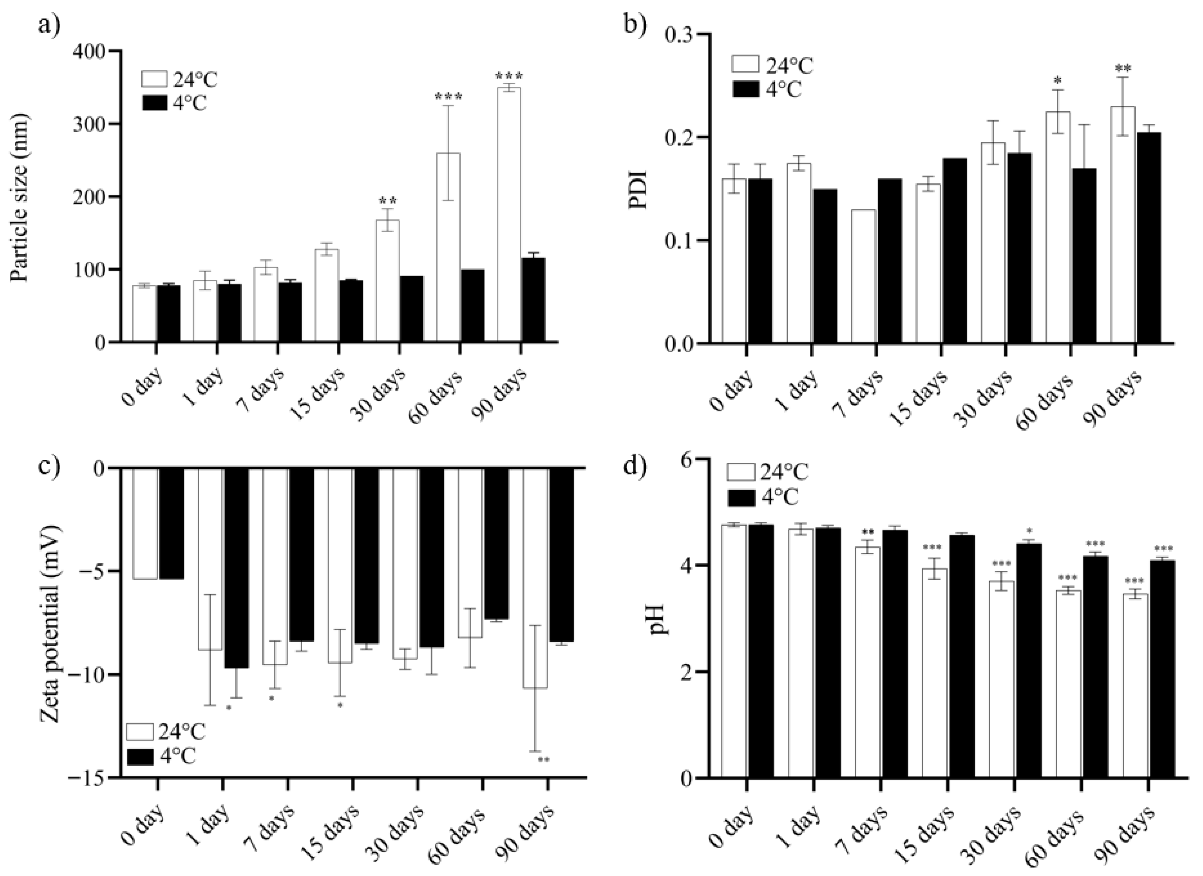
The stability study was conducted to evaluate the physicochemical integrity of NECE over different time points (0, 1, 7, 15, 30, 60, and 90 days after preparation). Stability was assessed by measuring mean particle size, PDI, zeta potential, and pH under two storage conditions: refrigerated (4 °C) and room temperature (24 °C).
The mean particle size of NECE at time 0 was 76.33 ± 0.014 nm under both storage conditions. After 90 days, the refrigerated NECE increased slightly to 115.66 ± 5.03 nm, remaining well below 200 nm and indicating stability. In contrast, NECE stored at room temperature reached 350 ± 5.65 nm after 90 days, reflecting instability. An increase in particle size was already observed at 15 days (133.66 ± 11.50 nm), suggesting the onset of droplet aggregation. Such increases are common in essential oil-containing NE but were effectively prevented by refrigeration (Figure 2a).
The PDI, which reflects formulation homogeneity as the ratio of the standard deviation to the mean particle size, remained below 0.3 under both storage conditions (Figure 2b). Maintaining a low PDI is essential to prevent phase separation or droplet coalescence, which is critical for the antimicrobial application of NECE [32].
Zeta potential values were also monitored (Figure 2c). Over 90 days, refrigerated NECE showed minimal variation (−5.38 ± 0 to −8.4 ± 0.14 mV), while room temperature NECE varied from −5.38 ± 0 to −10.67 ± 3.05 mV. More negative zeta potential values indicate stronger electrostatic repulsion, creating a higher energy barrier between droplets and helping to prevent aggregation [33].
pH values were monitored throughout the storage period (Figure 2d). Refrigerated NECE remained stable (4.76 ± 0.035 to 4.01 ± 0.05), whereas room temperature NECE showed a significant decrease, from 4.79 to 3.46 ± 0.09 (p < 0.001), starting at 15 days (3.94 ± 0.17) and continuing to 90 days. This acidification may result from free fatty acids generated by hydrolysis or oxidation of essential oil esters [26].
Overall, the refrigerated NECE demonstrated superior stability, maintaining all physicochemical parameters within the recommended ranges throughout the evaluation period.

3.4. Antimicrobial Activity: MIC and MBC

The antimicrobial activity of NECE and the FO was evaluated against P. aeruginosa and S. aureus, with results presented in Table 2. For P. aeruginosa (PA01), NECE exhibited MIC and MBC values of 2.5 mg/mL, indicating strong bactericidal activity, which was comparable to that of FO. The FO showed an MIC of 1.25 mg/mL and an MBC of 2.5 mg/mL, indicating slightly higher potency than NECE for this strain. Against S. aureus, both NECE and FO exhibited MIC and MBC values of 0.625 mg/mL. Overall, the antimicrobial properties of NECE and FO were comparable.
The MIC and MBC of NECE were reassessed after 90 days of storage at 4 °C, confirming that its antimicrobial potency remained stable over time. The antimicrobial effects of free C. flexuosus and eugenol are well-documented. Eugenol disrupts bacterial cells by causing membrane rupture and inhibiting DNA synthesis, while C. flexuosus can induce intracellular coagulation, leading to spheroplast formation [10]. However, the application of these compounds in their free form is limited by low solubility, susceptibility to oxidation, and volatility [9,34].
The previously reported NE containing 2% (v/v) eugenol required relatively high surfactant concentrations (20–22% w/v) [35]. The MIC against P. aeruginosa was 3.13 mg/mL, and the MBC was 6.25 mg/mL. The NECE developed in this study achieves better antimicrobial activity against P. aeruginosa without the need for such elevated surfactant levels, highlighting its improved efficiency and potential for safer applications. Gündel et al. (2018) [18] developed a nanoemulsion similar to the one prepared in this study, containing 5% C. flexuosus, 2% Span 80, and 2% Tween 80. For this nanoemulsion, the MBC against P. aeruginosa was 11.33 mg/mL, while against S. aureus it was 0.58 mg/mL. This result demonstrates that our NECE, with the combination of bioactive compounds, exhibits superior bactericidal activity against P. aeruginosa and a similar activity against S. aureus.
Combining different essential oils in a single formulation can potentiate their individual effects due to interactions among the various bioactive constituents. In this context, nanoencapsulation emerges as a promising strategy to overcome these limitations. In this study, a nanoemulsion system was developed to encapsulate C. flexuosus oil and eugenol. The combination of two essential oils with distinct antimicrobial mechanisms enables bacterial killing via multiple pathways, which is crucial for preventing antimicrobial resistance. The results demonstrate that nanoencapsulation allows the integration of the individual properties of each oil into a stable formulation.

3.5. Biological Assay

3.5.1. MTT

The MTT assay was performed to evaluate the safety profile of NECE in PBMCs, with its activity compared to 4% free oil (FO). Assessing the biological behavior of NECE in PBMCs is essential to ensure its safety, efficacy, and potential applicability in vivo systems.
Figure 3a,b shows the percentage of cell viability after 24 h of exposure. NECE increased cell viability at concentrations from 20 to 5 mg/mL, while lower concentrations did not produce statistically significant effects. These results indicate that NECE does not reduce cell viability at any tested concentration, confirming its biocompatibility [36]. FO at 4% also increased cell viability, observed at concentrations from 20 to 5 mg/mL. However, at lower concentrations (1.25 to 0.039 mg/mL), a significant decrease in cell viability was observed after 24 h of exposure (* p < 0.05; ** p < 0.01; *** p < 0.001) (Figure 3b). This decrease highlights the cytotoxicity of FO, which was prevented by nanoencapsulation.

3.5.2. Genotoxicity Assay

PicoGreen® dye exhibits high fluorescence, specificity, and sensitivity for detecting double-stranded DNA (dsDNA). An increase in dsDNA can indicate potential cell disruption and death.
After 24 h (Figure 4a), NECE induced significant increases in dsDNA only at 20 mg/mL, 10 mg/mL (** p < 0.001), and 0.312 mg/mL (p < 0.05). At other concentrations, dsDNA levels did not differ significantly from the negative control. At 48 h (Figure 5a), only 20 mg/mL and 10 mg/mL showed significant increases (** p < 0.001), with other concentrations showing no effect. After 72 h (Figure 6a), substantial increases were observed at 20 mg/mL, 10 mg/mL, and 5 mg/mL (** p < 0.001). Overall, NECE did not induce dsDNA damage at antimicrobial concentrations (>2.5 mg/mL), supporting the safety of the therapeutic dosage.
In contrast, FO showed higher dsDNA levels compared to NECE at 24 h (Figure 4b), 48 h (Figure 5b), and 72 h (Figure 6b). After 24 h, FO significantly increased dsDNA release at almost all concentrations (* p < 0.05–** p < 0.001). At 48 h, dsDNA levels were similar to those observed at 24 h. Prolonged exposure (72 h) further increased FO toxicity, with highly significant increases at concentrations from 20 mg/mL to 1.25 mg/mL (** p < 0.001) and significant increases at 0.625 mg/mL (* p < 0.01).
These dsDNA assay results are consistent with the MTT assay, confirming that NECE exhibits superior biocompatibility compared to FO, which caused cell damage even at antimicrobial concentrations (2.5–0.625 mg/mL). However, this assay may have some limitations, such as the samples can have a high affinity for DNA, which may interfere with PicoGreen binding to dsDNA, potentially affecting the accuracy of fluorescence-based DNA quantification [37]. Moreover, PicoGreen is not recommended for quantifying dsDNA in the presence of DNA-intercalating compounds, especially when studying interstrand crosslinks or other drug–DNA interactions. Moreover, reductions in DNA content are likely due to interference from polyphenols (present in C. flexuosus), which can bind DNA, inhibit amplification, and affect PicoGreen fluorescence, rather than reflecting true cytotoxicity. Accumulation of polyphenols in the cell sheet matrix may further impair measurements, as indicated by persistent color changes in the samples after digestion and DNA extraction [38,39].
Given these limitations, further genotoxicity assays are warranted to validate and strengthen the findings.

3.5.3. DCFH-DA Assay

ROS production is often associated with cellular damage; however, intracellular ROS generation is also closely linked to cell proliferation signaling [40,41]. It has been proposed that ROS inhibit protein tyrosine phosphatases, which are involved in suppressing cell proliferation. These proteins contain cysteine residues that are inactivated by ROS oxidation, which increases phosphoinositide 3-kinase (PI3K) activity, leading to Akt activation, a positive regulator of the PI3K pathway, thereby promoting proliferation [42].
NECE increased ROS production after 24, 48, and 72 h of exposure. For NECE, this increase is likely related to high cell metabolism and proliferation, as the MTT assay demonstrated no decrease in cell viability. A similar pattern was observed for FO; however, at concentrations of 1.25–0.156 mg/mL, the increased ROS production correlated with cell damage, as shown in the MTT assay. Results of the DCFH assay are presented in Figure 7, Figure 8 and Figure 9.
In vascular smooth muscle cells, insulin-like growth factor I induces epidermal growth factor receptor transactivation through ROS, mediated by Src activation. This leads to extracellular signal-regulated kinase 1/2 phosphorylation and promotes cell proliferation. In this case, the ROS increase is associated with signaling and proliferation rather than cell death, highlighting their role as non-cytotoxic signaling molecules [43]. Moderate levels of reactive oxygen species promote platelet activation, angiogenesis, and cell proliferation, supporting wound healing, while excessive ROS impairs cell function and tissue repair. Maintaining ROS within a beneficial range is crucial for effective wound recovery [44].
These findings highlight the potential of NECE, which not only improves the physicochemical properties of the free oils but also enhances their biocompatibility. The increase in reactive oxygen species observed at certain concentrations in this study was not associated with decreased cell proliferation, but rather with enhanced cell growth. Furthermore, these ROS levels may support wound healing by promoting cellular proliferation necessary for tissue repair.

4. Conclusions

In this study, a NECE was successfully developed and characterized for the first time. The formulation presented suitable physicochemical properties, with an average particle size below 200 nm, a polydispersity index below 0.3, and negative zeta potential values. Stability analysis demonstrated that storage at 4 °C for 90 days preserved the integrity and antimicrobial potency of the system. NECE exhibited strong antimicrobial activity against P. aeruginosa and S. aureus, confirming that nanoencapsulation maintains the efficacy of free oils while enhancing their stability. Moreover, NECE improved the biocompatibility of the free oils in PBMCs, suggesting a favorable profile for biomedical applications.
Overall, these findings highlight NECE as a promising and stable antimicrobial strategy, particularly for the treatment and prevention of skin infections caused by multidrug-resistant pathogens such as P. aeruginosa and S. aureus. Enhanced safety and biological activity suggest potential applications in wound healing formulations and topical therapeutics. Further research should investigate the mechanisms underlying the antimicrobial and regenerative effects, as well as evaluate in vivo efficacy and safety, to advance the translation of NECE into clinical and pharmaceutical applications.

Author Contributions

F.B.N.: Formal analysis, Methodology, Investigation, Writing—Original Draft, Visualization, and Software. R.B., L.d.S.S., L.V.Z., M.R.S. and V.F.B.N.: Investigation. A.G.: resource: characterization. A.F.O., C.R.B.R., M.R.S. and R.C.V.S.: Conceptualization, Writing—Review and Editing, Supervision. All authors have read and agreed to the published version of the manuscript.

Funding

Health–INCT _3D-Saúde, funded by CNPq, Brazil (Grant #406436/2022-3). RCVS thanks CNPq (process 305913/2022-0) and FAPERGS (process 24/2551-0001467-5).

Data Availability Statement

The data presented in this study are available on request from the corresponding author.

Acknowledgments

This study is part of the National Institute of Science and Technology. In 3D printing and Advanced Materials Applied to Human and Veterinary.

Conflicts of Interest

The authors declare no competing interests.

References

  1. Rahul, R.; Damodharan, N.; Leela, K.V.; Datchanamoorthy, M.; Gopinathan, A.; Rahul, R.; Damodharan, N.; Leela, K.V.; Datchanamoorthy, M.; Gopinathan, A. Antimicrobial Resistance: A One Health Perspective in India. In Antimicrobial Stewardship—New Insights; Mustafa, G., Ed.; IntechOpen: London, UK, 2023. [Google Scholar] [CrossRef]
  2. World Health Organization (WHO). Available online: https://www.who.int/ (accessed on 29 July 2025).[Green Version]
  3. Sun, J.; Deng, Z.; Yan, A. Bacterial Multidrug Efflux Pumps: Mechanisms, Physiology and Pharmacological Exploitations. Biochem. Biophys. Res. Commun. 2014, 453, 254–267. [Google Scholar] [CrossRef]
  4. Jibril, A.H.; Bawa, H.; Mohammed, K.; Nuhu, A.; Uhuami, A.O. High Risk of Pseudomonas aeruginosa Infection in Patients Attending Public Hospitals in Sokoto, Nigeria. Microbe 2025, 6, 100271. [Google Scholar] [CrossRef]
  5. Lapeyre, R.; Rezk, N.; McClean, S.; Moore, A.C. Systematic Mapping Review of Preclinical and Clinical Studies of Staphylococcus aureus Vaccines. Vaccine 2025, 61, 127333. [Google Scholar] [CrossRef] [PubMed]
  6. Nogrady, B. The Fight against Antimicrobial Resistance. Nature 2023, 624, S30–S32. [Google Scholar] [CrossRef]
  7. Sosa-Portugal, S.; Dale, L.; Devaney, J.; Sharp, A.; Malalana, F.; Timofte, D. Effectiveness of Steam Cleaning Technology on Reducing the Occurrence of ESKAPE Organisms and Escherichia coli in the Stables of an Equine Referral Hospital. J. Equine Vet. Sci. 2025, 151, 105636. [Google Scholar] [CrossRef]
  8. Kaur, G.; Panigrahi, C.; Agarwal, S.; Khuntia, A.; Sahoo, M. Recent Trends and Advancements in Nanoemulsions: Production Methods, Functional Properties, Applications in Food Sector, Safety and Toxicological Effects. Food Phys. 2024, 1, 100024. [Google Scholar] [CrossRef]
  9. Thakur, D.; Rana, P.; Singh, S.K.; Bakshi, M.; Kumar, S.; Singh, S. Nanoemulsion Edible Coating for Shelf-Life Improvement and Quality Control in Perishable Products. Plant Nano Biol. 2024, 10, 100114. [Google Scholar] [CrossRef]
  10. Haque, A.N.M.A.; Remadevi, R.; Naebe, M. Lemongrass (Cymbopogon): A Review on Its Structure, Properties, Applications and Recent Developments. Cellulose 2018, 25, 5455–5477. [Google Scholar] [CrossRef]
  11. Abd El-Fattah, A.A.; Hamid Sadik, N.A.; Shahin, A.M.; Shahin, N.N. Simvastatin and Eugenol Restore Autophagic Flux and Alleviate Oxidative, Inflammatory, and Fibrotic Perturbations in an Arginine-Induced Chronic Pancreatitis Rat Model. Arch. Biochem. Biophys. 2025, 768, 110357. [Google Scholar] [CrossRef]
  12. Mwithiga, G.; Maina, S.; Muturi, P.; Gitari, J. Lemongrass (Cymbopogon flexuosus) Growth Rate, Essential Oil Yield and Composition as Influenced by Different Soil Conditioners under Two Watering Regimes. Heliyon 2024, 10, e25540. [Google Scholar] [CrossRef] [PubMed]
  13. Tavvabi-Kashani, N.; Hasanpour, M.; Baradaran Rahimi, V.; Vahdati-Mashhadian, N.; Askari, V.R. Pharmacodynamic, Pharmacokinetic, Toxicity, and Recent Advances in Eugenol’s Potential Benefits against Natural and Chemical Noxious Agents: A Mechanistic Review. Toxicon 2024, 238, 107607. [Google Scholar] [CrossRef]
  14. Kusuma, I.Y.; Perdana, M.I.; Vágvölgyi, C.; Csupor, D.; Takó, M. Exploring the Clinical Applications of Lemongrass Essential Oil: A Scoping Review. Pharmaceuticals 2024, 17, 159. [Google Scholar] [CrossRef]
  15. Abd El-Hack, M.E.; El-Saadony, M.T.; Saad, A.M.; Salem, H.M.; Ashry, N.M.; Abo Ghanima, M.M.; Shukry, M.; Swelum, A.A.; Taha, A.E.; El-Tahan, A.M.; et al. Essential Oils and Their Nanoemulsions as Green Alternatives to Antibiotics in Poultry Nutrition: A Comprehensive Review. Poult. Sci. 2022, 101, 101584. [Google Scholar] [CrossRef] [PubMed]
  16. Chen, H.; Zhong, Q. Physical and Antimicrobial Properties of Self-Emulsified Nanoemulsions Containing Three Synergistic Essential Oils. Int. J. Food Microbiol. 2022, 365, 109557. [Google Scholar] [CrossRef] [PubMed]
  17. Sharma, A.; Sharma, N.K.; Srivastava, A.; Kataria, A.; Dubey, S.; Sharma, S.; Kundu, B. Clove and Lemongrass Oil Based Non-Ionic Nanoemulsion for Suppressing the Growth of Plant Pathogenic Fusarium oxysporum f. sp. lycopersici. Ind. Crops Prod. 2018, 123, 353–362. [Google Scholar] [CrossRef]
  18. da Silva Gündel, S.; de Souza, M.E.; Quatrin, P.M.; Klein, B.; Wagner, R.; Gündel, A.; de Almeida Vaucher, R.; Santos, R.C.V.; Ourique, A.F. Nanoemulsions Containing Cymbopogon Flexuosus Essential Oil: Development, Characterization, Stability Study and Evaluation of Antimicrobial and Antibiofilm Activities. Microb. Pathog. 2018, 118, 268–276. [Google Scholar] [CrossRef] [PubMed]
  19. De Godoi, S.N.; Quatrin, P.M.I.; Sagrillo, M.R.; Nascimento, K.; Wagner, R.; Klein, B.; Santos, R.C.V.; Ourique, A.F. Evaluation of Stability and In Vitro Security of Nanoemulsions Containing Eucalyptus Globulus Oil. Biomed. Res. Int. 2017, 2017, 2723418. [Google Scholar] [CrossRef]
  20. Clinical and Laboratory Standards Institute (CLSI). Performance Standards for Antimicrobial Susceptibility Testing, 28th ed.; CLSI supplement M100; CLSI: Wayne, PA, USA, 2018. [Google Scholar]
  21. Mosmann, T. Rapid Colorimetric Assay for Cellular Growth and Survival: Application to Proliferation and Cytotoxicity Assays. J. Immunol. Methods 1983, 65, 55–63. [Google Scholar] [CrossRef]
  22. Ho, T.M.; Abik, F.; Mikkonen, K.S. An Overview of Nanoemulsion Characterization via Atomic Force Microscopy. Crit. Rev. Food Sci. Nutr. 2022, 62, 4908–4928. [Google Scholar] [CrossRef]
  23. Prasad, J.; Das, S.; Maurya, A.; Soni, M.; Yadav, A.; Singh, B.; Dwivedy, A.K. Encapsulation of Cymbopogon khasiana × Cymbopogon pendulus Essential Oil (CKP-25) in Chitosan Nanoemulsion as a Green and Novel Strategy for Mitigation of Fungal Association and Aflatoxin B1 Contamination in Food System. Foods 2023, 12, 722. [Google Scholar] [CrossRef]
  24. Ramadan, M.M.; El Haggar, E.F.; Mohamed, R.S.; Mahmoud, K.F.; Mabrouk, A.M.; Hussien, A.G.; Mahmoud, A.E.; Mohawed, O.A.M.; El-Messery, T.M. Development of a Functional Cake with Probiotics and Micro-Encapsulated Essential Oils: Evaluation of Nutritional Properties, Liver Protection, and Immune Boosting. Heliyon 2024, 10, e22918. [Google Scholar] [CrossRef] [PubMed]
  25. Mohammadi-Jam, S.; Greenwood, R.W.; Waters, K.E. An Overview of the Temperature Dependence of the Zeta Potential of Aqueous Suspensions. Results Eng. 2025, 27, 105698. [Google Scholar] [CrossRef]
  26. da Silva Gündel, S.; Velho, M.C.; Diefenthaler, M.K.; Favarin, F.R.; Copetti, P.M.; de Oliveira Fogaça, A.; Klein, B.; Wagner, R.; Gündel, A.; Sagrillo, M.R.; et al. Basil Oil-Nanoemulsions: Development, Cytotoxicity and Evaluation of Antioxidant and Antimicrobial Potential. J. Drug Deliv. Sci. Technol. 2018, 46, 378–383. [Google Scholar] [CrossRef]
  27. Tinh, N.Q.; Van Thanh, D.; Van Thu, N.; Quynh Nhung, B.T.; Ngoc Huyen, P.; Phu Hung, N.; Thi Thuy, N.; Dieu Thuy, P.; Hoa Mi, N.; Thi Tam, K. Preparation of Nanoemulsions from Elsholtzia kachinensis and Elsholtzia ciliata Essential Oils via Ultrasonic Homogenization and Their Antibacterial and Anticancer Activities. RSC Adv. 2025, 15, 11243–11256. [Google Scholar] [CrossRef]
  28. Basumatary, I.B.; Kumar, S. Effect of Surfactant Concentration on Physicochemical and Antibacterial Properties of Eugenol Nanoemulsions. Curr. Indian. Sci. 2023, 2, e2210299X262244. [Google Scholar] [CrossRef]
  29. Devi, L.S.; Mukherjee, A.; Das, M.R.; Kumar, S. Antimicrobial Efficacy of Eugenol and Neem Oil Nanoemulsions Against Foodborne Pathogens and Food Spoilage Fungi. Food Bioeng. 2025, 4, 101–112. [Google Scholar] [CrossRef]
  30. Putra, P.S.U.; Adhika, D.R.; Genecya, G.; Al Madanie, M.S.; Asri, L.A.T.W. Evaluation of Chitosan-Encapsulated Lemongrass (Cymbopogon citratus) Essential Oil Nanoemulsion for Fruit Edible Coating. OpenNano 2025, 24, 100246. [Google Scholar] [CrossRef]
  31. Mohd Daud, I.S.; Mahmud Ab Rashid, N.K.; Palmer, J.; Flint, S. Characterization, Antibacterial Activity, and Stability of Supercritical Fluid Extracted Lemongrass Nanoemulsion on Bacillus cereus. Food Biosci. 2025, 68, 106526. [Google Scholar] [CrossRef]
  32. Shabani, P.; Davati, N.; Emamifar, A. Effect of Zataria multiflora Boiss L. Essential Oil and Its Nanoemulsion on Microbial Growth, Physicochemical and Organoleptic Properties of Doogh during Refrigerated Storage. Appl. Food Res. 2025, 5, 101003. [Google Scholar] [CrossRef]
  33. Showkat, S.; Anjum, N.; Ayaz, Q.; Mustafa, S.; Malik, A.R.; Beigh, M.A.; Banday, N.; Gulzar, B.; Wani, S.M. Enhancing Shelflife of Plum Fruit by Chitosan-Based Nanoemulsion Coating Incorporated with Ginger Essential Oil. Appl. Food Res. 2025, 5, 100768. [Google Scholar] [CrossRef]
  34. Sobhy, M.; Abdelkarim, E.A.; Hussein, M.A.; Aziz, T.; Al-Asmari, F.; Alabbosh, K.F.; Cui, H.; Lin, L. Essential Oils as Antibacterials against Multidrug-Resistant Foodborne Pathogens: Mechanisms, Recent Advances, and Legal Considerations. Food Biosci. 2025, 64, 105937. [Google Scholar] [CrossRef]
  35. Hassan, M.; Alijaniha, M.; Jafari, S.; Ghafari, A. Physicochemical Properties and Antimicrobial Efficacy of Eugenol Nanoemulsion Formed by Spontaneous Emulsification. Chem. Pap. 2025, 79, 1155–1163. [Google Scholar] [CrossRef]
  36. Nunes, F.B.; da Silva Bruckmann, F.; Viana, A.R.; da Rosa Salles, T.; Zancanaro, L.V.; Rhoden, D.S.B.; Franco, C.; Schuch, A.P.; Dotto, G.L.; Silva, L.F.O.; et al. Removal of Selective Serotonin Reuptake Inhibitor Using Magnetic Graphene Oxide Derivatives: Adsorption Study in Low Drug Concentration Using HPLC Quantification, in Vitro Safety, and Phytotoxicity. J. Environ. Chem. Eng. 2024, 12, 112336. [Google Scholar] [CrossRef]
  37. Cometta, S.; Hutmacher, D.W. Assessing the Biocompatibility of Tannic Acid-Based Biomaterials: Addressing Challenges in Standard Cytotoxic Assays. Bioengineering 2025, 12, 660. [Google Scholar] [CrossRef]
  38. Koba, M.; Szostek, A.; Konopa, J. Limitation of Usage of PicoGreen Dye in Quantitative Assays of Double-Stranded DNA in the Presence of Intercalating Compounds. Acta Biochem. Polim. 2007, 54, 883–886. [Google Scholar] [CrossRef]
  39. Ban, E.; Kim, A. PicoGreen Assay for Nucleic Acid Quantification—Applications, Challenges, and Solutions. Anal. Biochem. 2024, 692, 115577. [Google Scholar] [CrossRef] [PubMed]
  40. Sauer, H.; Wartenberg, M.; Hescheler, J. Reactive Oxygen Species as Intracellular Messengers During Cell Growth and Differentiation. Cell. Physiol. Biochem. 2001, 11, 173–186. [Google Scholar] [CrossRef] [PubMed]
  41. Ray, P.D.; Huang, B.W.; Tsuji, Y. Reactive Oxygen Species (ROS) Homeostasis and Redox Regulation in Cellular Signaling. Cell Signal 2012, 24, 981–990. [Google Scholar] [CrossRef]
  42. Diebold, L.; Chandel, N.S. Mitochondrial ROS Regulation of Proliferating Cells. Free Radic. Biol. Med. 2016, 100, 86–93. [Google Scholar] [CrossRef]
  43. Meng, D.; Shi, X.; Jiang, B.H.; Fang, J. Insulin-like Growth Factor-I (IGF-I) Induces Epidermal Growth Factor Receptor Transactivation and Cell Proliferation through Reactive Oxygen Species. Free Radic. Biol. Med. 2007, 42, 1651–1660. [Google Scholar] [CrossRef]
  44. Wang, G.; Yang, F.; Zhou, W.; Xiao, N.; Luo, M.; Tang, Z. The Initiation of Oxidative Stress and Therapeutic Strategies in Wound Healing. Biomed. Pharmacother. 2023, 157, 114004. [Google Scholar] [CrossRef] [PubMed]
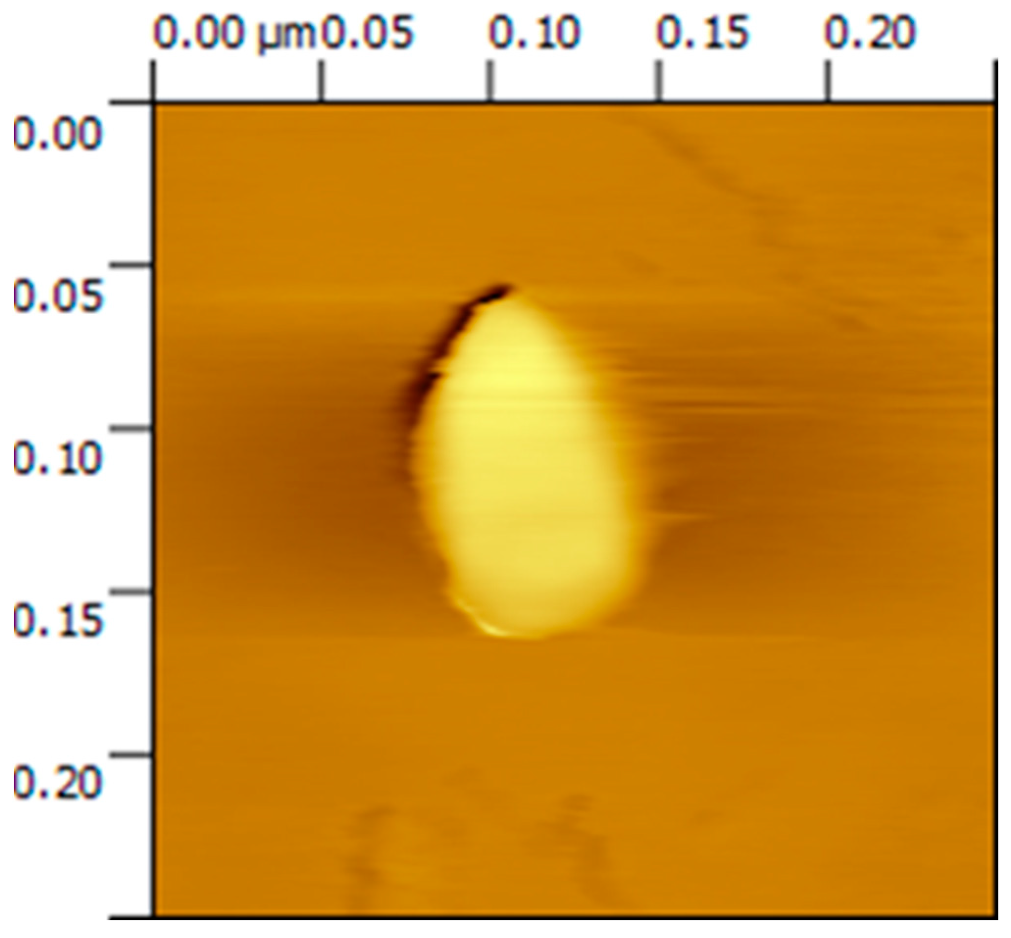
Figure 1. Atomic Force Microscopy of NECE.
Applsci 15 10214 g001
Figure 2. (a) Particle size of NECE for 90 days under different storage conditions: 24 °C and 4 °C. (b) PDI of NECE for 90 days under different storage conditions: 24 °C and 4 °C. (c) Zeta potential of NECE for 90 days under different storage conditions: 24 °C and 4 °C. (d) pH of NECE for 90 days under different storage conditions: 24 °C and 4 °C. p-value * p < 0.05; ** p < 0.01; *** p < 0.001.
Applsci 15 10214 g002
Figure 3. shows the percentage of cell viability after 24 h of exposure, for NECE (a) and FO (b). Abbreviations: NC (negative control), PC (positive control), NECE (nanoemulsion containing Cymbopogon flexuosus and eugenol), FO (Free oils at 4%), and p-value; ** p < 0.01; *** p < 0.001. The positive control consisted of 100 mM hydrogen peroxide.
Applsci 15 10214 g003
Figure 4. shows the results of the genotoxicity assay after 24 h of exposure for NECE (a) and FO (b), indicating the levels of double-stranded DNA (dsDNA) in treated cells. The vertical axis represents the dsDNA level, expressed as a percentage relative to the untreated control. The positive control consisted of hydrogen peroxide. Abbreviations: NC (negative control), PC (positive control), NECE (nanoemulsion containing Cymbopogon flexuosus and eugenol), FO (Free oils at 4%), and p-value * p < 0.05; ** p < 0.01; *** p < 0.001.
Applsci 15 10214 g004
Figure 5. shows the results of the genotoxicity assay after 48 h of exposure for NECE (a) and FO (b), indicating the levels of double-stranded DNA (dsDNA) in treated cells. The vertical axis represents the dsDNA level, expressed as a percentage relative to the untreated control. The positive control consisted of hydrogen peroxide. Abbreviations: NC (negative control), PC (positive control), NECE (nanoemulsion containing Cymbopogon flexuosus and eugenol), FO (Free oils at 4%), and p-value * p < 0.05; ** p < 0.01; *** p < 0.001.
Applsci 15 10214 g005
Figure 6. shows the results of the genotoxicity assay after 72 h of exposure for NECE (a) and FO (b), indicating the levels of double-stranded DNA (dsDNA) in treated cells. The vertical axis represents the dsDNA level, expressed as a percentage relative to the untreated control. The positive control consisted of hydrogen peroxide. Abbreviations: NC (negative control), PC (positive control), NECE (nanoemulsion containing Cymbopogon flexuosus and eugenol), FO (Free oils at 4%), and p-value ** p < 0.01; *** p < 0.001.
Applsci 15 10214 g006
Figure 7. shows the DCFH assay after 24 h of exposure, for NECE (a) and FO (b). The positive control consisted of 100 mM hydrogen peroxide. Abbreviations: NC (negative control), PC (positive control), NECE (nanoemulsion containing Cymbopogon flexuosus and eugenol), FO (Free oils at 4%), and p-value * p < 0.05; ** p < 0.01; *** p < 0.001.
Applsci 15 10214 g007
Figure 8. shows the DCFH assay after 48 h of exposure, for NECE (a) and FO (b). The positive control consisted of 100 mM hydrogen peroxide. Abbreviations: NC (negative control), PC (positive control), NECE (nanoemulsion containing Cymbopogon flexuosus and eugenol), FO (Free oils at 4%), and p-value ** p < 0.01; *** p < 0.001.
Applsci 15 10214 g008
Figure 9. shows the DCFH assay after 72 h of exposure, for NECE (a) and FO (b). The positive control consisted of 100 mM hydrogen peroxide. Abbreviations: NC (negative control), PC (positive control), NECE (nanoemulsion containing Cymbopogon flexuosus and eugenol), FO (Free oils at 4%), and p-value; *** p < 0.001.
Applsci 15 10214 g009
Table 1. Zeta potential, PDI, mean particle size, and pH of NECE.
NECE Characterization
Zeta Potential (Mv)PDIMean Particle Size (nm)pH
−5.38 ± 00.16 ± 0.01476.33 ± 0.0144.76 ± 0.035
Table 2. Antimicrobial activity of NECE and FO against P. aeruginosa and S. aureus.
Antimicrobial Activity of NECE Against P. aeruginosa
MIC NECE mg/mLMBC NECE mg/mLMIC FO mg/mLMBC FO mg/mL
P. aeruginosa Pa012.52.51.252.5
S. aureus ATCC 292130.6250.6250.6250.625
Disclaimer/Publisher’s Note: The statements, opinions and data contained in all publications are solely those of the individual author(s) and contributor(s) and not of MDPI and/or the editor(s). MDPI and/or the editor(s) disclaim responsibility for any injury to people or property resulting from any ideas, methods, instructions or products referred to in the content.