Article

Advances in the Treatment of Midface Fractures: Innovative CAD/CAM Drill Guides and Implants for the Simultaneous Primary Treatment of Zygomatic-Maxillary-Orbital-Complex Fractures

1
Department of Oral and Plastic Maxillofacial Surgery, Military Hospital Ulm, Academic Hospital of the University of Ulm, Oberer Eselsberg 40, 89081 Ulm, Germany
2
Department of Oral and Plastic Maxillofacial Surgery, University of Ulm, Albert-Einstein-Alee 10, 89081 Ulm, Germany
*
Author to whom correspondence should be addressed.
Appl. Sci. 2025, 15(18), 10194; https://doi.org/10.3390/app151810194
Submission received: 12 July 2025 / Revised: 14 September 2025 / Accepted: 16 September 2025 / Published: 18 September 2025
(This article belongs to the Special Issue Advances in Orthodontics and Dentofacial Orthopedics)

Abstract

Background: Midfacial trauma involving the zygomatic-maxillary-orbital (ZMO) complex poses significant reconstructive challenges due to anatomical complexity and the necessity for high-precision alignment. Traditional manual reduction techniques often result in inconsistent outcomes, necessitating revisions. Methods: This feasibility study presents two clinical cases treated using a novel, fully digital workflow incorporating computer-aided design and manufacturing (CAD/CAM) of patient-specific osteosynthesis plates and surgical drill guides. Following virtual fracture reduction and implant design, drill guides and implants were fabricated using selective laser melting. Surgical procedures included intraoral and transconjunctival approaches with intraoperative 3D imaging (mobile C-arm CT) to verify implant positioning. Postoperative results were compared to the virtual plan through image fusion. Results: Both cases demonstrated precise fit and anatomical restoration. The “one-position-fits-only” orbital implant design enabled highly accurate orbital wall reconstruction. Key procedural refinements between cases included enhanced interdisciplinary collaboration and improved guide designs, resulting in decreased planning-to-surgery intervals (<7 days) and seamless intraoperative application. Image fusion confirmed near-identical congruence between planned and achieved outcomes. Conclusions: The presented method demonstrates that fully digital, CAD/CAM-based midface reconstruction is feasible in the primary trauma setting. The technique offers reproducible precision, reduced intraoperative time, and improved functional and aesthetic outcomes. It may represent a paradigm shift in trauma care, particularly for complex ZMO fractures. Broader clinical adoption appears viable as production speed and workflow integration continue to improve.

1. Introduction

Fractures of the zygomatic-maxillary-orbital (ZMO) complex are among the most frequent and functionally significant injuries in maxillofacial trauma [1]. Due to their anatomical location and involvement of key facial buttresses and the orbital frame, they carry a high risk of functional impairments (e.g., diplopia, enophthalmos, malocclusion) and aesthetic deformities (e.g., midfacial flattening, asymmetry) if not reconstructed with precision [2]. Accurate three-dimensional realignment and stabilization of bone fragments therefore remains a central surgical challenge, particularly in complex or multi-fragmentary orbital trauma [3].
Traditional reduction and fixation techniques rely largely on the surgeon’s intraoperative judgment. Although transcutaneous and intraoral approaches provide access, manual repositioning under limited visual control often leads to variable outcomes and a frequent need for secondary corrections [4].
In recent years, computer-aided design and manufacturing (CAD/CAM) technologies have transformed cranio-maxillofacial surgery. Initially established in elective orthognathic and oncologic procedures, CAD/CAM enables virtual surgical planning, digital fracture simulation, and the production of patient-specific implants (PSIs) and cutting or drilling guides [5]. These tools improve accuracy, reproducibility, and intraoperative efficiency [6].
Despite these advantages, application in acute trauma surgery has been limited. Barriers have included the time required for planning, dependence on industrial-grade additive manufacturing, and the need for close coordination between surgical and engineering teams [7]. Recent advances in digital segmentation, rapid prototyping, and selective laser melting (SLM), however, allow production of surgical guides and PSIs within 3–5 working days, making their use in primary trauma care feasible.
In orbital reconstruction, PSIs have proven superior in restoring orbital volume, reducing enophthalmos, and lowering revision rates [8]. Similarly, virtual planning and backwards-designed osteosynthesis plates have improved outcomes in mandibular and midfacial reconstructions by providing biomechanical accuracy and topographical precision [9].
To date, however, an integrative digital workflow—combining virtual fracture reduction, patient-specific osteosynthesis plates, and individualized drill guides for simultaneous midface and orbital reconstruction—has rarely been applied in trauma settings. Such a workflow could improve anatomical accuracy, standardize complex procedures, reduce intraoperative time, and enhance long-term functional and aesthetic results. The additional use of intraoperative 3D imaging (e.g., mobile cone-beam CT) may further optimize outcomes.
This manuscript presents a feasibility study and case series on the primary treatment of ZMO fractures using CAD/CAM drill guides and patient-specific implants. What differentiates this study from previous work is the integration of virtual fracture reduction, CAD/CAM-guided drilling templates, and patient-specific osteosynthesis plates into a single workflow applied in the acute trauma setting. While CAD/CAM solutions are well established in elective surgery, their use for primary management of complex midfacial fractures remains rare. This feasibility study therefore aims to demonstrate the technical implementation and clinical applicability of such an integrative digital workflow in the treatment of zygomatic-maxillary-orbital-complex fractures.

2. Methods and Materials

2.1. Study Design and Patient Selection

All patients underwent a CT scan of the facial skull in 1 mm slice thickness for diagnosis and indication. Pre- and postoperative ophthalmologic check-ups were performed. ReOss® GmbH (LLC) (Filderstadt, Germany) was chosen as the implant manufacturer for the CAD/CAM implants. The operation was performed by an intraoral approach to reconstruct the midfacial pillars. Repositioning in the area of the orbital rim and insertion of the CAD/CAM orbital implants was performed via a retroseptal, transconjunctival approach. A 3D-C-arm image of the midface was taken intraoperatively for immediate intraoperative repositioning control and to assess the reduction and the position of the implants. Afterwards, the preoperative plans were matched and compared with the results.

2.2. Pre-Operative Procedure

The DICOM data of the Computed tomography (CT) scans were uploaded to the collaborating company. This formed the basis for virtual planning. After bone segmentation the fractures were digitally repositioned and the PSIs and drill guides were designed in form of backwards planning (see respective cases for figures and explanation).
This procedure determines the repositioning of the bone fragments via the design and position of the patient-specific osteosynthesis plate.
Following the surgeon’s final approval of the planning and design, the surgical guides and the PSI were manufactured using selective laser melting technique.

2.3. Surgical Procedure

The surgical procedure was conducted in accordance with the internal clinic standard for patient identification, correct anatomical positioning of the patient, intraoral germ reduction using CHX mouthwash, intravenous administration of 3 g Unacid® (Ampicillin/Sulbactam) as a single dose antibiotic and 250 mg Solu-Decortin®H as a prophylactic measure for swelling, sterile washing and draping of the patient, and a subsequent team time-out. For details of specific surgical procedures, the authors refer to the case presentations below.

3. Results

3.1. Case 1

A 45-year-old male patient presented with a typical zygomatic-maxillary-orbital-complex fracture following a traffic accident as a scooter driver.
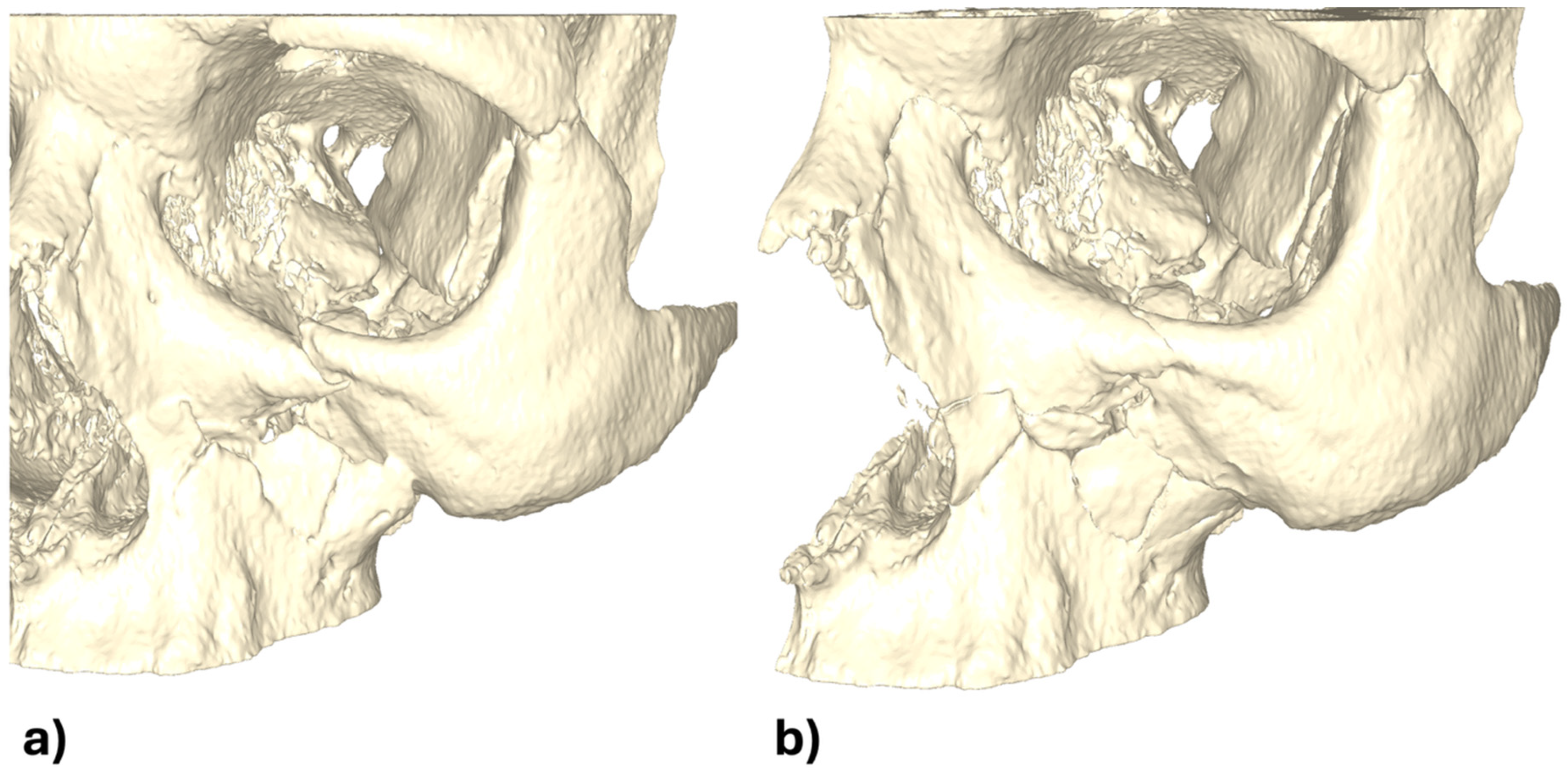
The initial assessment showed facial asymmetry, periorbital swelling and diplopia. Clinical examination revealed depression of the zygomatic arch on the left side and restricted ocular movement. CT scan confirmed fractures of the zygomatic-maxillary-orbital complex, with significant displacement of the zygomatic bone and herniation of orbital contents (Figure 1).
In this case, the digital reduction in the entire midface was performed in accordance with the aforementioned scheme (Figure 2).
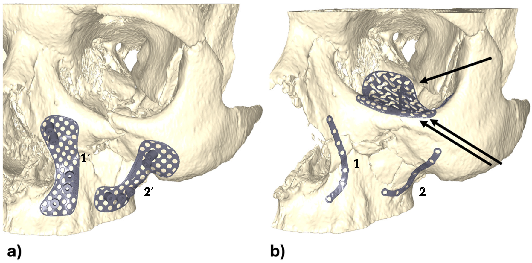
After virtual reduction the PSIs for reconstruction were designed. In this case we planned mini-plates at the lateral und medial buttress for stabilization of the midfacial pillars and a third plate on the infraorbital rim. For orbital wall reconstruction a CAD/CAM orbital wall PSI with our ‘One Position Fits Only’ design was planned as well (Figure 3b). In our workflow, the orbital implant was designed according to a “one-position-fits-only” principle, meaning that the implant geometry and footprint allow only a single, anatomically correct position during insertion. This design prevents rotational errors and ensures precise alignment with the surrounding osteosynthesis plates. Then the screw hole position of the two mini-plates were memorized and transferred backwards to the none-repositioned situation. Based on this information, two surgical-drill-guides with drill sleeves were designed (Figure 3a).
After the surgeon’s final approval of the planning and design the guides und the implants were manufactured.
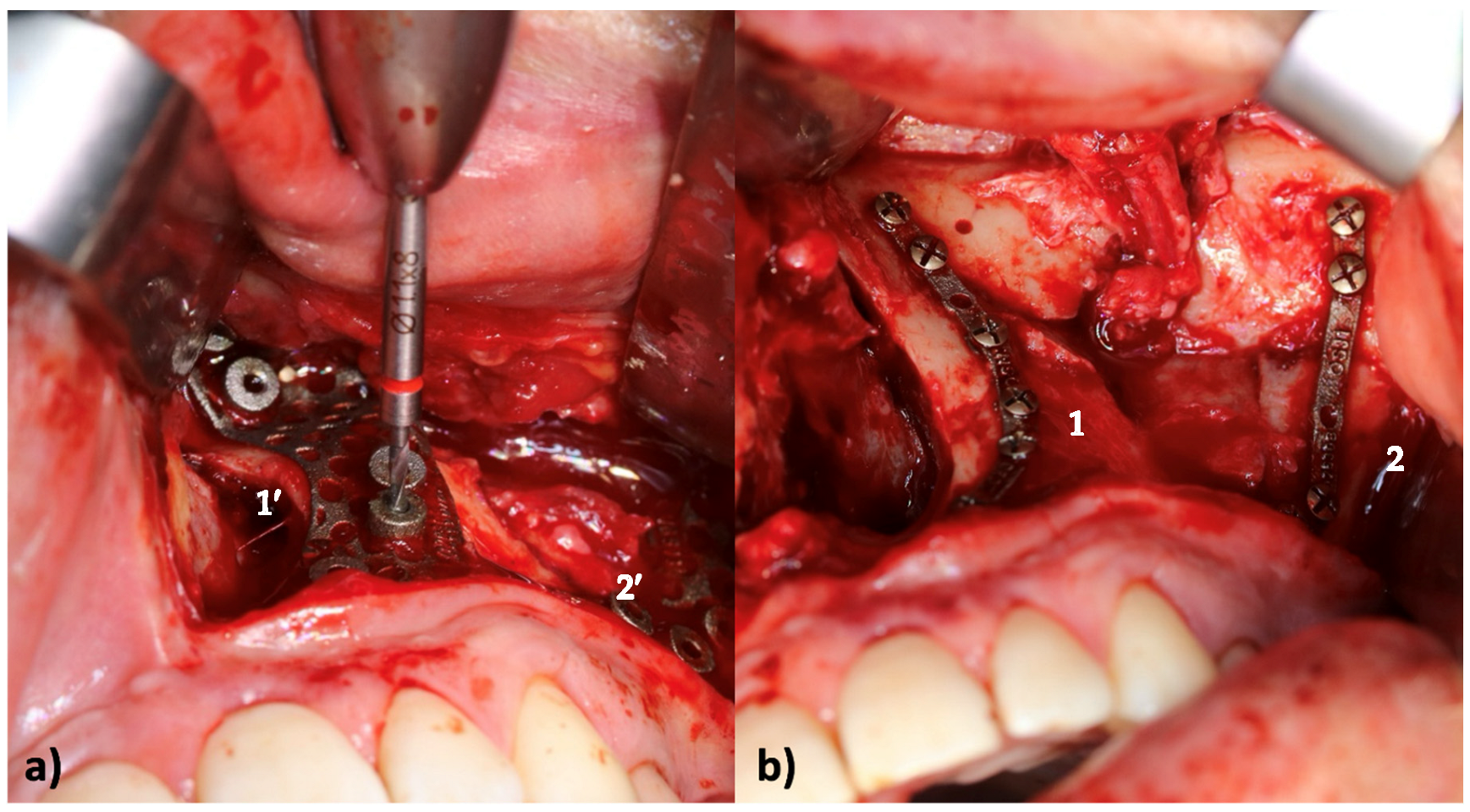
During surgery, first the drill guides were positioned and fixed with two mini-screws on the fractured situs on the paranasal and zygomatic buttresses. This was followed by guided drilling of the screw holes according to the (drill-)guide. After removal of the guides, the reduction in the facture was performed and the CAD/CAM mini plates were fixed according the pre-drilled holes (Figure 4).
Afterwards a transconjunctival approach to the infraorbital rim and orbital floor was performed. The reduction was checked at the infraorbital rim and the third patient-specific mini-plate was fixed. The correct fit of the implant eliminated possible rotational errors. The orbital implant was then inserted via the transconjunctival approach (Figure 5a). The orbital implant was designed in our one-position-fits-only design with an anterior, lateral footprint extension which allows a distinctive positioning of the orbital implant. The implant fitted perfectly to the osteosynthesis plate of the orbital rim from the dorsal side (Figure 5a).
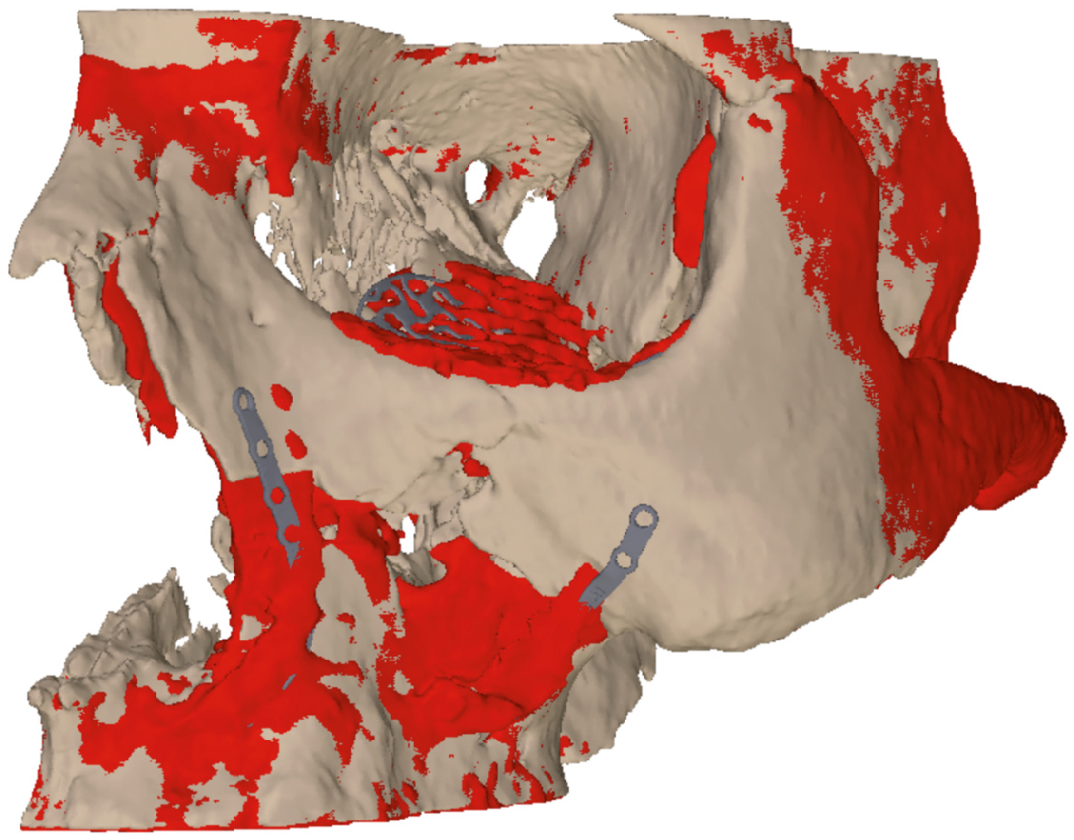
After repositioning and fixation of the four implants, an intraoperative 3D-Scan with a mobile 3D-C-arm device (Siemens Cios Spin®, Munich, Germany) was employed to prove reduction and implant position (Figure 5b,c). For postoperative evaluation we fused the 3D planning and the received result (Figure 6).

3.2. Case 2

A 33-year-old male patient presented with a zygomatic-maxillary-orbital-complex fracture after a brawl.
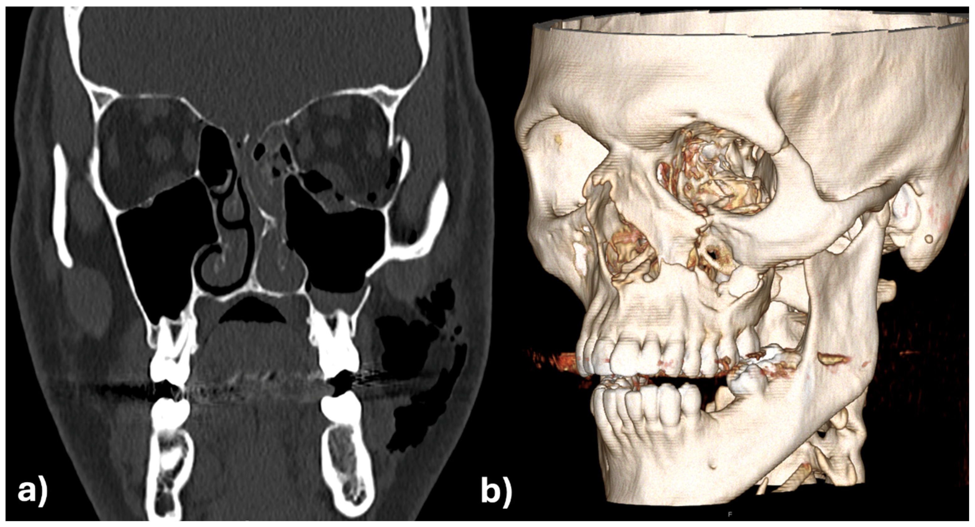
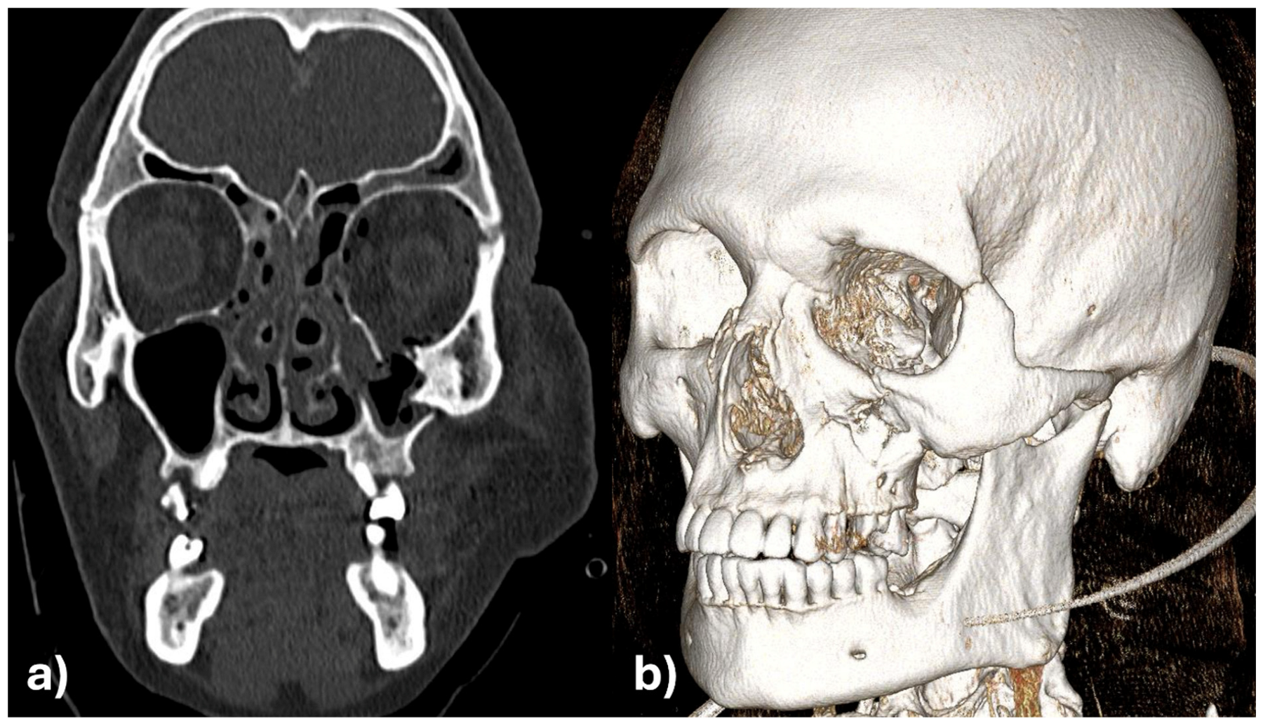
The initial assessment showed facial asymmetry, periorbital swelling, and diplopia. Clinical examination revealed depression of the zygomatic arch on the left side and restricted ocular movement. CT scan confirmed fractures of the zygomatic-maxillary-orbital-complex, with significant displacement of the zygomatic bone and herniation of orbital contents into the maxillary sinus (Figure 7a,b).
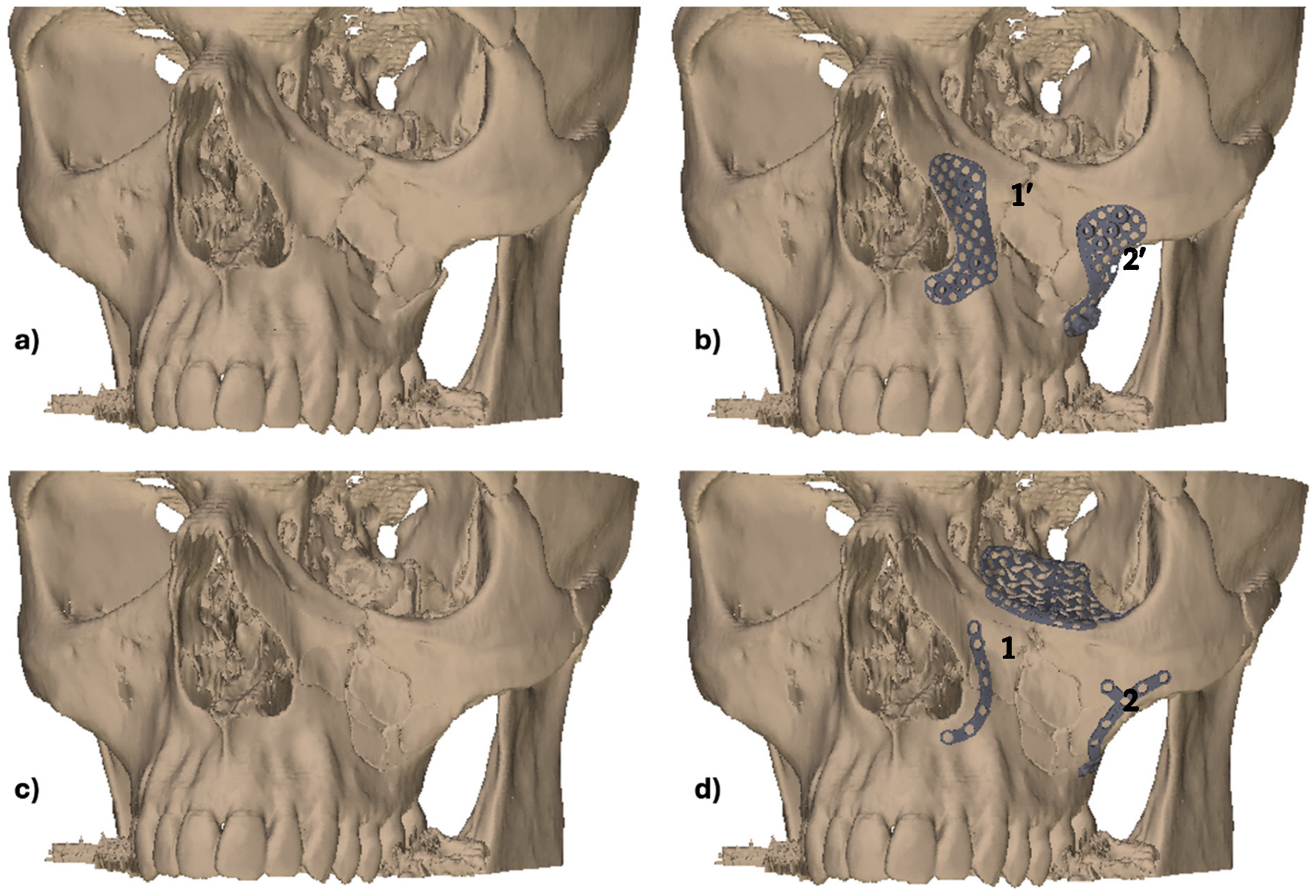
Virtual planning with digital reduction and design of the drill guides and the patient specific implants was performed employing the same workflow as shown in case 1 (Figure 8a–d).
Also, the surgical intervention was performed like in case 1 with primary fixation of the drill guides on the medial and lateral buttresses via an intraoral approach and pre-drilling the screw holes. After guide removal open reduction and fixation of the mini plates was performed in accordance with the plan. Afterwards the infraorbital rim was stabilized by the third mini-plate and orbital floor was reconstructed with the orbital implant via a transconjunctival approach (Figure 9a–e).
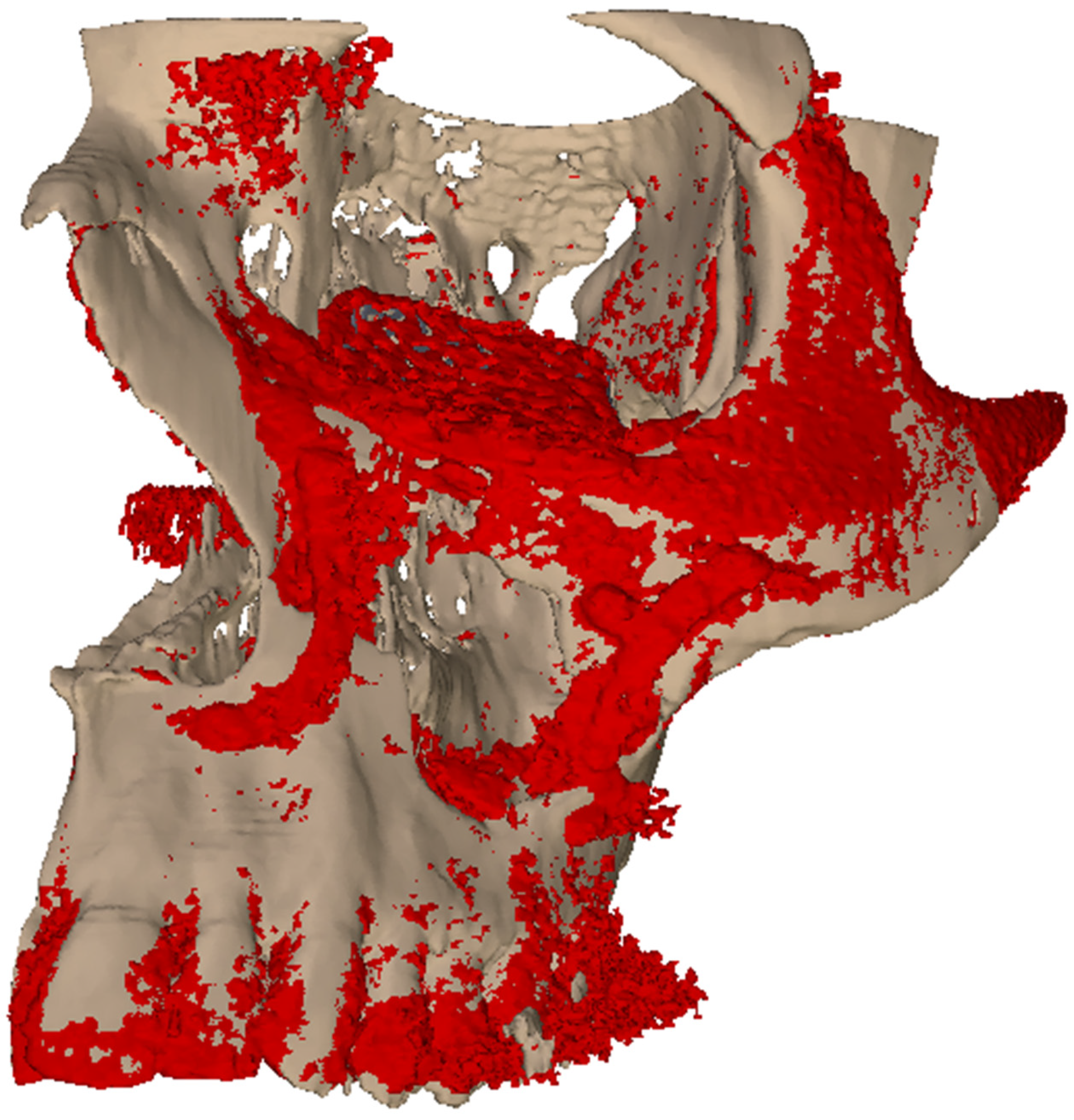
Figure 10a,b are showing the images of the intraoperative 3D C-arm scan and Figure 11 shows the image fusion between plan and achieved result.

4. Discussion

4.1. Specific Case Discussion

These two cases provides promising early evidence in the evolving application of CAD/CAM technology for the primary treatment of maxillofacial fractures. Specifically, they demonstrate the technical feasibility and clinical utility of employing patient-specific surgical guides and implants for midfacial reconstruction, with a particular emphasis on the orbital and zygomatic regions.
In the initial case, the intraoperative fit of the CAD/CAM-designed guides and implants in the midface was found to be optimal. The additional orbital rim implant enabled highly accurate reconstruction of the orbital frame, and the “one-position-fits-only” design of the orbital implant facilitated precise placement on the dorsal aspect of the orbital rim plate. The precise alignment between the two implants was found to be a distinct advantage in achieving an anatomically correct and functionally stable reconstruction.
However, the case also highlighted challenges in the early stages of implementation. The close and repeated collaborations between surgeons and medical engineers were necessary but difficult to coordinate due to time constraints. Although orbital reconstruction has now become relatively routine for experienced engineers, the importance of collaborative planning remains paramount, particularly in complex trauma cases. In this instance, the development and revision of multiple design proposals was necessary due to unanticipated intraoperative challenges. Notably, the enoral approach was insufficiently considered in the design phase, resulting in technical difficulties during the insertion of drills into the guide, especially for cranial drill holes in the zygomatic and paranasal areas. Furthermore, the drill sleeves exhibited inadequate continuity for the corresponding drill, resulting in minor titanium abrasion. Additionally, the penetrability of the implants for osteosynthesis screws necessitated intraoperative adjustments.
The insights gained from this inaugural experience served as a foundation for the subsequent approach to the second case. The insights and feedback from prior planning processes were leveraged to significantly streamline digital bone repositioning and implant design. The open and constructive collaboration between the surgical and engineering teams resulted in rapid production, with surgical guides and implants being delivered within just five days. Intraoperatively, the components could be applied without complications, and the planned repositioning was achieved with a precise implant fit. In this instance, the drilling templates and screw holes demonstrated optimal consistency, reflecting tangible improvements in design and manufacturing workflows.
However, a high degree of mobility in the zygomatic body during the surgical approach and fracture exposure did pose some challenges to the positioning of the surgical guides, particularly in comparison to more stable areas such as the paranasal region. This finding serves to underscore the expectation that in cases involving smaller or more mobile bone fragments, deviations from the planned position may be more likely—an issue that is less prominent in orthognathic procedures on non-osteotomized jaws. Notwithstanding these challenges, the insertion and pre-drilling procedures were successfully executed. The orbital rim plate was instrumental in correcting minor inaccuracies in the orbital frame region. Of course, it would be possible to combine the infraorbital plate with orbital implant. However, from our perspective we believe that it is beneficial to split this off for higher precision of the entire reconstruction.
The analysis presented here demonstrates the potential of CAD/CAM technology to achieve reliable, anatomically accurate, and functional reconstructions of complex midfacial fractures. Of particular note is the necessity of effective and timely interdisciplinary communication. A well-coordinated team approach, particularly during the design and planning phase, can prevent time-consuming revisions and optimize surgical outcomes. The sharing of original drills and screws with the implant manufacturer for inspection and certification was also a key step toward seamless integration of surgical requirements into the production process.
The ongoing refinement of CAD/CAM workflows, informed by real-world clinical experience, is resulting in reduced planning and production times, as well as enhanced surgical outcomes. Consequently, the method is gaining traction in clinical practice, with additional cases already in preparation. These developments affirm the value of digital, patient-specific reconstruction in modern maxillofacial trauma care.

4.2. General Discussion

The integration of CAD/CAM technology in primary midfacial reconstruction represents a beneficial advance in the field of oral and maxillofacial surgery, particularly regarding the precise repositioning of fractures. Conventional techniques frequently necessitate the surgeon’s intraoperative assessment and manual realignment of fractures, a process that is not only time-consuming but also susceptible to inaccuracies. Such inaccuracies can result in complications, including malocclusion, enophthalmos and facial asymmetry, which can necessitate complicated revision procedures and re-osteotomies. In contrast, our approach utilizing CAD/CAM technology permits meticulous preoperative planning, thereby facilitating the creation of surgical guides that enable the precise and reproducible repositioning of fractures, which could contribute to future improvements in this field.
Perhaps the most transformative aspect of our method is the ability to digitally reposition fractures before the surgical procedure commences. This digital process entails the creation of a virtual model of the patient’s skull, thereby enabling the surgeon to manipulate the fractured segments to their optimal anatomical positions. Once the optimal alignment is determined, CAD/CAM technology is used to create customized surgical guides that are customized to the patient’s particular anatomy. These guides are produced with such precision that they guarantee the bones can be repositioned during surgery in a manner that aligns them with the configuration depicted in the digital model. This “one position fits only” design represents a significant advancement over traditional methods, where the final positioning frequently relies on the surgeon’s visual and tactile assessment during the operation.
This digital repositioning offers notable advantages over conventional care. Firstly, it ensures that the fractures are aligned in the most anatomically correct position, thereby reducing the risk of post-surgical complications. The precision of the CAD/CAM surgical guides eliminates the need for guesswork associated with traditional fracture fixation, thereby ensuring more consistent and reliable outcomes. Furthermore, this approach has the potential to significantly reduce the surgical time. In the standard care approach, a significant portion of the operative time is dedicated to achieving proper alignment of the fracture segments, which often necessitates multiple adjustments. The pre-planned digital alignment enables more expeditious and efficacious surgical procedures, as the surgeon can promptly position the fracture segments using the guides, obviating the necessity for extensive intraoperative corrections.
Moreover, the enhanced precision afforded by the CAD/CAM guides diminishes the probability of requiring subsequent corrective surgeries. In the context of conventional methodologies, it is not uncommon for postoperative imaging to reveal suboptimal fracture alignment, thereby necessitating further interventions. The capacity of our method to achieve the correct alignment on the first attempt serves to minimize these risks, thereby facilitating a more expedient recovery process and enhancing long-term outcomes for patients. The combination with intraoperative 3D imaging continues to be of particular value here.
The true landmark in this field is the way CAD/CAM guides transform the approach of surgeons to fracture repositioning. In contrast to the reliance on intraoperative adjustments, which can be imprecise and subjective, our method provides a pre-determined, scientifically grounded approach to fracture alignment. This results in not only improved functional and aesthetic outcomes but also a more predictable and streamlined surgical process.
The present report is limited by the very small sample size of only two cases, which restricts the strength of the conclusions that can be drawn. Although both cases demonstrated precise anatomical restoration and technical feasibility, the evidence level remains low, and results cannot be generalized to the wider patient population. Further studies including larger case series and ideally controlled trials are necessary to assess reproducibility, cost-effectiveness, and long-term outcomes. Until such evidence is available, the presented workflow should be regarded as a proof-of-concept approach rather than a broadly established clinical standard.
The primary objective of this feasibility study was to assess the outcomes, which was primarily achieved through a descriptive analysis. The focus of this analysis was on intraoperative handling, implant fit, and congruence between the virtual plan and postoperative imaging. The utilisation of standardised postoperative functional or aesthetic measures (e.g., diplopia testing, exophthalmometry, or validated patient-reported outcome questionnaires) was not systematically recorded. Nevertheless, both patients underwent routine follow-up examinations, during which no subjective complaints (e.g., diplopia, sensory disturbances, discomfort) or clinical abnormalities were noted. While this provides preliminary reassurance, the lack of standardised functional and aesthetic outcome measures represents a limitation of the present report. Future studies with larger cohorts should systematically compare this digital workflow to standard approaches in terms of operative time, complication rates, revision surgery, and long-term functional outcomes.
While the feasibility of CAD/CAM-guided fracture management has been demonstrated in the present cases, its widespread implementation in emergency trauma care is still limited by cost and resource availability. The fabrication of patient-specific implants and guides necessitates specialised infrastructure, collaboration with engineering partners, and additional preoperative planning time. The present study has identified a number of factors that are currently resulting in higher treatment costs when compared to conventional methods. Furthermore, these factors may not be available in all trauma centres, particularly in cases of urgent intervention. However, with ongoing improvements in production speed, cost reduction through streamlined workflows, and increasing availability of in-house or regional manufacturing facilities, broader accessibility is expected in the near future.
Further developments may offer more sophisticated and artificial intelligence supported digital planning tools that permit even finer adjustments to the fracture alignment, as well as the integration of real-time feedback during surgery to further enhance precision.
5. Conclusions

In conclusion, this feasibility study demonstrates that a fully digital workflow using CAD/CAM-guided drilling templates and patient-specific osteosynthesis plates is technically feasible for the primary treatment of zygomatic-maxillary-orbital-complex fractures. The approach allowed precise repositioning of large bone fragments and was successfully implemented within a clinically acceptable timeframe. While these results are encouraging, they are based on only two cases, and thus generalizability is limited. Larger prospective studies are required to validate reproducibility, assess cost-effectiveness, and confirm long-term clinical benefits. Nevertheless, these findings highlight the potential of digitally guided workflows to improve surgical accuracy and reproducibility in midfacial trauma care.

Author Contributions

Conceptualization, F.W.; methodology, F.W. and A.S. (Alexander Schramm); investigation, M.E., S.P. and A.S. (Andreas Sakkas); data curation, S.P. and S.K.; writing—original draft preparation, M.E. and S.P.; writing—review and editing, A.S. (Andreas Sakkas), S.K., A.S. (Alexander Schramm), M.S. and F.W.; supervision, A.S. (Alexander Schramm) and F.W. All authors have read and agreed to the published version of the manuscript.

Funding

This research received no external funding.

Institutional Review Board Statement

Ethical review and approval were waived for this study by the Institutional Review Board (IRB) of the University of Ulm due to its nature as a retrospective case series.

Informed Consent Statement

Informed consent was obtained from all participants for inclusion in the study and for publication of their clinical data and images.

Data Availability Statement

The data presented in this study are available on request from the corresponding author. The data are not publicly available due to patient confidentiality.

Conflicts of Interest

The authors declare no conflicts of interest.

References

  1. Yamsani, B.; Gaddipati, R.; Vura, N.; Ramisetti, S.; Yamsani, R. Zygomaticomaxillary Complex Fractures: A Review of 101 Cases. J. Maxillofac. Oral Surg. 2016, 15, 417–424. [Google Scholar] [CrossRef] [PubMed]
  2. Kaukola, L.; Snäll, J.; Roine, R.; Sintonen, H.; Thorén, H. Health-related quality of life of patients with zygomatic fracture. Med. Oral Patol. Oral Cir. Bucal 2017, 22, e636–e642. [Google Scholar] [CrossRef] [PubMed]
  3. Krasovsky, A.; Hija, A.; Zeineh, N.; Capucha, T.; Blanc, O.; Emodi, O.; Rachmiel, A.; Shilo, D. Three-Dimensional Reconstruction of Orbital Fractures. J. Vis. Exp. 2025, 219, e67450. [Google Scholar] [CrossRef] [PubMed]
  4. Chodroff, J.; Elias, N.; Whitcomb, M.; Vo, C.; Bell, R.B. Secondary Correction of Midface Fractures. Atlas Oral Maxillofac. Surg. Clin. 2021, 29, 139–150. [Google Scholar] [CrossRef] [PubMed]
  5. Zoabi, A.; Redenski, I.; Oren, D.; Kasem, A.; Zigron, A.; Daoud, S.; Moskovich, L.; Kablan, F.; Srouji, S. 3D Printing and Virtual Surgical Planning in Oral and Maxillofacial Surgery. J. Clin. Med. 2022, 11, 2385. [Google Scholar] [CrossRef] [PubMed]
  6. Pietzka, S.; Fink, J.; Winter, K.; Wilde, F.; Schramm, A.; Ebeling, M.; Kasper, R.; Sakkas, A. Dental Root Injuries Caused by Osteosynthesis Screws in Orthognathic Surgery—Comparison of Conventional Osteosynthesis and Osteosynthesis by CAD/CAM Drill Guides and Patient-Specific Implants. J. Pers. Med. 2023, 13, 706. [Google Scholar] [CrossRef] [PubMed]
  7. Unsal, G.; Turkyilmaz, I.; Lakhia, S. Advantages and limitations of implant surgery with CAD/CAM surgical guides: A literature review. J. Clin. Exp. Dent. 2020, 12, e409–e417. [Google Scholar] [CrossRef] [PubMed]
  8. González, L.V.; López, J.P.; Orjuela, M.P.; Díaz-Báez, D. Technology Guided Management of Orbital Trauma: A Systematic Review. J. Maxillofac. Oral Surg. 2025, 24, 343–352. [Google Scholar] [CrossRef] [PubMed]
  9. El-Mahallawy, Y.; Abdelrahman, H.H.; Al-Mahalawy, H. Accuracy of virtual surgical planning in mandibular reconstruction: Application of a standard and reliable postoperative evaluation methodology. BMC Oral Health 2023, 23, 119. [Google Scholar] [CrossRef] [PubMed]
Figure 1. Zygomatic-maxillary-orbital-complex fracture of the left side. (a) Coronal view, (b) 3D reconstruction.
Applsci 15 10194 g001
Figure 2. Pre-operative zygomatic-maxillary-orbital-complex fracture e without (a) and after digital repositioning (b).
Applsci 15 10194 g002
Figure 3. CAD/CAM surgical guides (1′ and 2′) (a) and CAD/CAM miniplates (1 and 2) and orbital wall implant (b) (Black arrow: orbital wall implant; Double black arrow: separate infraorbital plate).
Applsci 15 10194 g003
Figure 4. (a) Drill guides (1′ and 2′) positioned on the fractured situs (b) after reduction and CAD/CAM implant placement (1 and 2).
Applsci 15 10194 g004
Figure 5. (a) Orbital implant inserted via a transconjunctival approach, (b,c) intraoperative 3D scan for implant placement control.
Applsci 15 10194 g005
Figure 6. Image fusion of 3D planning (beige) and post-operative situation (red) to illustrate the difference between virtual planning and achieved result.
Applsci 15 10194 g006
Figure 7. Zygomatic-maxillary-orbital-complex fracture of the left side in coronary view (a) and as 3D reconstruction (b).
Applsci 15 10194 g007
Figure 8. (a) Pre-operative situation (b) with patient-specific CAD/CAM surgical guides (1′ and 2′) (c) after digital repositioning of the zygomatic bone and orbital wall fractures (d) with patient-specific CAD/CAM implants (1 and 2).
Applsci 15 10194 g008
Figure 9. (a) Intraoperative view of CAD/CAM surgical guides and drilling (1′ and 2′). (b) Pre-drilled holes after removal of surgical guides and before repositioning. (c) Insertion of CAD/CADM plates after repositioning of the zygomatic fracture (2). (d) Inserted and fixed CAD/CAM implants (1 and 2). (e) View of the infraorbital rim and fixed orbital wall implant.
Applsci 15 10194 g009
Figure 10. 3D (a) and in coronal view (b) of the intraoperative 3D scan which shows an almost perfect anatomic reconstruction and fit of the patient specific implants.
Applsci 15 10194 g010
Figure 11. Image fusion of 3D planning (beige) and post-operative situation (red) to illustrate the difference between virtual planning and achieved result.
Applsci 15 10194 g011
Disclaimer/Publisher’s Note: The statements, opinions and data contained in all publications are solely those of the individual author(s) and contributor(s) and not of MDPI and/or the editor(s). MDPI and/or the editor(s) disclaim responsibility for any injury to people or property resulting from any ideas, methods, instructions or products referred to in the content.