Article

Effects of Rapid Maxillary Expansion on Pulmonary Function in Adolescents: A Spirometric Evaluation

1
Department of Orthodontics, Faculty of Dentistry, Kocaeli Health and Technology University, 41275 Kocaeli, Turkey
2
Department of Orthodontics, Private Clinics, 16000 Bursa, Turkey
3
Department of Orthodontics, Private Clinics, 27310 Gaziantep, Turkey
4
Department of Orthodontics, Faculty of Dentistry, Harran University, 63050 Şanlıurfa, Turkey
5
Department of Physiology, Faculty of Medicine, Ankara Medipol University, 06570 Ankara, Turkey
*
Author to whom correspondence should be addressed.
Appl. Sci. 2025, 15(18), 10189; https://doi.org/10.3390/app151810189
Submission received: 4 August 2025 / Revised: 7 September 2025 / Accepted: 10 September 2025 / Published: 18 September 2025
(This article belongs to the Section Applied Dentistry and Oral Sciences)

Abstract

Objectives: Rapid maxillary expansion (RME) is widely used in orthodontics to correct transverse maxillary deficiencies. Beyond its skeletal and dental effects, RME may influence upper airway dimensions and respiratory function, particularly in growing individuals. This study aimed to evaluate the impact of RME on pulmonary function in adolescents using spirometric measurements. Materials and Methods: Fifteen adolescent patients (8 females, 7 males; mean age: 13.93 ± 2.89 years) diagnosed with maxillary transverse constriction underwent orthodontic treatment with acrylic-bonded RME appliances over a mean duration of 3.56 ± 0.67 months. Respiratory function was assessed via spirometry at baseline (T0) and one day after appliance removal (T1). Parameters recorded included peripheral oxygen saturation (SpO2), forced expiratory volume in one second (FEV1), forced vital capacity (FVC), FEV1/FVC ratio, and vital capacity (VC). Data were analyzed using the paired-samples t-test (for normally distributed variables) or the Wilcoxon signed-rank test (for non-normal distributions), with statistical significance set at p < 0.05. Results: Following RME treatment, all respiratory parameters showed a consistent upward trend but did not reach statistical significance. SpO2 increased from 96.98 ± 0.96% to 97.01 ± 0.98% (p = 0.925). VC rose from 2.86 ± 1.07 L to 3.03 ± 0.80 L (p = 0.626). The FEV1/FVC ratio improved from 90.88 ± 12.17% to 92.34 ± 7.37% (p = 0.742). Mean FEV1 increased from 2.61 ± 0.72 L to 2.72 ± 0.68 L (p = 0.518), while FVC rose from 2.87 ± 0.75 L to 2.96 ± 0.69 L (p = 0.547). No adverse effects were reported during the treatment period. Conclusions: This study identified a non-significant but consistent trend toward improved pulmonary function following RME in adolescents. These preliminary findings should be considered hypothesis-generating rather than confirmatory evidence, as none of the outcomes reached statistical significance. While the observed upward trends in oxygen saturation, lung volumes, and expiratory performance suggest potential respiratory benefits, larger-scale, controlled, and long-term studies incorporating both spirometric and anatomical airway assessments are needed to validate these observations.

1. Introduction

Rapid maxillary expansion (RME) is not only an established orthopedic intervention for correcting transverse maxillary deficiencies, but also a procedure with potential systemic effects due to its influence on the upper airway morphology and function. The maxilla forms the floor of the nasal cavity, and its transverse dimension directly affects the width and patency of the nasal airway. A constricted maxilla is frequently associated with a high-arched palate, reduced nasal floor width, and increased nasal airway resistance, which can contribute to chronic mouth breathing and altered craniofacial growth patterns in children and adolescents [1,2]. From a physiological perspective, the nasal airway is the primary pathway for respiration, contributing to air filtration, humidification, and warming. Restriction in nasal airflow increases the work of breathing and can negatively impact sleep quality, exercise tolerance, and overall respiratory health [3,4].
Several recent systematic reviews and meta-analyses have confirmed that RME leads to measurable increases in nasal cavity width and upper airway volume, with corresponding reductions in nasal airway resistance [5,6,7,8]. Seif-Eldin et al. found significant post-treatment gains in transverse skeletal width accompanied by improved nasal breathing capacity in both pre- and post-pubertal patients [1]. Piełunowicz et al. demonstrated in a 2025 systematic review that RME, particularly when combined with functional orthodontic appliances, significantly enhanced upper airway dimensions in children with sleep-disordered breathing [9]. Similarly, Xie et al. highlighted RME’s role in improving pediatric obstructive sleep apnea outcomes, emphasizing multidisciplinary collaboration for optimal results [10]. In addition, numerous studies have indicated that RME produces significant skeletal and dentoalveolar effects, including spontaneous occlusal changes and mandibular adaptations [11,12,13,14].
From an anatomical perspective, the maxilla constitutes the floor of the nasal cavity; therefore, maxillary transverse deficiency often results in a narrowed nasal airway and increased nasal resistance. This restriction forces patients to rely more on oral breathing, which alters normal airflow dynamics, bypasses nasal conditioning functions, and can increase the work of breathing. Chronic mouth breathing is associated with reduced ventilatory efficiency, altered thoracic biomechanics, and in some cases impaired sleep quality. Functionally, these changes may manifest as lower spirometric values, including reduced FEV1, FVC, and oxygen saturation. By expanding the maxilla, RME increases the nasal floor width, reduces airway resistance, and facilitates more efficient nasal respiration. These structural and functional improvements provide a plausible mechanism linking correction of maxillary constriction to measurable gains in pulmonary performance [5,6,9,10,15].
The impact of RME on adjacent anatomical regions has been assessed using various imaging modalities, including posteroanterior cephalometric radiographs, acoustic rhinometry, and three-dimensional cone-beam computed tomography (CBCT) [8,16]. These tools have facilitated accurate assessments of changes in airway volume and nasal resistance. In addition to anatomical modifications, RME may influence tongue posture, nasopharyngeal airflow, and other physiological functions [9,10,17,18,19]. Warren et al. reported a 45% improvement in nasal cross-sectional area following RME, indicating a potential enhancement in airflow through the nasal passage [15].
Respiratory function testing plays a pivotal role in the functional evaluation of airway changes [10]. Spirometry, a widely accepted method for assessing pulmonary function, offers several advantages: it is non-invasive, repeatable, and easily applicable in both pediatric and adult populations [15,20]. Parameters such as oxygen saturation, forced vital capacity (FVC), forced expiratory volume in one second (FEV1), and the FEV1/FVC ratio provide insights into both ventilatory efficiency and potential airway obstruction. Interestingly, spirometry has been utilized in dental research to assess the influence of edentulism and prosthodontic appliances on respiratory performance [21]. Notably, a strong association has been established between periodontal disease and decreased lung volumes in adult populations [22,23], further highlighting the interrelationship between oral and respiratory health.
While the influence of orthognathic surgery and functional appliances on pulmonary parameters has been explored [24,25,26], there remains limited literature focusing specifically on the functional respiratory effects of RME—especially when evaluated via spirometry in growing individuals. Most existing studies have concentrated on the anatomical or volumetric changes of the upper airway following RME using radiographic imaging techniques [25,26]. Furthermore, recent investigations have demonstrated the efficacy of RME in managing pediatric obstructive sleep apnea (OSA), suggesting a potential role for RME in improving airway patency and breathing efficiency [27,28,29,30,31].
Despite these promising findings, there is a distinct lack of studies that assess the functional respiratory outcomes of RME using objective pulmonary measures such as spirometry, particularly in adolescents. Given the close anatomical and physiological connections between the maxillary structures and the upper respiratory tract, it is plausible that correcting maxillary constriction through RME could lead to measurable improvements in pulmonary function.
Although numerous studies have demonstrated the anatomical changes of the nasal cavity and upper airway following RME, very few have focused on the functional respiratory outcomes using spirometric evaluation, especially in adolescents. Given the anatomical and physiological relationship between the maxilla and upper airway structures, it is plausible that correcting maxillary constriction could yield measurable improvements in pulmonary function. However, the literature still lacks sufficient objective evidence on this issue. Therefore, this study was designed to specifically investigate the short-term effects of RME on spirometric parameters in adolescents, aiming to bridge this important gap.

2. Materials and Methods

2.1. Study Design and Ethical Approval

This investigation was a single-arm, before–after observational study designed and reported in line with the STROBE recommendations (Supplementary Material). Consecutive adolescent patients with maxillary transverse constriction underwent acrylic-bonded rapid maxillary expansion (RME). Spirometric outcomes were recorded at baseline before appliance placement (T0) and one day after appliance removal (T1). Measurements were performed in a controlled clinical environment between 09:00 and 11:00 to minimize diurnal variation, following American Thoracic Society/European Respiratory Society standards, and by the same experienced physiologist to reduce measurement bias.
The study was conducted in accordance with the Declaration of Helsinki and approved by the Ethics Committee of Gaziantep University (Approval No: 2011-12, Date: 11 December 2011). Informed Consent Statement: Written informed consent was obtained from the parents or legal guardians of all participants; assent was obtained from adolescents where applicable.

2.2. Participants and Eligibility Criteria

A total of 15 adolescents (8 females, 7 males; mean age: 13.93 ± 2.89 years) diagnosed with maxillary transverse constriction were included in the study.
The inclusion criteria were as follows: bilateral posterior crossbite; no prior orthodontic treatment; good oral hygiene and compliance; no history of adenoidectomy, tonsillectomy, or nasal surgery; absence of systemic or respiratory diseases that could influence pulmonary function. In addition, only adolescent patients were included, within an age range where midpalatal suture opening with conventional tooth-borne RME is still considered clinically feasible. Cases with advanced skeletal maturation or poor prognosis for skeletal expansion were excluded to minimize the risk of undesired dentoalveolar tipping instead of true skeletal expansion.
Exclusion criteria included active upper respiratory tract infection, current use of medication affecting respiratory performance, craniofacial syndromes, poor compliance with treatment or measurement protocols, or conditions likely to predispose to relapse after expansion.

2.3. Intervention: Appliance Design and Activation Protocol

All participants were treated with acrylic-bonded rapid maxillary expansion (RME) appliances incorporating a maxillary expansion screw (Leone A0620-11, 11 mm, Florence, Italy) positioned along the midpalatal suture and parallel to the occlusal plane. The appliances were cemented with multi-cure glass ionomer cement (3M Unitek, Monrovia, CA, USA) according to the manufacturer’s instructions.
Parents were instructed to activate the expansion screw by one-quarter turn twice daily (0.25 mm per turn) until a cusp-to-cusp relationship between the maxillary palatal cusp of the first molar and the buccal cusp of the mandibular first molar was achieved. At that point, the screw was stabilized with light-cure composite resin, and the appliance was left in place passively for retention over 3 months. The mean treatment duration was 3.56 ± 0.67 months.
This conventional tooth-borne bonded expander was selected for ethical and clinical feasibility reasons. Although skeletal anchorage-assisted expanders (e.g., MARPE or bone-borne devices) may provide greater skeletal effects in older adolescents, these options were not applied in the present cohort.

2.4. Outcomes and Measurements

The primary outcomes were spirometric parameters reflecting pulmonary function, namely peripheral oxygen saturation (SpO2, %), forced expiratory volume in one second (FEV1, L), forced vital capacity (FVC, L), vital capacity (VC, L), and the FEV1/FVC ratio (%).
Respiratory function was assessed using a portable digital spirometer with integrated pulse oximetry (Spirolab III, MIR, Rome, Italy). The device was calibrated daily with a 3 L calibration syringe according to the manufacturer’s instructions and American Thoracic Society/European Respiratory Society (ATS/ERS) standards [30]. Calibration checks included low-, medium-, and high-flow verification to ensure accuracy within ±3% of reference values.
All tests were conducted in a quiet clinical examination room under controlled environmental conditions (temperature: 22–24 °C; relative humidity: 40–60%). To minimize diurnal variation, all measurements were performed between 09:00 and 11:00 a.m. Participants were instructed to avoid caffeine for at least 4 h, vigorous physical activity for 2 h, heavy meals for 2 h, and smoking or second-hand smoke exposure for 12 h before testing.
During testing, participants were seated comfortably with nasal airflow occluded by soft nasal clips to ensure exclusive oral breathing through the mouthpiece, and disposable bacterial/viral filters were used for infection control. For each spirometric parameter, at least three acceptable and reproducible maneuvers were obtained according to ATS/ERS criteria, and the highest value was used for analysis. Measurements were performed at two time points: T0, before initiation of RME treatment, and T1, one day after removal of the expansion appliance to avoid discomfort during testing. All measurements were carried out by the same experienced physiologist to minimize inter-operator variability and reduce measurement bias (Figure 1).

2.5. Efforts to Reduce Bias

Several measures were implemented to minimize potential sources of bias. All spirometric assessments were performed by the same experienced physiologist to eliminate inter-operator variability. Testing conditions were standardized by conducting measurements in a controlled environment (temperature 22–24 °C, humidity 40–60%) and at a consistent time of day (09:00–11:00 a.m.) to reduce environmental and diurnal influences. The spirometer was calibrated daily according to ATS/ERS standards, with accuracy verified within ±3% of reference values. All spirometric maneuvers were carried out in accordance with ATS/ERS reproducibility and acceptability criteria, and the best of at least three trials was recorded. Potential confounding effects were minimized by excluding participants with systemic or respiratory diseases, recent infections, prior surgeries (such as adenoidectomy or tonsillectomy), or poor compliance. Finally, the short interval between T0 and T1 measurements minimized variability due to external or lifestyle-related changes, ensuring that observed differences could be attributed primarily to the RME intervention.

2.6. Statistical Analysis

Statistical analysis was performed using IBM SPSS Statistics for Windows, Version 26.0 (IBM Corp., Armonk, NY, USA). Descriptive statistics (mean ± standard deviation for continuous variables; frequencies and percentages for categorical variables) were calculated for demographic and clinical characteristics. The Shapiro–Wilk test was applied to assess normality of data distribution for all continuous outcomes. For normally distributed variables, paired-samples t-tests were used to compare pre-treatment (T0) and post-treatment (T1) values. For non-normally distributed variables, the Wilcoxon signed-rank test was applied. A two-tailed significance level of p < 0.05 was considered statistically significant. Given the exploratory nature and limited sample size of the study, no formal power analysis was conducted a priori. Results are therefore interpreted as preliminary findings that require validation in larger, adequately powered studies.

3. Results

A total of 15 patients (8 females and 7 males) with a mean age of 13.93 ± 2.89 years were included in the study. The mean height and weight of the participants were 154.00 ± 12.07 cm and 48.33 ± 11.93 kg, respectively. The mean body mass index (BMI) was calculated as 20.18 ± 3.39 kg/m2. The average duration of RME treatment, including both the active expansion phase and the subsequent 3-month retention period, was 3.56 ± 0.67 months. Detailed descriptive statistics are presented in Table 1.
The spirometric data obtained before (T0) and after (T1) the RME procedure are presented in Table 2. All paired analyses showed non-significant changes, despite a consistent numerical trend toward improvement. Specifically, SpO2 increased slightly from 96.98 ± 0.96% to 97.01 ± 0.98% (p = 0.925), VC rose from 2.86 ± 1.07 L to 3.03 ± 0.80 L (p = 0.626), FEV1 increased from 2.61 ± 0.72 L to 2.72 ± 0.68 L (p = 0.518), FVC rose from 2.87 ± 0.75 L to 2.96 ± 0.69 L (p = 0.547), and the FEV1/FVC ratio improved from 90.88 ± 12.17% to 92.34 ± 7.37% (p = 0.742). No adverse effects or complications related to the RME intervention were observed during the study period (Table 2, Figure 2).
Individual patient changes in peripheral oxygen saturation (SpO2), vital capacity (VC), and FEV1/FVC ratio before (T0) and after (T1) RME are shown in Figure 3. Most patients demonstrated a post-treatment increase in all three parameters, with particularly noticeable upward shifts in VC values. However, a small number of patients showed minimal change or slight decreases. Overall, the plots reveal a consistent but modest trend toward improvement in pulmonary function following RME, supporting the possibility of functional respiratory benefits despite the absence of statistically significant differences (Figure 3).

4. Discussion

The primary aim of this study was to evaluate whether RME produces measurable short-term changes in pulmonary function in adolescents, using spirometric outcomes. Although our cohort demonstrated consistent, yet non-significant improvements in SpO2, VC and FEV1/FVC, these findings should be interpreted in the context of prior literature that spans distinct age groups, appliances/protocols (tooth-borne, tooth-bone-borne/MARPE, bone-borne, SARME), methodologies (spirometry, acoustic rhinometry, CBCT, CFD), and broad geographic settings.
Spirometry provides a low-risk, repeatable functional readout that complements anatomic airway assessments [21,22]. Case–control data in mixed pediatric samples have reported post-RME improvements in spirometric indices, especially among mouth-breathers versus nasal-breathers, though magnitudes vary and follow-up is often short [32]. In contrast, our adolescents (mean age ≈ 14 years) exhibited small effect sizes that did not reach significance—likely reflecting a combination of (i) modest baseline impairment; (ii) small sample (n = 15); and (iii) very short T1 timing (1-day post-removal), which may precede soft-tissue/neuromuscular adaptation. It should also be noted that the mean age of our participants was approximately 14 years, which lies near the upper limit of the age range where conventional RME is most effective skeletally. At this developmental stage, the midpalatal suture may be partially fused and skeletal resistance increased, leading to a greater proportion of dentoalveolar changes relative to true skeletal expansion. This factor may have attenuated the magnitude of functional improvements observed in our spirometric outcomes. Notably, meta-analytic work in pediatric OSA shows consistent short-term improvements in AHI and nadir SpO2 after RME but emphasizes the need for more randomized trials and longer follow-up to disentangle growth effects [26,27,33,34]. Our results are therefore directionally aligned with the functional trajectory suggested by OSA literature, albeit underpowered to confirm significance. It is important to note, however, that many of the studies cited above primarily evaluated outcomes related to the upper airway, such as nasal cavity dimensions, nasopharyngeal patency, or OSA severity. These parameters, while relevant to airway physiology, are not directly equivalent to the spirometric indices of pulmonary function assessed in our study. Therefore, comparisons between our findings and those studies should be interpreted with caution. While improvements in upper airway patency may provide an indirect rationale for better respiratory efficiency, our results represent an initial step in objectively assessing pulmonary outcomes through spirometry. Future investigations integrating both upper airway imaging and pulmonary function testing would allow a more direct correlation between anatomical airway changes and measurable lung performance.
A substantial body of work using acoustic rhinometry (AR) and rhinomanometry indicates RME reduces nasal airway resistance (NAR) by increasing minimal cross-sectional area of the nasal cavity [10,15,17]. Classic prospective AR studies in children (~13 years; n ≈ 20–30) found significant reductions in NAR during/after expansion with bonded appliances [15,17]. More recently, a randomized clinical trial in children compared tooth-tissue-borne, tooth-borne, and bone-borne expanders with AR and again showed appliance-specific improvements in nasal patency [33,35,36]. Our spirometric findings are more conservative than AR results, which is plausible because spirometry is distal and global, while AR captures proximal nasal geometry (earlier to change). These modality differences reinforce the value of pairing spirometry with AR/CBCT in future work.
Numerous pediatric CBCT studies from Europe and the Middle East have reported increases in nasal cavity and nasopharyngeal volumes after RME, with effect sizes moderated by age, sex, growth pattern, and appliance design [18,22,30,37,38]. Recent pediatric OSA cohorts similarly show nasal/nasopharyngeal enlargement and improved respiratory parameters after RME on CBCT and clinical outcomes [28]. Complementarily, computational fluid dynamics (CFD) analyses suggest medium-term reductions in pressure drop and velocity changes in the upper airway after RME in children and adolescents, supporting a mechanistic link to reduced nasal resistance [26,39]. The anatomic/CFD data thus provide a plausible substrate for the modest functional gains we observed, which may mature over time beyond our immediate post-treatment timepoint.
Across geographies (e.g., Turkey, Spain, Italy, China), studies comparing tooth-borne vs. tooth-bone-borne (MARPE) vs. bone-borne (BAME) devices report that while all can enlarge the nasal airway, the pattern and location of change may differ: some series show greater nasopharyngeal/oropharyngeal area gains with bone-anchored devices (BAME) but larger minimal nasal cross-section increases with MARPE [40,41,42]. Three-dimensional, long-term data in growing children indicate tooth-bone-borne designs may balance skeletal expansion efficiency with fewer dentoalveolar side-effects than pure tooth-borne appliances [21,43]. In non-growing patients or when suture opening is limited, surgically assisted RME (SARME) remains indicated, with demonstrated skeletal and airway benefits, albeit with surgical morbidity considerations [4,44]. These heterogeneities in age, craniofacial maturity, appliance anchorage, and protocol partly explain the variability in functional outcomes—and underscore the importance of stratified analyses.
Systematic reviews and meta-analyses indicate that RME can reduce AHI and improve oxygenation in children with OSA, particularly in the short term (<3 years) [35]; newer syntheses (including mixed adult/pediatric samples) also report improvement trends, though heterogeneity (age, OSA severity, co-therapies) remains high [10]. Importantly, randomized controlled evidence directly contrasting RME with adenotonsillectomy and/or combined approaches in pediatric OSA is emerging, supporting a multidisciplinary algorithm where craniofacial expansion complements ENT interventions when maxillary constriction coexists [45]. Our non-OSA adolescent sample and short follow-up likely attenuated observable spirometric effects; however, the direction of change we observed is consistent with the OSA literature’s functional improvements when RME targets a recognized anatomical bottleneck.
Several design factors likely contributed to non-significance: (1) small sample size (n = 15) reduces power to detect 3–6% changes in VC/FEV1/FVC; (2) immediate T1 timing may precede stabilization of soft-tissue tone and neuromuscular breathing patterns; (3) no control group limits causal inference; (4) spirometry alone lacks regional sensitivity to nasal-pharyngeal changes detectable by AR/CBCT. Future trials should (a) enroll larger samples (power-based) and include controls; (b) add intermediate (e.g., 1–3 months) and long-term (≥6–12 months) spirometry; (c) integrate AR (nasal patency), CBCT (regional volumes), and optionally CFD; and (d) stratify by appliance (tooth-borne vs. TBB/MARPE vs. BAME vs. SARME) and craniofacial maturity.
The potential applications of RME extend well beyond the correction of transverse maxillary deficiency. In pediatric populations with maxillary constriction, RME can serve as an adjunct or alternative therapy for obstructive sleep apnea (OSA), enhancing nasal patency and improving sleep indices, particularly when combined with appropriate ENT interventions and myofunctional therapy [27,28,30,35,45]. In the surgical setting, pre-orthognathic expansion of the maxilla may optimize posterior airway space and peri-operative respiratory function, thereby complementing established surgical protocols [24,25,41]. Furthermore, within the framework of airway-centric orthodontics, early expansion in selected adolescents has the potential to normalize nasal breathing and promote favorable craniofacial growth patterns; however, the durability of these effects must be confirmed through long-term follow-up using objective measures such as acoustic rhinometry (AR), cone-beam computed tomography (CBCT), and polysomnography [37,42,46].
Long-term effects and follow-up. While short-term enlargement of nasal/upper airway dimensions is well documented, durability into late adolescence/adulthood—and its translation into sustained pulmonary gains—remain open questions. Emerging long-term pediatric data suggest persistent symptom improvements at 3–5 years in selected cohorts, but randomized, multi-center trials with standardized, race-neutral spirometry equations and harmonized imaging would strengthen causal inference [30,47]. Finally, incorporating patient-reported outcomes (sleep quality, daytime function) alongside objective metrics will better define clinically meaningful benefit.
Limitations of the study.
This study has some limitations. First, the relatively small sample size (n = 15) reduced the statistical power to detect subtle but clinically meaningful changes in respiratory function. Second, the very short-term follow-up (spirometric evaluation performed only one day after appliance removal) may not have allowed sufficient time for medium- or long-term physiological adaptations to become evident. Third, the absence of a control group limits the ability to establish causal inference, as potential improvements might also be influenced by growth- or time-related changes rather than solely by RME. In addition, the relatively high mean age of the cohort (≈14 years) may have limited the skeletal effects of RME, since partial midpalatal suture fusion and increased skeletal resistance are expected at this stage, potentially leading to more dentoalveolar than skeletal expansion. Additionally, in adolescents approaching skeletal maturity, tooth-borne expanders may produce dentoalveolar tipping rather than true skeletal expansion, and the possibility of relapse remains a concern. Since our study only evaluated short-term outcomes, we were unable to assess relapse over time, which represents an additional limitation. Furthermore, skeletal anchorage-assisted RME (e.g., MARPE or bone-borne appliances) may provide different and potentially more stable results in this age group, and future comparative studies are warranted. Finally, the study relied solely on spirometric data without correlating anatomical airway changes through radiologic assessment or functional data from sleep studies such as polysomnography. It should also be noted that the reported treatment duration (3.56 ± 0.67 months) included both the active expansion phase and the subsequent 3-month retention period. As spirometric outcomes were assessed one day after appliance removal, this timing may have influenced the observed results and should be considered when comparing with studies using different evaluation schedules. Future research with larger, controlled cohorts and extended follow-up intervals, ideally integrating both anatomical and functional assessments, will be necessary to validate and expand on these preliminary findings.

5. Conclusions

In conclusion, no statistically significant changes were observed in pulmonary function following RME in adolescents. The procedure was well tolerated, with no adverse effects reported. These preliminary findings highlight the need for larger-scale, long-term studies integrating both anatomical and functional assessments to clarify the clinical impact of RME on respiratory physiology.

Supplementary Materials

The following supporting information can be downloaded at: https://www.mdpi.com/article/10.3390/app151810189/s1, the STROBE checklist for cohort, case-control, and cross-sectional observational studies.

Author Contributions

Conceptualization, Y.A. and R.O.; Methodology, Y.A., R.O., O.S. and T.D.; Validation, O.S. and N.E.I.; Formal analysis, O.S. and N.E.I.; Investigation, Y.A., R.O., N.E.I. and T.D.; Resources, Y.A., R.O., O.S., N.E.I. and T.D.; Data curation, Y.A., R.O., N.E.I. and T.D.; Writing—original draft, Y.A.; Writing—review and editing, R.O., O.S., N.E.I. and T.D.; Visualization, O.S. and N.E.I.; Supervision, O.S., N.E.I. and T.D.; Project administration, Y.A. and T.D.; Funding acquisition, Y.A. and T.D. All authors have read and agreed to the published version of the manuscript.

Funding

This research received no external funding.

Institutional Review Board Statement

The study was conducted in accordance with the Declaration of Helsinki and approved by the Ethics Committee of Gaziantep University (Approval No: 2011-12, Date: 11 December 2011). Informed Consent Statement: Written informed consent was obtained from the parents or legal guardians of all participants; assent was obtained from adolescents where applicable.

Data Availability Statement

The data supporting the findings of this study are available from the corresponding author upon reasonable request.

Conflicts of Interest

The authors declare no conflicts of interest.

References

  1. Seif-Eldin, N.F.; Elkordy, S.A.; Fayed, M.S.; Elbeialy, A.R.; Eid, F.H. Transverse Skeletal Effects of Rapid Maxillary Expansion in Pre and Post Pubertal Subjects: A Systematic Review. Open Access Maced. J. Med. Sci. 2019, 7, 467–477. [Google Scholar] [CrossRef] [PubMed]
  2. Inchingolo, A.D.; Ferrara, I.; Viapiano, F.; Netti, A.; Campanelli, M.; Buongiorno, S.; Latini, G.; Carpentiere, V.; Ciocia, A.M.; Ceci, S.; et al. Rapid Maxillary Expansion on the Adolescent Patient: Systematic Review and Case Report. Children 2022, 9, 1046. [Google Scholar] [CrossRef] [PubMed]
  3. Kanwal, L.; Qabool, H.; Idrees, W.; Sukhia, R.H.; Fida, M. Skeletal and dental changes after bone-borne versus tooth-borne surgically assisted rapid palatal expansion in subjects with maxillary transverse deficiency: A systematic review and meta-analysis. Dent. Med. Probl. 2025, 62, 333–341. [Google Scholar] [CrossRef] [PubMed]
  4. Bortolotti, F.; Solidoro, L.; Bartolucci, M.L.; Incerti Parenti, S.; Paganelli, C.; Alessandri-Bonetti, G. Skeletal and dental effects of surgically assisted rapid palatal expansion: A systematic review of randomized controlled trials. Eur. J. Orthod. 2020, 42, 434–440. [Google Scholar] [CrossRef]
  5. McNamara, J.A.; Lione, R.; Franchi, L.; Angelieri, F.; Cevidanes, L.H.S.; Darendeliler, M.A.; Cozza, P. The role of rapid maxillary expansion in the promotion of oral and general health. Prog. Orthod. 2015, 16, 33. [Google Scholar] [CrossRef]
  6. Kan, H.; Sözen, T.; Öğretmenoğlu, O.; Ciğer, S. Evaluation of the Effects of Orthopedic Treatment on the Dentofacial Structure and Upper Airway of Subjects with Skeletal Class III Malocclusion. Turk. J. Orthod. 2024, 37, 153–161. [Google Scholar] [CrossRef]
  7. Yalcin-Gungor, A.; Turkkahraman, H.; Baykul, T.; Alkis, H. Comparison of the Effects of Rapid Maxillary Expansion and Surgically Assisted Rapid Maxillary Expansion in the Sagittal, Vertical, and Transverse Planes. Med. Oral Patol. Oral Cir. Bucal 2012, 17, e311–e319. [Google Scholar] [CrossRef]
  8. Militi, A.; Maio, A.; Nucera, R.; Bellocchio, A.M.; Fiorillo, L.; Galletti, F.; Portelli, M. Effects of rapid maxillary expansion in pediatric patients affected by obstructive sleep apnea syndrome: A literature review. Minerva Dent. Oral Sci. 2025, 74, 114–119. [Google Scholar] [CrossRef]
  9. Piełunowicz, M.; Kotuła, J.; Kotuła, K.; Więckiewicz, M.; Lis, J.; Kawala, B.; Kuc, A.E.; Sarul, M. Effects of rapid maxillary expansion and functional orthodontic treatment in children with sleep disordered breathing: A systematic review. BMC Oral Health 2025, 25, 1059. [Google Scholar] [CrossRef]
  10. Xie, B.; Zhang, L.; Lu, Y. The role of rapid maxillary expansion in pediatric obstructive sleep apnea: Efficacy, mechanism and multidisciplinary collaboration. Sleep Med. Rev. 2023, 67, 101733. [Google Scholar] [CrossRef]
  11. Ugolini, A.; Abate, A.; Donelli, M.; Gaffuri, F.; Bruni, A.; Maspero, C.; Lanteri, V. Spontaneous Mandibular Dentoalveolar Changes after Rapid Maxillary Expansion (RME), Slow Maxillary Expansion (SME), and Leaf Expander-A Systematic Review. Children 2024, 11, 501. [Google Scholar] [CrossRef] [PubMed]
  12. Bucci, R.; D’Anto, V.; Rongo, R.; Valletta, R.; Martina, R.; Michelotti, A. Dental and skeletal effects of palatal expansion techniques: A systematic review of the current evidence from systematic reviews and meta-analyses. J. Oral Rehabil. 2016, 43, 543–564. [Google Scholar] [CrossRef] [PubMed]
  13. Ugolini, A.; Bruni, A.; Abate, A.; Pistoni, F.; Donelli, M.; Quinzi, V.; Silvestrini Biavati, F.; Lanteri, V. Effects On Palatal Surface Area In Mixed Dentition Patients Treated With Leaf Expander And Rapid Palatal Expander, Compared To Untreated Subjects: A Randomised Clinical Trial. Eur. J. Paediatr. Dent. 2025, 26, 48–54. [Google Scholar] [CrossRef] [PubMed]
  14. Abate, A.; Ugolini, A.; Bruni, A.; Quinzi, V.; Lanteri, V. Three-dimensional assessment on digital cast of spontaneous upper first molar distorotation after Ni-ti leaf springs expander and rapid maxillary expander: A two-centre randomized controlled trial. Orthod. Craniofac Res. 2025, 28, 104–115. [Google Scholar] [CrossRef]
  15. Warren, D.W.; Hershey, G.; Turvey, T.A.; Hinton, V.A.; Hairfield, W.M. The nasal airway following maxillary expansion. Am. J. Orthod. Dentofac. Orthop. 1987, 91, 111–116. [Google Scholar] [CrossRef]
  16. Sökücü, O.; Doruk, C.; Uysal, Ö.İ. Comparison of the effects of RME and fan-type RME on nasal airway by using acoustic rhinometry. Angle Orthod. 2010, 80, 870–875. [Google Scholar] [CrossRef]
  17. Almuzian, M.; Ju, X.; Almukhtar, A.; Ayoub, A.; Al-Muzian, L.; McDonald, J.P. Does rapid maxillary expansion affect nasopharyngeal airway? A prospective Cone Beam Computerised Tomography (CBCT) based study. Surgeon 2018, 16, 1–11. [Google Scholar] [CrossRef]
  18. Iwasaki, T.; Saitoh, I.; Takemoto, Y.; Inada, E.; Kakuno, E.; Kanomi, R.; Hayasaki, H.; Yamasaki, Y. Tongue posture improvement and pharyngeal airway enlargement as secondary effects of rapid maxillary expansion: A cone-beam computed tomography study. Am. J. Orthod. Dentofac. Orthop. 2013, 143, 235–245. [Google Scholar] [CrossRef]
  19. Panetti, B.; Federico, C.; Sferrazza Papa, G.F.; Di Filippo, P.; Di Ludovico, A.; Di Pillo, S.; Chiarelli, F.; Scaparrotta, A.; Attanasi, M. Three Decades of Managing Pediatric Obstructive Sleep Apnea Syndrome: What’s Old, What’s New. Children 2025, 12, 919. [Google Scholar] [CrossRef]
  20. Adoni, V.V.; Indrakumar, H.S.; Venkatesh, D.; Kashyap, R.; Jayanthi, D.; Prakash, N. Spirometric Assessment of Impact of Complete Dentures on Respiratory Performance: An in vitro Study. J. Contemp. Dent. Pract. 2018, 19, 177–180. [Google Scholar] [CrossRef]
  21. Weber Santos, B.; Scalco, J.C.; Parazzi, P.L.F.; Schivinski, C.I.S. Compatibility of the global lung function 2012 spirometry reference values in children, adolescents and young adults: A systematic review. Expert Rev. Respir. Med. 2024, 18, 883–892. [Google Scholar] [CrossRef]
  22. Agusti, A.; Fabbri, L.M.; Baraldi, E.; Celli, B.; Corradi, M.; Faner, R.; Martinez, F.D.; Melén, E.; Papi, A. Spirometry: A practical lifespan predictor of global health and chronic respiratory and non-respiratory diseases. Eur. J. Intern. Med. 2021, 89, 3–9. [Google Scholar] [CrossRef] [PubMed]
  23. Neves, B.R.; Leao, R.S.; da Silva, A.S.C.; Junior, J.F.S.; do Egito Vasconcelos, B.C.; Pellizzer, E.P.; Moraes, S.L.D. Influence of Complete Denture Use on Respiratory Capacity: A Systematic Review. J. Contemp. Dent. Pract. 2021, 22, 1197–1205. [Google Scholar] [PubMed]
  24. Foltan, R.; Hoffmannova, J.; Pavlikova, G.; Hanzelka, T.; Klima, K.; Horka, E.; Adamek, S.; Sedy, J. The influence of orthognathic surgery on ventilation during sleep. Int. J. Oral. Maxillofac. Surg. 2011, 40, 146–149. [Google Scholar] [CrossRef] [PubMed]
  25. Grinberg, N.; Whitefield, S.; Kleinman, S.; Ianculovici, C.; Feldman, S.; Peleg, O. Posterior airway compromise following orthognathic surgery in skeletal class III patient—A systematic review and meta-analysis. Sleep Med. 2025, 129, 192–199. [Google Scholar] [CrossRef]
  26. Segna, E.; Goker, F.; Tirelli, G.; Del Fabbro, M.; Giannì, A.B.; Beltramini, G.A.; Rossi, D.S. Maxillomandibular Advancement with the Use of Virtual Surgical Planning and the CAD/CAM Technology in OSA Surgery: Volumetric Analysis of the Posterior Airway Space. Medicina 2025, 61, 179. [Google Scholar] [CrossRef]
  27. Alan, A.; Ugurlu, M.; Bayrakdar, İ.S.; Gonuldas, F.; de Castro Lopes, S.L.P.; Ferreira Costa, A.L.; Orhan, K. Evaluation of Respiratory Conditions in Individuals Undergoing Rapid Maxillary Expansion: A Computational Fluid Dynamics Study. Diagnostics 2025, 15, 527. [Google Scholar] [CrossRef]
  28. Diab, A.M.I.; Mohammed, B.B.H.; Ghoneim, M.M.; Ali, M.A.M.; Özdemir, S.; Shendy, M.A.M.; Boufahja, F.; Ali, M.M.M. Effect of Slow Maxillary Expansion and Alternative Rapid Maxillary Expansion Protocols on Airway Volume in Cleft Palate Cases: A Cone Beam Computed Tomography Based Study. Cureus 2024, 16, e59534. [Google Scholar] [CrossRef]
  29. Pirelli, P.; Fiaschetti, V.; Mampieri, G.; Condo, R.; Ubaldi, N.; Pachi, F.; Giancotti, A. Effect of rapid maxillary expansion on nasomaxillary structure and sleep disordered breathing in children with obstructive sleep apnoea. Aust. Dent. J. 2024, 69, S112–S120. [Google Scholar] [CrossRef]
  30. Schiavi, E.; Ryu, M.H.; Martini, L.; Balasubramanian, A.; McCormack, M.C.; Fortis, S.; Regan, E.A.; Bonini, M.; Hersh, C.P. Application of the European Respiratory Society/American Thoracic Society Spirometry Standards and Race-Neutral Equations in the COPDGene Study. Am. J. Respir. Crit. Care Med. 2024, 210, 1317–1328. [Google Scholar] [CrossRef]
  31. Zreaqat, M.; Hassan, R.; Alforaidi, S.; Kassim, N.K. Effects of rapid maxillary expansion on upper airway parameters in OSA children with maxillary restriction: A CBCT study. Pediatr. Pulmonol. 2024, 59, 2490–2498. [Google Scholar] [CrossRef]
  32. Shah Bukhari, J.A.; Sudan, S.; Bangar, B.; Kumar, N.; Bhatia, P.; Duggal, R. Assessment of the Effect of Complete Dentures on Respiratory Performance: A Spirometric Analysis. J. Pharm. Bioallied Sci. 2021, 13, S440–S443. [Google Scholar] [CrossRef]
  33. Abate, A.; Cavagnetto, D.; Fama, A.; Matarese, M.; Lucarelli, D.; Assandri, F. Short term effects of rapid maxillary expansion on breathing function assessed with spirometry: A case-control study. Saudi Dent. J. 2021, 33, 538–545. [Google Scholar] [CrossRef] [PubMed]
  34. Monini, S.; Malagola, C.; Villa, M.P.; Tripodi, C.; Tarentini, S.; Malagnino, I.; Marrone, V.; Lazzarino, A.I.; Barbara, M. Rapid Maxillary Expansion for the Treatment of Nasal Obstruction in Children Younger Than 12 Years. Arch. Otolaryngol.—Head Neck Surg. 2009, 135, 22–27. [Google Scholar] [CrossRef] [PubMed]
  35. Camacho, M.; Chang, E.T.; Song, S.A.; Abdullatif, J.; Zaghi, S.; Pirelli, P.; Certal, V.; Guilleminault, C. Rapid maxillary expansion for pediatric obstructive sleep apnea: A systematic review and meta-analysis. Laryngoscope 2017, 127, 1712–1719. [Google Scholar] [CrossRef] [PubMed]
  36. Gokce, G.; Gode, S.; Ozturk, A.; Kirazli, T.; Veli, I. Evaluation of the effects of different rapid maxillary expansion appliances on airway by acoustic rhinometry: A randomized clinical trial. Int. J. Pediatr. Otorhinolaryngol. 2022, 155, 111074. [Google Scholar] [CrossRef]
  37. Caruso, S.; Lisciotto, E.; Caruso, S.; Marino, A.; Fiasca, F.; Buttarazzi, M.; Sarzi Amade, D.; Evangelisti, M.; Mattei, A.; Gatto, R. Effects of Rapid Maxillary Expander and Delaire Mask Treatment on Airway Sagittal Dimensions in Pediatric Patients Affected by Class III Malocclusion and Obstructive Sleep Apnea Syndrome. Life 2023, 13, 673. [Google Scholar] [CrossRef]
  38. Smith, T.; Ghoneima, A.; Stewart, K.; Liu, S.; Eckert, G.; Halum, S.; Kula, K. Three-dimensional computed tomography analysis of airway volume changes after rapid maxillary expansion. Am. J. Orthod. Dentofac. Orthop. 2012, 141, 618–626. [Google Scholar] [CrossRef]
  39. Lo Giudice, A.; Polizzi, A.; Lagravere, M.; Flores-Mir, C.; Isola, G.; Ronsivalle, V.; Leonardi, R. Changes in upper airway airflow after rapid maxillary expansion considering normal craniofacial development as a factor: A retrospective study using computer fluid dynamics. Eur. J. Orthod. 2024, 47, cjae077. [Google Scholar] [CrossRef]
  40. Echarri-Nicolas, J.; Gonzalez-Olmo, M.J.; Echarri-Labiondo, P.; Romero, M. Short-term outcomes in the upper airway with tooth-bone-borne vs bone-borne rapid maxillary expanders. BMC Oral Health 2023, 23, 714. [Google Scholar] [CrossRef]
  41. Gokce, S.M.; Gorgulu, S.; Gokce, H.S.; Bengi, O.; Sabuncuoglu, F.; Ozgen, F.; Bilgic, H. Changes in posterior airway space, pulmonary function and sleep quality, following bimaxillary orthognathic surgery. Int. J. Oral Maxillofac. Surg. 2012, 41, 820–829. [Google Scholar] [CrossRef] [PubMed]
  42. Iwasaki, T.; Papageorgiou, S.N.; Yamasaki, Y.; Ali Darendeliler, M.; Papadopoulou, A.K. Nasal ventilation and rapid maxillary expansion (RME): A randomized trial. Eur. J. Orthod. 2021, 43, 283–292. [Google Scholar] [CrossRef] [PubMed]
  43. Bazargani, F.; Knode, V.; Plaksin, A.; Magnuson, A.; Ludwig, B. Three-dimensional comparison of tooth-borne and tooth-bone-borne RME appliances: A randomized controlled trial with 5-year follow-up. Eur. J. Orthod. 2023, 45, 690–702. [Google Scholar] [CrossRef] [PubMed]
  44. Røsland, A.; Bertelsen, R.J.; Bunæs, D.F.; Drengenes, C.; Engström, G.; Klinge, B.; Lie, S.A.; Nilsson, P.M.; Jönsson, D.; Malinovschi, A. Periodontitis is associated with airflow obstruction in the Malmö Offspring Dental Study. J. Clin. Periodontol. 2023, 51, 86–96. [Google Scholar] [CrossRef]
  45. Aksilp, C.; Pechpongsai, P.; Intakorn, P.; Chaweewannakorn, C.; Boonpratham, S.; Satravaha, Y.; Anuwongnukroh, N.; Peanchitlertkajorn, S. A randomized controlled trial comparing treatment efficacy between rapid maxillary expansion and adenotonsillectomy in pediatric obstructive sleep apnea. Sleep Breath. 2025, 29, 256. [Google Scholar] [CrossRef]
  46. Caprioglio, A.; Meneghel, M.; Fastuca, R.; Zecca, P.A.; Nucera, R.; Nosetti, L. Rapid maxillary expansion in growing patients: Correspondence between 3-dimensional airway changes and polysomnography. Int. J. Pediatr. Otorhinolaryngol. 2014, 78, 23–27. [Google Scholar] [CrossRef]
  47. Hariharan, A.; Muwaquet Rodriguez, S.; Hijazi Alsadi, T. The Role of Rapid Maxillary Expansion in the Management of Obstructive Sleep Apnoea: Monitoring Respiratory Parameters-A Systematic Review and Meta-Analysis. J. Clin. Med. 2024, 14, 116. [Google Scholar] [CrossRef]
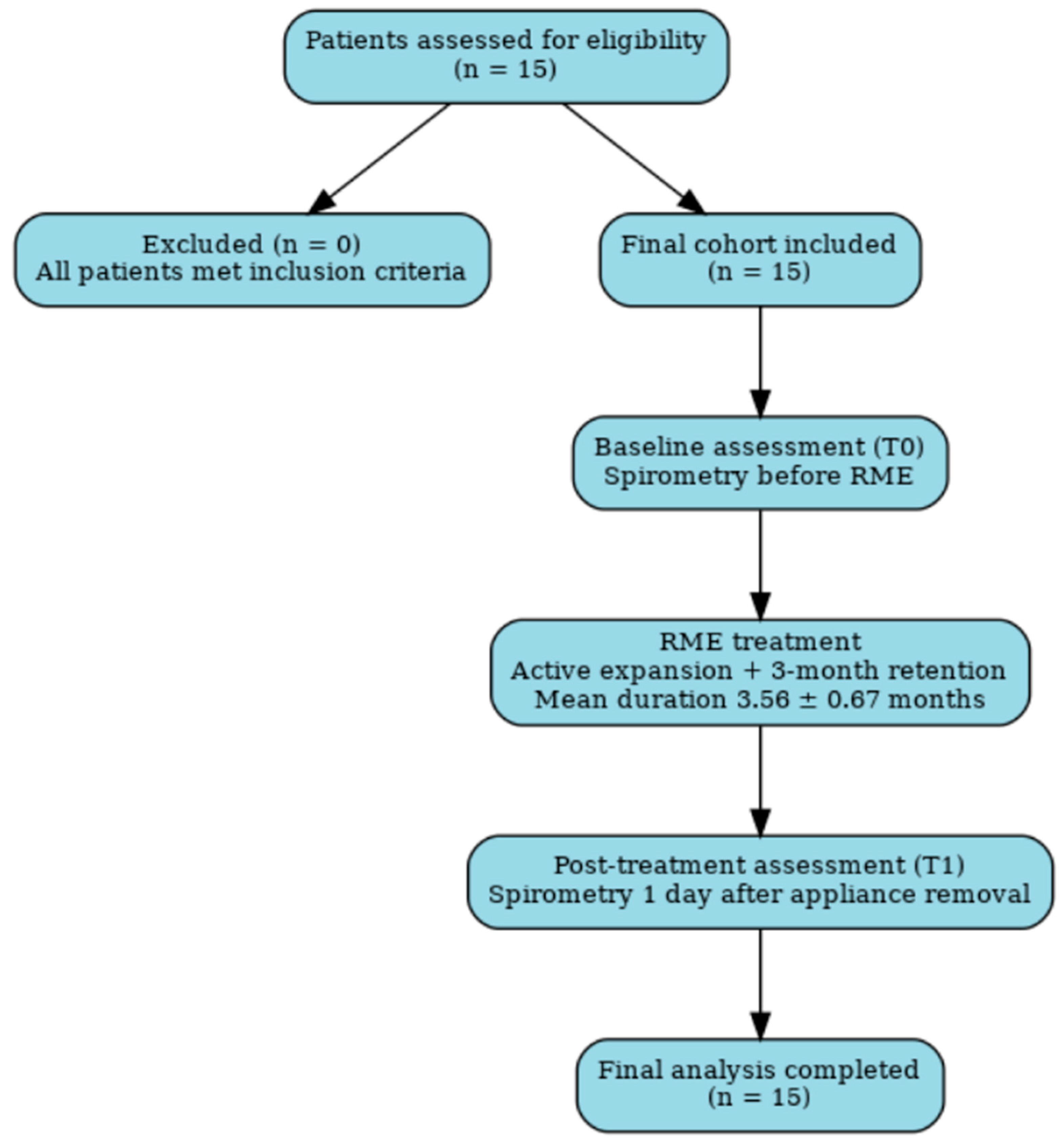
Figure 1. Flowchart of the Study Design.
Applsci 15 10189 g001
Figure 2. Spirometric outcomes before (T0) and after (T1) rapid maxillary expansion (RME). Each panel displays group means ± SD with paired comparisons: (A) peripheral oxygen saturation (SpO2, %), (B) vital capacity (VC, L), (C) forced expiratory volume in 1 s (FEV1, L), (D) forced vital capacity (FVC, L), and (E) FEV1/FVC ratio (%). p-values are derived from paired-samples t-test or Wilcoxon signed-rank tests, as appropriate.
Applsci 15 10189 g002
Figure 3. Individual patient trajectories (spaghetti plots) for pulmonary outcomes before (T0) and after (T1) rapid maxillary expansion (RME). Each gray line represents one patient, illustrating within-subject changes; the red line indicates the group mean at each timepoint. Panels display: peripheral oxygen saturation (SpO2, %), vital capacity (VC, L), forced expiratory volume in 1 s (FEV1, L), forced vital capacity (FVC, L), and FEV1/FVC ratio (%).
Applsci 15 10189 g003
Table 1. Descriptive Statistics of the Study Group.
ParameterValue
Number of subjects (n)15
Gender (Female/Male)8/7
Mean age ± SD (years)13.93 ± 2.89
Mean height ± SD (cm)154.00 ± 12.07
Mean weight ± SD (kg)48.33 ± 11.93
Body mass index (BMI) ± SD (kg/m2)20.18 ± 3.39
Mean RME treatment time ± SD (months)3.56 ± 0.67
Table 2. Comparison of Respiratory Parameters Before (T0) and After (T1) RME Treatment.
VariableT0 Mean ± SDT1 Mean ± SDp Value *
SpO2 (%)96.98 ± 0.9697.01 ± 0.980.925
VC (L)2.86 ± 1.073.03 ± 0.800.626
FEV1 (L)2.61 ± 0.722.72 ± 0.680.518
FVC (L)2.87 ± 0.752.96 ± 0.690.547
FEV1/FVC (%)90.88 ± 12.1792.34 ± 7.370.742
* p value from paired-samples t-test or Wilcoxon signed-rank test, as appropriate.
Disclaimer/Publisher’s Note: The statements, opinions and data contained in all publications are solely those of the individual author(s) and contributor(s) and not of MDPI and/or the editor(s). MDPI and/or the editor(s) disclaim responsibility for any injury to people or property resulting from any ideas, methods, instructions or products referred to in the content.