Article

Orally Delivered Hyaluronic Acid Tetrasaccharide Improves Skin Barrier Function in UVB-Irradiated Mice: A Bioactive Approach for Cosmetic and Nutritional Applications

1
Laboratory of Dermatological Physiology, Faculty of Pharmacy and Pharmaceutical Sciences, Josai University, 1-1 Keyakidai, Sakado 350-0295, Saitama, Japan
2
Sunsho Pharmaceutical Co., Ltd., 12 Nanryo, Fujinomiya 418-0019, Shizuoka, Japan
3
Laboratory of Cosmetic Sciences, Institute of Ocean Energy, Saga University, 1 Honjo, Saga 840-8502, Saga, Japan
*
Author to whom correspondence should be addressed.
Appl. Sci. 2025, 15(18), 10182; https://doi.org/10.3390/app151810182
Submission received: 24 July 2025 / Revised: 12 September 2025 / Accepted: 17 September 2025 / Published: 18 September 2025

Abstract

Hyaluronic acid (HA), which is present in various foods, has been the subject of various claims about its ability to relieve dry skin. In this study, the intestinal absorption of hyaluronic acid tetrasaccharide (HA4) and its ability to protect the skin from UV after oral administration were compared with those of high-molecular-weight HA. Intestinal absorption was evaluated by the Caco-2 cell monolayer mem-brane permeability assay. HA4 permeated the Caco-2 monolayer, reaching 2.67 µg/cm2 after 120 min, whereas HA did not. HA or HA4 was orally administered to UVB-irradiated mice, and the effects were evaluated using transepidermal water loss (TEWL), water content of the stratum corneum (SC), and epidermal thickness. HA4 permeated the Caco-2 monolayer. On day 26, TEWL significantly increased by 17.5 ± 3.1 g/m2/h in the Control group but only 8.0 ± 1.7 g/m2/h in the HA4 group compared to the Normal group, but no significant difference was observed. Water content of SC decreased by 25.7 ± 1.5 arbitrary units (a.u.) in the Control group; the decrease was attenuated in the HA4 group (17.5 ± 0.7 a.u.) (p < 0.05 vs. Control). On day 28, epidermal thickness reached 69.5 ± 10.8 µm in the Control group and was significantly lower in the HA4 group (43.5 ± 5.1 µm) (p < 0.01 vs. Control). These findings indicate that orally administered HA4 is efficiently absorbed and significantly attenuates UVB-induced skin barrier impairment, suggesting its promise as a functional food ingredient for improving dry skin.

1. Introduction

The skin is the largest organ in the human body, which functions to retain water and act as a barrier against external stimuli. Visible light and ultraviolet (UV) radiation are included among such external stimuli, with UV radiation being of particular importance because it can induce skin damage. UV radiation is classified into UVA, UVB, and UVC according to the wavelength. Most UV radiation that reaches the ground is UVA and UVB, with UVA constituting as much as 97% of this. Nonetheless, UVB is highly energetic and can seriously damage the skin by causing the overgrowth of keratinocytes, induction of erythema, and deterioration of skin barrier function [1,2]. Decreased skin barrier function causes dryness and inflammation of the skin. As a potential option to alleviate this, hyaluronic acid (HA) added to foods has been suggested to function in alleviating skin dryness [3,4].
HA is a glycosaminoglycan in which D-glucuronic acid and N-acetyl-D-glucosamine are alternately linearly linked by β-1,4 or β-1,3 glycosidic bonds. It was isolated from the vitreous of the bovine eye in 1934 [5]. HA is also used to treat joint diseases in horses and dogs. Both oral administration and intra-articular injection have been reported [6,7]. Orally administered HA is absorbed into the body and transferred to the skin [8,9,10]. It has been reported that HA has different physiological activities depending on its molecular weight. HA with a molecular weight of 800 kDa or more has been reported to suppress the production of inflammatory cytokines and matrix metalloproteinases (MMPs), inhibit the activation of nuclear factor kappa beta (NF-κB), and suppress angiogenesis [11,12,13,14]. By contrast, HA of 10–500 kDa exhibits inflammatory and angiogenic effects [15,16,17,18]. Meanwhile, it has been reported that the oral administration of HA of less than 10 kDa to hairless mice improves epidermal thickening and reduced skin water content resulting from UVB irradiation and increases HA synthase (HAS)2 gene expression [19]. In addition, in humans with dry skin, the oral administration of 80, 300, or 800 kDa HA for 6 weeks increased skin water content [3,20]. Moreover, the oral administration of 2 and 300 kDa HA to humans was found to reduce wrinkles [21]. Recent studies have highlighted the biological activities of oligosaccharides and their roles in maintaining skin homeostasis through modulation of hydration, inflammation, and barrier integrity [22]. Hyaluronic acid fragments, including tetrasaccharides, have gained attention not only for their physicochemical properties but also for their potential as signaling molecules in skin biology [23,24,25].
In recent years, increasing attention has been directed toward HA oligosaccharides, particularly tetrasaccharides (HA4). HA4 has a defined structure (776 Da) and distinct bioactivities, including the promotion of keratinocyte differentiation via CD44 and stimulation of ceramide production [26,27]. Topical application of HA4 has been reported to improve transepidermal water loss (TEWL), stratum corneum (SC) hydration, and epidermal thickness in UV-irradiated skin [28]. However, to date, there have been no studies examining whether orally administered HA4 can be absorbed in the intestine and subsequently improve skin barrier function in vivo.
Therefore, the objective of this study was to investigate the intestinal absorption of HA4 using a Caco-2 cell monolayer assay and to evaluate whether oral administration of HA4 could improve UVB-induced impairment of skin barrier function in mice, compared with high-molecular-weight HA.

2. Materials and Methods

2.1. Materials

HA4 (776 Da) was purchased from Biomimetics Sympathies Inc. (Tokyo, Japan). High-molecular-weight HA (>1200 kDa) was purchased from Kikkoman Biochemifa Co. (Tokyo, Japan). Isoflurane inhalation anesthetic was purchased from Mylan Pharmaceutical Co., Ltd. (Osaka, Japan). Dulbecco’s Modified Eagle’s Medium (DMEM), Hank’s Balanced Salt Solution (HBSS), non-essential amino acids, sodium acetate, ammonium acetate, acetic acid, methanol, Meyer hematoxylin solution, eosin Y, chloroform, ethanol, methanol, acetic acid, N,N-dimethylformamide, hexane, xylene, cholesterol, palmitic acid, diethyl ether, phosphoric acid, copper (II) sulfate pentahydrate, and 10% formalin solution were purchased from FUJIFILM Wako Pure Chemical Corp. (Osaka, Japan). Hyaluronidase (EC3.2.1.35) was purchased from Nacalai Tesque Inc. (Kyoto, Japan).

2.2. Cell Culture

Human colon cancer Caco-2 cells were purchased from Riken Gene Bank (Tsukuba, Ibaraki, Japan). Caco-2 cells were cultured on a 100 mm dish at 2 × 10⁵ cells/dish. The medium used was MEM containing 20% FBS and 0.1 mM non-essential amino acids, and culture was performed under conditions of 37 °C and 5% CO2. Caco-2 cells were seeded at a density of 6.0 × 10⁴ cells/cm2 on the apical side of Millicell® Hanging Cell Culture Inserts (Corning, Corning, NY, USA). Caco-2 cell monolayers were prepared by cultivating Caco-2 cells on polycarbonate membrane filters for 28 days. The medium was changed every other day.

2.3. Cell Permeation Test

Caco-2 cells were used in this test at passages 55 to 60. Transepithelial electrical resistance (TEER) of the Caco-2 cell monolayers was measured using a Millicell® ERS-2 voltameter (Millipore Corporation, Bedford, MA, USA), to confirm their integrity. Cell monolayers with TEER values above 250 Ω·cm2 were used for the permeation test [29]. The monolayers were rinsed with HBSS (pH6.0). HBSS was then added to the basolateral side (receiving phase). A permeation test was performed by adding a 5 mg/mL HA4 or HA solution prepared with HBSS (pH6.0) to the apical side (feed phase). After 30, 60, 90, and 120 min, an aliquot of the receiving phase (200 µL) was taken and 200 µL of fresh HBSS was added to the receiving phase to maintain a constant volume. The integrity of the cell monolayers was confirmed by monitoring TEER values before and after the experiments.

2.4. Quantitative Determination of HA and HA4

The HA that permeates Caco-2 cell monolayers is decomposed to HA4 by hyaluronidase (EC3.2.1.35) [30]. HA was quantified by measuring the decomposed HA4 in accordance with a previously reported procedure [31]. Decomposed HA4 was obtained using 40 U/mL hyaluronidase prepared with sodium acetate buffer (pH 5.0) at 37 °C for 24 h with HA. Quantitative determination of HA4 was performed using a liquid chromatography/tandem mass spectrometry (LC-MS/MS) system. Chromatographic analysis was performed using a 4000 QTRAP® LC-MS/MS system (system controller: CBM-20A; Shimazu Corporation, Kyoto, Japan) (Thermo Fisher Scientific, Waltham, MA, USA) and an InertSustain® C18 column (3.0 × 250 mm; GL Sciences Inc., Tokyo, Japan). The mobile phase was a 20% methanol mixed solution of 10 mM ammonium acetate solution, the flow rate was 200 µL/min, and 192.9/775.2 (product/precursor) were detected in the negative ionization mode. A calibration curve was prepared from the analytical values of HA4 obtained by decomposing 0.005, 0.025, 0.05, 0.25, and 0.5 µg/mL HA solutions using hyaluronidase. Representative chromatograms and calibration plots used for quantification are provided in the Supplementary Materials (Figures S1 and S2).

2.5. Animals

Male hairless mice (HR-1) aged 7-to-8 weeks old were purchased from Hoshino Laboratory Animals, Inc. (Ibaraki, Japan). All experimental animals had free access to food (Labo MR Stock; Nosan Corporation, Kanagawa, Japan) and water, and were housed in rooms where the lighting was automatically regulated on a 12 h light/dark cycle and constant temperature (25 ± 2 °C) and humidity (50 ± 10%). All animal experiments and maintenance were performed under conditions approved by the animal research committee of Josai University (approval numbers: H29096 and JU18094).

2.6. Oral Administration to Mice and UVB Irradiation

Aqueous solutions of 0.1 mg/mL HA and HA4 were prepared and orally administered to hairless mice at a dose of 10 mL/kg daily for 28 days. In terms of the molecular weight, HA and HA4 differ by approximately 1500-fold, so here we made the concentrations uniform. The same volume of purified water was orally administered to the Control group. The UVB lamp used was a Philips Broadband TL 20W/12RS (Philips, Amsterdam, The Netherlands) with a peak wavelength of 302 nm. UVB was irradiated on the left back of the hairless mice in the HA, HA4, and Control group three times a week. The UVB irradiation dose was 30, 60, 90, and 120 mJ/cm2/day in the 1st, 2nd, 3rd, and 4th weeks, respectively. The UVB irradiation conditions were determined based on the reports of Cho et al. [32] and Tanaka et al. [33]. The water content of the SC was measured with a Corneometer® (Courage + Khazaka, Cologne, Germany). TEWL was measured with VAPOSCAN AS-VT100RS (Asahi Techno Lab, Kanagawa, Japan). The water content of SC and TEWL were measured five times a week. At the time of measurement, anesthesia was performed via the inhalation of isoflurane using an animal experimental anesthesia device (SN-487-OT Air; Shinano Manufacturing Co., Ltd., Tokyo, Japan). On the final day of the animal experiment, hairless mice were sacrificed by cervical dislocation and the skin at the irradiated site was collected.

2.7. Histological Procedures

Cryosections were prepared from tissue samples embedded in optimal cutting temperature (OCT) compound. OCT compound is a water-soluble embedding medium used to prepare tissue samples for frozen sectioning. Frozen sections were prepared using LEICA CM3050S (Leica Biosystems Nussloch GmbH, Nussloch, Germany). Sliced sections were prepared under the following conditions: internal temperature −25 °C, pedestal temperature −25 °C, and section thickness 5 µm. A 10% formalin solution, hematoxylin-eosin (HE), and Multimount 480 (Matsunami Glass Ind. Co., Ltd., Osaka, Japan) were used for staining and encapsulation of the sections. HE-stained skin sections were observed with an inverted all-in-one fluorescence microscope (BZ-710; Keyence, Osaka, Japan). The thickness of the epidermis was calculated from the microscopic images by measuring it at 20 points for each tissue section and obtaining the average.

2.8. Statistical Analysis

Statistical analysis was performed using JMP® v.13.1 from SAS (SAS Institute Inc., Cary, NC, USA). Indicated p-values were derived from Tukey’s post hoc multiple comparison test.

3. Results and Discussion

3.1. HA4 Was Absorbed Intact in the Intestine

A Caco-2 cell monolayer membrane permeation assay was performed to predict the intestinal absorption of HA and HA4. Figure 1 shows the results from evaluating the permeability of HA4 through a Caco-2 single-layer membrane. In the HA4 group, the amount of HA4 that permeated increased in a time-dependent manner and was 2.67 µg/cm2 after 120 min. Meanwhile, HA permeation could not be confirmed in the HA group (Figure 1). The TEER values of the HA and HA4 group were 529.6 and 502.6 Ω·cm2 before the permeation test and 417.6 and 422.3 Ω·cm2 after it. TEER exceeded 200 Ω·cm2 before and after the permeation assay, indicating that the monolayer was intact [34]. Caco-2 cells express various transporters, and the results of assays of permeation across their monolayer correlate with intestinal absorption, providing a useful index for predicting intestinal absorption [35]. It has been reported that HA with a larger molecular weight permeates less through a Caco-2 cell monolayer membrane [34]. Hisada et al. evaluated the membrane permeability of hyaluronic acid of various molecular weights using Caco-2 cell monolayer membranes [36]. They showed that HA with a smaller molecular weight permeates more across membranes and found that HA oligosaccharides passed through the Caco-2 cell monolayer membrane. However, whether the skin conditions change after the intestinal absorption of HA oligosaccharides has not been evaluated. The reported intestinal absorption of HA oligosaccharides is consistent with our results. Our Caco-2 cell permeation test, HA4 was quantified from the basolateral side (receiving phase) solution using LC-MS/MS, and it was revealed that HA4 is absorbed intact in the intestine (Figure 1). When HA is administered orally, it is degraded and then absorbed in the intestinal tract. In the Caco-2 cell permeation test, since there are no digestive enzymes, it is natural that HA remains on the apical side and does not permeate the Caco-2 cell monolayer membrane. The digestion and absorption of HA have been investigated in several papers. It has been reported that oral administration of 99mTc-labeled 1000 kDa HA to rats and dogs resulted in its distribution in the blood, skin, and joints [8]. In addition, Kimura et al. reported that, after orally administering 300 kDa HA to rats, disaccharide in the blood was detected at 4 h and peaked at 6 to 8 h [10]. In addition, disaccharide, tetrasaccharide, and polysaccharide HA were distributed in rat skin and were detectable from 6 h onwards, with maximum amounts detected after 8 h. HA is broken down into oligosaccharides in the cecum and absorbed in the large intestine. These reports suggest that HA4 is likely to be absorbed intact into the intestinal tract in vivo.

3.2. Oral Administration of HA4 Suppresses the Deterioration of Water Content of SC and Improves Barrier Function Caused by UVB Irradiation

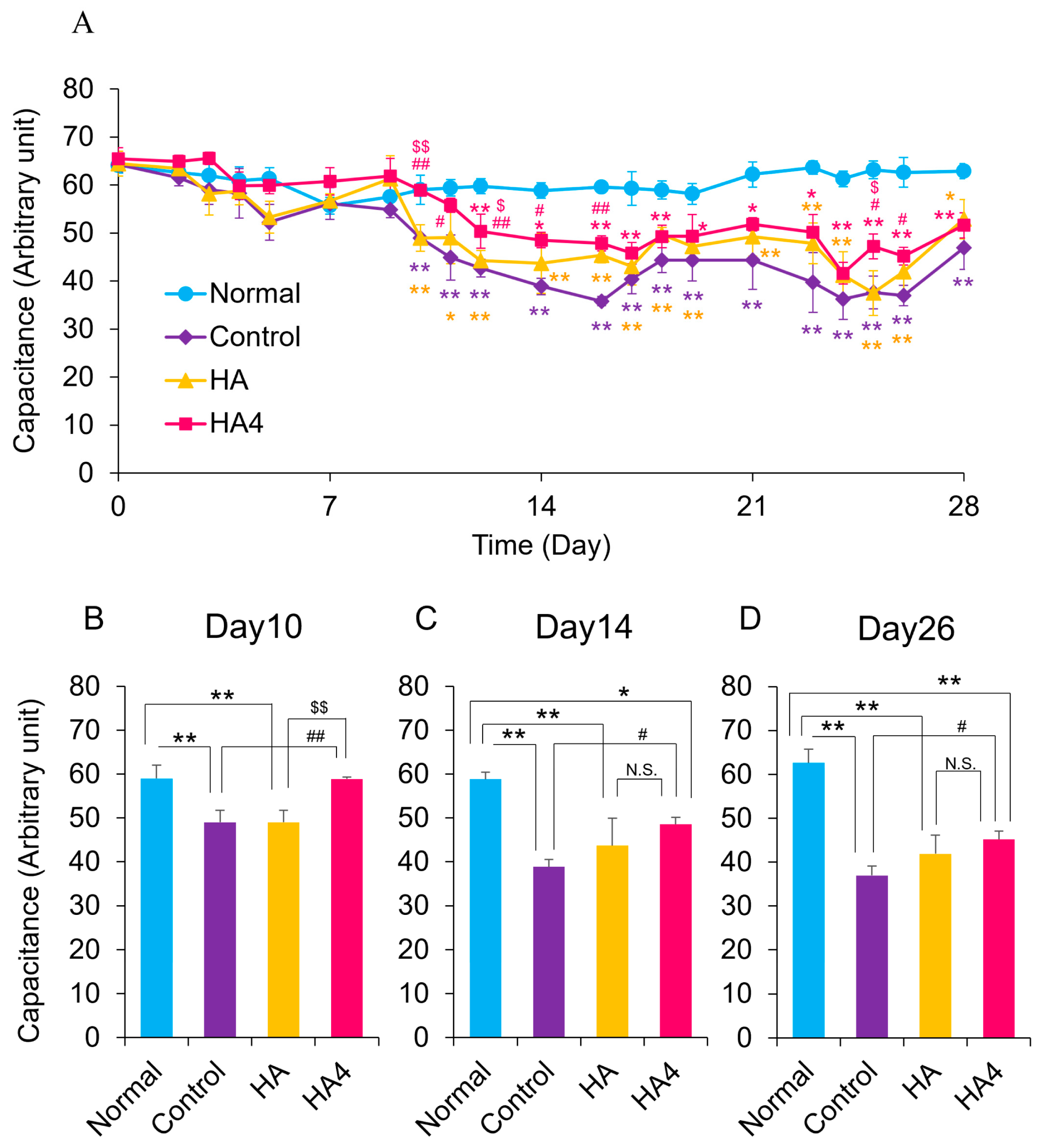
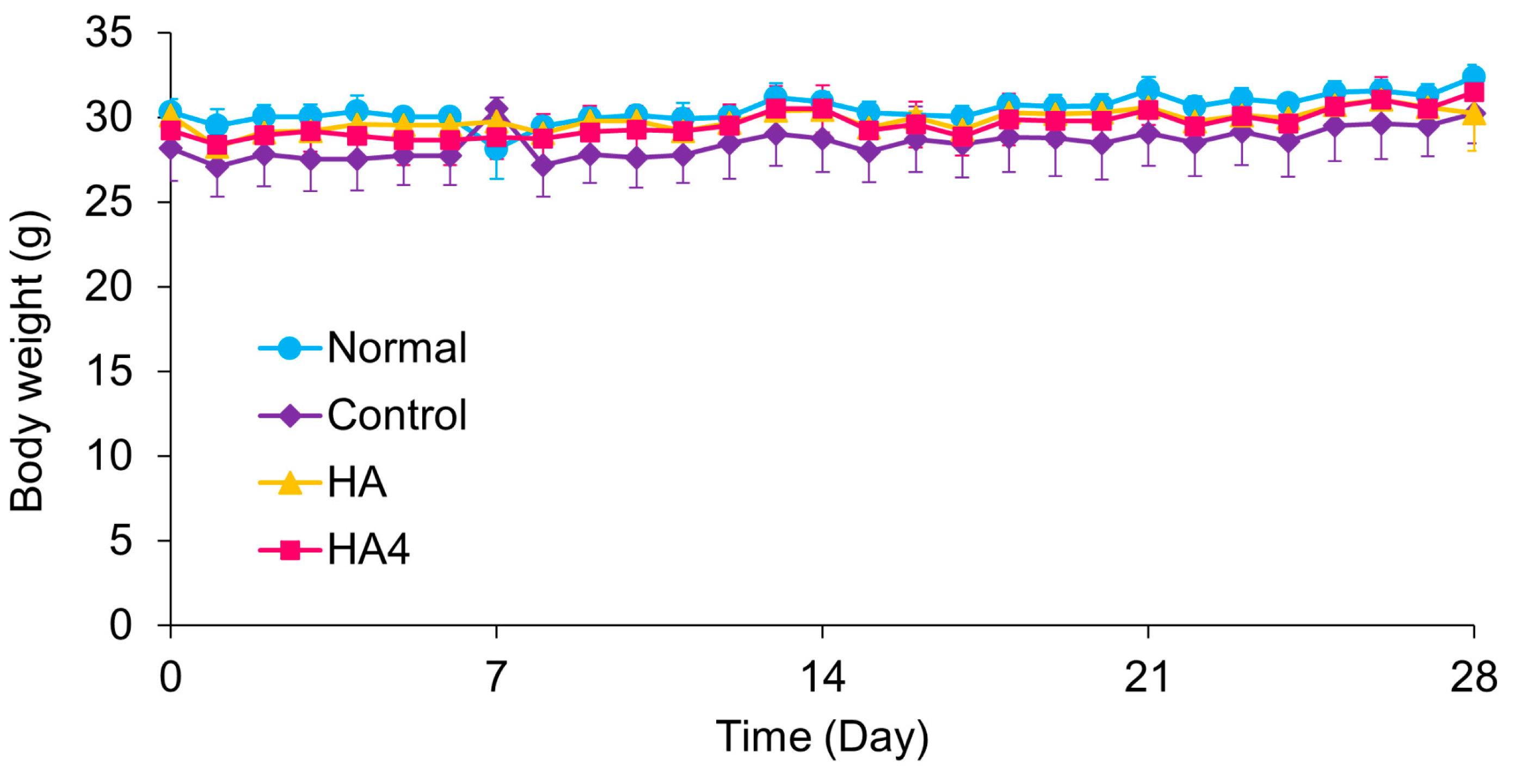
HA4 improves skin damage caused by UVA irradiation when treated to the skin [28]. If HA4 is absorbed from the intestinal tract without being digested, it may have a beneficial effect on the skin earlier than HA. Next, we evaluated the effects of orally administered HA and HA4 on improving the skin of UVB-irradiated mice by measuring the TEWL and water content of SC. Regarding changes in the TEWL, TEWL in the Control and HA groups increased by 8.0 ± 1.1 and 4.6 ± 0.2 g/m2/h compared with that in the Normal group on the day 12. At this time, the HA4 group increased by 3.2 ± 0.1 g/m2/h, but the difference was not significant. Additionally, the level in the HA4 group was significantly lower than in the Control group. In the HA group, there was also significant suppression of the increase in TEWL caused by UVB, although not to the same extent as in the HA4 group (Figure 2B). UVB irradiation at 120 mJ/cm2/day was started on day 22, and the TEWL value of the Control and HA group further increased by 17.5 ± 3.0 and 15.4 ± 4.2 g/m2/h on day 26. Meanwhile, the HA4 group was increased by 8.0 ± 1.7 g/m2/h, but the increase was slightly suppressed compared to the Control and HA groups (Figure 2C). The HA4 group showed no significant increase over the 28 days compared with the Normal group, and from day 12 onwards the levels remained significantly lower than those of the Control group (Figure 2). The water content of SC was significantly decreased by 10 ± 0.6 arbitrary units (a.u.) in both the Control and HA groups compared with that in the Normal group on day 10. Moreover, that in the HA4 group was significantly higher than those in the Control and HA groups and was comparable to that of the Normal group (Figure 3B). On day 14, compared to the Normal group, the decrease was 20 ± 0.9 a.u. in the Control group, 16.1 ± 2.2 a.u. in the HA group, and 10.3 ± 0.3 a.u. in the HA4 group. The HA group had recovered to the same level as the HA4 group had (Figure 3C), and thereafter the HA group showed values similar to those of the HA4 group (Figure 3D). The water content of SC was not significantly affected by the first UVB irradiation but decreased with the second UVB irradiation. Meanwhile, the decrease was slightly suppressed in the HA4 group (Figure 3). Body weight did not differ among all groups, as confirmed by statistical analysis using Tukey’s post hoc multiple comparison test (p > 0.05). (Figure 4). It has been reported that TEWL increased, water content decreased, and the epidermis was thickened by UVB irradiation of the skin of hairless mice [37,38]. Oral administration of HA4 suppressed the deterioration of TEWL and water content of SC caused by UVB irradiation. In the HA group, the deterioration was also suppressed compared with that in the Control group, although not as much as in the HA4 group (Figure 2 and Figure 3). These results indicate that the oral administration of HA4 improved the functionality of skin with impaired barrier function resulting from UVB irradiation. When HA with molecular weights of 8, 10, and 300 kDa was orally administered to UV-irradiated hairless mice, the water content of SC was restored after 4 weeks, and when the difference in molecular weight was examined, the smaller the molecular weight, the more significantly the water content was restored [19]. Oral intake of HA is a recognized functional food, as it helps retain moisture in the skin and relieves dryness. It is thought that ingested HA is broken down by intestinal bacteria [39], and HA at the oligosaccharide level is absorbed in the large intestine and transferred to the bloodstream and skin. HA4 is absorbed from the intestinal tract without being degraded, so it may be rapidly absorbed into the blood and skin.

3.3. Oral Administration of HA4 Suppresses UVB-Induced Epidermal Thickening

Oral administration of HA4 improved the water content of SC and TEWL that had deteriorated due to UVB irradiation, suggesting that HA4 may have a physiological effect on epidermal keratinocytes. Observation of skin morphology on the final day revealed that the epidermis had thickened due to UVB irradiation (Control group, Figure 5B). In addition, the epidermal morphology of the HA group was similar to that of the control group (Figure 5C). On the other hand, epidermal thickening was suppressed in the HA4 group (Figure 5D). The epidermal thicknesses of the Normal, Control, HA, and HA4 group were 22.0 ± 1.7, 69.5 ± 10.8, 65.7 ± 9.8, and 43.5 ± 5.1 µm, respectively (Figure 6). Compared with the Normal group, the thickness was 3.2-folds greater in the Control group, 3.0-fold greater in the HA group, and 2.0-folds greater in the HA4 group. The epidermis was thickened by UVB irradiation. However, epidermal thickness was significantly decreased in the HA4 group compared with that in the Control group. There was no difference in epidermal thickness between the Control and HA group. Kawada et al. irradiated hairless mice with UV rays at a dose of 200 mg/kg body weight per day for six weeks and measured the condition of their skin. The results showed that the HA group had significantly less UV damage to the skin than the control group [19]. The mechanism of epidermal thickening caused by UV irradiation is thought to involve increased cell death of keratinocytes, increased filaggrin production, and increased epidermal growth factor in the MAPK pathway [40,41,42]. The high molecular weight of HA reduces UV-induced apoptosis in human corneal epithelial cells [43]. This suggests that orally administered HA4 and HA may also suppress UV-induced apoptosis in keratinocytes and prevent an increase in epidermal thickness. Our data showed that although skin condition improved in the HA group, epidermal thickness remained unchanged. HA has often been evaluated based on long-term intake, and the effects of HA may only be in the intermediate stages in a 28-day evaluation. Since the HA4 group showed an improvement in skin condition and suppression of epidermal thickening, it is possible that the effects of HA4 appeared more quickly than those of HA.
Since a reduction in skin damage leads to a reduction in skin wrinkles, oral intake of hyaluronic acid, especially ultra-low molecular weight HA such as HA4, is expected to be effective in reducing skin wrinkles. There are several surgical treatments using fillers as symptomatic treatments for skin wrinkles, but these are expensive and involve risks such as pain and swelling [44]. Topical treatments such as HA injections are effective quickly, but the effect gradually fades [45]. On the other hand, supplements take time to improve wrinkles, but the effects will last if you take them regularly. For this reason, demand for supplements is increasing because they are easy to take continuously and provide support from within the body. There have been many reports of HA intake improving dry skin and wrinkles [46,47,48,49]. Kim et al. treated 52 Korean women aged 30 years or older with crow’s feet with HA (molecular weight 75 kDa, 240 mg/day) for 8 weeks. The HA group had significantly fewer wrinkles than the placebo group [50]. Watanabe et al. treated 28 Japanese women aged 30–49 with crow’s feet wrinkles with HA (molecular weight 38 kDa, 240 mg/day) for 8 weeks. The HA group showed a significant reduction in maximum mean wrinkle depth compared to the placebo group [51]. Oe et al. treated Japanese men and women aged 22 to 59 with HA (molecular weight 2 and 300 kDa, 120 mg/day) for 12 weeks. The HA group showed better values for sulcus volume ratio, wrinkle area ratio, and wrinkle volume ratio compared to the placebo group [52]. These reports suggest that HA4 may also be effective in improving wrinkles. It has been reported that HA binds to CD44 present on the surface of keratinocytes and normalizes skin function through signal transduction [53]. HA4 induces the differentiation of epidermal keratinocytes through the activation of CD44 [24]. It is thus speculated that orally administered HA and HA4 improved epidermal thickening and skin barrier function by improving the abnormal differentiation of the epidermis caused by UVB. It also increases expression of the HAS gene in fibroblasts [54]. In addition to our report, it has been confirmed that HA promotes the proliferation of dermal fibroblasts and HA synthesis in fibroblasts [55,56]. It is believed that some of the orally ingested HA promotes HA synthesis in dermal fibroblasts and is involved in maintaining normal skin and preventing wrinkles. Although this study did not evaluate its effect on wrinkles, it is likely that it may be effective against wrinkles. There is no conclusive evidence as to whether oral administration of HA4 changes the HA content in the skin in vivo or whether it has any effect on wrinkles. Further research is needed on these points.
It was also suggested that the beneficial effect on skin with damaged barrier function resulting from UVB irradiation was higher in the HA4 group than in the HA group because HA4 with a low molecular weight was absorbed more by the intestine and more of it was transferred to the skin. However, blood and skin concentrations were not measured, and it was not evaluated whether absorbed HA4 acts directly on the skin or whether it affects the skin of UV-damaged model mice via other factors. Furthermore, although bacterial degradation of HA in the intestine has been previously reported, our study did not directly investigate this process. The potential contribution of microbiota-mediated degradation to HA4 absorption and systemic effects remains to be elucidated. This should be considered a limitation of the current study and warrants further investigation using isotope-labeled HA4 and gut microbiota analysis. In the future, it will be necessary to investigate the decomposition of these substances by intestinal bacteria and their transferability to tissues such as blood and skin. Furthermore, with the aim of enabling oral administration of HA4 to protect the skin from UV rays, we would like to evaluate the concentration in the skin and the expression levels of related factors, as well as conduct research on optimizing the dosage of HA4. These findings suggest that HA4 may serve as a model compound for studying carbohydrate-based bioactivity beyond classical polysaccharide functions. Given its defined structure and measurable effects, HA4 could also provide a benchmark for evaluating intestinal glycan absorption and its systemic dermatological benefits. Further studies utilizing structure–activity relationships and tissue distribution profiling are warranted to determine whether specific oligosaccharide configurations confer enhanced bioavailability or skin-targeting properties. This suggests further investigation into its effects on skin barrier regulation and protection against UV-induced damage is warranted. Taken together, our results support the potential of orally administered HA4 as a novel bioactive ingredient for cosmeceutical application.

4. Conclusions

In this study, orally administered HA4 improved skin barrier function and epidermal thickening in the skin of mice whose barrier function had been damaged by UVB irradiation, compared with the effects of the oral administration of HA. It was suggested that this difference in beneficial effects was due to the difference in intestinal absorption between HA4 and HA. HA4 can be expected to be useful for foods aimed at improving dry skin and wrinkles. Future studies should aim to elucidate the detailed absorption mechanisms, tissue distribution, and long-term efficacy of HA4, as well as its potential clinical applications in humans.

Supplementary Materials

The following supporting information can be downloaded at https://www.mdpi.com/article/10.3390/app151810182/s1, Figure S1: Representative LC-MS/MS chromatogram of HA4.; Figure S2: Calibration curve of HA4 used for quantification.

Author Contributions

Conceptualization, M.K., M.O. and Y.T.; Methodology, M.K., M.O. and T.A.; Software, M.K., M.O. and T.A.; Validation, M.O. and T.A.; Formal Analysis, M.O. and T.A.; Resources, M.O. and Y.T.; Data Curation, M.O. and T.A.; Writing—Original Draft Preparation, M.K., M.O., T.A. and Y.T.; Writing—Review and Editing, M.K., M.O. and Y.T.; Visualization, M.O. and T.A.; Supervision, Y.T.; Project Administration, M.O. and Y.T.; Funding Acquisition, Y.T. All authors have read and agreed to the published version of the manuscript.

Funding

Yoshihiro Tokudome received a research grant from Sunsho Pharmaceutical Co. Ltd.

Institutional Review Board Statement

All animal experiments and maintenance were performed under conditions approved by the animal research committee of Josai University (Approval number; H29096/6 April 2017) and JU18094/3 April 2018).

Informed Consent Statement

Not applicable.

Data Availability Statement

The data used to support the findings of this study are available from the corresponding author upon request.

Acknowledgments

We are grateful to Yoshiharu Matahira and Masahiro Kurono for helpful discussions.

Conflicts of Interest

Masaki Okawara was employed by Sunsho Pharmaceutical Co., Ltd. The remaining authors declare that the research was conducted in the absence of any commercial or financial relationships that could be construed as a potential conflict of interest.

References

  1. Abe, T.; Mayuzumi, J. The change and recovery of human skin barrier functions after ultraviolet light irradiation. Chem. Pharm. Bull. 1979, 27, 458–462. [Google Scholar] [CrossRef]
  2. Haratake, A.; Uchida, Y.; Schmuth, M.; Tanno, O.; Yasuda, R.; Epstein, J.H.; Elias, P.M.; Holleran, W.M. UVB-induced alterations in permeability barrier function: Roles for epidermal hyperproliferation and thymocyte-mediated response. J. Investig. Dermatol. 1997, 108, 769–775. [Google Scholar] [CrossRef] [PubMed]
  3. Kawada, C.; Yoshida, T.; Yoshida, H.; Sakamoto, W.; Odanaka, W.; Sato, T.; Yamasaki, T.; Kanemitsu, T.; Masuda, Y.; Urushibata, O. Ingestion of hyaluronans (molecular weights 800 k and 300 k) improves dry skin conditions: A randomized, double blind, controlled study. J. Clin. Biochem. Nutr. 2015, 56, 66–73. [Google Scholar] [CrossRef] [PubMed]
  4. Hsu, T.F.; Su, Z.R.; Hsieh, Y.H.; Wang, M.F.; Oe, M.; Matsuoka, R.; Masuda, Y. Oral hyaluronan relieves wrinkles and improves dry akin: A 12-week double-blinded, placebo-controlled study. Nutrients 2021, 13, 2220. [Google Scholar] [CrossRef]
  5. Meyer, K.; Palmer, J.W. The polysaccharide of the vitreous humor. J. Biol. Chem. 1934, 107, 629–634. [Google Scholar] [CrossRef]
  6. da Silva Xavier, A.A.; da Rosa, P.P.; de Brum Mackmill, L.; Roll, V.F.B. An assessment of the effectiveness of hyaluronic acid and polyacrylamide hydrogel in horses with osteoarthritis: Systematic review and network meta-analysis. Res. Vet. Sci 2021, 134, 42–50. [Google Scholar] [CrossRef]
  7. Serra Aguado, C.I.; Ramos-Plá, J.J.; Soler, C.; Segarra, S.; Moratalla, V.; Redondo, J.I. Effects of Oral Hyaluronic Acid Administration in Dogs Following Tibial Tuberosity Advancement Surgery for Cranial Cruciate Ligament Injury. Animals 2021, 11, 1264. [Google Scholar] [CrossRef]
  8. Balogh, L.; Polyak, A.; Mathe, D.; Kiraly, R.; Thuroczy, J.; Terez, M.; Janoki, G.; Ting, Y.; Bucci, L.R.; Schauss, A.G. Absorption, uptake and tissue affinity of high-molecular-weight hyaluronan after oral administration in rats and dogs. J. Agric. Food Chem. 2008, 56, 10582–10593. [Google Scholar] [CrossRef] [PubMed]
  9. Oe, M.; Mitsugi, K.; Odanaka, W.; Yoshida, H.; Matsuoka, R.; Seino, S.; Kanemitsu, T.; Masuda, Y. Dietary hyaluronic acid migrates into the skin of rats. Sci. World J. 2014, 2014, 378024. [Google Scholar] [CrossRef]
  10. Kimura, M.; Maeshima, T.; Kubota, T.; Kurihara, H.; Masuda, Y.; Nomura, Y. Absorption of Orally Administered Hyaluronan. J. Med. Food 2016, 19, 1172–1179. [Google Scholar] [CrossRef]
  11. Nakamura, K.; Yokohama, S.; Yoneda, M.; Okamoto, S.; Tamaki, Y.; Ito, T.; Okada, M.; Aso, K.; Makino, I. High, but not low, molecular weight hyaluronan prevents T-cell-mediated liver injury by reducing proinflammatory cytokines in mice. J. Gastroenterol. 2004, 39, 346–354. [Google Scholar] [CrossRef]
  12. Spessotto, P.; Rossi, F.M.; Degan, M.; Francia, R.D.; Perris, R.; Colombatti, A.; Gattei, V. Hyaluronan-CD44 interaction hampers migration of osteoclast-like cells by down-regulating MMP-9. J. Cell Biol. 2002, 158, 1133–1144. [Google Scholar] [CrossRef] [PubMed]
  13. Takahashi, K.; Goomer, R.S.; Harwood, F.; Kubo, T.; Hirasawa, Y.; Amiel, D. The effects of hyaluronan on matrix metal-loproteinase-3 (MMP-3), interleukin-1beta(IL-1beta), and tissue inhibitor of metalloproteinase-1 (TIMP-1) gene expression during the development of osteoarthritis. Osteoarthr. Cartil. 1999, 7, 182–190. [Google Scholar] [CrossRef]
  14. Neumann, A.; Schinzel, R.; Palm, D.; Riederer, P.; Munch, G. High molecular weight hyaluronic acid inhibits advanced glycation endproduct-induced NF-kappaB activation and cytokine expression. FEBS Lett. 1999, 453, 283–287. [Google Scholar] [CrossRef]
  15. Hodge-Dufour, J.; Noble, P.W.; Horton, M.R.; Bao, C.; Wysoka, M.; Burdick, M.D.; Strieter, R.M.; Trinchieri, G.; Pure, E. Induction of IL-12 and chemokines by hyaluronan requires adhesion-dependent priming of resident but not elicited macrophages. J. Immunol. 1997, 159, 2492–2500. [Google Scholar] [CrossRef]
  16. McKee, C.M.; Penno, M.B.; Cowman, M.; Burdick, M.D.; Strieter, R.M.; Bao, C.; Noble, P.W. Hyaluronan (HA) fragments induce chemokine gene expression in alveolar macrophages. The role of HA size and CD44. J. Clin. Investig. 1996, 98, 2403–2413. [Google Scholar] [CrossRef]
  17. Noble, P.W.; McKee, C.M.; Cowman, M.; Shin, H.S. Hyaluronan fragments activate an NF-kappa B/I-kappa B alpha autoregulatory loop in murine macrophages. J. Exp. Med. 1996, 183, 2373–2378. [Google Scholar] [CrossRef] [PubMed]
  18. West, D.C.; Hampson, I.N.; Arnold, F.; Kumar, S. Angiogenesis induced by degradation products of hyaluronic acid. Science 1985, 228, 1324–1326. [Google Scholar] [CrossRef] [PubMed]
  19. Kawada, C.; Kimura, M.; Masuda, Y.; Nomura, Y. Oral administration of hyaluronan prevents skin dryness and epidermal thickening in ultraviolet irradiated hairless mice. J. Photochem. Photobiol. B 2015, 153, 215–221. [Google Scholar] [CrossRef]
  20. Kawada, C.; Yoshida, T.; Yoshida, H.; Matsuoka, R.; Sakamoto, W.; Odanaka, W.; Sato, T.; Yamasaki, T.; Kanemitsu, T.; Masuda, Y.; et al. Ingested hyaluronan moisturizes dry skin. Nutr. J. 2014, 13, 70. [Google Scholar] [CrossRef]
  21. Yun, M.K.; Lee, S.J.; Song, H.J.; Yu, H.J.; Rha, C.S.; Kim, D.O.; Choe, S.Y.; Sohn, J. Protein expression level of skin wrinkle-related factors in hairless mice fed hyaluronic acid. J. Med. Food 2017, 20, 420–424. [Google Scholar] [CrossRef]
  22. Lee, S.H.; Kim, J.W.; Hyunc, J.W.; Yoon, S.-P. Biological effects of chitosan oligosaccharides on the skin and their clinical applicability. Carbohydr. Res. 2025, 555, 109588. [Google Scholar] [CrossRef]
  23. Gruber, J.V.; Holtz, R.; Riemer, J. Hyaluronic acid (HA) stimulates the in vitro expression of CD44 proteins but not HAS1 proteins in normal human epidermal keratinocytes (NHEKs) and is HA molecular weight dependent. J. Cosmet. Dermatol. 2022, 21, 1193–1198. [Google Scholar] [CrossRef]
  24. Stern, R.; Asari, A.; Sugahara, K. Hyaluronan fragments: An information-rich system. Eur. J. Cell Biol. 2006, 85, 699–715. [Google Scholar] [CrossRef]
  25. Campo, G.M.; Avenoso, A.; D’Ascola, A.; Scuruchi, M.; Prestipino, V.; Calatroni, A.; Campo, S. Hyaluronan in part mediates IL-1beta-induced inflammation in mouse chondrocytes by up-regulating CD44 receptors. Gene 2012, 494, 24–35. [Google Scholar] [CrossRef]
  26. Kage, M.; Tokudome, Y.; Matsunaga, Y.; Hariya, T.; Hashimoto, F. Effect of hyaluronan tetrasaccharides on epidermal differentiation in normal human epidermal keratinocytes. Int. J. Cosmet. Sci. 2014, 36, 109–115. [Google Scholar] [CrossRef]
  27. Kage, M.; Tokudome, Y. Hyaluronan tetrasaccharides stimulate ceramide production through upregulated mRNA expression of ceramide synthesis-associated enzymes. Arch. Dermatol. Res. 2016, 308, 95–101. [Google Scholar] [CrossRef]
  28. Kage, M.; Tokudome, Y.; Hashimoto, F. Permeation of hyaluronan tetrasaccharides through hairless mouse skin: An in vitro and in vivo study. Arch. Dermatol. Res. 2013, 305, 69–77. [Google Scholar] [CrossRef]
  29. Delie, F.; Rubas, W. A human colonic cell line sharing similarities with enterocytes as a model to examine oral absorption: Advantages and limitations of the Caco-2 model. Crit. Rev. Ther. Drug Carr. Syst. 1997, 14, 221–286. [Google Scholar] [CrossRef]
  30. Meyer, K.; Rapport, M.M. Hyaluronidases. Adv. Enzymol. Relat. Subj. Biochem. 1952, 13, 199–236. [Google Scholar]
  31. Chun, L.E.; Koob, T.J.; Eyre, D.R. Quantitation of hyaluronic acid in tissues by ion-pair reverse-phase high-performance liquid chromatography of oligosaccharide cleavage products. Anal. Biochem. 1988, 171, 197–206. [Google Scholar] [CrossRef] [PubMed]
  32. Cho, H.S.; Lee, M.H.; Lee, J.W.; No, K.O.; Park, S.K.; Lee, H.S.; Kang, S.; Cho, W.G.; Park, H.J.; Oh, K.W.; et al. Anti-wrinkling effects of the mixture of vitamin C, vitamin E, pycnogenol and evening primrose oil, and molecular mechanisms on hairless mouse skin caused by chronic ultraviolet B irradiation. Photodermatol. Photoimmunol. Photomed. 2007, 23, 155–162. [Google Scholar] [CrossRef] [PubMed]
  33. Tanaka, M.; Koyama, Y.; Nomura, Y. Effects of collagen peptide ingestion on UV-B-induced skin damage. Biosci. Biotechnol. Biochem. 2009, 73, 930–932. [Google Scholar] [CrossRef] [PubMed]
  34. Bock, U.; Flötotto, T.; Haltner, E. Validation of cell culture models for the intestine and the blood-brain barrier and comparison of drug permeation. ALTEX—Altern. Anim. Exp. 2004, 3, 57–64. [Google Scholar]
  35. Yamashita, S.; Tanaka, Y.; Endoh, Y.; Taki, Y.; Sakane, T.; Nadai, T.; Sezaki, H. Analysis of drug permeation across Caco-2 monolayer: Implication for predicting in vivo drug absorption. Pharm. Res. 1997, 14, 486–491. [Google Scholar] [CrossRef]
  36. Hisada, N.; Satsu, H.; Mori, A.; Totsuka, M.; Kamei, J.; Nozawa, T.; Shimizu, M. Low-molecular-weight hyaluronan permeates through human intestinal Caco-2 cell monolayers via the paracellular pathway. Biosci. Biotechnol. Biochem. 2008, 72, 1111–1114. [Google Scholar] [CrossRef]
  37. Yoo, J.H.; Kim, J.K.; Yang, H.J.; Park, K.M. Effects of egg shell membrane hydrolysates on UVB-radiation-induced wrinkle formation in SKH-1 hairless mice. Korean J. Food Sci. Anim. Resour. 2015, 35, 58–70. [Google Scholar] [CrossRef]
  38. Takagi, Y.; Nakagawa, H.; Kondo, H.; Takema, Y.; Imokawa, G. Decreased levels of covalently bound ceramide are associated with ultraviolet B-induced perturbation of the skin barrier. J. Investig. Dermatol. 2004, 123, 1102–1109. [Google Scholar] [CrossRef]
  39. Kawai, K.; Kamochi, R.; Oiki, S.; Murata, K.; Hashimoto, W. Probiotics in human gut microbiota can degrade host glycosaminoglycans. Sci. Rep. 2018, 8, 10674–10687. [Google Scholar] [CrossRef]
  40. Lee, J.K.; Kim, J.H.; Nam, K.T.; Lee, S.H. Molecular events associated with apoptosis and proliferation induced by ultraviolet-B radiation in the skin of hairless mice. J. Dermatol. Sci. 2003, 32, 171–179. [Google Scholar] [CrossRef]
  41. Kambayashi, H.; Yamashita, M.; Odake, Y.; Takada, K.; Funasaka, Y.; Ichihashi, M. Epidermal changes caused by chronic low-dose UV irradiation induce wrinkle formation in hairless mouse. J. Dermatol. Sci. 2001, 27 (Suppl. 1), S19–S25. [Google Scholar] [CrossRef] [PubMed]
  42. El-Abaseri, T.B.; Putta, S.; Hansen, L.A. Ultraviolet irradiation induces keratinocyte proliferation and epidermal hyperplasia through the activation of the epidermal growth factor receptor. Carcinogenesis 2006, 27, 225–231. [Google Scholar] [CrossRef]
  43. Pauloin, T.; Dutot, M.; Joly, F.; Warnet, J.M.; Rat, P. High molecular weight hyaluronan decreases UVB-induced apoptosis and inflammation in human epithelial corneal cells. Mol. Vis. 2009, 15, 577–583. [Google Scholar]
  44. Haneke, E. Adverse effects of fillers and their histopathology. Facial Plast. Surg. 2014, 30, 599–614. [Google Scholar] [CrossRef]
  45. Mashiko, T.; Kinoshita, K.; Kanayama, K.; Feng, J.; Yoshimura, K. Perpendicular strut injection of hyaluronic acid filler for deep wrinkles. Plast. Reconstr. Surg. Glob. Open 2015, 3, e567. [Google Scholar] [CrossRef] [PubMed]
  46. Kajimoto, O.; Odanaka, W.; Sakamoto, W.; Yoshida, K.; Takahashi, T. Clinical effects of hyaluronic acid for dry skin. J. New Rem. Clin. 2001, 50, 548–560. [Google Scholar]
  47. Sato, T.; Sakamoto, W.; Odanaka, W.; Yoshida, K.; Urushibata, O. Clinical effects of hyaluronic acid diet for dry and rough skin. Aesthetic Dermatol. 2002, 12, 109–120. [Google Scholar]
  48. Sato, T.; Yoshida, T.; Kanemitsu, T.; Yoshida, K.; Hasegawa, M.; Urushibata, O. Clinical effects of hyaluronic acid diet for moisture content of dry skin. Aesthetic Dermatol. 2007, 17, 33–39. [Google Scholar]
  49. Yoshida, T.; Kanemitsu, T.; Narabe, O.; Tobita, M. Improvement of dry skin by a food containing hyaluronic acids derived from microbial fermentation. J. New Rem. Clin. 2009, 58, 143–155. [Google Scholar]
  50. Kim, H.K.; Moon, T.K.; Kim, N.S. Effect of hyaluronan on wrinkle. Food Style 2007, 11, 42–46. [Google Scholar]
  51. Watanabe, M.; Matsui, K.; Kondo, S. Effects of low molecular weight hyaluronic acid by oral intake to beautify skin -placebo-controlled double-blind comparative study. Jpn. Pharmacol. Ther. 2015, 43, 57–64. [Google Scholar]
  52. Oe, M.; Sakai, S.; Yoshida, H.; Okado, N.; Kaneda, H.; Masuda, Y.; Urushibata, O. Oral hyaluronan relieves wrinkles: A double-blinded, placebo-controlled study over a 12-week period. Clin. Cosmet. Investig. Dermatol. 2017, 10, 267–273. [Google Scholar] [CrossRef] [PubMed]
  53. Bourguignon, L.Y. Matrix hyaluronan-activated CD44 signaling promotes keratinocyte activities and improves abnormal epidermal functions. Am. J. Pathol. 2014, 184, 1912–1919. [Google Scholar] [CrossRef] [PubMed]
  54. Kage, M.; Tokudome, Y.; Matsunaga, Y.; Hariya, T.; Hashimoto, F. Expression of hyaluronan synthase and collagen type I mRNA by hyaluronan tetrasaccharides in normal human dermal fibroblasts. J. Jpn. Cosmet. Sci. Soc. 2013, 37, 1–5. [Google Scholar]
  55. Greco, R.M.; Iocono, J.A.; Ehrlich, H.P. Hyaluronic acid stimulates human fibroblast proliferation within a collagen matrix. J. Cell. Physiol. 1998, 177, 465–473. [Google Scholar] [CrossRef]
  56. Lüke, H.J.; Prehm, P. Synthesis and shedding of hyaluronan from plasma membranes of human fibroblasts and metastatic and non-metastatic melanoma cells. Biochem. J. 1999, 343, 71–75. [Google Scholar] [CrossRef]
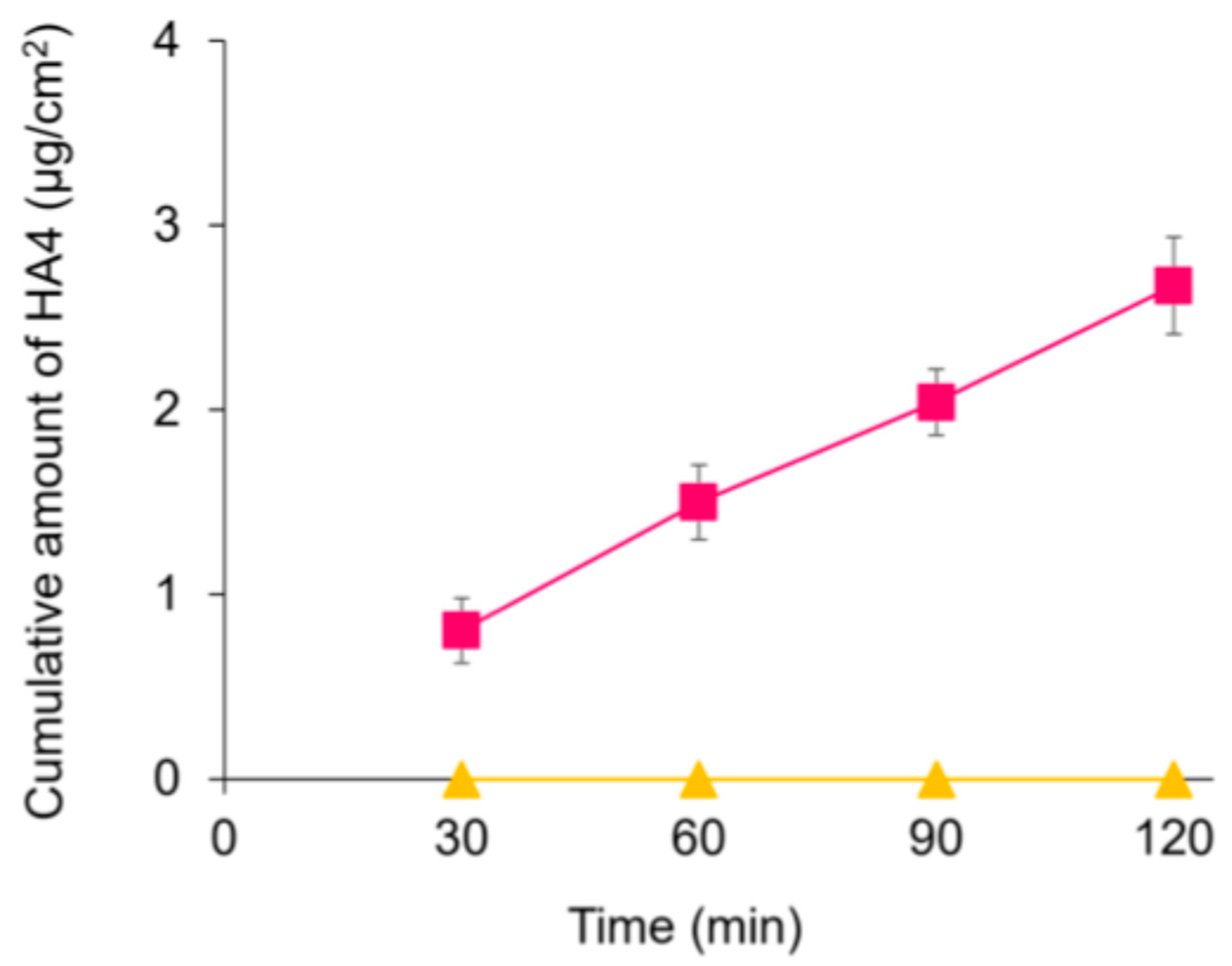
Figure 1. Evaluation of HA4 permeability across a Caco-2 single-layer membrane. Caco-2 cells were seeded on the inserts and cultured. Cell monolayers with TEER values above 250 Ω·cm2 were used for the permeation test. The integrity of the cell monolayers was confirmed by measuring TEER values before and after the experiments. HA or HA4 solution was added to the apical side, and the amount of permeation was quantified and evaluated. Each point represents mean ± S.D. (n = 3). Symbols: HA (▲) and HA4 (■).
Applsci 15 10182 g001
Figure 2. Change in TEWL of mice after HA4 oral administration and UVB irradiation. HA and HA4 were orally administered daily for 28 days. UVB irradiation was administered at four increasing doses, and TEWL was measured five times a week. (A) Time course of TEWL. Values of TEWL on day 12 (B) and day 26 (C). Values represent the mean ± S.D. (n = 3–4). * p < 0.05, ** p < 0.01 (versus Normal group), # p < 0.05, ## p < 0.01 (versus Control group), $ p < 0.05 (versus HA group), Tukey’s post hoc multiple comparison test. N.S.: not significant. Symbols: Normal (●), Control (◆), HA (▲), and HA4 (■) group.
Applsci 15 10182 g002
Figure 3. Change in water content of SC after HA4 oral administration and UVB irradiation. HA and HA4 were orally administered daily for 28 days. UVB irradiation was administered at four increasing doses, and water content of SC was measured five times a week. (A) Time course of water content of SC. Values of water content of SC on day 10 (B), day 14 (C) and day 26 (D). Values represent the mean ± S.D. (n = 3–4). * p < 0.05, ** p < 0.01 (versus Normal group), # p < 0.05, ## p < 0.01 (versus Control group), $ p < 0.05, $$ p < 0.01 (versus HA group), Tukey’s post hoc multiple comparison test. N.S.: not significant. Symbols: Normal (●), Control (◆), HA (▲), and HA4 (■) group.
Applsci 15 10182 g003
Figure 4. Change in body weight after HA4 oral administration and UVB irradiation. HA and HA4 were orally administered daily for 28 days. UVB irradiation was administered at four increasing doses, and body weight was measured daily. Values represent the mean ± S.D. (n = 3–4). Symbols: Normal (●), Control (◆), HA (▲), and HA4 (■) group.
Applsci 15 10182 g004
Figure 5. Change in skin morphology after HA4 oral administration and UVB irradiation. HA and HA4 were orally administered daily for 28 days. Skin samples were fixed and stained with HE. HE-stained sections (scale bar, 50 µm) (AD). (A) Normal, (B) Control, (C) HA, and (D) HA4 group.
Applsci 15 10182 g005
Figure 6. Change in epidermal thickness after HA4 oral administration and UVB irradiation. HA and HA4 were orally administered daily for 28 days. Skin samples were fixed and stained with HE. HE-stained sections were examined under a microscope and epidermal thickness was assessed. Values represent the mean ± S.D. (n = 3–4). * p < 0.05, ** p < 0.01 (versus Nor-mal group), ## p < 0.01 (versus Control group), $ p < 0.05 (versus HA group), Tukey’s post hoc multiple comparison test.
Applsci 15 10182 g006
Disclaimer/Publisher’s Note: The statements, opinions and data contained in all publications are solely those of the individual author(s) and contributor(s) and not of MDPI and/or the editor(s). MDPI and/or the editor(s) disclaim responsibility for any injury to people or property resulting from any ideas, methods, instructions or products referred to in the content.