Article

Membrane-Active Phenolic Compounds from Cephalaria uralensis (Murray) Roem. & Schult.: Isolation, Structural Characterization, and Antioxidant Potential

by
Anna Berecka-Rycerz
1,
Małgorzata Chrząszcz-Wróbel
2,
Arkadiusz Paweł Matwijczuk
3,
Piotr Hołowiński
4,
Sebastian Granica
5 and
Katarzyna Dos Santos Szewczyk
2,*
1
Department of Medicinal Chemistry, Medical University of Lublin, Jaczewskiego 4, 20-090 Lublin, Poland
2
Department of Pharmaceutical Botany, Medical University of Lublin, Chodźki 1, 20-093 Lublin, Poland
3
Department of Biophysics, Faculty of Environmental Biology, University of Life Sciences in Lublin, Akademicka 13, 20-950 Lublin, Poland
4
Department of Chromatography, Institute of Chemical Sciences, Faculty of Chemistry, Maria Curie Sklodowska University in Lublin, 20-031 Lublin, Poland
5
Microbiota Lab, Department of Pharmaceutical Biology, Medical University of Warsaw, 1 Banacha St., 02-097 Warsaw, Poland
*
Author to whom correspondence should be addressed.
Appl. Sci. 2025, 15(17), 9585; https://doi.org/10.3390/app15179585
Submission received: 12 August 2025 / Revised: 28 August 2025 / Accepted: 29 August 2025 / Published: 30 August 2025

Abstract

In this study, we isolated and identified six major phenolic constituents from Cephalaria uralensis. The compounds—quercetin 6-C-β-glucopyranoside, isoorientin, swertiajaponin, 3,5-dicaffeoylquinic acid, 4,5-dicaffeoylquinic acid, and chlorogenic acid—were characterized by LC–MS and NMR. All isolates exhibited strong free-radical scavenging ability and significant interaction with lipid monolayers (Δπ up to ~6.5–7 mN/m), suggesting dual antioxidant and membrane-perturbing activities. In antioxidant assays, isoorientin, showed the lowest IC50 among the isolates. Notably, 4,5-dicaffeoylquinic acid caused the largest increase in monolayer surface pressure, indicating a particularly strong tendency to integrate with lipid bilayers. In fact, chlorogenic acid, isoorientin, and swertiajaponin are well-documented natural antioxidants, and related phenolic acids have been shown to possess potent antimicrobial activity. Thus, the C. uralensis phenolics identified in our study likely underlie the extract’s bioactivity. These findings highlight C. uralensis as a source of membrane-active polyphenols with potential applications in skin-related oxidative and microbial conditions.

1. Introduction

Oxidative stress and skin inflammation underlie many dermatological conditions. Plant phenolics—including flavonoids and caffeoylquinic acids—play key roles in mitigating these effects. Polyphenols can protect the skin by scavenging reactive oxygen species (ROS) and by bolstering endogenous antioxidant defenses [1]. For example, phenolics facilitate direct radical quenching, enhance enzymes like catalase and superoxide dismutase, chelate redox-active metals, and regenerate other antioxidants (e.g., tocopherol) [1]. These mechanisms help maintain dermal fibroblast function and collagen production under oxidative stress. Moreover, many polyphenols absorb UV radiation and act as natural photoprotective agents—reducing UV-induced skin damage [1]. Inflammation is another major factor in skin pathology; flavonoids often inhibit NF-κB and cytokine signaling [1], providing an anti-inflammatory layer of protection. Thus, dietary or topical polyphenols have been proposed as skin-protective agents in conditions from photoaging to acne [1].
The genus Cephalaria (F. Caprifoliaceae) comprises about 94 species of annual and perennial herbs distributed in Europe, the Mediterranean, East Asia, Central Africa, and the Middle East [2,3,4,5]. Cephalaria species are traditionally used in Europe and Asia for inflammatory and skin ailments. For example, Cephalaria setosa and C. syriaca were traditionally applied to treat skin ailments such as seborrheic conditions [6,7]. However, despite these historical uses, many Cephalaria plants remain poorly studied. Phytochemical investigations reveal that Cephalaria species produce a diverse array of secondary metabolites, including triterpenoid saponins, iridoid glycosides, alkaloids, and polyphenols (especially flavonoids and caffeic acid derivatives) [8,9,10,11,12,13,14,15]. These compounds underpin various biological activities observed in the genus—for instance, cytotoxic, immunomodulatory, antibacterial, antifungal, and hypoglycemic effects have all been documented from Cephalaria extracts or isolated constituents [3,11,15,16,17,18,19]. Such findings suggest that Cephalaria spp. could be a rich source of bioactive molecules with therapeutic potential.
A recent study demonstrated that a crude aerial extract of C. uralensis exhibited moderate antibacterial effects against common skin pathogens—Staphylococcus aureus, S. epidermidis, and Cutibacterium acnes, without fibroblast toxicity (therapeutic index > 10) [15]. These Gram-positive bacteria are key contributors to acne and wound infections. The phenolics isolated here (flavonoid glycosides and caffeoylquinic acids) likely underlie that activity. Indeed, similar compounds are known to disrupt bacterial membranes and inhibit resistance mechanisms. For example, chlorogenic acid dramatically increases bacterial membrane permeability, and 4,5-dicaffeoylquinic acid is a potent efflux-pump inhibitor, potentially synergizing with antibiotics.
These findings highlight C. uralensis as a promising source of pharmacologically active compounds, particularly for dermatological applications such as acne treatment. Nevertheless, the bioactive constituents responsible for these effects and their mechanisms of action remain to be fully elucidated.
Among C. uralensis metabolites, phenolic compounds (e.g., flavonoid glycosides and caffeoylquinic acids) are of special interest due to their dual antioxidant and antimicrobial potentials. Plant-derived phenolics are well known as potent antioxidants that can quench free radicals and chelate redox-active metal ions, thereby mitigating oxidative stress in biological systems. Indeed, chlorogenic acid, isoorientin, and swertiajaponin—all present in Cephalaria—are well-documented natural antioxidants with strong radical-scavenging effects [20,21,22]. At the same time, many phenolics also exhibit direct antimicrobial properties. They may attack microbes through mechanisms distinct from conventional antibiotics, which is particularly relevant in the context of rising multidrug resistance (MDR) among pathogens. One key mechanism is the interaction of phenolics with microbial cell membranes. Phenolics can adsorb to and penetrate lipid bilayers, disrupting membrane integrity or function [23,24,25]. For example, certain flavonoid glycosides have been shown to bind bacterial membranes and cause leakage of cellular contents (a bactericidal effect) [24]. Similarly, chlorogenic acid—a caffeoylquinic acid abundant in many plants—increases the permeability of bacterial envelopes and induces loss of barrier function, ultimately leading to cell death [26]. By targeting the cytoplasmic membrane, such compounds can circumvent typical resistance mechanisms and even potentiate conventional antibiotics [23]. In addition, phenolics that embed in lipid membranes might serve a protective role in host tissues by preventing lipid peroxidation and reinforcing the cellular oxidative defense.
Despite these clues, the specific active principles of C. uralensis were unknown. To address this gap, we undertook a phytochemical and biophysical study: First, we fractionated an ethanolic extract of C. uralensis and isolated its main constituents. Second, we fully characterized each isolate’s structure (LC–MS, NMR) and measured its antioxidant capacity and membrane-binding behavior. Evaluating pure compounds (rather than crude mixtures) is essential for bioactivity assignment and for understanding structure-activity relationships. Third, we assessed how these compounds interact with lipid membranes, using a Langmuir monolayer model to simulate one leaflet of a cell membrane. Taken together, this approach reveals how C. uralensis phenolics may protect skin—by neutralizing oxidative stress and by directly compromising microbial membranes.

2. Materials and Methods

2.1. Chemicals and Reagents

All chemical reagents were purchased from various commercial suppliers and were of the highest purity available. 2,2-diphenyl-1-picrylhydrazyl (DPPH) radical, (±)-6-hydroxy-2,5,7,8-tetramethylchromane-2-carboxylic acid (Trolox), 2,2′-azino-bis(3-ethylbenzothiazoline-6-sulfonic acid) (ABTS+), and ascorbic acid were purchased from Sigma-Aldrich Fine Chemicals (St. Louis, MO, USA). LC-grade acetonitrile, methanol, formic acid for HPLC, and DMSO-d6 were purchased from Sigma (Poznan, Poland). Water was purified on a Milli-Q system (Millipore, Bedford, MA, USA). Other chemicals used for preparation of the extracts were of analytical grade and were obtained from Polish Reagents (Avantor Performance Materials Poland, Gliwice, Poland). Reference standards were sourced from ChromaDex (Irvine, CA, USA).

2.2. Plant Material

The aerial parts of Cephalaria uralensis (Murray) Roem. & Schult., were collected from the Botanical Garden of Maria Curie-Skłodowska University (UMCS) in Lublin, Poland, at an elevation of 181 m a.s.l. (coordinates: 51°15′40″ N, 22°30′51″ E) in August 2022. Taxonomic identification was confirmed by Dr. A. Cwener, a botanist from the UMCS Botanical Garden.

2.3. Extraction Procedure and Isolation

A total of 3000 g of air-dried aerial parts of C. uralensis were extracted with 30% ethanol (3 × 2500 mL). Extraction was carried out by shaking at room temperature for 24 h. The resulting extracts were combined, filtered, concentrated under reduced pressure, and lyophilized after freezing using a vacuum concentrator (Free Zone 1, Labconco, Kansas City, KS, USA) to obtain dry residue (yield: 11.30 g dry; 3.8% w/w of plant material). The crude extract was then re-dissolved in hot distilled water (200 mL) and successively partitioned with chloroform (5 × 300 mL), ethyl acetate (7 × 300 mL), and n-butanol (5 × 300 mL), yielding 4.27 g, 9.35 g, and 16.18 g of dried fractions, respectively. The dried n-butanol fraction (16.18 g) was chromatographed on Diaion HP-20 column (90 × 2.5 cm) eluting with mixtures of methanol-water (0:100/100:0 v/v in six steps) to give 86 fractions pooled into 10 main fractions CB1–CB10. Fraction CB4 (5.02 g) was chromatographed on Sephadex LH-20 column (45 × 1.8 cm) eluting with mixtures of MeOH-H2O (60:40 to 100:0, v/v in 4 steps) to give 45 fractions pooled into 6 main fractions CS1–CS6. Fraction CS3 (728.59 mg) was subjected to the preparative HPLC system [(Shimadzu LC-20AP, Kyoto, Japan) equipped with an autosampler (SIL-10A), a UV detector (SPD-10), and a fraction collector (FRC-10)] under the following conditions: mobile phase A—0.1% formic acid in water; mobile phase B—0.1% formic acid in acetonitrile; flow rate 20 mL/min; column temperature 25 °C; detection at 280 nm; gradient: 0 min—6% B, 50 min—23% B. This yielded pure compounds: 1 (16.2 mg; 0.14% of crude), 2 (89.5 mg; 0.79%), 3 (9.7 mg; 0.09%), 4 (75.1 mg; 0.67%), 5 (79.2 mg; 0.70%) and 6 (20.5 mg; 0.18%).
C. uralensis aerial parts were extracted with 30% aqueous ethanol to solubilize polar phenolic compounds. A sequential partitioning scheme (chloroform, ethyl acetate, n-butanol) was employed to separate metabolites by polarity: nonpolar lipids were removed in chloroform, moderately polar phenolics (e.g., mono-caffeoylquinic acids) partitioned into ethyl acetate, and highly polar glycosides remained in the butanol phase. This approach is standard for phenolic extracts: polar solvents yield more phenolic content. Thus, 30% ethanol was chosen to maximize recovery of both hydrophilic and lipophilic antioxidants, and subsequent partitioning ensured enrichment of flavonoid glycosides and caffeoylquinic acids in the butanol fraction for isolation.

2.4. NMR Analysis

1H and 13C NMR spectra were acquired at 298 K using an ASCEND 600 MHz instrument (Bruker, Bremen, Germany). Samples for NMR analysis were dissolved in DMSO-d6 and transferred to the NMR tubes.

2.5. LC-MS Analysis

Chromatographic analysis and high-resolution mass spectrometry (HRMS) measurements were performed on LC-MS system equipped with ultra-high-performance liquid chromatography instrument (UltiMate 3000; Dionex, Sunnyvale, CA, USA) and linear trap quadrupole-Orbitrap mass spectrometer (LTQ-Orbitrap Velos; Thermo Fisher Scientific, San Jose, CA, USA) with electrospray ionization operating in the negative ion mode. Chromatographic separation was performed on a Gemini C18 column (100 × 4.6 mm, 3 µm; Phenomenex, Torrance, CA, USA). Mobile phase consisted of 25 mM formic acid in water (A) and 25 mM formic acid in acetonitrile (B). Gradient program starting at 5% B, increasing to 90% B for 40 min was employed. Mobile phase flow rate was 0.4 mL/min. The MS conditions were as follows: spray voltage, 4.5 kV; sheath gas, 40 arbitrary units; auxiliary gas, 10 arbitrary units; sweep gas, 10 arbitrary units; capillary temperature, 320 °C; nitrogen (>99.98%) as sheath, auxiliary and sweep gas. MS analysis for scan range of 100–1000 m/z was performed with the resolution of 70.000. Mass calibration was conducted 24 h prior to measurements using Pierce calibration solution (Thermo Fisher Scientific, San Jose, CA, USA).

2.6. UV–Vis

UV–Vis spectra were recorded using a Cary 300 Bio UV–Vis spectrophotometer (Varian, Santa Clara, CA, USA) equipped with a thermostated 6 × 6 multicell holder controlled by a Peltier block. Temperature monitoring was performed with a thermoprobe (Cary Series II, Varian, Palo Alto, CA, USA) placed directly in the sample [27,28].

2.7. Lipid Monolayer Experiment

The lipid monolayer experiment was carried out using a Minitrough 2 Langmuir-Blodgett system (KSV Instruments, Helsinki, Finland), placed in a laminar flow hood continuously purged with nitrogen gas (relative humidity 80%) during measurements. Monomolecular layers were formed in a Teflon trough (282 mm × 75 mm), equipped with two symmetric hydrophilic barriers and a Wilhelmy plate made of platinum used as a surface pressure sensor. The monolayers of the mixture were formed at the air–water interface. After each measurement, the trough surfaces were thoroughly cleaned. Solutions were applied to the aqueous subphase using a Hamilton microsyringe (accuracy ± 0.1 μL). The solvent was allowed to evaporate for 15 min, and the monolayer was then compressed at a barrier speed of 75 cm2/min. The subphase temperature (24 ± 1 °C) was maintained using a PolyScience thermostatic circulator [29]. As a control experiment, a sample containing only the solvent was injected beneath the monolayer surface; the selected volume did not induce any changes in the measured pressure, allowing for rapid pressure stabilization.

2.8. Antioxidant Activity Assays

The antioxidant assays were carried out in 96-well plates (Nunclon, Nunc, Roskilde, Denmark) using an Infinite Pro 200 F Elisa Reader (Tecan Group Ltd., Männedorf, Switzerland). Each experiment was conducted in triplicate. Antioxidant activity was expressed as IC50 values, representing the extract concentration required to reduce the initial DPPH/ABTS absorbance by 50%.

2.8.1. DPPH Assay

The 2,2-diphenyl-1-picrylhydrazyl (DPPH) radical scavenging capacity was determined following a previously described modified method [30]. DPPH solution (180 μL of freshly prepared 0.07 mg/mL solution) was mixed with 20 μL of the examined samples in various concentrations. The decrease in DPPH absorbance induced by samples was monitored at 517 nm after 30 min of incubation at 28 °C in the dark. Ascorbic acid was used as the control.

2.8.2. ABTS●+ Assay

The ABTS●+ decolorization assay was the second method used to evaluate antioxidant activity [31]. The radical cation was prepared by reacting 7 mM ABTS with 2.45 mM potassium persulfate (24 h, dark); this solution was diluted to an absorbance ~0.70 at 734 nm. Samples were added and absorbance read at 734 nm after 6 min. Trolox was used as a positive control.

2.9. Statistical Analysis

All assays were repeated at least three times. Data are presented as mean values ± standard deviation (SD) based on three independent experiments. IC50 values of the tested extracts were determined from concentration–inhibition curves. Statistical significance between treatment and control was evaluated (Student’s t-test or one-way ANOVA with post hoc Tukey’s test where appropriate), with p < 0.05 considered significant (GraphPad Prism 5, version 5.04).

3. Results and Discussion

3.1. Isolation and Structural Characteristics of C. uralensis Phenolics

Six major phenolic compounds were isolated from Cephalaria uralensis. The compounds were identified as: (1) quercetin-6-C-β-glucopyranoside [32], (2) isoorientin [33], (3) swertiajaponin [34], (4) 3,5-dicaffeoylquinic acid [35], (5) 4,5-dicaffeoylquinic acid [35], and (6) chlorogenic acid [36]. Two sets of resonance signals were observed for swertiajaponin, which can be attributed to the presence of two stable rotamers [34]. Table 1 shows summary of structural characterization techniques used in our study. The structures (Figure S1) were fully assigned by HRMS (Table 2) and 1H and 13C NMR (Table S1). Notably, the isolated flavonoids are C-glycosylated analogues of luteolin and quercetin, which tend to be more resistant to hydrolysis and sometimes show distinct bioactivities compared to their O-glycosylated counterparts [35]. The caffeoylquinic acids (CQAs) include both mono- and di-caffeoyl esters of quinic acid, commonly known as chlorogenic acids.
The yield and relative concentration of each phenolic depended strongly on solvent polarity. Polar extractions favor glycosides and dicaffeoyl esters: we observed that the n-butanol fraction (most polar) contained the majority of flavonoid glycosides, while EtOAc (intermediate polarity) was rich in mono-caffeoyl acids. This matches reports that aqueous or highly polar extracts have greater total phenolics: for instance, water extracts of L. japonica had higher total phenols, chlorogenic acid, and luteolin glycosides than ethanol extracts [37]. In our study, polar fractions (water/butanol) yielded substantially more material than the chloroform fraction, consistent with literature (e.g., 33% yield for aqueous vs. ~5% for chloroform [38]). These observations underline that extraction method (solvent choice, polarity, temperature) can significantly alter the phenolic profile and must be optimized for target compounds.
Each of isolated compounds has distinct bioactivity profiles. Quercetin 6-C-glucoside is a polyhydroxyflavonol with the quercetin aglycone. Its multiple free hydroxyl groups confer strong antioxidant capacity [39]. By analogy to quercetin 3-O-glycosides, it likely scavenges free radicals and chelates metals. Glycosylation at C-6 increases solubility but still preserves antioxidant potency [39]. Quercetin glycosides also show antimicrobial effects (membrane disruption and efflux inhibition) in other studies. Isoorientin (luteolin-6-C-glucoside) has a luteolin core with two ortho-dihydroxy groups in the A-ring. It is a well-known antioxidant and anti-inflammatory agent (reported strong DPPH and ABTS radical scavenging). Isoorientin also can inhibit bacterial growth and biofilm formation by disrupting membranes, similar to other flavone glycosides. Swertiajaponin, an apigenin derivative, exhibits exceptionally strong antioxidant and anti-melanogenic activity [40]. It has been shown to be among the most potent tyrosinase inhibitors of natural flavonoids, reducing melanin synthesis in skin cells [41]. Its pronounced radical-scavenging ability also suggests anti-aging cosmetic potential. Chlorogenic acid (5-CQA) is a ubiquitous caffeoylquinic acid with broad bioactivity. As a cinnamic acid ester, CGA is a powerful antioxidant and anti-inflammatory natural product [41]. It also perturbs microbial membranes: studies indicate CGA increases bacterial envelope permeability and inhibits pathogen growth [41]. Chlorogenic acid has documented skin benefits, including anti-acne effects by suppressing inflammation and lipid production in Cutibacterium acnes models. 3,5-Dicaffeoylquinic acid (3,5-DCQA) and 4,5-Dicaffeoylquinic acid (4,5-DCQA) each contain two caffeoyl groups. These dicaffeoylquinic acids are among the most potent antioxidants known, strongly inducing cellular antioxidant defenses [42]. They also have anti-inflammatory properties and can inhibit bacterial growth. Notably, 4,5-DCQA has been reported as a potent bacterial efflux pump inhibitor, potentially reversing antibiotic resistance. Both DCQAs thus synergize radical scavenging with antimicrobial action.
The phenolic profile of C. uralensis is consistent with patterns seen in related Caprifoliaceae species. For example, Lonicera japonica—another medicinal Caprifoliaceae—contains high levels of chlorogenic acid and flavone glycosides (e.g., luteolin-7-O-glucoside) which contribute to its antioxidant and anti-inflammatory effects [37]. Likewise, other Cephalaria studies have noted abundant caffeic acid derivatives and C-glycosyl flavonoids [43]. Thus, our findings (caffeoylquinic acids + flavonoid C-glycosides) fit the broader chemotaxonomic profile of this family, reflecting conserved biosynthetic pathways for these phenolics [37,43].
The compounds identified are promising for dermatological applications. The strong antibacterial activity of Cephalaria extract against S. aureus, S. epidermidis, and C. acnes has been attributed to its phenolic content. In particular, chlorogenic acid and caffeoylquinic acids likely inhibit C. acnes by both reducing inflammation and disrupting bacterial membranes. In fact, chlorogenic acid has been reported to diminish C. acnes-induced skin inflammation and lipid overproduction, improving acne symptoms in models [44]. Likewise, the antibiofilm capacity of plant polyphenols is well documented [45], suggesting our phenolics could prevent biofilm formation by acne pathogens or other skin bacteria. In the cosmetic field, swertiajaponin stands out: its dual action as a free-radical scavenger and tyrosinase inhibitor makes it an attractive skin-whitening and anti-aging agent [40]. More generally, potent antioxidants such as isoorientin, chlorogenic acid, and DCQAs can protect skin cells from UV- and pollution-induced oxidative damage. Thus, these phenolics not only elucidate the bioactivity of C. uralensis but also point to their applied potential in anti-acne formulations, antibiofilm strategies, and cosmeceuticals (e.g., antioxidants for skincare) [40,45].

3.2. Antioxidant Activity

Plant-derived secondary metabolites, particularly phenolics, offer a promising strategy for protecting the skin against oxidative damage. Excessive production of reactive oxygen species (ROS) and resultant oxidative stress disrupt the cellular redox balance, triggering signaling pathways that lead to protein, lipid, and DNA damage. A reduction in fibroblast activity is a major contributor to the visible signs of skin aging. However, polyphenolic antioxidants have been shown to restore fibroblast functionality and stimulate collagen synthesis, thereby mitigating these degenerative effects [46,47].
In this study, antioxidant activity was evaluated using microplate-based, cell-free assays. The results revealed that both the crude extract of C. uralensis and the isolated active compounds exhibited a potent ability to scavenge ABTS●+ and DPPH free radicals.
All isolates and the crude CUE extract showed strong antioxidant (radical-scavenging) activity. Figure 1 summarizes the IC50 values from the DPPH and ABTS●+ assays (mean ± SD, n = 3). The IC50 for DPPH ranged from 2.17 (isoorientin) to 6.05 mg/mL (chlorogenic acid) and for ABTS●+ from 0.44 to 1.11 mg/mL. However, these values were higher than this for ascorbic acid (0.48 mg/mL) and Trolox (0.09 mg/mL) used as a positive control. Our results are consistent with numerous literature reports describing these molecules as potent antioxidants. Chlorogenic acid, for example, is well known for its radical-scavenging ability (indeed, it is often a major contributor to the antioxidant activity of coffee and many plants). Flavonoids like luteolin and quercetin glycosides are likewise effective antioxidants due to their conjugated ring systems and multiple hydroxyl groups that can donate hydrogen atoms to quench radicals. Our findings are consistent with those of Chrząszcz et al., who noted that Cephalaria extracts rich in chlorogenic acid, isoorientin, and swertiajaponin displayed robust activity in DPPH/ABTS assays [15]. In our study, the DPPH and ABTS●+ results showed that both the raw extract of C. uralensis and the active compounds isolated from it have similar, strong antioxidant properties. Qualitatively, the scavenging efficiency follows the compounds’ structures. The strongest effect was observed for isoorientin. The obtained results highlight the strong antioxidant effectiveness of phenolic compounds from Cephalaria. Mechanistically, the antioxidant activity stems from the phenolics ability to stabilize unpaired electrons—the catechol (o-dihydroxy) structure on the caffeoyl moieties and B-ring of flavonoids is particularly important for radical stabilization. Isoorientin (a 3′,4′-dihydroxylated flavone) and swertiajaponin (its 7-methylated analog) both have the catechol B-ring, explaining their high radical-scavenging efficiency [48]. Quercetin glycosides possess an additional 3-OH and 4-oxo group that contribute to an extended conjugation, further enhancing antioxidant potency [48]. Thus, from a structure-activity perspective, the polyhydroxylated and conjugated architecture of these phenolics is ideal for quenching free radicals and chelating metal ions, which likely contributes to the mitigation of oxidative stress in biological contexts. The selected monolayer model allowed for detailed observation of phenolic interactions with lipid membranes at the molecular level. Utilizing the Langmuir trough technique, we monitored real-time changes in surface pressure as each compound was introduced beneath the monolayer. This approach provided valuable insights into the penetration, insertion, and perturbation capabilities of phenolics like 4,5-dicaffeoylquinic acid and flavonoids such as isoorientin. These findings significantly contribute to our understanding of their membrane activity, as documented in numerous literature reports [49,50,51,52].
The comparability of findings from different reports is limited due to variations in experimental conditions. In addition, current knowledge regarding the antioxidant activity of Cephalaria species remains incomplete. Azab [53] demonstrated that aqueous, ethanolic, and ethyl acetate extracts of the aerial parts of C. jopponsis exhibit strong antioxidant activity (41.1, 30.1, and 20.7 mg ascorbic acid g−1 dry extract, respectively). Chrząszcz et al. [15] found that different extracts from the aerial parts and flowers from C. uralensis and C. gigantea demonstrate strong radical scavenging activity. Knautia (F. Caprifoliaceae) extracts showed similar activity. The strongest DPPH radical scavenging activity was observed for K. drymeia extract (IC50 = 0.50 mg/mL), and the weakest effect was noted for K. macedonica extract (IC50 = 4.04 mg/mL). Karalija et al. [54] assessed the antioxidant potential of methanolic shoot extracts of K. sarajevensis cultured in Murashige and Skoog, using peroxidase activity and the DPPH assay. The DPPH results indicated that the radical scavenging activity of shoot cultures ranged between 10 and 90 µg/mL. Moreover, the methanolic extract of K. integrifolia showed inhibition rates of 77.5% and 75.07% at concentrations of 1000 µg/mL and 500 µg/mL, respectively [55].

3.3. Membrane Activity—Monolayer Insertion Assays

Injection of each of the isolated compounds beneath a lipid monolayer composed of DPPC and formed at the air–water interface resulted in a rapid increase in surface pressure (Δπ), indicating the compounds’ ability to penetrate and perturb the lipid film. In contrast, no significant changes in Δπ were observed upon injection into the subphase in the absence of a lipid monolayer, confirming that the recorded effects arise specifically from interactions with the lipid interface rather than from experimental artifacts. Notably, clear differences were observed in the extent of interaction between the tested compounds and the lipid membrane. Isoorientin (ISO), a highly polar C-glucosylated flavone derivative, induced the smallest surface pressure increase (~2 mN/m). By comparison, 4,5-dicaffeoylquinic acid (4,5-DCQA) consistently produced the highest Δπ, exceeding 6.5–7 mN/m across all measurements. The isomer 3,5-DCQA showed an intermediate effect (~3.5 mN/m). Interestingly, the crude ethanolic extract from Cephalaria uralensis (CUE), which contains a mixture of these phenolics, caused a Δπ of approximately 5.5 mN/m—higher than that of isoorientin or 3,5-DCQA, but still noticeably lower than pure 4,5-DCQA. Accordingly, the observed order of membrane activity was as follows: isoorientin < 3,5-DCQA < CUE extract < 4,5-DCQA. The data are presented in Table 3 and in Figure S2.
These results indicate that C. uralensis phenolics have an inherent affinity for lipid interfaces, but the strength of this interaction is strongly structure-dependent. In this context, membrane activity is understood as the compound’s ability to undergo con-formational changes within the lipid environment, as well as the number of hydroxyl groups present in its molecular structure. Although the latter factor is undoubtedly important, for the compounds studied here, it appears to be secondary to the former. The fact that 4,5-DCQA penetrated the DPPC monolayer most effectively suggests that its molecular features optimize amphiphilicity and membrane binding. Dicaffeoylquinic acids possess two aromatic (caffeoyl) moieties that can insert into the hydrophobic lipid phase, while their multiple polar hydroxyls and carboxyl groups can anchor to the phospholipid headgroup region. The 4,5-DCQA isomer may adopt a conformation that allows both caffeoyl groups to engage the membrane simultaneously (perhaps one intercalating into the lipid tail region and the other interacting at the interface), thereby producing a larger disruption of monolayer packing. By contrast, 3,5-DCQA, which differs only in the placement of one caffeoyl group on the quinic acid backbone, showed a markedly lower Δπ. This suggests a structure—activity relationship where small changes in the substitution pattern on quinic acid alter the molecule’s orientation or depth of penetration in the lipid layer. Literature reports similarly note that 4,5-DCQA can be more bioactive than its isomers; for instance, 4,5-DCQA from Artemisia absinthium was a potent inhibitor of bacterial efflux pumps, whereas 3,5-DCQA was ineffective in that regard [56]. Such differential behavior underscores how the spatial arrangement of functional groups modulates interactions with biological targets like membranes or proteins.
Isoorientin’s weak membrane insertion can be rationalized by its high hydrophilicity—as a flavone C-glycoside with a sugar moiety, it likely resides at the water—lipid interface and only shallowly penetrates the monolayer. In general, flavonoid glycosides are less lipophilic than aglycones and may cause minimal perturbation to tightly packed phospholipids [23].
Interestingly, swertiajaponin (an O-methylated isoorientin) might be expected to interact slightly more with membranes due to the methylation reducing one hydrophilic hydroxyl, but its effect in our assay was not explicitly reported. Quercetin 6-C-glucoside, another highly hydroxylated C-glycoside, was similarly not singled out in the monolayer results; it likely has membrane behavior analogous to isoorientin (quercetin and luteolin share the same flavone backbone except for an extra 3-OH in quercetin).
The crude extract’s relatively strong Δπ (second only to pure 4,5-DCQA) is notable. It could indicate additive or synergistic effects: multiple phenolics in CUE may collectively insert into the monolayer, or certain minor constituents in the extract (in addition to the six major compounds) might contribute to membrane perturbation. Such cooperative interactions are plausible in a complex mixture and often underpin the efficacy of whole-plant extracts in biological systems.

3.4. Comparison to Other Medicinal Plants

The phenolic profile and bioactivities of C. uralensis share parallels with many other medicinal plants rich in similar compounds. Echinacea species, for instance, contain abundant caffeoylquinic acids (including 3,5-DCQA and 4,5-DCQA) and are used worldwide for their immunomodulatory and antioxidant properties. A recent review noted that dicaffeoylquinic acids are key contributors to Echinacea pharmacological effects—exhibiting antioxidative, anti-inflammatory, antimicrobial, and even antiviral activities [57]. Hedera helix is another example where DCQAs in the extract add to its therapeutic profile [57]. Our identification of 4,5-DCQA as a potent membrane—active compound resonates with studies on other herbs: Artemisia absinthium yielded the same 4,5-DCQA, which was found to enhance antibiotic efficacy by inhibiting bacterial drug-efflux pumps in Gram-positive pathogens [56]. This efflux-pump inhibitory activity is a valuable strategy to combat MDR bacteria, suggesting that Cephalaria’s 4,5-DCQA might impart similar benefits (e.g., synergizing with conventional antibiotics or helping to overcome bacterial resistance mechanisms). Flavonoid C-glycosides like isoorientin and swertiajaponin are likewise widespread in the plant kingdom and known for diverse bioactivities. For example, both compounds occur in Cymbopogon citratus leaves, where they were reported to contribute to the plant’s anti-inflammatory and enzyme-inhibitory effects [57]. Isoorientin in lemongrass showed high radical-scavenging activity and was implicated in the herb’s traditional medicinal uses [58]. In another study, Graptophyllum flavonoid glycosides (structurally similar to isoorientin) were isolated and found to exert bactericidal effects by damaging the bacterial membrane permeability and causing leakage of intracellular material [23,24]. Rutin (quercetin-3-O-rutinoside), a flavonoid glycoside common in many plants, has been shown to synergize with antibiotics; for instance, rutin combined with gentamicin induced oxidative stress and cell-wall disruption in drug-resistant P. aeruginosa, leading to improved bacterial killing [23]. These examples from recent literature illustrate that the phenolics found in Cephalaria are part of a larger group of plant metabolites with multifaceted biological actions. Their membrane-targeting ability and antioxidant capacity are recurrent themes, underpinning antibacterial, anti-biofilm, anti-inflammatory, and even antiparasitic effects reported across different species.

3.5. Mechanistic Implications

The dual membrane-active and antioxidant nature of C. uralensis phenolics suggests they could combat microbial infections on two fronts: by physically perturbing microbial membranes and by mitigating oxidative stress (which can both enhance immune response and directly exert pro-oxidant lethal effects on microbes). Chlorogenic acid exemplifies this duality—it can breach bacterial membranes and also scavenge free radicals, thereby possibly protecting host tissues from inflammation while attacking pathogens [26]. Our monolayer data indicate that 4,5-DCQA and related compounds have a strong propensity to associate with lipid bilayers. In a cellular context, such insertion could lead to increased membrane permeability, depolarization, and leakage of vital metabolites or ions from microbial cells [26]. Electron microscopy in previous studies has indeed visualized membrane damage and cytoplasmic content release in bacteria treated with chlorogenic acid [26]. Additionally, membrane binding may allow phenolics to accumulate at the site of lipid peroxidation and interrupt radical chain reactions, thus protecting membrane lipids from oxidative damage in host cells. Interestingly, some flavonoids can also act as pro-oxidants under specific conditions, generating reactive oxygen species that contribute to killing pathogens [23]. For example, the combination of rutin and gentamicin triggered excess ROS in bacteria, leading to oxidative burst and cell death [23]. While our study focused on antioxidant assays in cell-free systems, it is plausible that Cephalaria phenolics might similarly induce lethal oxidative stress within microbes when used at higher concentrations or in the presence of redox-active metals. This context-dependent behavior (antioxidant in host environments vs. pro-oxidant in microbial environments) is a known phenomenon for polyphenols and could be an avenue for further research. From a structure—activity relationship (SAR) perspective, our findings reinforce that achieving an optimal balance between hydrophilicity and lipophilicity is crucial for membrane-interacting compounds. 4,5-DCQA, with its large di-caffeoyl structure, has sufficient hydrophobic character to deeply insert into lipid layers, yet enough polar functionality to remain at the interface—this “amphiphilic balance” likely underpins its pronounced membrane activity. Flavonoids like isoorientin, on the other hand, are very polar (due to the sugar and multiple hydroxyls) and therefore mostly stay at the surface of the bilayer, causing limited disruption. Modifying such molecules can shift this balance, e.g., adding a methyl group (swertiajaponin vs. isoorientin) or removing a sugar (aglycones vs. glycosides) tends to increase lipophilicity, which may enhance membrane permeability but sometimes at the cost of water-solubility or antioxidant capacity. Indeed, C-glycosides of flavones often retain considerable polarity but are more stable than O-glycosides; they might require specific transport or membrane conditions to penetrate cells. Future SAR studies could systematically compare the membrane affinity of these phenolics and analogues (for instance, comparing quercetin aglycone to quercetin-6-C-glucoside, or luteolin vs. isoorientin vs. swertiajaponin) to delineate how each functional group influences activity. Moreover, the stark difference between 4,5-DCQA and 3,5-DCQA in both our monolayer assay and literature reports on efflux pump inhibition [56] suggests that positional isomerism can significantly affect the conformational flexibility and target binding of CQAs. This could be due to differing intramolecular hydrogen bonding patterns or steric factors that make one isomer more suited to interacting with a protein pocket (such as an efflux pump transporter) or inserting into a lipid domain than another [56]. In summary, the experimental evidence combined with comparative literature analysis paints C. uralensis phenolics as membrane-active antioxidants. They are capable of lodging into lipid membranes and perturbing their structure—an action that likely translates into anti-microbial effects (through membrane disruption or enhanced uptake of other antimicrobials)—while concurrently neutralizing free radicals and reactive species. This unique combination of activities is highly desirable in the search for new therapeutic agents, especially given the modern challenges of antibiotic resistance and inflammatory oxidative damage.
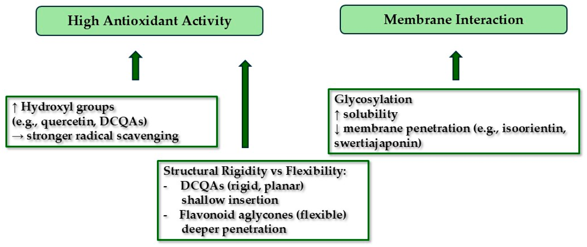
The structure—activity relationship among these compounds is highlighted by their degree of hydroxylation, glycosylation, and flexibility. Multiple free hydroxyl groups (as in quercetin glycosides and caffeoyl acids) enhance radical-scavenging power [39]. In contrast, glycosylation increases water solubility and may reduce membrane insertion relative to aglycones, but C-glycosides (like isoorientin) still integrate into membranes sufficiently to disrupt bacteria. Similarly, the rigid esterified structure of dicaffeoylquinic acids can insert into lipid layers and donate hydrogen, whereas more flexible flavonoid aglycones (with fewer sugar moieties) may penetrate membranes more deeply. Figure 2 illustrates how increasing hydroxylation correlates with antioxidant activity, while glycosylation and backbone rigidity modulate membrane interaction and bioavailability.
More hydroxyl (-OH) groups increase antioxidant capability (via free radical donation) [39]. Glycosylation enhances water solubility but can reduce membrane affinity. Rigid, planar structures (e.g., di-caffeoylquinic acids) favor shallow membrane association, whereas more flexible aglycones (flavonols) penetrate bilayers more deeply. These SAR features help explain the observed antioxidant and membrane-disrupting activities of each compound.

3.6. Novelty, Limitations, and Future Work

This work is novel in directly linking the chemical constituents of C. uralensis to their biophysical properties. It is the first report to use a Langmuir monolayer model to quantify how C. uralensis phenolics insert into lipid films. We demonstrate that certain plant phenolics combine membrane activity with potent radical scavenging—a combination desirable for skin therapeutics. We identified for the first time that 4,5-diCQA from C. uralensis has exceptional membrane affinity, and we confirmed the presence of medically relevant flavonoid C-glycosides in this species. These findings fill the literature gap on C. uralensis phytochemistry and mechanisms.
Our assays were in vitro, with isolated lipid films and chemical radicals, not in living cells. We did not directly measure bacterial inhibition of the pure compounds. Therefore, whether the same membrane effects occur in bacterial or skin cells needs confirmation. The yields of pure isolates were modest (<1% each of extract); minor constituents (not captured here) might also contribute to activity.
To build on these insights, the next steps will include: (1) Microbiological assays—determine MICs/MBCs of each isolate against skin pathogens (Gram-positive and Gram-negative), and test synergy with antibiotics. (2) Cell-based models—assess cytoprotective or pro-oxidant effects in human keratinocytes/fibroblasts and infected skin models. (3) In vivo or ex vivo skin tests, e.g., wound-healing assays or acne models to evaluate therapeutic potential. These steps will clarify how to exploit C. uralensis phenolics in practical formulations (e.g., topical creams for infected wounds or acne) while addressing any solubility/bioavailability issues.

4. Conclusions

We isolated six phenolic compounds from Cephalaria uralensis and demonstrated that each combines membrane-perturbing ability with strong antioxidant capacity. In antioxidant assays, all isolates outperformed crude extract, with isoorientin and 4,5-diCQA showing especially low IC50 values. In Langmuir monolayers, 4,5-diCQA caused the greatest surface-pressure increase, indicating superior membrane insertion. These dual activities suggest C. uralensis phenolics could protect skin by neutralizing oxidative stress and combat microbes by destabilizing their membranes or efflux pumps. This is the first study to characterize the lipid-interaction behavior of C. uralensis phenolics, contributing new insight into their mode of action. The main limitation is that we have not yet confirmed these effects in live cells or organisms. Future work will focus on microbial assays, skin models, and optimizing these compounds for therapeutic use. Overall, our findings establish C. uralensis as a valuable source of multifunctional antioxidants, with potential applications in dermatology and infection control.

Supplementary Materials

The following supporting information can be downloaded at: https://www.mdpi.com/article/10.3390/app15179585/s1, Figure S1: Structures of the isolated compounds 16 from C. uralensis.; Figure S2: The increase in surface pressure (SP) over time following a single injection of: ISO—isoorientin, 3,5-diCQA—3,5-dicaffeoylquinic acid, 4,5-diCQA—4,5-dicaffeoylquinic acid, and the crude extract CUE—ethanolic extract from the aerial parts of C. uralensis, all dissolved in water and injected beneath the lipid monolayer; Table S1: 1H and 13C NMR spectroscopic data for phenolic compounds isolated from Cephalaria uralensis (compounds 16).

Author Contributions

Conceptualization, K.D.S.S.; methodology, K.D.S.S., A.P.M., A.B.-R., P.H. and S.G.; software, K.D.S.S., A.P.M. and P.H.; formal analysis, K.D.S.S., M.C.-W., A.P.M. and S.G.; investigation, K.D.S.S., A.P.M.; M.C.-W., P.H. and S.G.; resources, K.D.S.S.; data curation K.D.S.S., A.P.M., P.H. and S.G.; visualization, A.B.-R.; writing—original draft preparation, K.D.S.S., A.P.M., P.H. and A.B.-R.; writing—review and editing, K.D.S.S.; supervision, K.D.S.S. and A.P.M. All authors have read and agreed to the published version of the manuscript.

Funding

This research was funded by the Ministry of Science and Higher Education in Poland DS45 project of the Medical University of Lublin.

Institutional Review Board Statement

Not applicable.

Informed Consent Statement

Not applicable.

Data Availability Statement

Data available on request.

Acknowledgments

We gratefully acknowledge the use of the facilities of the Ecotech-Complex in Lublin, project number UDA-POIG.02.01.00–06-212/09–03.

Conflicts of Interest

The authors declare no conflicts of interest.

References

  1. Farhan, M. The promising role of polyphenols in skin disorders. Molecules 2024, 29, 865. [Google Scholar] [CrossRef]
  2. WFO Plant List. Cephalaria Schrad. Available online: https://wfoplantlist.org/taxon/wfo-4000007238-2025-06 (accessed on 24 July 2025).
  3. Sarıkahya, N.B.; Kırmızıgül, S. Antimicrobially active hederagenin glycosides from Cephalaria elmaliensis. Planta Med. 2012, 78, 828–833. [Google Scholar] [CrossRef]
  4. Sumer, G.; Sarikahya, N.B.; Kirmizigul, S. Phytochemical and biological investigations on Cephalaria anatolica. Rec. Nat. Prod. 2017, 11, 497–507. [Google Scholar] [CrossRef]
  5. Kayce, P.; Kırmızıgül, S. Chemical constituents of two endemic Cephalaria species. Rec. Nat. Prod. 2010, 4, 141–148. [Google Scholar]
  6. Baydoun, S.; Lamis, C.; Dalleh, H.; Nelly, A. Ethnopharmacological survey of medicinal plants used in traditional medicine by the communities of Mount Hermon, Lebanon. J. Ethnopharmacol. 2015, 173, 139–156. [Google Scholar] [CrossRef] [PubMed]
  7. Dalar, A.; Mukemre, M.; Unal, M.; Ozgokce, F. Traditional medicinal plants of Ağrı Province, Turkey. J. Ethnopharmacol. 2018, 226, 56–72. [Google Scholar] [CrossRef]
  8. Alankuş-Çalışkan, Ö.; Anil, H. A bidesmodic triterpene saponin from Cephalaria transsyvanica. Phytochemistry 1995, 38, 1493–1495. [Google Scholar] [CrossRef] [PubMed]
  9. Celenk, V.U.; Sarikahya, N.B.; Kirmizigul, S. Isolation and structural studies on saponins from three Cephalaria species from Anatolia. Chem. Nat. Compd. 2020, 56, 180–182. [Google Scholar] [CrossRef]
  10. Kırmızıgül, S.; Anıl, H.; Uçar, F.; Akdemir, K. Antimicrobial and antifungal activities of three new triterpenoid glycosides. Phytoter. Res. 1996, 10, 274–276. [Google Scholar] [CrossRef]
  11. Top, H.; Sarıkahya, N.B.; Nalbantsoy, A.; Kırmızıgül, S. Immunomodulatory, hemolytic properties and cytotoxic activity potent of triterpenoid saponins from Cephalaria balansae. Phytochemistry 2017, 137, 139–147. [Google Scholar] [CrossRef] [PubMed]
  12. Mustafayeva, K.; Elias, R.; Balansard, G.; Suleimanov, T.; Mayu-Lede, V.; Kerimov, Y. Iridoid glycosides from Cephalaria kotschyi roots. Chem. Nat. Compd. 2008, 44, 132–133. [Google Scholar] [CrossRef]
  13. Aliev, A.M.; Movsumov, I.S.; Bagirov, E.K. Alkaloids from certain Cephalaria species. Khim. Prir. Soedin. 1975, 5, 667. [Google Scholar]
  14. Movsumov, I.S.; Garaev, E.A.; Isaev, M.I. Flavonoids from Cephalaria grossheimii. Chem. Nat. Compd. 2009, 45, 422–423. [Google Scholar] [CrossRef]
  15. Chrząszcz, M.; Miazga-Karska, M.; Klimek, K.; Granica, S.; Tchórzewska, D.; Ginalska, G.; Szewczyk, K. Extracts from Cephalaria uralensis (Murray) Roem. & Schult. and Cephalaria gigantea (Ledeb.) Bobrov as potential agents for treatment of acne vulgaris: Chemical characterization and in vitro biological evaluation. Antioxidants 2020, 9, 796. [Google Scholar] [CrossRef]
  16. Kayce, P.; Sarikahya, B.N.; Pekmez, M.; Arda, N.; Kırmızıgül, S. The structure and cytotoxic activity of a new saponin: Cephoside A from Cephalaria elazigensis var. purpurea. Turk. J. Chem. 2017, 41, 345–353. [Google Scholar] [CrossRef]
  17. Sarıkahya, N.B.; Kırmızıgül, S. Antimicrobial triterpenoid glycosides from Cephalaria scoparia. J. Nat. Prod. 2010, 73, 825–830. [Google Scholar] [CrossRef] [PubMed]
  18. Kırmızıgül, S.; Anıl, H.; Rose, M.E. Triterpenoid saponins from Cephalaria transsylvanica. J. Nat. Prod. 1996, 59, 415–418. [Google Scholar] [CrossRef] [PubMed]
  19. Hamdan, I.I.; Afifi, F.U. Studies on the in vitro and in vivo hypoglycemic activities of some medicinal plants used in treatment of diabetes in Jordanian traditional medicine. J. Ethnopharmacol. 2004, 93, 117–121. [Google Scholar] [CrossRef] [PubMed]
  20. Naveed, M.; Hejazi, V.; Abbas, M.; Kamboh, A.A.; Khan, G.J.; Shumzaid, M.; Ahmad, F.; Babazadeh, D.; Xia, F.; Modarresi-Ghazani, F.; et al. Chlorogenic acid (CGA): A pharmacological review and call for further research. Biomed. Pharmacother. 2018, 97, 67–74. [Google Scholar] [CrossRef]
  21. Yuan, L.; Wang, J.; Wu, W.; Liu, Q.; Liu, X. Effect of isoorientin on intracellular antioxidant defence mechanisms in hepatoma and liver cell lines. Biomed. Pharmacother. 2016, 81, 356–362. [Google Scholar] [CrossRef]
  22. Moon, K.M.; Lee, B.; Cho, W.K.; Lee, B.S.; Kim, C.Y.; Ma, J.Y. Swertiajaponin as an anti-browning and antioxidant flavonoid. Food Chem. 2018, 30, 207–214. [Google Scholar] [CrossRef]
  23. Ecevit, K.; Barros, A.A.; Silva, J.M.; Reis, R.L. Preventing microbial infections with natural phenolic compounds. Future Pharmacol. 2022, 2, 460–498. [Google Scholar] [CrossRef]
  24. Tagousop, C.N.; Tamokou, J.-D.; Ekom, S.E.; Ngnokam, D.; Voutquenne-Nazabadioko, L. Antimicrobial activities of flavonoid glycosides from Graptophyllum grandulosum and their mechanism of antibacterial action. BMC Complement. Altern. Med. 2018, 18, 252. [Google Scholar] [CrossRef] [PubMed]
  25. Reis, A.; Perez-Gregorio, R.; Mateus, N.; de Freitas, V. Interactions of dietary polyphenols with epithelial lipids: Advances from membrane and cell models in the study of polyphenol absorption, transport and delivery to the epithelium. Crit. Rev. Food Sci. Nutr. 2021, 61, 3007–3030. [Google Scholar] [CrossRef]
  26. Lou, Z.; Wang, H.; Zhu, S.; Ma, C.; Wang, Z. Antibacterial activity and mechanism of action of chlorogenic acid. J. Food Sci. 2011, 76, 398–403. [Google Scholar] [CrossRef]
  27. Czernel, G.; Budziak, I.; Oniszczuk, A.; Karcz, D.; Pustuła, K.; Górecki, A.; Matwijczuk, A.; Gładyszewska, B.; Gagoś, M.; Niewiadomy, A.; et al. ESIPT-related origin of dual fluorescence in the selected model 1,3,4-thiadiazole derivatives. Molecules 2020, 25, 4168. [Google Scholar] [CrossRef] [PubMed]
  28. Matwijczuk, A.; Budziak-Wieczorek, I.; Czernel, G.; Karcz, D.; Barańska, A.; Jedlińska, A.; Samborska, K. Classification of honey powder composition by FTIR spectroscopy coupled with chemometric analysis. Molecules 2022, 27, 3800. [Google Scholar] [CrossRef]
  29. Gagoś, M.; Arczewska, M. FTIR spectroscopic study of molecular organization of the antibiotic amphotericin B in aqueous solution and in DPPC lipid monolayers containing the sterols cholesterol and ergosterol. Eur. Biophys. J. 2012, 41, 663–673. [Google Scholar] [CrossRef] [PubMed]
  30. Szewczyk, K.; Bogucka-Kocka, A.; Vorobets, N.; Grzywa-Celińska, A.; Granica, S. Phenolic composition of the leaves of Pyrola rotundifolia L. and their antioxidant and cytotoxic activity. Molecules 2020, 25, 1749. [Google Scholar] [CrossRef]
  31. Pieczykolan, A.; Pietrzak, W.; Dos Santos Szewczyk, K.; Gawlik-Dziki, U.; Nowak, R. LC-ESI-MS/MS polyphenolic profile and in vitro study of cosmetic potential of Aerva lanata (L.) Juss. herb extracts. Molecules 2022, 27, 1259. [Google Scholar] [CrossRef]
  32. Siddiqui, J.A.; Sharan, K.; Swarnkar, G.; Rawat, P.; Kumar, M.; Manickavasagam, L.; Maurya, R.; Pierroz, D.; Chattopadhyay, N. Quercetin-6-C-β-D-glucopyranoside isolated from Ulmus wallichiana planchon is more potent than quercetin in inhibiting osteoclastogenesis and mitigating ovariectomy-induced bone loss in rats. Menopause 2011, 18, 198–207. [Google Scholar] [CrossRef]
  33. Kumazawa, T.; Minatogawa, T.; Matsuba, S.; Sato, S.; Onodera, J. An effective synthesis of isoorientin: The regioselective synthesis of a 6-C-glucosylflavone. Carbohydr. Res. 2000, 329, 507–513. [Google Scholar] [CrossRef] [PubMed]
  34. Nakayama, M.; Koshioka, M.; Kondo, T.; Imizu, K. Flavone C-glucosides responsible for yellow pigmentation induced by low temperature in bracts of Zantedeschia aethiopica. Nat. Prod. Commun. 2015, 10, 425–427. [Google Scholar] [CrossRef]
  35. Tchoumtchoua, J.; Mathiron, D.; Pontarin, N.; Gagneul, D.; van Bohemen, A.I.; Otogo N’nang, E.; Mesnard, F.; Petit, E.; Fontaine, J.X.; Molinié, R.; et al. Phenolic profiling of flax highlights contrasting patterns in winter and spring varieties. Molecules 2019, 24, 4303. [Google Scholar] [CrossRef] [PubMed]
  36. Zhao, J.G.; Yan, Q.Q.; Xue, R.Y.; Zhang, J.; Zhang, Y.Q. Isolation and identification of colourless caffeoyl compounds in purple sweet potato by HPLC-DAD-ESI/MS and their antioxidant activities. Food Chem. 2014, 161, 22–26. [Google Scholar] [CrossRef] [PubMed]
  37. Hsu, H.F.; Hsiao, P.C.; Kuo, T.C.; Chiang, S.T.; Chen, S.L.; Chiou, S.J.; Ling, X.H.; Liang, M.T.; Cheng, W.Y.; Houng, J.Y. Antioxidant and anti-inflammatory activities of Lonicera japonica Thunb. var. sempervillosa Hayata flower bud extracts prepared by water, ethanol and supercritical fluid extraction techniques. Ind. Crops Prod. 2016, 89, 543–549. [Google Scholar] [CrossRef]
  38. Nakamura, M.; Ra, J.H.; Jee, Y.; Kim, J.S. Impact of different partitioned solvents on chemical composition and bioavailability of Sasa quelpaertensis Nakai leaf extract. J. Food Drug Anal. 2017, 25, 316–326. [Google Scholar] [CrossRef]
  39. Materska, M. Quercetin and its derivatives: Chemical structure and bioactivity-a review. Pol. J. Food Nutr. Sci. 2008, 58, 407–413. [Google Scholar]
  40. Lee, B.; Moon, K.M.; Lee, B.S.; Yang, J.H.; Park, K.I.; Cho, W.K.; Ma, J.Y. Swertiajaponin inhibits skin pigmentation by dual mechanisms to suppress tyrosinase. Oncotarget 2017, 8, 95530–95541. [Google Scholar] [CrossRef]
  41. Nguyen, V.; Taine, E.G.; Meng, D.; Cui, T.; Tan, W. Chlorogenic acid: A systematic review on the biological functions, mechanistic actions, and therapeutic potentials. Nutrients 2024, 16, 924. [Google Scholar] [CrossRef]
  42. Hufnagel, M.; Rademaekers, A.; Weisert, A.; Häberlein, H.; Franken, S. Pharmacological profile of dicaffeoylquinic acids and their role in the treatment of respiratory diseases. Front. Pharmacol. 2024, 15, 1371613. [Google Scholar] [CrossRef]
  43. Chrząszcz, M.; Szewczyk, K.; Tchórzewska, D. Biotechnological potential of Cephalaria uralensis (Murray) Roem. & Schult. and C. gigantea (Ledeb.) Bobrov—Comparative analysis of plant anatomy and the content of biologically active substances. Plants 2021, 10, 986. [Google Scholar] [CrossRef]
  44. Luo, J.; He, W.; Li, X.; Ji, X.; Liu, J. Anti-acne vulgaris effects of chlorogenic acid by anti-inflammatory activity and lipogenesis inhibition. Exp. Dermatol. 2021, 30, 865–871. [Google Scholar] [CrossRef]
  45. Slobodníková, L.; Fialová, S.; Rendeková, K.; Kováč, J.; Mučaji, P. Antibiofilm activity of plant polyphenols. Molecules 2016, 21, 1717. [Google Scholar] [CrossRef]
  46. Kanak, S.; Klimek, K.; Miazga-Karska, M.; Dybowski, M.P.; Typek, R.; Olech, M.; Dos Santos Szewczyk, K. Unveiling the ethnomedicinal potential of Alchemilla speciosa Buser: An underexplored source of bioactive compounds for skin health. J. Ethnopharmacol. 2025, 351, 120068. [Google Scholar] [CrossRef]
  47. Merecz-Sadowska, A.; Sitarek, P.; Kucharska, E.; Kowalczyk, T.; Zajdel, K.; Cegliński, T.; Zajdel, R. Antioxidant properties of plant-derived phenolic compounds and their effect on skin fibroblast cells. Antioxidants 2021, 10, 726. [Google Scholar] [CrossRef]
  48. Ghozzi, I.; Fontaine, J.X.; Molinié, R.; Elboutachfaiti, R.; Akkouche, L.; Sebei, K.; Mathiron, D.; Hano, C.; Garros, L.; Choque, E.; et al. Relationship between the structure of the flavone C-glycosides of linseed (Linum usitatissimum L.) and their antioxidant activity. Molecules 2024, 29, 5829. [Google Scholar] [CrossRef]
  49. Hossain, S.I.; Saha, S.C.; Deplazes, E. Phenolic compounds alter the ion permeability of phospholipid bilayers via specific lipid interactions. Phys. Chem. Chem. Phys. 2021, 23, 22352–22366. [Google Scholar] [CrossRef]
  50. Wang, R.; Peng, J.; Shi, X.; Cao, S.; Xu, Y.; Xiao, G.; Li, C. Change in membrane fluidity induced by polyphenols is highly dependent on the position and number of galloyl groups. Biochim. Biophys. Acta Biomembr. 2022, 1864, 184015. [Google Scholar] [CrossRef] [PubMed]
  51. Costa, V.; Costa, M.; Rebelo, R.; Arques, F.; Ferreira, M.; Gameiro, P.; Barros, T.; Geraldo, D.; Monteiro, L.S.; Paiva-Martins, F. Phytyl phenolipids: Structurally modified antioxidants with superior lipid membrane interaction. Molecules 2025, 30, 2193. [Google Scholar] [CrossRef] [PubMed]
  52. Tejuca, M.; Serra, M.D.; Ferreras, M.; Lanio, M.E.; Menestrina, G. Mechanism of membrane permeabilization by sticholysin I, a cytolysin isolated from the venom of the sea anemone Stichodactyla helianthus. Biochemistry 1996, 35, 14947–14957. [Google Scholar] [CrossRef] [PubMed]
  53. Azab, A. Total phenolic content, antioxidant capacity and antifungal activity of extracts of Carthamus tenuis and Cephalaria joppensis. Eur. Chem. Bull. 2018, 7, 156–161. [Google Scholar] [CrossRef]
  54. Karalija, E.; Ćavar Zeljković, S.; Tarkowski, P.; Muratović, E.; Parić, A. The effect of cytokinins on growth, phenolics, antioxidants and antimicrobial potential in liquid agitated shoot cultures of Knautia sarajevensis. Plant Cell Tissue Organ Cult. 2017, 131, 347–357. [Google Scholar] [CrossRef]
  55. Kılınc, H. Phytochemical profiles, antimicrobial and antioxidant activity of Knautia integrifolia (L.) Bertol. subsp. integrifolia. Plants 2025, 14, 466. [Google Scholar] [CrossRef]
  56. Fiamegos, Y.C.; Kastritis, P.L.; Exarchou, V.; Han, H.; Bonvin, A.M.; Vervoort, J.; Lewis, K.; Hamblin, M.R.; Tegos, G.P. Antimicrobial and efflux pump inhibitory activity of caffeoylquinic acids from Artemisia absinthium against gram-positive pathogenic bacteria. PLoS ONE 2011, 6, e18127. [Google Scholar] [CrossRef]
  57. Orrego, R.; Leiva, E.; Cheel, J. Inhibitory effect of three C-glycosylflavonoids from Cymbopogon citratus (Lemongrass) on human low density lipoprotein oxidation. Molecules 2009, 14, 3906–3913. [Google Scholar] [CrossRef] [PubMed]
  58. Nam, T.G.; Kim, D.O.; Eom, S.H. Effects of light sources on major flavonoids and antioxidant activity in common buckwheat sprouts. Food Sci. Biotechnol. 2017, 27, 169–176. [Google Scholar] [CrossRef] [PubMed]
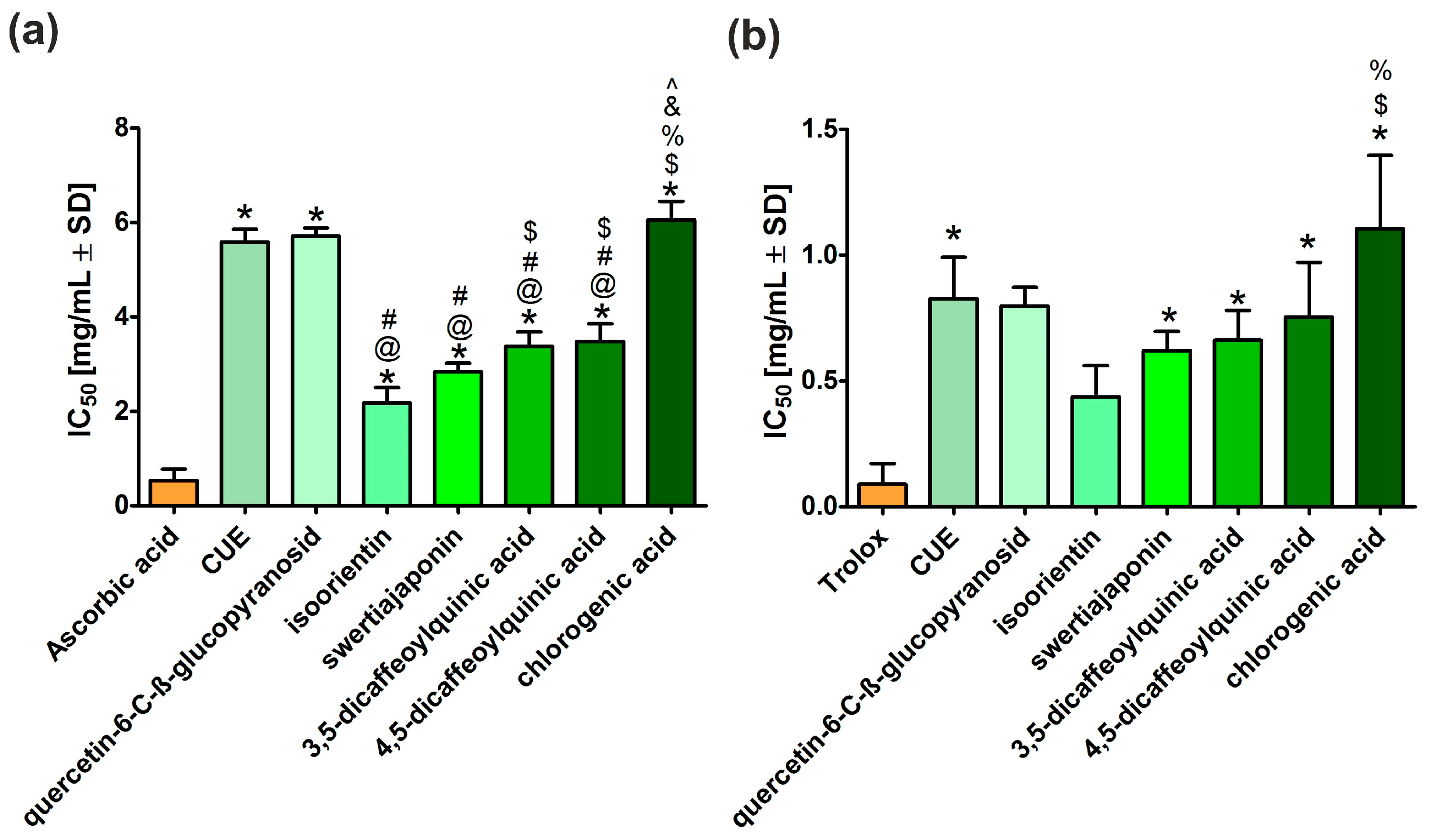
Figure 1. The IC50 values determined in antioxidant tests. (a) DPPH radical scavenging assay; (b) antiradical capacity determination with ABTS●+. The values are expressed as the mean ± standard deviation from three independent measurements. *—Statistically significant differences compared to reference substances—ascorbic acid or Trolox; @—Statistically significant differences compared to CUE; #—Statistically significant differences compared to quercetin-6-C-β-glucopyranoside; $—Statistically significant differences compared to isoorientin; %—Statistically significant differences compared to swertiajoponin; &—Statistically significant differences compared to 3,5-dicaffeoylquinic acid; ^—Statistically significant differences compared to 4,5-dicaffeoylquinic acid (a One-Way ANOVA test, followed by a Tukey’s multiple comparison test, p < 0.05, GraphPad Prism 5, Version 5.04 Software).
Applsci 15 09585 g001
Figure 2. Structure-activity relationship (SAR) features of phenolics from C. uralensis.
Applsci 15 09585 g002
Table 1. Summary of structural characterization techniques used.
TechniqueInformation Obtained
High-Resolution LC–MSExact masses and molecular formulas; fragmentation patterns (e.g., loss of sugar moieties) to infer glycosylation. Essential for assigning molecular weight and initial formula hypotheses.
NMRDetailed proton and carbon connectivity, confirming flavonoid vs. caffeoyl structure, position of sugar attachment, and ring substitution. Enabled unambiguous structural elucidation of each glycoside (e.g., identifying C-glycosidic linkages)
Table 2. High-Resolution Mass Spectrometry (HRMS) data of phenolic compounds isolated from Cephalaria uralensis (experimental vs. theoretical mass, error values, and elemental composition).
No.Experimental Mass [M-H] (Da)Theoretical Mass
[M-H] (Da)
Δ mDaΔ ppmElemental
Composition
1463.08831463.088200.110.24C21H19O12
2447.09336447.093290.070.16C21H19O11
3461.10885461.10894−0.090.20C22H21O11
4515.11985515.119500.350.68C25H23O12
5515.11964515.119500.140.27C25H23O12
6353.08760353.08781−0.210.59C16H17O9
(1) quercetin-6-C-β-glucopyranoside; (2) isoorientin; (3) swertiajaponin; (4) 3,5-dicaffeoylquinic acid; (5) 4,5-dicaffeoylquinic acid; (6) chlorogenic acid; Da—Dalton (unit of molecular mass); Δ mDa—Mass difference in millidaltons; Δ ppm—Mass accuracy error in parts per million; [M–H]—Deprotonated molecular ion detected in negative ion mode.
Table 3. Increase in surface pressure for individual compounds resulting from a single injection beneath the surface of a lipid monolayer formed from DPPC. ISO—isoorientin, 3,5-diCQA—3,5-dicaffeoylquinic acid, 4,5-diCQA—4,5-dicaffeoylquinic acid, and the crude extract CUE—ethanolic extract from the aerial parts of C. uralensis.
CompoundIncrease in Surface Pressure (SP)
[mN/m]
ISO~2
3,5-diCQA~3.5
4,5-diCQA6.5–7
CUE~5.5
Disclaimer/Publisher’s Note: The statements, opinions and data contained in all publications are solely those of the individual author(s) and contributor(s) and not of MDPI and/or the editor(s). MDPI and/or the editor(s) disclaim responsibility for any injury to people or property resulting from any ideas, methods, instructions or products referred to in the content.