Article

The Impact of Damon Self-Ligating Orthodontic Therapy on Soft Tissue and Incisor Inclination: Extraction vs. Non-Extraction Treatment

1
Department of Orthodontics, Johann-Wolfgang Goethe University, Theodor-Stern-Kai 7, 60596 Frankfurt, Germany
2
Private Dental Office, 65812 Bad Soden am Taunus, Germany
3
Department of Prosthodontics, Johann-Wolfgang Goethe University, 60596 Frankfurt, Germany
*
Author to whom correspondence should be addressed.
Appl. Sci. 2025, 15(17), 9265; https://doi.org/10.3390/app15179265
Submission received: 13 June 2025 / Revised: 20 July 2025 / Accepted: 20 August 2025 / Published: 22 August 2025
(This article belongs to the Special Issue Advances in Dentistry and Dental Status Improvement)

Abstract

(1) Background: The present study aimed to evaluate the effects of fixed orthodontic treatment using the Damon self-ligating system on incisor inclination and soft tissue facial profile. Additionally, the study compared the outcomes of premolar extraction versus non-extraction approaches on these parameters. (2) Methods: This retrospective study included 72 patients who were treated with the Damon self-ligating bracket system (Damon, Ormco, Orange, CA, USA). Thirty-five patients (mean age 12.5 years; 20 girls and 15 boys) underwent premolar extraction (Group Ex), while 37 patients (mean age 12.8 years; 16 girls and 21 boys) were treated without extractions (Group NonEx). Lateral cephalometric radiographs were taken pre- and posttherapeutically, and measurements were conducted using OnyxCeph3™ (Image Instruments, Chemnitz, Germany). (3) Results: Following orthodontic treatment, the upper incisors in group Ex were significantly more retruded (p < 0.01), while the mandibular incisors in group NonEx were significantly more proinclined (p < 0.01). Both lips in group Ex, and the upper lip in the group NonEx, exhibited a greater distance from the esthetic line post-therapy (p < 0.01). The position of the lips relative to the SR line did not significantly change in group Ex, whereas group NonEx showed a significantly increased lip protrusion (p < 0.01). A weak negative correlation (−0.37) was observed between the increased retroclination of the upper incisors and the retrusion of the upper lip relative to the SR line. (4) Conclusions: Premolar extraction, as part of a treatment strategy for cases with severe crowding, combined with Damon™ self-ligating braces, does not adversely affect the soft tissue profile.

1. Introduction

Premolar extraction has long been a fundamental yet contentious approach in orthodontic treatment, particularly when addressing moderate to severe crowding and insufficient space in the dental arch [1,2]. In patients with severe crowding, this method is often employed as the only viable solution to achieve a harmonious alignment of the teeth within a stable dental arch and to obtain lasting stability of the therapy results [3]. In borderline cases, however, clinicians face greater complexity in decision-making due to the delicate balance between treatment benefits and potential drawbacks [4]. Specifically, the aesthetic implications of premolar extraction have been a subject of critical debate, particularly concerning its potential to cause undesirable effects, such as retroinclination of the incisors and a reduction in the lip prominence [1,5]. This phenomenon, often described as a Dished-In Face, refers to a decrease in the prominence of the upper and lower lip regions, while the nose and chin retain their prominence [6,7].
It is important to note that, during growth, the natural prominence of the lips tends to diminish slightly as the nose and chin keep on growing more prominently [7,8]. In an era where dental, smile, and facial aesthetics are highly valued, the aesthetic outcomes of orthodontic treatment have become a critical measure of success. Patients’ motivation to undergo orthodontic interventions is often esthetic-driven rather than due to functional concerns related to chewing [9,10,11]. This raises the question of whether orthodontic treatment involving premolar extraction in borderline cases with moderate to severe crowding can also meet the aesthetic expectations necessary for a harmonious facial profile. Additionally, the decline in the popularity of extraction therapy has been influenced in part by claims from manufacturers of passive self-ligating systems, which assert that these systems can create sufficient space for non-extraction treatments through posterior physiologic arch expansion. This posterior expansion may potentially reduce the need for anterior proclination, thereby enabling non-extraction therapy without causing lip protrusion. However, the expansion effect of this system might result in further lip retrusion, potentially exacerbating the “dished-in” facial appearance often associated with extraction cases.
To the best of our understanding, no previous investigation has directly compared the effects of extraction versus non-extraction treatments using self-ligation systems on the soft tissue profile in growing adolescents.
Therefore, the objective of this scientific research was to evaluate and compare the effects of extraction vs. nonextraction therapies on soft tissue profile following orthodontic treatment of 72 borderline cases (mean age of 12.7 years) using Damon™ self-ligating system (Damon, Ormco, Orange, CA, USA).

2. Materials and Methods

2.1. Design of the Study and Ethical Approval

This monocentric, retrospective study was conducted at the Orthodontics department of J. W. Goethe University Frankfurt and received ethical approval from the medical university’s ethics committee (No.: 20-686). All procedures adhered to the guidelines set forth by the Declaration of Helsinki and its latest revision in 2013.

2.2. Patient Population

All patient records were obtained from the archives of the Department of Orthodontics and Orofacial Orthopedics. The study included only those patients with moderate space deficiencies in either upper jaw or both upper and lower jaws, who were treated with self-ligating fixed appliances in both jaws (Damon, Ormco, Orange, CA, USA; 0.022 slot size). All patients were considered borderline extraction cases.
Two patient groups were analyzed. Group Ex (n = 35) included patients who underwent extraction of either all four first premolars (two per jaw) or only the upper first premolars. The mean space deficiency was 6.15 mm (SD 3.28 mm) in the maxillary arch and 3.37 mm (SD 2.88 mm) in the mandibular arch. Group NonEx (n = 37) consisted of patients treated without permanent tooth extraction. Their mean space deficiency was 4.46 mm (SD 1.31 mm) in the maxillary arch and 2.4 mm (SD 1.98 mm) in the mandibular arch.

2.3. Treatment Duration and Growth Considerations

The average treatment duration was similar between groups. Patients in the extraction group began treatment at a mean age of 12.5 years and completed therapy at a mean age of 15.9 years, while those in the non-extraction group had a mean age of 12.8 years at the start of the treatment and finished at 16.3 years old. Thus, both groups underwent treatment during a comparable growth window, minimizing the likelihood that differences in maturational changes could have systematically influenced the soft tissue outcomes.

2.4. Inclusion and Exclusion Criteria

Inclusion criteria for the extraction group required treatment with fixed self-ligating appliances (Damon, Ormco, Orange, CA, USA; 0.022 slot size) in both jaws, extraction of either two premolars in the upper jaw or four premolars (two in each jaw). The non-extraction group included patients treated without premolar extraction. Exclusion criteria for both groups were the presence of craniofacial anomalies, dental aplasia (other than third molar teeth), and the use of expansion appliances such as Hyrax expander or Quadhelix. All patients were treated using the following archwire sequence: Damon CuNiti archwires with dimensions of 0.014, 0.016, 0.016 × 0.025, 0.018 × 0.025, and Damon SS archwire 0.019 × 0.025 (Ormco, Orange, CA, USA). Only patients with digital high-quality lateral cephalograms prior to (T0) and after (T1) treatment were included in the present investigation.

2.5. Data Collection and Analysis

T0 and T1 cephalograms were analyzed using OnyxCeph3™ software (Version 2.1., Image Instruments GmbH, Chemnitz, Germany) (Figure 1). The used landmarks and measurements are presented in Table 1.

2.6. Statistical Analysis

Statistical analysis of the present study was conducted with the assistance of the Institute for Biostatistics and Mathematical Modeling at the Medical Faculty of J. W. Goethe University in Frankfurt am Main, using BiAS software (Version 11.12, Hans Ackermann BiAS for Windows) [12].
The power was analyzed based on the research by Bishara et al. [13]. The calculated statistical power was 82%, which resulted in a sample size of 29 patients in group Ex and 37 patients in the group NonEx to achieve a significance level of 0.05.
Using the Kolmogorov–Smirnov-Lilliefors the normality of data distribution was tested. Inter-group comparisons were analyzed at T0 and T1 using the two-sample t-test for parameters with normal distribution, and the Wilcoxon–Mann–Whitney U test for non-normally distributed parameters. Effect sizes were calculated using Cohen’s d for the t-test and Rosenthal’s R for the Wilcoxon–Mann–Whitney U test [14,15]. Cohen’s d was interpreted as 0.2 = small, 0.5 = medium, and 0.8 = large, while Rosenthal’s R was categorized as 0.1 = small, 0.3 = medium, 0.5 = large, and >0.7 = very large.
Intra-group comparisons at T0 and T1 were performed using the one-sample t-test for data with normal distribution and the Wilcoxon matched pairs test for data with non-normal distribution, with corresponding effect sizes calculated using Cohen’s d and Rosenthal’s R [14,15].
To assess correlations, Pearson’s linear correlation was applied to normally distributed parameters [16]. The Evans correlation coefficient (r) was used for interpretation: r < 0.2 indicating very weak, 0.2–0.4, weak, 0.4–0.6, moderate, 0.6–0.8, strong, and >0.8 very strong correlation.
The measurements were all carried out by the same researcher. To assess the reliability of cephalometric measurements, intra-rater reliability was calculated using the Intraclass Correlation Coefficient (ICC). A random subset of 20 lateral cephalograms was traced and measured for a second time by the same researcher after a four-week interval. The ICC values for all linear and angular measurements ranged from 0.87 to 0.96, corresponding with good to excellent reliability. These results support the consistency and reproducibility of the cephalometric assessments used in this study.

3. Results

The descriptive results are presented in Table 2. Group Ex (n = 35) consisted of 20 girls and 15 boys, while Group NonEx (n = 37) included 16 girls and 21 boys. Additionally, within Group Ex, 20 patients were treated with the extraction of upper and lower first premolars and 15 patients underwent the extraction of only upper first premolars.

3.1. Intra-Group Comparisons (T0–T1)

3.1.1. Group Ex

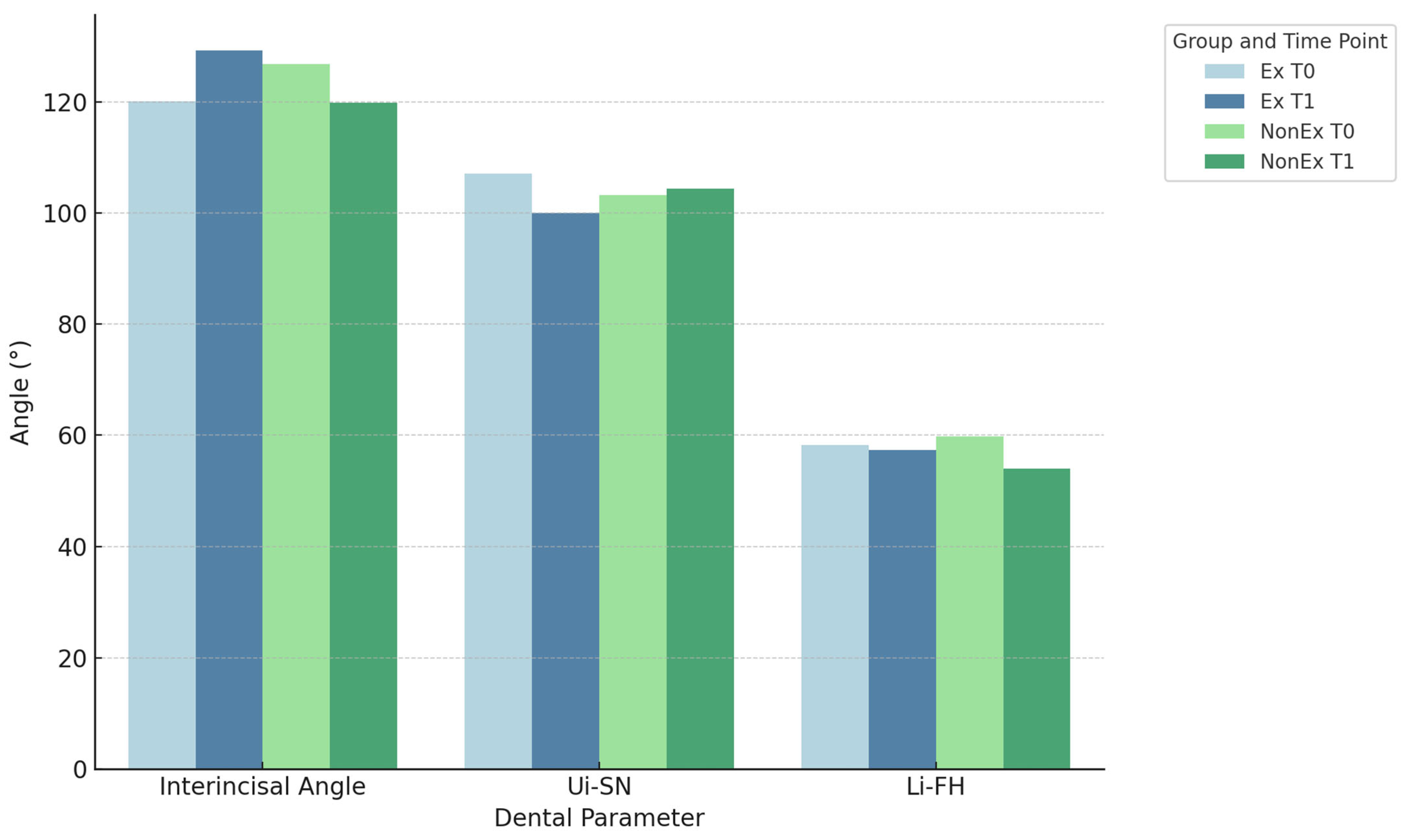
A statistically significant reduction in upper incisors inclination was seen following extraction at T1, which was manifested in the enlargement of interincisal angle (p < 0.01) and maxillary incisor to SN plane angle (p < 0.01). However, no statistically significant differences were observed in the inclination of lower incisors, measured by the angles between lower incisor and Frankfurt plane (LiFH), and mandibular incisor and mandibular plane (LiMeTmh).
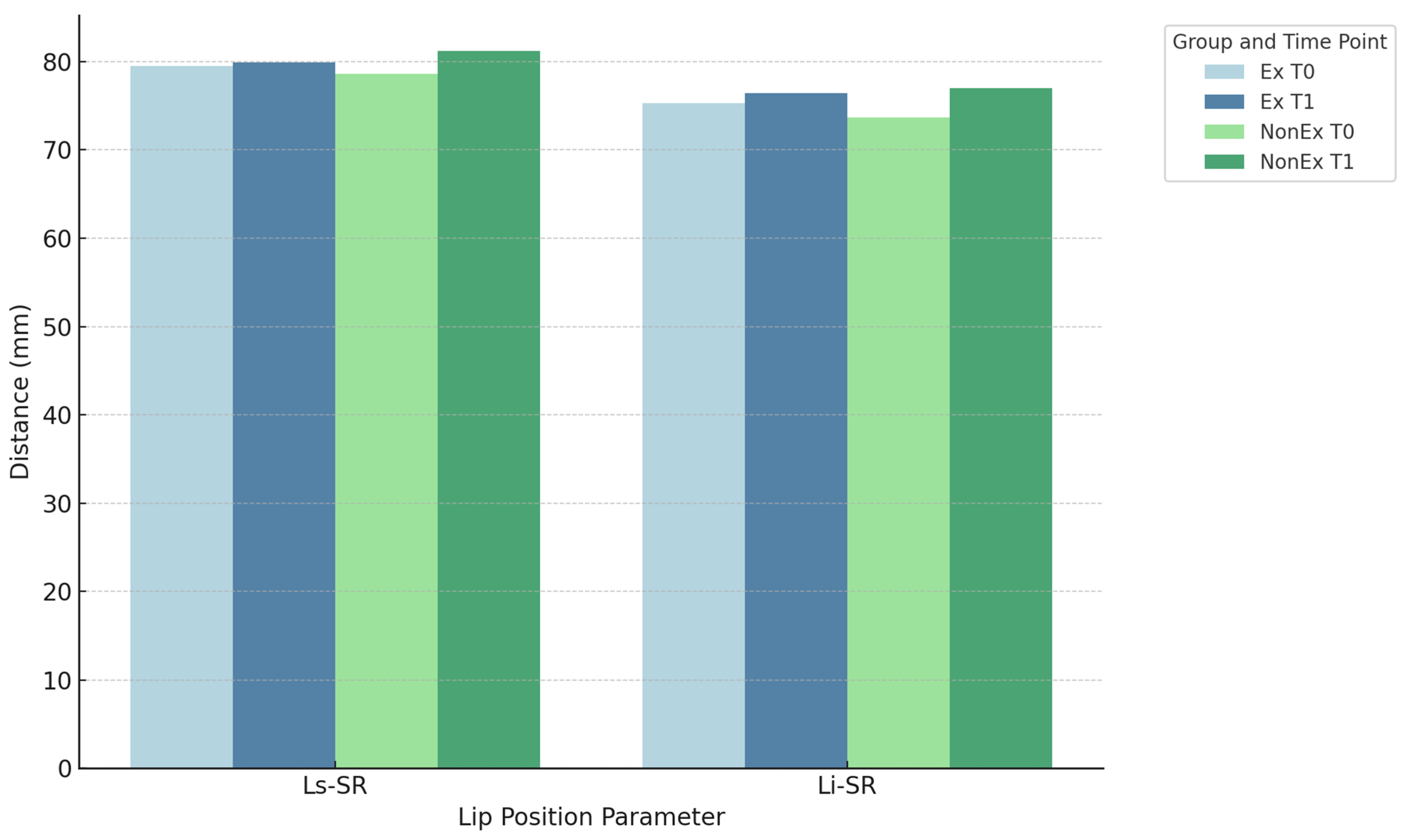
A statistically significant reduction in upper and lower lip prominence was perceived in the extraction group, with both the upper lip to Esthetic Line (Ls-E) and lower lip to Esthetic Line (Li-E) distances increasing significantly (p < 0.01). However, the distances from the upper lip to the SR line (Ls-SR) and lower lip to the SR line (Li-SR) did not display significant changes following extraction treatment (Table 3).

3.1.2. Group NonEx

In group NonEx, the interincisal angle showed a significant reduction (p < 0.01), indicating anterior incisor protrusion. Given that no statistically significant changes were perceived in the inclination of the maxillary incisors, this decrease is primarily due to the protrusion of the lower incisors. This is further supported by significant changes in the angles between the mandibular incisors and the Frankfurt plane, as well as the lower incisors and the mandibular plane (both p < 0.01) (Figure 2).
Interestingly, a significant increase in the distance of the upper lip to the esthetic line was observed following the nonextraction treatment (p < 0.01), whereas the distance of the lower lip to the esthetic line remained unchanged.
On the other hand, statistically significant increase in the distances of the upper and lower lips to the SR line was seen following the nonextraction treatment (p < 0.01) (Table 4) (Figure 3).

3.2. Inter-Group Comparison (T0–T1 Changes)

The difference in the change in values of the interincisal angle before and after therapy between the groups was significant (p < 0.01). These differences were attributed to the changes in the inclination of both maxillary and mandibular incisors.
The differences between the groups regarding the changes in the angle between maxillary incisors and cranial base (UiNS) as well as upper incisors and palatinal plane (UiSNAPm) turned out to be statistically significant (both p < 0.01).
Similar results were found regarding the changes in the lower incisor inclination measurements with lower incisor to Frankfurt line angle at p = 0.01 and lower incisor to mandibular plane angle at p < 0.01.
Regarding the changes in distances of upper and lower lips to the esthetic line, no statistically significant differences were observed between groups Ex and NonEx. However, significant differences were found regarding the changes in the distances of the upper lip (p < 0.01) and lower lip (p = 0.05) to the SR (Table 5) (Figure 2).

3.3. Comparison of Two-Premolar Extraction in the Upper Arch and Four-Premolar Extraction (2 Each in the Upper and Lower Arch) in Group Ex

Significant differences were obtained between the two types of extractions in the Ex group. The interincisal angle increase was significant in cases where all four first premolars were extracted, indicating greater retrusion of the anterior incisors. In contrast, no significant change in the interincisal angle was seen following the extraction of only two maxillary premolars, suggesting that four-premolar extractions resulted in more pronounced retrusion. This effect was primarily due to the marked retrusion of the lower incisors in the four-premolar extraction cases, whereas no significant retrusion of the mandibular incisors was found in cases where only the upper first premolars were extracted (Table 6).

4. Discussion

The present evaluation demonstrated that premolar extraction in borderline cases in combination with Damon self-ligating braces (Damon, Ormco, Orange, CA, USA) did not adversely affect the soft tissue profile. While the inclination of both the maxillary and mandibular incisors exhibited increased retroclination following extraction, the correlation between incisor inclination and lip prominence was weak. Consequently, the increased retroclination did not result in a significant retrusion of the lips. Furthermore, no significant changes were observed in the nasolabial angle following extraction therapy.
The lack of increased retroclination of the mandibular incisors after extraction therapy can be explained by the extraction pattern within the study cohort. The extraction group included 20 subjects who had four premolars extracted and 15 subjects with only two premolars extracted in the upper arch. In the subgroup with four premolars extracted, a stronger retrusion of the mandibular incisors was observed. Conversely, in the subgroup with only upper premolar extractions, the angles showed no significant changes, diluting the overall effect of extraction. Therefore, preserving the premolars in the lower jaw and limiting extractions to the upper jaw in a borderline patient can be considered a viable therapeutic option when maintaining the incisor inclination is a high priority in the treatment plan.
The present study utilized two reference lines to assess the effects of premolar extraction on the soft tissue profile. The SR line represents a bony structure, while the E line is based entirely on soft tissue landmarks, which can undergo substantial growth-related changes over extended periods. The nose in particular grows more prominently forward in the course of facial growth compared to other components of the profile [2,17]. The increased growth of the nose and chin affects the perceived change in lip prominence, specifically relative to the Esthetic Line. Consequently, relying solely on the E Line does not allow an accurate assessment of changes in lip position in growing individuals.
After extraction therapy, no significant changes were noted in the positions of both lips relative to the SR line. This result was expected for the lower lip, as there was no significant change in the inclination of the mandibular incisors. A comparison between the subgroups with four premolars and two premolars extracted also showed no difference in lip position. However, the lack of significant change in the upper lip position was surprising, given the significant retroclination of the maxillary incisors. The weak correlation between maxillary incisor retroclination and upper lip retrusion supports the theory that lip position is influenced not solely by incisor inclination but also by lip morphology and tension [6,18,19,20]. It has also been indicated that the selection of a particular extraction pattern did not significantly affect lip response, which appears to be more strongly associated with soft tissue characteristics than dental or skeletal structures. Specifically, vermilion fullness emerged as the most influential factor contributing to the extent of lip retraction [18,19].
In group NonEx, a significant protrusion of the lower incisors took place (p < 0.01). Thus, the use of the Damon self-ligating system did not adequately preserve the initial mandibular incisor inclination and was unable to prevent mandibular incisor protrusion in non-extraction treatment of borderline patients. Even though the upper incisors showed only a slight, non-significant protrusion, both lips were significantly more protruded relative to the SR line in the NonEx group (p < 0.01). This further confirms that lip position does not strictly follow the incisor inclination of their responding jaw. The protrusion of the lower lip appeared to have affected the upper lip position as well.
In comparing extraction and non-extraction therapy, lip position did not change significantly in group Ex, while in group NonEx, both lips became more protruded. Therefore, the lips demonstrated greater sensitivity to an increase in incisor inclination than to a decrease.
The results of the present investigation are partially in agreement with those of a similar study [2], comparing extraction vs. non-extraction modalities. The authors reported post-treatment lip retrusion in both groups, with greater retrusion obtained in the extraction group [2]. It is noteworthy that their evaluation relied on the E-Line. Similarly, our findings showed increased retrusion of both lips in relation to the E-Line, with more pronounced retrusion in the extraction group. Additionally, both studies involved growing subjects, which likely contributed to the anterior shift in the Esthetic Line during treatment. However, contrary to our study, they did not use a bony reference line such as SR-line to isolate the lip position from other soft tissue structures. The lack of lip protrusion in the non-extraction group of their study [2] could be due to the differences in their treatment duration or method of anchorage.
Recent evidence continues to underscore the nuanced effects of dental extractions on soft tissue outcomes in adolescents. Ekstam et al. conducted a retrospective cephalometric study using conventional bracket systems and found significant decreases in both upper and lower lip prominence regarding the E-line following extraction therapy in adolescents [21]. Unlike our findings—where non-extraction treatment showed an increase in upper lip position to the E-line—their extraction group demonstrated greater lip retrusion. These differences may be attributable to variations in bracket type and mechanics or the lack of skeletal-based reference line measurements in their analysis.
A comprehensive meta-analysis by Elias et al. revealed that four-premolar extraction is consistently associated with upper and lower lip retraction when measured against the E-plane, summarizing results across three upper-lip and five lower-lip studies [22]. This aligns with soft tissue changes observed in adult extraction cases, as seen in Choi et al., who reported significant reductions in lip-closing force and notable lip retraction post-extraction [23].
Furthermore, a systematic review by Konstantonis et al. confirmed a general trend toward more pronounced lip retraction, increased nasolabial angle, and profile flattening following extraction therapy—effects that were more evident when four premolars were extracted—highlighting the importance of treatment extent on soft tissue outcomes [24]. In contrast, studies comparing extraction vs. non-extraction therapies using 3D measurements such as vermilion morphology (e.g., Liu et al.) suggest that extraction may also enhance aesthetic features like vermilion volume without altering proportions [25].
Taken together, these recent findings support our conclusions that while extraction therapy using self-ligating systems does impact soft tissue profiles, the extent and clinical significance of these effects depend on the specific reference method, bracket system, and individual patient characteristics.
The weak correlation between lip position and incisor inclination observed in our study is consistent with findings from other studies, which suggest that the impact of extraction on profile changes is limited and that maturational changes play a more significant role [6,7,20]. Furthermore, the non-extraction philosophy, which advocates for passive posterior expansion using the Damon self-ligating system, could not be substantiated in the present study. Significant incisor protrusion was observed in the non-extraction treatment of borderline cases. Consequently, the anticipated negative effects of extraction therapy, as well as the effectiveness of passive self-ligating systems in avoiding extractions while preserving anterior inclination, were not demonstrated in the present study.

Limitations

This study’s demographic profile of models and participants was relatively narrow, which adversely limits the broader applicability of the findings. Blinding of the clinician was not achievable, and the ongoing growth of the participants may have influenced the outcomes. Additionally, measurement errors cannot be ruled out, as the tension in soft tissues during cephalometric analysis could not be controlled.

5. Conclusions

The findings of the current investigation revealed no adverse impacts on the soft tissue profile following premolar extraction as a treatment strategy in borderline extraction cases managed with Damon self-ligating bracket system. The use of Damon self-ligating bracket system could not avoid the lower incisor protrusion in the non-extraction group. Even though a reduction in upper and lower incisors’ inclination was registered in the extraction group, only a weak correlation was found between the incisor inclination and lip position.
This study provides clinically significant evidence that premolar extraction, when performed in borderline cases using the Damon™ self-ligating system, does not adversely affect the soft tissue profile, alleviating a common concern among clinicians and patients. The comparative design and detailed cephalometric evaluation offer practical insights for treatment planning, particularly in selecting extraction vs. non-extraction protocols. Furthermore, the use of both skeletal and soft-tissue reference lines enhances diagnostic precision and strengthens the applicability of the findings in routine orthodontic practice.

Author Contributions

Conceptualization, S.B. (Sarah Bühling), S.K. and S.S.; methodology, S.B. (Sarah Bühling), S.B. (Silvia Brandt) and S.E.; software, N.P. and B.S.; validation, S.B. (Sarah Bühling) and S.S.; formal analysis, S.S., S.B. (Sarah Bühling), B.S., S.K. and S.E.; investigation, S.B. (Sarah Bühling) and S.B. (Silvia Brandt); resources, S.B. (Sarah Bühling) and B.S.; data curation, S.S., S.B. (Sarah Bühling) and S.E.; writing—original draft preparation, S.B. (Sarah Bühling); writing—review and editing, N.P.; visualization, S.K. and S.B. (Sarah Bühling); supervision, S.K., S.B. (Sarah Bühling), B.S. and S.S.; project administration, S.B. (Sarah Bühling) and S.K. All authors have read and agreed to the published version of the manuscript.

Funding

This research received no external funding.

Institutional Review Board Statement

The study was approved by the ethics committee of the medical department of J. W. Goethe University in Frankfurt am Main (Approval Nr 20-686, Approval date 5 May 2020). Written consent was obtained from the patients for publication of their anonymized data. Furthermore, the study was carried out in accordance with the Helsinki Declaration and its revisions (the last one in 2013).

Informed Consent Statement

Informed consent was obtained from all parents and legal guardians of children involved in the study.

Data Availability Statement

The data are available upon request from the corresponding author due to data privacy regulations of the J. W. Goethe University.

Conflicts of Interest

The authors declare that they have no conflicts of interest.

References

  1. Sayahpour, B.; Lau, D.; Eslami, S.; Buehling, S.; Kopp, S.; Jamilian, A.; Chhatwani, S. Posttreatment stability following therapy using passive self-ligating brackets: Extraction vs. nonextraction. J. Orofac. Orthop. 2023, 86, 1–9. [Google Scholar] [CrossRef] [PubMed]
  2. Kirschneck, C.; Proff, P.; Reicheneder, C.; Lippold, C. Short-term effects of systematic premolar extraction on lip profile, vertical dimension and cephalometric parameters in borderline patients for extraction therapy—A retrospective cohort study. Clin. Oral Investig. 2016, 20, 865–874. [Google Scholar] [CrossRef] [PubMed]
  3. Bowman, S.J. The Esthetic Impact of Extraction and Nonextraction Treatments on Caucasian Patients. Angle Orthod. 2000, 70, 3–10. [Google Scholar] [CrossRef] [PubMed]
  4. Tepedino, M.; Esposito, R.; Potrubacz, M.I.; Xhanari, D.; Ciavarella, D. Evaluation of the relationship between incisor torque and profile aesthetics in patients having orthodontic extractions compared to non-extractions. Clin. Oral Investig. 2023, 27, 5233–5248. [Google Scholar] [CrossRef] [PubMed]
  5. Buhling, S.; Schmied, S.; Eslami, S.; Brandt, S.; Plein, N.; Kopp, S.; Sayahpour, B. Changes in the arch width and buccal corridor after fixed orthodontic treatment with Damon self-ligating system: Premolar extraction vs. non-extraction. Dent. Press. J. Orthod. 2024, 29, e2423159. [Google Scholar] [CrossRef]
  6. Erdinc, A.E.; Nanda, R.S.; Dandajena, T.C. Profile changes of patients treated with and without premolar extractions. Am. J. Orthod. Dentofac. Orthop. 2007, 132, 324–331. [Google Scholar] [CrossRef] [PubMed]
  7. Zierhut, E.C.; Joondeph, D.R.; Artun, J.; Little, R.M. Long-Term Profile Changes Associated with Successfully Treated Extraction and Nonextraction Class II Division 1 Malocclusions. Angle Orthod. 2000, 70, 208–219. [Google Scholar] [CrossRef] [PubMed]
  8. Kocadereli, I. Changes in soft tissue profile after orthodontic treatment with and without extractions. Am. J. Orthod. Dentofac. Orthop. 2002, 122, 67–72. [Google Scholar] [CrossRef] [PubMed]
  9. Birnie, D.; Harradine, N. The Extraction Non-Extraction Decision. In Excellence in Orthodontics Course Manual Chapter 6; Excellence in Orthodontics: Solihull, UK, 2016; pp. 79–92. [Google Scholar]
  10. Iiksal, E.; Hazar, S.; Akyalin, S. Smile esthetics: Perception and comparison of treated and untreated smiles. Am. J. Orthod. Dentofac. Orthop. 2006, 129, 8–16. [Google Scholar] [CrossRef]
  11. Janson, G.; Mendes, L.M.; Junqueira, C.H.Z.; Garib, D.G. Soft-tissue changes in Class II malocclusion patients treated with extractions: A systematic review. Eur. J. Orthod. 2016, 38, 631–637. [Google Scholar] [CrossRef]
  12. Ackermann, H. BiAS. f \ u r Windows Handbuch Version 11.12. 2020. Available online: https://support.microsoft.com/en-us/topic/windows-10-update-history-53c270dc-954f-41f7-7ced-488578904dfe (accessed on 20 July 2025).
  13. Bishara, S.E.; Cummins, D.M.; Jakobsen, J.R.; Zaher, A.R. Dentofacial and soft tissue changes in Class II, Division 1 cases treated with and without extractions. Am. J. Orthod. Dentofac. Orthop. 1995, 107, 28–37. [Google Scholar] [CrossRef] [PubMed]
  14. Cohen, C.; Cohen, P.; West, S.; Aiken, L. Applied Multiple Regression/Correlation Analysis for the Behavioral Sciences; Routledge: Oxfordshire, UK, 2013. [Google Scholar]
  15. Rosenthal, R. Meta-Analytic Procedures for Social Research; Sage: Beverly Hills, CA, USA, 1991. [Google Scholar]
  16. Evans, J. Straightforward Statistics for the Behavioral Sciences; Thomson Brooks/Cole Publishing Co.: Monterey, CA, USA, 1996. [Google Scholar]
  17. Bishara, S.E.; Jakobsen, J.R.; Hession, T.J.; Treder, J.E. Soft tissue profile changes from 5 to 45 years of age. Am. J. Orthod. Dentofac. Orthop. 1998, 114, 698–706. [Google Scholar] [CrossRef] [PubMed]
  18. Albertini, P.; Barbara, L.; Albertini, E.; Willeit, P.; Lombardo, L. Soft-tissue profile changes in adult patients treated with premolar extractions. Am. J. Orthod. Dentofac. Orthop. 2024, 166, 171–178. [Google Scholar] [CrossRef] [PubMed]
  19. Wholley, C.J.; Woods, M.G. The effects of commonly prescribed premolar extraction sequences on the curvature of the upper and lower lips. Angle Orthod. 2003, 73, 386–395. [Google Scholar] [CrossRef] [PubMed]
  20. Basciftci, F.A.; Usumez, S. Effects of extraction and nonextraction treatment on class I and class II subjects. Angle Orthod. 2003, 73, 36–42. [Google Scholar] [CrossRef] [PubMed]
  21. Ekstam, A.; Sonesson, M.; Hellén-Halme, K. Effects of Premolar Extraction and Orthodontic Treatment in Adolescents: A Retrospective Cephalometric Study. Acta Odontol. Scand. 2024, 82, 56–64. [Google Scholar] [CrossRef] [PubMed]
  22. Elias, K.G.; Sivamurthy, G.; Bearn, D.R. Extraction vs. Nonextraction Orthodontic Treatment: A Systematic Review and Meta-Analysis. Angle Orthod. 2024, 94, 83–106. [Google Scholar] [CrossRef] [PubMed]
  23. Choi, T.H.; Kim, S.H.; Kim, C.; Kook, Y.A.; Larson, B.E.; Lee, N.K. Changes in Maximum Lip-Closing Force after Extraction and Nonextraction Orthodontic Treatments. Korean J. Orthod. 2020, 50, 120–128. [Google Scholar] [CrossRef] [PubMed]
  24. Konstantonis, D.; Vasileiou, D.; Papageorgiou, S.N.; Eliades, T. Soft Tissue Changes Following Extraction vs. Nonextraction Orthodontic Fixed Appliance Treatment: A Systematic Review and Meta-Analysis. Eur. J. Oral Sci. 2018, 126, 43–53. [Google Scholar] [CrossRef] [PubMed]
  25. Liu, Y.; Zhao, J.; Gao, L.; Zhang, C. Three-Dimensional Changes in Lip Vermilion Morphology After Extraction and Non-Extraction Orthodontic Treatment: A CBCT-Based Study. J. Dent. 2019, 85, 100–109. [Google Scholar] [CrossRef]
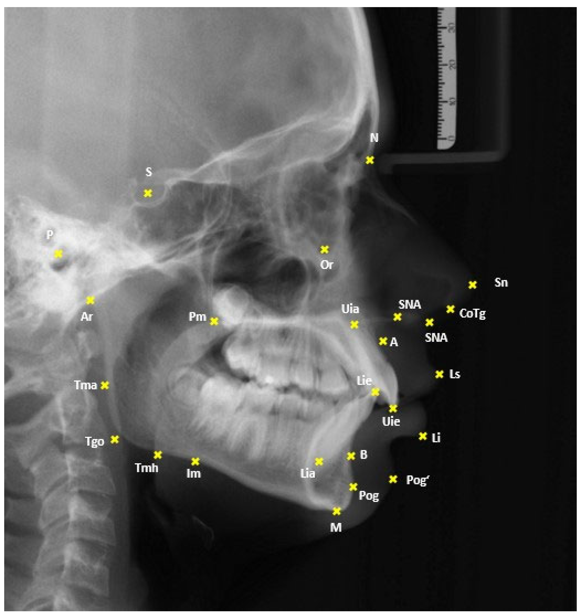
Figure 1. Landmarks for the cephalometric analysis.
Applsci 15 09265 g001
Figure 2. Changes in dental inclination parameter pre- (T0) and post-therapeutically (T1).
Applsci 15 09265 g002
Figure 3. Changes in lip position before and after treatment.
Applsci 15 09265 g003
Table 1. Landmarks used in the present study.
Landmark/MeasurementAbbreviationDefinition
SellaSCenter of the pituitary fossa
NasionNMost anterior point along the nasofrontal suture
Anterior Nasal SpineANSTip of the anterior nasal spine
Pterygomaxillary FissurePmIntersection of the posterior margin of the maxillary body with the contour of both soft and hard palates
Point AADeepest point of the concavity on the frontal contour of the alveolar process of the maxilla
Upper Incisal EdgeUieIncisal tip of the most anterior upper central incisor
Upper Incisal ApexUiaRoot apex of the most anterior upper central incisor
Lower Incisal EdgeLieIncisal tip of the most anterior lower central incisor
Lower Incisal ApexLiaRoot apex of the most anterior lower central incisor
Point BBDeepest point of the concavity on the frontal contour of the alveolar process of the mandible
MentonMLowest point of the mandibular symphysis
Masseteric NotchImPoint of the deepest cranial concavity of the horizontal mandibular ramus
ArticulareArIntersection of the posterior contour of the mandibular condyle with the clivus of the cranial base
Tagent Menton HorizontalTmhTangential point from Menton to the horizontal ramus at the mandibular angle
Tagent Menton AscendanceTmaTangential point from Articulare to the ascending ramus at the region of the mandibular angle
Gonion Tangential PointTgoIntersection of the lines Me-Tmh and Ar-Tma
PorionPMost superior point of the external auditory meatus
OrbitaleOrMost inferior point of the bony orbit
Upper LipLsPoint of greatest anterior projection of the upper lip
Lower LipLiPoint of greatest anterior projection of the lower lip
Nasal TipNsMost anterior point of the nasal tip
Soft Tissue PogonionPog’Most anterior point of the soft tissue chin
SubnasaleSnPoint where the upper lip meets the nasal columella
Columella Tangent PointCoTgTangential point from Subnasale to the nasal columella
SNA AngleSNAThis angle is made by the reference lines S-N and N-A; it provides information about the positional relationship of the maxilla to the cranial base.
SNB AngleSNBThis angle is formed by the reference lines S-N and N-B. It allows assessment of the sagittal position of the mandible.
ANB AngleANBThis angle is formed by the reference lines Nasion-Point A and Nasion-Point B. It provides insight into the sagittal relationship between the mandible and maxilla.
Interincisal AngleUiLiThis angle is built by the axes of the upper incisors (Uie-Uia) and the lower incisors (Lie-Lia). It is measured dorsally and shows the relationship between the axes of the anterior teeth. It is also a good indicator of bimaxillary dental protrusion. A smaller angle indicates more protruded anterior teeth.
Upper Incisor AngleUiSNAPmThis angle is constructed between the axis of the upper incisors and the maxillary plane (Spa-Pm). It provides information about the inclination of the first upper incisors in relation to the basal plane of the maxilla.
Lower Incisor AngleLiMeTmhThis angle is between the axis of the lower incisors and the mandibular plane (Me-Tmh). It shows the inclination of the first mandibular incisors relative to the mandibular plane.
Upper Incisor to Nasion Line AngleUiNSThis angle is formed by the axis of the upper incisors and the NS line. It provides information about the inclination of the first upper incisors relative to the N-S line.
Mandibular Incisor to Frankfurt Horizontal Angle LiFHThis angle is formed by the axis of the lower incisors and the Frankfurt Horizontal (P-Or). It shows the inclination of the first lower incisors relative to the Frankfurt Horizontal plane.
Distance Ls-Esthetic PlaneLs-EThis distance is measured from the upper lip to the Ricketts Esthetic Plane (Ns-Pog‘) at a 90-degree angle. It assesses the harmonious appearance of the lip profile relative to the soft tissue components of the nose and chin.
Distance Li-Esthetic PlaneLi-EThis distance is measured from the lower lip to the Esthetic Plane according to Ricketts, vertical to the Esthetic Plane. It also provides insight into the harmonious appearance of the lip profile relative to the soft tissue components of the nose and chin.
Distance Ls-SR LineLs-SRThis distance is measured from the upper lip to the SR Line (a perpendicular to the Nasion–Sella line, minus seven degrees at the Sella). It helps in evaluating the lip position relative to a bony reference plane.
Distance Li-SR LineLi-SRThis distance is measured from the lower lip to the SR Line (a perpendicular to the Nasion–Sella line, minus seven degrees at the Sella). It helps in evaluating the lip position relative to a bony reference plane.
Nasolabial AngleLsSnCoTgThis angle is formed by the upper lip tangent (Ls-Sn) and the columella tangent (Sn-CoTg).42 It gives information about the upper part of facial convexity, specifically the prominence of the upper lip in profile.
Anterior Face HeightAFHThis distance is measured from the Nasion to the Menton. It describes the anterior facial height.
Posterior Face HeightPFHThis distance is measured from the Sella to the Gonion Tangent Point. It describes the posterior facial height.
Facial Height RatioS-Tgo/N-MeThis is calculated using the formula Ratio = (Distance S-Tgo * 100)/Distance N-Me. The ratio indicates the proportion of posterior to anterior facial height.
Table 2. Descriptive analysis of participants in groups NonEx (without extraction) and Ex (with extraction).
GroupsExNonEx
Time PointT0T1T0T1
MeasurementsAverage/Median *SD/1. -3. Quartile **Average/Median *SD/1. -3. Quartile **Average/Median *SD/1. -3. Quartile **Average/Median *SD/1. -3. Quartile **
SNA (°)79.493.8779.414.1280.743.6680.543.84
SNB (°)75.563.8775.683.9876.43.6776.863.83
ANB (°)3.922.343.732.24.371.983.662
Interincisal angle (°)120.0712.08129.23 *122.31–133.48 **126.849.03119.85 *114.73–123.85 **
Nasiolabial angle (°)99.2112.52101.25 *99.08/104.9899.978.7101.4 *92.98–106.6 **
Ui-SN (°)107.19.0799.986.35103.187.1104.367.57
Li-FH (°)58.256.7757.4 *54.23–62.98 **59.777.3754.05 *51.03–59.9 **
Ui—SNA-Pm (°)65.95 *58.1–70.25 **71.785.9168.95 *64.58–71.4 **67.117.02
Li-Me-Tmh (°)82.66.5984.198.23836.679.538.29
Ls-E (mm)−1.183.2−3.132.8−1.252.09−2.842.35
Li-E (mm)1.183.74−0.093.560.532.540.152.72
Ls-SR (mm)79.535.7579.95.8978.615.2681.25.68
Li-SR (mm)75.296.8876.46.7873.685.3777.026.92
AFH (mm)107.1 *105–109.7 **110.58 *108.69–114.54 **106.8 *103.3–111.85 **114.85 *109.23–122.43 **
PFH (mm)67.375.2970.955.8770.836.1177.046.5
S-Tgo/N-Me63.365.2364.385.4865.644.9766.275.26
Cephalometric measurements prior to (T0) and after (T1) orthodontic therapy. The normally distributed data is shown as the average and SD (standard deviation), whereas the non-normally distributed data is reported as Median * and 1. -3. Quartile **.
Table 3. Comparison of treatment changes before (T0) and after (T1) therapy within group Ex (treated with extraction).
Group Ex
MeasurementsAverage/Median *SD/1. -3. Quartile **p-ValueEffect Size Cohen/Rosenthal Rating
SNA (°)0.151.40.550.11
SNB (°)−0.171.280.440.141
ANB (°)0.391.240.080.321
Interincisal angle (°)−9.1412.1<0.010.762
Nasiolabial angle (°)−2.6610.280.150.261
Ui-SN (°)7.017.98<0.010.883
Li-FH (°)−1.067.760.430.141
Ui—SNA-Pm (°)−7.387.6<0.010.973
Li-Me-Tmh (°)−1.687.790.220.221
Ls-E (mm)−2.191.07<0.012.053
Li-E (mm)−1.251.94<0.010.652
Ls-SR (mm)−0.15 *−7.3–3.7 **0.770.05 0
Li-SR (mm)−1.543.640.060.421
AFH (mm)−3.65 *−13.5–−0.1 **<0.010.62 3
PFH (mm)−3.933.14<0.011.33
S-Tgo/N-Me−0.971.47<0.010.662
The data with normal distribution is shown as average and SD (standard deviation), whereas the non-normally distributed data is reported as Median * and 1. -3. Quartile **. p-value < 0.05 is considered statistically significant.
Table 4. Comparison of treatment changes before (T0) and after (T1) therapy within group NonEx (treated without extraction).
Group NonEx
MeasurementsAverage/Median *SD/1. -3. Quartile **p-ValueEffect Size Cohen/Rosenthal Rating
SNA (°)−0.1 *−2.4–2.9 **0.730.04 0
SNB (°)−0.451.430.060.321
ANB (°)0.651.31<0.010.52
Interincisal angle (°)5.058.23<0.010.612
Nasiolabial angle (°)0.226.780.850.031
Ui-SN (°)−0.886.280.40.141
Li-FH (°)3.575.34<0.010.672
Ui—SNA-Pm (°)1.35.970.190.221
Li-Me-Tmh (°)3.465.39<0.010.642
Ls-E (mm)−2 *−4.8–4.7 **<0.010.5 3
Li-E (mm)−0.421.450.090.291
Ls-SR (mm)−2.62.7<0.010.953
Li-SR (mm)−3.43.25<0.011.053
AFH (mm)−8.516.01<0.011.413
PFH (mm)−6.243.93<0.011.593
S-Tgo/N-Me−0.691.33<0.010.522
The normally distributed data is shown as average and SD (standard deviation), whereas the non-normally distributed data is reported as Median * and 1. -3. Quartile **. p-value < 0.05 is considered statistically significant.
Table 5. Inter-group comparison of T0–T1 changes following the orthodontic therapy.
Measurementsp-ValueEffect Size Cohen/Rosenthal Rating
SNA (°)0.770.04 1
SNB (°)0.390.21
ANB (°)0.390.211
Interincisal angle (°)<0.011.383
Nasiolabial angle (°)0.180.331
Ui-SN (°)<0.011.113
Li-FH (°)0.010.72
Ui—SNA-Pm (°)<0.011.283
Li-Me-Tmh (°)<0.010.772
Ls-E (mm)0.280.14 1
Li-E (mm)0.060.512
Ls-SR (mm)<0.010.39 2
Li-SR (mm)0.050.552
AFH (mm)0.020.29 1
PFH (mm)0.020.632
S-Tgo/N-Me0.40.21
p-value < 0.05 is considered statistically significant.
Table 6. Comparisons of T0–T1 changes following the orthodontic therapy within group Ex (treated with extraction) between extraction of four first premolars and extraction of only two upper premolars.
Measurementsp-ValueEffect Size Cohen/Rosenthal Rating
SNA (°)0.470.251
SNB (°)0.170.491
ANB (°)0.020.833
Interincisal angle (°)0.020.863
Nasiolabial angle (°)0.430.281
Ui-SN (°)0.960.020
Li-FH (°)<0.010.47 2
Ui—SNA-Pm (°)0.670.150
Li-Me-Tmh (°)<0.011.23
Ls-E (mm)0.020.49 2
Li-E (mm)0.030.943
Ls-SR (mm)0.120.34 2
Li-SR (mm)0.340.411
AFH (mm)0.090.37 2
PFH (mm)0.011.183
S-Tgo/N-Me<0.010.45 2
p-value < 0.05 is considered statistically significant.
Disclaimer/Publisher’s Note: The statements, opinions and data contained in all publications are solely those of the individual author(s) and contributor(s) and not of MDPI and/or the editor(s). MDPI and/or the editor(s) disclaim responsibility for any injury to people or property resulting from any ideas, methods, instructions or products referred to in the content.