Article

Reconstructive Therapy in Patients with Peri-Implantitis in a University Dental Hospital: A Preliminary Retrospective Case Series Focusing on Complications

by
Ahmad Alahmari
1,2,
Xavier Costa-Berenguer
1,
Rui Figueiredo
1,3,*,
Eduard Valmaseda-Castellón
1,3,
Alba Sánchez-Torres
1,3,* and
Marta García-García
1,3
1
Oral Surgery and Implantology, Faculty of Medicine and Health Sciences, University of Barcelona, 08028 Barcelona, Spain
2
Department of Periodontics and Community Dental Sciences, Division of Periodontics, College of Dentistry, King Khalid University, Abha 61413, Saudi Arabia
3
Odontological and Maxillofacial Pathology and Therapeutics Group, Institut d’Investigació Biomèdica de Bellvitge (IDIBELL Institute), 08907 Barcelona, Spain
*
Authors to whom correspondence should be addressed.
Appl. Sci. 2025, 15(14), 8040; https://doi.org/10.3390/app15148040
Submission received: 19 June 2025 / Revised: 9 July 2025 / Accepted: 14 July 2025 / Published: 18 July 2025
(This article belongs to the Special Issue Recent Advances in Digital Dentistry and Oral Implantology)

Abstract

Peri-implantitis is an inflammatory disease-causing bone loss around dental implants, often requiring reconstructive surgical therapies to reduce probing depth and regenerate bone. However, such surgeries are frequently complicated by postoperative issues. This retrospective case series aimed to identify the main postoperative complications following the reconstructive treatment of peri-implant bone defects in peri-implantitis patients. Data from 14 patients with 21 affected implants were analyzed, including demographics, oral hygiene, surgical techniques, and complications such as wound dehiscence, membrane exposure, and infections. Wound dehiscence was measured using Image J® software version 1.54. Descriptive and bivariate analyses were performed. The results showed that 11 implants (52.4%; 95% confidence interval (95%CI): 29% to 76%) in nine patients (57.1%; 95%CI = 27% to 87%) developed soft tissue dehiscence after one week, with membrane exposure observed in 4 implants. Dehiscence was significantly associated with mandibular implant location (p = 0.003), poor interproximal hygiene (p = 0.008), and membrane exposure (p = 0.034). No postoperative infections were recorded. In conclusion, more than half of peri-implantitis patients undergoing reconstructive surgery experience wound dehiscence, particularly in cases involving mandible, poor hygiene, and membrane exposure. This complication might compromise bone regeneration and reduce the treatment success rate. These results should be interpreted cautiously due to study design limitations (retrospective design, lack of a control group, and small sample size).

1. Introduction

In recent decades, an important number of patients have been successfully treated with dental implants in order to restore edentulous spaces and to recover masticatory function [1]. However, these devices are not exempt from complications. Biological complications such as peri-implant mucositis and peri-implantitis can significantly reduce the success of dental implants and may result in their failure [2,3]. According to the 2017 World Workshop on the Classification of Periodontal and Peri-Implant Diseases and Conditions, peri-implant mucositis is an inflammatory pathological condition of the peri-implant mucosa without accompanying bone loss. Clinical signs include pus formation, redness, swelling, and bleeding on probing (BOP) [4,5]. On the other hand, in peri-implantitis, inflammation affects soft and hard tissues, causing peri-implant bone loss. This can be observed clinically as increased probing depth (PD), significant progressive bone loss beyond the margin of measurement error (approximately 0.5 mm), and the presence of pus and/or bleeding on probing [5,6]. This biological complication affects approximately one out of every five patients with dental implants and is one of the most common causes of implant failure [7]. A systematic review with meta-analysis reported subject-based estimated weighted mean prevalences and ranges for peri-implant mucositis and peri-implantitis. According to these authors, peri-implant mucositis has a prevalence of 43% (range from 19% to 65%), while peri-implantitis is slightly less common with a prevalence of 22% (range from 1% to 47%) [8]. Additionally, Mir-Mari et al. [9] concluded that the patient-based prevalence of peri-implantitis in a private practice located in Spain was 9.1% (16.3% of implants). In this sample, peri-implant mucositis affected 38.8% of the patients and 21.6% of the implants. The real prevalence may be higher since the available diagnostic tests have limitations and require improvements [10,11,12].
Several risk factors for peri-implantitis have been identified [13]. These include the history of periodontitis, inadequate oral hygiene, smoking, diabetes, and lack of regular supportive peri-implant therapy. A comprehensive risk assessment and personalized preventive strategies are important to minimize complications and ensure long-term implant success [14]. Regular dental check-ups and periodontal and peri-implant supportive therapy are also crucial for maintaining health. Other factors that have been suggested as risk factors for the onset and/or progression of peri-implant diseases are the absence of peri-implant keratinized mucosa (PIKM), occlusal overload, the presence of titanium particles within peri-implant tissues, bone compression necrosis, overheating, micromotion, or biocorrosion [15,16].
Different therapies have been proposed for the treatment of peri-implantitis [17]. Most reports state that a non-surgical approach appears to be insufficient to solve this biological complication [18]. However, these conservative therapies can remove the oral biofilm, modify the risk indicators, and improve the peri-implant tissues prior to the surgical phase [19]. Surgical treatments appear to provide more promising outcomes. The type of surgical approach is usually determined by clinical factors like the peri-implant defect anatomy [20], the amount of keratinized mucosa, and the esthetic component [6].
In 2007, Schwarz et al. [20] classified peri-implant defects into non-contained or suprabony defects (type II) or intraosseous defects (type I). Later, Monje et al. [21] modified this classification to include the severity of the defect. These classifications are currently used for decision-making when performing surgical treatment.
Most authors describe three major techniques: resective, reconstructive, and combined [6,22,23]. Resective approaches are used in uncontained defects with a suprabony component [24]. Romeo et al. [25,26] introduced the concept of implantoplasty as an additional technique to improve the outcomes of resective surgeries. Additionally, Lang et al. [27] emphasized that smoothing the implant surface enhances peri-implant tissue stability and promotes long-term maintenance. Resective approaches have been assessed in recent years, with acceptable results. However, recurrence of the disease occurs in 39–59.5% of the treated implants [28,29,30].
A reconstructive approach is usually indicated in self-containing defects that are usually found in the initial stages of peri-implantitis and seem to provide good results [18,31]. Combined approaches have also been proposed, consisting of implantoplasty of the supra-osseous portion of the defect and reconstruction of the infrabony portion using guided bone regeneration (GBR) techniques [32]. Schwarz et al. [33] showed that combined surgery significantly decreased inflammation, probing depth (PD), and bleeding on probing (BOP).
However, the treatment of peri-implantitis poses significant challenges, and postsurgical complications are common mostly due to the chronic inflammation of peri-implant tissues and the presence of biofilm. The recently published clinical guidelines for the prevention and treatment of peri-implant diseases by the European Federation of Periodontology [34] recommended the use of bone substitutes for reconstructive therapy, with no apparent differences between the different available materials. On the other hand, the use of membranes is a matter of debate, and the available evidence is still scarce to recommend the systematic use of these devices [31,35]. Among the most common postoperative complications following the reconstructive treatments of implants with peri-implantitis are postoperative infections, wound dehiscence, and membrane or biomaterial exposures [36,37]. Proper soft tissue healing is essential to ensure treatment success, preventing bacterial contamination and improving the esthetic outcomes [38]. A recent randomized clinical trial found a considerable incidence of complications in cases treated with a xenograft and a non-resorbable membrane, with 19% of patients presenting soft tissue dehiscence, 9.5% with membrane exposures, and 4.8% with bone substitute exposure [35]. However, the available data on this topic are still scarce. For this reason, a study with the aim of describing the main postoperative complications associated with reconstructive therapy of peri-implant bone defects in patients with peri-implantitis was performed.

2. Materials and Methods

2.1. Study Design and Study Population

A preliminary retrospective case series was carried out in patients diagnosed with peri-implantitis and requiring surgical reconstructive and combined treatment at the Implant Maintenance Unit (IMU) of the Dental Hospital of the University of Barcelona between 2022 and 2023. All patients were treated by students of the Master’s Degree Program in Oral Surgery and Implantology at the University of Barcelona, supervised by two experienced professionals (MGG and XCB). Patients with incomplete pre- or postoperative clinical and radiographic records were excluded from this study. Likewise, the lack of adequate postoperative photographic records was an exclusion criterion.

2.2. Peri-Implant Treatment and Maintenance Protocols

All patients diagnosed with peri-implantitis were treated according to the same protocol.

2.2.1. Diagnosis of Peri-Implantitis

The diagnostic criteria for peri-implantitis defined in the 2017 World Workshop on the Classification of Periodontal and Peri-Implant Diseases and Conditions [5] were applied: presence of bleeding and/or suppuration on gentle probing; probing depth ≥6 mm; and bone levels ≥3 mm apical to the most coronal portion of the intraosseous part of the implant. All clinical measurements (probing, bleeding on probing) were recorded without prosthesis.

2.2.2. Non-Surgical Treatment

After the diagnosis, specific oral hygiene instructions were explained to the patients. After removing the prosthesis, a non-surgical approach consisting of mechanical debridement using titanium curettes (AEIIN128-L5X American Eagle; Missoula, MT, USA) was performed and, if necessary, a correction of the prosthetic design was made to allow adequate access for oral hygiene.

2.2.3. Surgical Treatment

After removing the prosthesis, a 4% articaine solution with 1:100,000 epinephrine (Artinibsa; Inibsa, Lliçà del Vallés, Barcelona, Spain) was injected, and a full-thickness flap was elevated, exposing the margin of the peri-implant defect. The granulation tissue was removed from the peri-implant defect with titanium curettes (AEIIN128-L5X American Eagle; Missoula, MT, USA). After that, the implant surface was chemically decontaminated with 0.12% chlorhexidine gluconate for 60 s. This additional process aimed to prevent microbiological contamination and increase surface cleanliness. Chlorhexidine was chosen because of its documented antibacterial effectiveness and widespread usage in peri-implantitis protocols, as emphasized by Renvert & Polyzois (2015) et al. [39], who demonstrated its importance as a practical adjuvant in both non-surgical and surgical therapy techniques. Implants with intrabony defects were treated with regenerative surgeries, while implants with mixed defects (suprabony and infrabony) were treated with a combined approach.
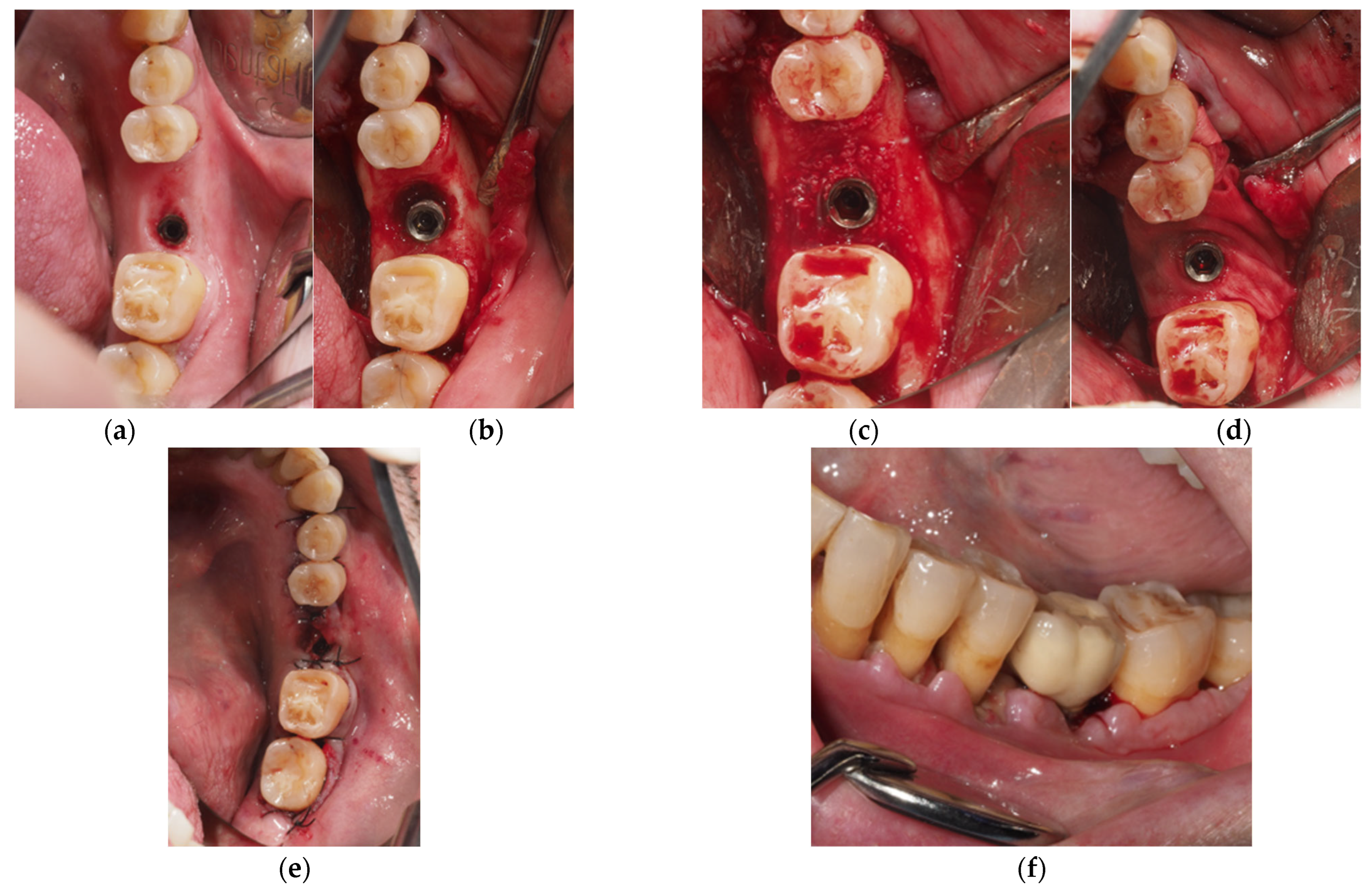
  • Regenerative Approach: Deproteinized bovine bone mineral (DBBM, Geistlich Bio-Oss 0.25–1 mm, Geistlich Biomaterials, Wolhusen, Switzerland) was used to fill the defect, and a resorbable membrane (Geistlich BioGide, Geistlich Biomaterials, Wolhusen, Switzerland) was adapted around the implant neck. Non-resorbable polyamide (Supramid 4/0, St. With, Belgium) and PTFE (Cytoplast 4/0; Osteogenics Biomedical, Lubbock, TX, USA) sutures were used to close the wound, allowing for transmucosal healing (Figure 1).
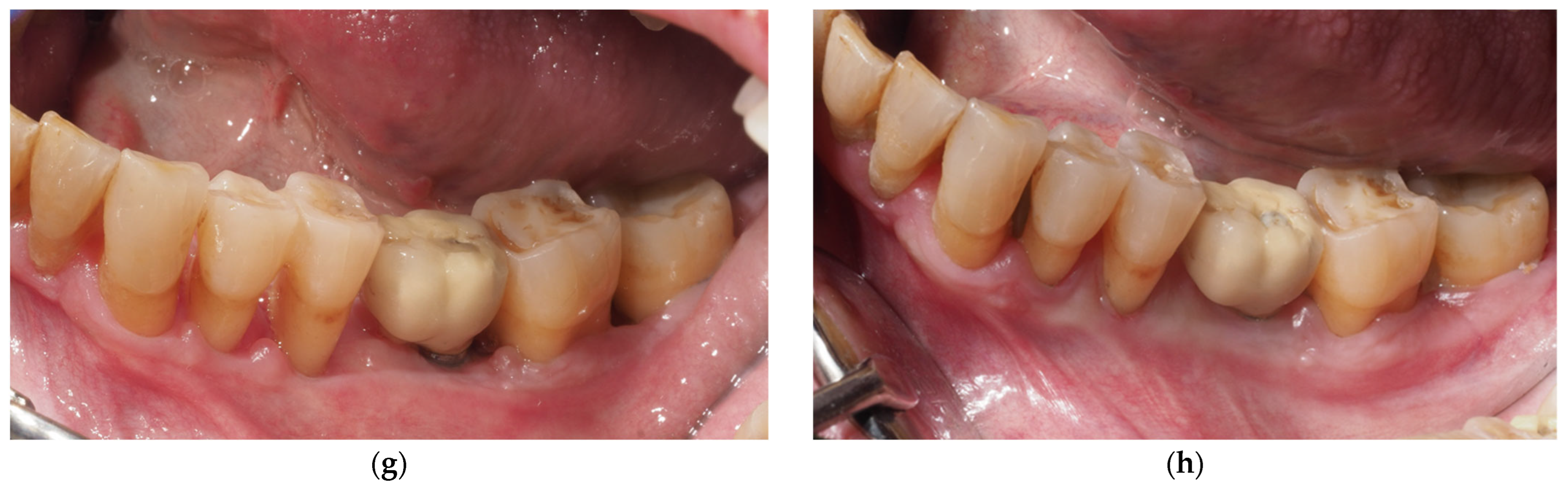
  • Combined Approach: The implant area located in the suprabony compartment of the defect was treated with implantoplasty, and the implant surface located in the infrabony defect was debrided with curettes. Threads were removed and polished with specific burs: an oval-shape tungsten carbide bur (H379 314 023; Komet Dental, Lemgo, Germany) was used to remove the implant threads, and the surface was sequentially polished with two silicon carbide polishers (9618 314 030 and 9608 314 030, Komet Dental, Lemgo, Germany) [40]. In cases of inadequate keratinized tissue or thin biotype, a connective tissue graft was added. Deproteinized bovine bone mineral (DBBM, Geistlich Bio-Oss 0.25–1 mm, Geistlich Bio-materials, Wolhusen, Switzerland) was used to fill the defect, and a resorbable membrane (Geistlich BioGide, Geistlich Biomaterials, Wolhusen, Switzerland) was adapted around the implant neck. Non-resorbable polyamide (Supramid 4/0, St. With, Belgium) and PTFE (Cytoplast 4/0; Osteogenics Biomedical, Lubbock, TX, USA) sutures were used to close the wound, allowing for transmucosal healing (Figure 2).

2.2.4. Postoperative Care

A nonsteroidal anti-inflammatory drug (generally ibuprofen 600 mg every 8 h for 5 days), an antibiotic (generally amoxicillin 750 mg every 8 h for 7 days), and a rescue analgesic (generally paracetamol 1 g every 8 h) were prescribed. Patients were instructed to use of a soft toothbrush and to gently rinse twice daily with a chlorhexidine solution (PerioAid 0.12%; Dentaid, Cerdanyola del Vallés, Barcelona, Spain) for two weeks. The sutures were removed two weeks after surgery.
Postoperative appointments were scheduled for one, two, and four weeks after surgery. Photographs were taken at all follow-up visits. Afterwards, patients were enrolled in regular supportive periodontal and peri-implant therapy (every 3 to 6 months, depending on the risk factors): after retrieving the prosthetic restorations, biofilm and calculus were removed, and teeth and abutments were polished. Residual pockets (probing pocket depth >4 mm) with bleeding on probing were retreated. Probing and mechanical debridement with titanium curettes or an ultrasonic device were performed at the sites surgically treated for peri-implantitis. At each session, patients were instructed on how to maintain adequate oral hygiene.

2.3. Data Collection

A single investigator (AA) collected the following variables from the patients’ clinical, radiological, and photographic records.
Patient-related variables: Sex, age, tobacco use, history of periodontitis, and oral hygiene habits (interproximal brushing, mouthwash).
Implant-related variables: Implant position (anterior: incisor, canine; posterior: premolar and molar) and type of prosthetic restoration (cement-retained, screw-retained, overdenture).
Surgical variables: Date of surgical procedure, surgical technique, number of treated implants, used biomaterials and membranes, and simultaneous connective tissue grafting and implantoplasty.
Postoperative variables (assessed on medical files and in the photographs): Postoperative infection (measured as presence of suppuration, pain, and redness in the wound), membrane exposure, sensory disturbances, and biofilm around sutures.
Wound dehiscence was considered the primary outcome variable. A single clinician (AA) measured this variable with ImageJ® software (Wayne Rasband; Bethesda, MD, USA) in the 7- or 15-day postoperative photograph. The final value was calculated as a coefficient between the length of the incision (measured in mm from the most mesial part to the most distal part of the incision) and the length of dehiscence (measured in mm from the most mesial part to the most distal part of the dehiscence). Dehiscence was considered when this value was higher than zero (Figure 3).

2.4. Statistical Analysis

All data were registered in Stata/IC v15.1 (StataCorp LLC, Lakeway Drive, TX, USA). A descriptive analysis was used to summarize patient-, implant-, and surgical-related variables. Relationships between wound dehiscence and the remaining covariables were explored with Pearson chi-square tests, t-tests, and one-way ANOVA. The 95% confidence intervals (CIs) were calculated for the incidences.

3. Results

A total of 14 patients with 21 implants were analyzed. The main study variables and their possible relation with the occurrence of wound dehiscence can be observed in Table 1. A total of 11 implants (52.4%; 95%CI: 29% to 76%) from nine patients (57.1%; 95%CI = 27% to 87%) had wound dehiscences.
The study population (n = 14) had a mean age of 62.9 years (SD = 10.7), with 57.1% male participants and 42.9% smokers. Periodontitis prevalence was 92.9%, and 14.3% regularly used mouthwash. Most cases (71.4%) were partial restorations (71.4%) located in the posterior area (71.4%).
Patients with mandibular implants (33% vs. 19%; p = 0.003) and who did not use interdental brushes (50% vs. 7.1%; p = 0.008) had a significantly higher incidence of dehiscences. Age, gender, smoking status, periodontitis history, mouthwash use, restoration type, and implant location showed no significant associations with the occurrence of dehiscences.
No postoperative infections or sensory alterations were registered. Table 2 provides a detailed evaluation of the surgical procedures and their outcomes.
Among postoperative factors, only membrane exposure showed a statistically significant association with dehiscence (p = 0.034). All cases with membrane exposure (19.1%) had associated dehiscences. In patients without exposure (80.9%), dehiscences were still found in 33.3% of cases.
Two patients had implants treated by both surgical techniques, regenerative and combined. The patient-based incidence of dehiscence for regenerative procedures was 50% (3 from 6 patients) and for the combined technique was 60% (6 from 10 patients). The total patient-based incidence was 64.3% (9 from 14 patients).
Implantoplasty (p = 0.284), connective tissue grafting (p = 0.525), and biofilm accumulation around the sutures (p = 0.130) showed no significant association with the development of dehiscences. There were no differences in the number, percentage, or millimeters of dehiscence according to the type of surgery (Table 3).

4. Discussion

The results of this retrospective study provide clinically useful information on postoperative complications occurring after peri-implant reconstructive therapy. Postoperative soft tissue dehiscences were the most frequent complication affecting 57.1% (95%CI = 27% to 87%) of patients and 52.4% (95%CI = 29% to 76%) of implants. Several reports are in line with these results, showing that wound dehiscences are common and can occur in approximately half of cases [35,36,41]. This complication might have an important impact on treatment outcomes, especially when membranes are used. Indeed, the scientific literature has shown that early membrane exposure can significantly compromise the regenerative outcome and increase the risk of secondary complications [42]. In the present report, membrane exposure was found in 19.1% of cases and showed a clear correlation with the presence of dehiscences (p = 0.034).
Peri-implant surgery seems to have a higher risk of would dehiscence in comparison with other procedures such a ridge augmentation (16.9%) and regenerative periodontal surgery (15%) [43,44]. This might be partially explained by the defect anatomy, since peri-implant defects usually have a supracrestal component [20,21] that hampers wound closure.
The dimension of the dehiscence is an important variable to consider since it provides objective data on the magnitude of this complication. In the present study, dehiscences had a mean length of 7.6 mm (SD = 2.0) and affected 21.4% of the total incision length. Previous authors have reported similar data, with mean lengths ranging from 4 to 7 mm, typically accounting for 15% to 25% of the total incision length [45,46]. It is notable that, despite the high frequency of dehiscences, no cases of postoperative infections were found. The absence of suppuration or infectious signs/symptoms might be related to the applied antibacterial postoperative protocols (prophylactic antibiotic administration, use of postoperative chlorhexidine mouthrinses, and mechanical cleaning of the wound with a soft toothbrush). These measures seemed to be sufficient, even though 57.1% of patients had visible biofilm in the surgical wound area. Although the association between the presence of biofilm and dehiscence did not reach statistical significance (p = 0.130), Tonetti et al. [47] emphasize that effective plaque control is critical during the initial weeks of healing and should be considered a key factor for successful regeneration.
Complications occurred in both surgical techniques. Regenerative approaches had a dehiscence rate of 50%, while combined approaches seemed to be more prone to this complication (60% of patients). The reported rates seem to vary considerably. While a recently published clinical trial observed that only 19% of implants with a regenerative submerged approach had dehiscences [35], Roos-Jansaker et al. found a much higher figure (62.5% of implants) after 2 weeks of healing using the same technique [48]. Factors that may influence the appearance of wound dehiscence may include flap tension [49], periosteal release incisions [50], use of membrane [40,51,52], suturing technique [53], or submerged or non-submerged healing [54]. The surgical treatment techniques described in the studies published to date are very heterogeneous and may justify the different reported incidences. In our sample, non-submerged healing that allowed for the immediate placement of the prosthesis was employed and periosteal releasing incisions were performed to reduce flap tension. One of the main advantages of this option is that the patient recovers their chewing ability and quality of life immediately after surgery. A study published by our team [55] found that regenerative peri-implant surgery negatively affected the patients’ quality of life. Thus, the immediate placement of the prosthesis may improve this outcome. It is unclear if a submerged approach provides better regenerative outcomes, and the literature on this topic is sparce [54,56,57,58]. On the one hand, a submerged approach may be more beneficial in terms of radiographic bone fill, but other variables such as comfort and impact on the patient’s quality of life should also be considered. In our sample, patients undergoing mandibular surgeries (p = 0.003) and that did not use interdental brushes (50% vs. 7.1%; p = 0.008) were significantly more prone to this complication. Mandibular sites seem to be anatomically susceptible to soft tissue dehiscences, probably due to the presence of a thinner keratinized mucosa, and restricted vascularization, all of which might affect wound stability and healing [59]. Furthermore, the absence of interdental hygiene promotes biofilm buildup and prolonged inflammation, which may contribute to mucosal recession [60]. Heitz-Mayfield et al. [61] and Figuero et al. [23] emphasized that adequate oral hygiene, including the interproximal areas, is paramount for the success of peri-implant reconstructive therapy. Other variables that might affect the complication rate, such as operation time, flap design, or prosthetic configurations, were not assessed in the present study and should be evaluated in future research.
Mechanical debridement with instruments such as titanium curettes and ultrasonic scalers may significantly modify implant surface topography [62,63]. These surface alterations might damage guided bone regeneration outcomes, as indicated by Kumar et al. (2024) [64] and Alabbad et al. (2025) [65], illustrating the necessity for careful instrument selection and procedural control.
In patients with a thin gingival biotype, particularly in the mandible, a staged approach that includes an initial soft tissue graft (e.g., CTG or tunnel method) followed by delayed bone augmentation might improve wound stability and the esthetic results [66]. Hadzik et al. (2023) [67] observed that connective tissue grafting prior to implant placement resulted in a stable increase in tissue thickness, promoting long-term peri-implant health.
The absence of an association between the occurrence of complications and several factors traditionally considered risk factors is also noteworthy. In our sample, tobacco consumption, systemic conditions, and history of periodontitis were not related to an increased risk of complications. This is probably related to the small sample size. Indeed, previous reports have suggested that smoking negatively affects wound healing and increases the risk of postoperative complications by 2.1 to 2.6 times [68]. Similarly, systemic conditions such as poorly controlled diabetes mellitus can significantly impair wound healing in regenerative procedures [69]. Thus, the results of the present study should be interpreted with caution.
The high prevalence of periodontal disease (92.9%) in advanced stages (46.2% in stage 3 and 38.5% in stage 4) in our sample suggests that the included patients had a significant involvement of the periodontal tissues. The scientific literature has established that the history of severe periodontitis is a predictor for the development and progression of peri-implantitis, with an odds ratio of 4.7 [5].
This study has several limitations that should be considered. As mentioned previously, the limited sample size—14 patients and 21 implants—limits the statistical power of the analysis and can affect the ability to detect significant associations between pre- and postoperative variables and the development of complications. Also, the use of both regenerative and combined approaches in a small number of patients may compromise the internal validity, and this does not allow us to distinguish the effects attributed to each modality Moreover, the lack of a control group and the retrospective nature of the study introduce inherent limitations, such as the restricted availability of clinical variables (e.g., postoperative pain and edema could not be assessed) and potential biases in data collection. To mitigate this, the researchers used standardized photographs and performed measurements using specific software. Additionally, the fact that all patients were treated in a university-based dental hospital may further restrict the applicability of the results to other clinical contexts. Despite these limitations, we believe that this study provides clinically relevant information to the literature, particularly given the scarcity of large patient series with long-term follow-up. For all of the abovementioned reasons, the present outcomes should be interpreted with caution. Large-sample randomized clinical trials with a control group and a longer follow-up are required to confirm the reported findings and to determine the effect of these complications on the implant success rates.

5. Conclusions

More than half of patients (57.1%; 95%CI = 27–87%) with peri-implantitis who undergo surgical reconstructive therapy may present wound dehiscences. This complication is associated with mandibular location, poor interproximal hygiene, and membrane exposure. The findings of this study should be interpreted with caution due to its retrospective design, the lack of a control group, and the small sample size. Future large-sample randomized clinical trials with a control group and a longer follow-up are required to confirm the reported findings.

Author Contributions

A.A.: study concept/design, data collection and curation, and drafting of the manuscript. X.C.-B.: study concept/design and data collection. R.F.: study concept/design, data interpretation, funding acquisition, critical review, and approval of the manuscript. E.V.-C.: study concept/design, data interpretation, supervision, critical review, and approval of the manuscript. A.S.-T.: study concept/design and data analysis and interpretation, statistics, critical review, and approval of the manuscript. M.G.-G.: study concept/design, data analysis/interpretation, supervision, and drafting and approval of the manuscript. All authors have read and agreed to the published version of the manuscript.

Funding

This research received no external funding.

Institutional Review Board Statement

The study protocol was approved by the Ethics Committee (CEIm) of the University of Barcelona Dental Hospital (Ref.: 41/2024; Approval date: 13 February 12025), and the study was conducted in accordance with the Declaration of Helsinki [70] on human studies. The patients were given full information about the surgical procedures and signed a written informed consent.

Informed Consent Statement

Patient consent was waived because we only reviewed the medical files, photographs, and radiographic images of the patients anonymously, and there was no intervention or investigation of humans directly.

Data Availability Statement

The original contributions presented in this study are included in the article. Further inquiries can be directed to the corresponding author.

Acknowledgments

This work was partially funded through a research agreement between the University of Barcelona and Dentaid SL (Càtedra UB-DENTAID).

Conflicts of Interest

The authors have no financial or other interests directly related to the products or information mentioned in this paper. Dr. Ahmad Alahmari and Dr. Xavier Costa-Berenguer report no conflicts of interest. Dr. Rui Figueiredo reports grants, personal fees, and non-financial support from MozoGrau (Valladolid, Spain), Avinent (Santpedor, Spain), Inibsa Dental (Lliçà de Vall, Spain), and Dentaid SL (Cerdanyola del Vallés, Spain), non-financial support from Nobel Biocare (Zürich, Switzerland), and personal fees from Geistlich Pharma AG (Wolhusen, Switzerland), BioHorizons Iberica (Madrid, Spain), Araguaney Dental (Barcelona, Spain), Septodont (Saint-Maur-des-fossés, France), Dentaid SL (Cerdanyola del Vallés, Spain), and Laboratorios Silanes (Mexico City, Mexico) outside of the submitted work. Dr. Figueiredo has also participated as a principal investigator in a randomized clinical trial sponsored by Mundipharma (Cambridge, UK), and in another clinical trial as a co-investigator for Menarini Richerche (Florence, Italy). Dr. Eduard Valmaseda-Castellón reports grants, personal fees, and non-financial support from MozoGrau (Valladolid, Spain), Avinent (Santpedor, Spain), Inibsa Dental (Lliçà de Vall, Spain), and Dentaid SL (Cerdanyola del Vallés, Spain), non-financial support from Nobel Biocare (Zürich, Switzerland), and personal fees from Laboratorios Silanes (Mexico City, Mexico) outside of the submitted work. Dr. Valmaseda-Castellón has also participated as an investigator in randomized clinical trials sponsored by Geistlich Pharma AG (Wolhusen, Switzerland), Mundipharma (Cambridge, UK), and Menarini Richerche (Florence, Italy). Dra. Alba Sanchez-Torres reports grants, personal fees, and nonfinancial support from MozoGrau (Valladolid, Spain), Mundipharma (Cambridge, UK), and Unither Pharmaceuticals (Paris, France) outside of the submitted work. Dra. Marta García García reports personal fees and non-financial support from Global Medical Implants (Barcelona, Spain) and reports grants, personal fees, and non-financial support from Dentaid SL (Cerdanyola del Vallés, Spain).

References

  1. Block, M.S. Dental Implants: The Last 100 Years. J. Oral Maxillofac. Surg. 2018, 76, 11–26. [Google Scholar] [CrossRef] [PubMed]
  2. Atieh, M.A.; AlAli, F.; Alsabeeha, N.H.M. Outcome of supportive peri-implant therapy on the rates of peri-implant diseases and marginal bone loss: A systematic review and meta-analysis. Quintessence Int. 2021, 52, 122–131. [Google Scholar] [PubMed]
  3. Atieh, M.A.; Almutairi, Z.; Amir-Rad, F.; Koleilat, M.; Tawse-Smith, A.; Ma, S.; Lin, L.; Alsabeeha, N.H. A retrospective analysis of biological complications of dental implants. Int. J. Dent. 2022, 2022, 1545748. [Google Scholar] [CrossRef] [PubMed]
  4. Heitz-Mayfield, L.J.A.; Salvi, G.E. Peri-implant mucositis. J. Periodontol. 2018, 89, S257–S266. [Google Scholar] [CrossRef] [PubMed]
  5. Berglundh, T.; Armitage, G.; Araujo, M.G.; Avila-Ortiz, G.; Blanco, J.; Camargo, P.M.; Chen, S.; Cochran, D.; Derks, J.; Figuero, E.; et al. Peri-implant diseases and conditions: Consensus report of workgroup 4 of the 2017 World Workshop on the Classification of Periodontal and Peri-Implant Diseases and Conditions. J. Periodontol. 2018, 89 (Suppl. S1), S237–S248. [Google Scholar] [CrossRef] [PubMed]
  6. Schwarz, F.; Derks, J.; Monje, A.; Wang, H. Peri-implantitis. J. Periodontol. 2018, 89 (Suppl. S1), S267–S290. [Google Scholar] [CrossRef] [PubMed]
  7. Berglundh, T.; Mombelli, A.; Schwarz, F.; Derks, J. Etiology, pathogenesis and treatment of peri-implantitis: A European perspective. Periodontol. 2000 2024, in press. [CrossRef] [PubMed]
  8. Derks, J.; Schaller, D.; Håkansson, J.; Wennström, J.L.; Tomasi, C.; Berglundh, T. Effectiveness of implant therapy analyzed in a swedish population. J. Dent. Res. 2016, 95, 43–49. [Google Scholar] [CrossRef] [PubMed]
  9. Mir-Mari, J.; Mir-Orfila, P.; Figueiredo, R.; Valmaseda-Castellón, E.; Gay-Escoda, C. Prevalence of peri-implant diseases. A cross-sectional study based on a private practice environment. J. Clin. Periodontol. 2012, 39, 490–494. [Google Scholar] [CrossRef] [PubMed]
  10. García-García, M.; Mir-Mari, J.; Figueiredo, R.; Valmaseda-Castellón, E. Probing single-tooth dental implants with and without prostheses: A cross-sectional study comparing healthy and peri-implant mucositis sites. J. Clin. Periodontol. 2021, 48, 581–589. [Google Scholar] [CrossRef] [PubMed]
  11. García-García, M.; Mir-Mari, J.; Benic, G.I.; Figueiredo, R.; Valmaseda-Castellón, E. Accuracy of periapical radiography in assessing bone level in implants affected by peri-implantitis: A cross-sectional study. J. Clin. Periodontol. 2016, 43, 85–91. [Google Scholar] [CrossRef] [PubMed]
  12. Guarnieri, R.; Reda, R.; Zanza, A.; Miccoli, G.; Di Nardo, D.; Testarelli, L. Can peri-implant marginal bone loss progression and a-MMP-8 be considered indicators of the subsequent onset of peri-implantitis? A 5-year study. Diagnostics 2022, 12, 2599. [Google Scholar] [CrossRef] [PubMed]
  13. Rebeiz, T.; Nasr, L.; Kassir, A.R.; Menassa, G.; Chakar, C. Assessment of the association between the Implant Disease Risk Assessment (IDRA) tool and peri-implantitis: A retrospective cohort study with up to 8 years of follow-up. Int. J. Oral Maxillofac. Surg. 2024, 53, 845–852. [Google Scholar] [CrossRef] [PubMed]
  14. Heitz-Mayfield, L.J.A.; Heitz, F.; Lang, N.P. Implant Disease Risk Assessment IDRA–a tool for preventing peri-implant disease. Clin. Oral Implant. Res. 2020, 31, 397–403. [Google Scholar] [CrossRef] [PubMed]
  15. Dreyer, H.; Grischke, J.; Tiede, C.; Eberhard, J.; Schweitzer, A.; Toikkanen, S.E.; Glöckner, S.; Krause, G.; Stiesch, M. Epidemiology and risk factors of peri-implantitis: A systematic review. J. Periodontal Res. 2018, 53, 657–681. [Google Scholar] [CrossRef] [PubMed]
  16. Giok, K.C.; Veettil, S.K.; Menon, R.K. Risk factors for peri-implantitis: An umbrella review of meta-analyses of observational studies and assessment of biases. J. Dent. 2024, 146, 105065. [Google Scholar] [CrossRef] [PubMed]
  17. Romandini, M.; Laforí, A.; Pedrinaci, I.; Baima, G.; Ferrarotti, F.; Lima, C.; Holtzman, L.P.; Aimetti, M.; Cordaro, L.; Sanz, M. Effect of sub-marginal instrumentation before surgical treatment of peri-implantitis: A multi-centre randomized clinical trial. J. Clin. Periodontol. 2022, 49, 1334–1345. [Google Scholar] [CrossRef] [PubMed]
  18. Ramanauskaite, A.; Daugela, P.; Juodzbalys, G. Treatment of peri-implantitis: Meta-analysis of findings in a systematic literature review and novel protocol proposal. Quintessence Int. 2016, 47, 379–393. [Google Scholar] [PubMed]
  19. Karlsson, K.; Derks, J.; Håkansson, J.; Wennström, J.L.; Petzold, M.; Berglundh, T. Interventions for peri-implantitis and their effects on further bone loss: A retrospective analysis of a registry-based cohort. J. Clin. Periodontol. 2019, 46, 872–879. [Google Scholar] [CrossRef] [PubMed]
  20. Schwarz, F.; Herten, M.; Sager, M.; Bieling, K.; Sculean, A.; Becker, J. Comparison of naturally occurring and ligature-induced peri-implantitis bone defects in humans and dogs. Clin. Oral Implant. Res. 2007, 18, 161–170. [Google Scholar] [CrossRef] [PubMed]
  21. Monje, A.; Pons, R.; Insua, A.; Nart, J.; Wang, H.L.; Schwarz, F. Morphology and severity of peri-implantitis bone defects. Clin. Implant. Dent. Relat. Res. 2019, 21, 635–643. [Google Scholar] [CrossRef] [PubMed]
  22. Schwarz, F.; Jepsen, S.; Obreja, K.; Galarraga-Vinueza, M.E.; Ramanauskaite, A. Surgical therapy of peri-implantitis. Periodontol. 2000 2022, 88, 145–181. [Google Scholar] [CrossRef] [PubMed]
  23. Figuero, E.; Graziani, F.; Sanz, I.; Herrera, D.; Sanz, M. Management of peri-implant mucositis and peri-implantitis. Periodontol. 2000 2014, 66, 255–273. [Google Scholar] [CrossRef] [PubMed]
  24. Serino, G.; Turri, A. Outcome of surgical treatment of peri-implantitis: Results from a 2-year prospective clinical study in humans. Clin. Oral Implant. Res. 2011, 22, 1214–1220. [Google Scholar] [CrossRef] [PubMed]
  25. Romeo, E.; Lops, D.; Chiapasco, M.; Ghisolfi, M.; Vogel, G. Therapy of peri-implantitis with resective surgery. A 3-year clinical trial on rough screw-shaped oral implants. Part II: Radiographic outcome. Clin. Oral Implant. Res. 2007, 18, 179–187. [Google Scholar] [CrossRef] [PubMed]
  26. Romeo, E.; Ghisolfi, M.; Murgolo, N.; Chiapasco, M.; Lops, D.; Vogel, G. Therapy of peri-implantitis with resective surgery: A 3-year clinical trial on rough screw-shaped oral implants. Part I: Clinical outcome. Clin. Oral Implant. Res. 2005, 16, 9–18. [Google Scholar]
  27. Lang, N.P.; Berglundh, T. Periimplant diseases: Where are we now? Consensus of the Seventh European Workshop on Periodontology. J. Clin. Periodontol. 2011, 38, 178–181. [Google Scholar] [PubMed]
  28. Carcuac, O.; Derks, J.; Abrahamsson, I.; Wennström, J.L.; Berglundh, T. Risk for recurrence of disease following surgical therapy of peri-implantitis—A prospective longitudinal study. Clin. Oral Implant. Res. 2020, 31, 1072–1077. [Google Scholar] [CrossRef] [PubMed]
  29. Serino, G.; Turri, A.; Lang, N.P. Maintenance therapy in patients following the surgical treatment of peri-implantitis: A 5-year follow-up study. Clin. Oral Implant. Res. 2015, 26, 950–956. [Google Scholar] [CrossRef] [PubMed]
  30. Romandini, M.; Bougas, K.; Alibegovic, L.; Hosseini, S.; Carcuac, O.; Berglundh, T.; Derks, J. Long-term outcomes and prognostic factors of surgical treatment of peri-implantitis—A retrospective study. Clin. Oral Implant. Res. 2023, 35, 321–329. [Google Scholar] [CrossRef] [PubMed]
  31. Tomasi, C.; Regidor, E.; Ortiz-Vigón, A.; Derks, J. Efficacy of reconstructive surgical therapy at peri-implantitis-related bone defects. A systematic review and meta-analysis. J. Clin. Periodontol. 2019, 46, 340–356. [Google Scholar] [CrossRef] [PubMed]
  32. González Regueiro, I.; Martínez Rodriguez, N.; Barona Dorado, C.; Sanz-Sánchez, I.; Montero, E.; Ata-Ali, J.; Duarte, F.; Martínez-González, J.M. Surgical approach combining implantoplasty and reconstructive therapy with locally delivered antibiotic in the treatment of peri-implantitis: A prospective clinical case series. Clin. Implant. Dent. Relat. Res. 2021, 23, 864–873. [Google Scholar] [CrossRef] [PubMed]
  33. Schwarz, F.; John, G.; Schmucker, A.; Sahm, N.; Becker, J. Combined surgical therapy of advanced peri-implantitis evaluating two methods of surface decontamination: A 7-year follow-up observation. J. Clin. Periodontol. 2017, 44, 337–342. [Google Scholar] [CrossRef] [PubMed]
  34. Herrera, D.; Berglundh, T.; Schwarz, F.; Chapple, I.; Jepsen, S.; Sculean, A.; Kebschull, M.; Papapanou, P.N.; Tonetti, M.S.; Sanz, M.; et al. Prevention and treatment of peri-implant diseases—The EFP S3 level clinical practice guideline. J. Clin. Periodontol. 2023, 50, 4–76. [Google Scholar] [CrossRef] [PubMed]
  35. Regidor, E.; Ortiz-Vigón, A.; Romandini, M.; Dionigi, C.; Derks, J.; Sanz, M. The adjunctive effect of a resorbable membrane to a xenogeneic bone replacement graft in the reconstructive surgical therapy of peri-implantitis: A randomized clinical trial. J. Clin. Periodontol. 2023, 50, 765–783. [Google Scholar] [CrossRef] [PubMed]
  36. Roos-Jansåker, A.M.; Lindahl, C.; Persson, G.R.; Renvert, S. Long-term stability of surgical bone regenerative procedures of peri-implantitis lesions in a prospective case-control study over 3 years. J. Clin. Periodontol. 2011, 38, 590–597. [Google Scholar] [CrossRef] [PubMed]
  37. Roos-Jansåker, A.; Persson, G.R.; Lindahl, C.; Renvert, S. Surgical treatment of peri-implantitis using a bone substitute with or without a resorbable membrane: A 5-year follow-up. J. Clin. Periodontol. 2014, 41, 1108–1114. [Google Scholar] [CrossRef] [PubMed]
  38. Jepsen, S.; Berglundh, T.; Genco, R.; Aass, A.M.; Demirel, K.; Derks, J.; Figuero, E.; Giovannoli, J.L.; Goldstein, M.; Lambert, F.; et al. Primary prevention of peri-implantitis: Managing peri-implant mucositis. J. Clin. Periodontol. 2015, 42, 152–157. [Google Scholar] [CrossRef] [PubMed]
  39. Renvert, S.; Polyzois, I.N. Clinical approaches to treat peri-implant mucositis and peri-implantitis. Periodontol. 2000 2015, 68, 369–404. [Google Scholar] [CrossRef] [PubMed]
  40. Costa-Berenguer, X.; García-García, M.; Sánchez-Torres, A.; Sanz-Alonso, M.; Figueiredo, R.; Valmaseda-Castellón, E. Effect of implantoplasty on fracture resistance and surface roughness of standard diameter dental implants. Clin. Oral Implant. Res. 2018, 29, 46–54. [Google Scholar] [CrossRef] [PubMed]
  41. Schwarz, F.; Sahm, N.; Schwarz, K.; Becker, J. Impact of defect configuration on the clinical outcome following surgical regenerative therapy of peri-implantitis. J. Clin. Periodontol. 2010, 37, 449–455. [Google Scholar] [CrossRef] [PubMed]
  42. Urban, I.; Sanz-Sánchez, I.; Monje, A.; Montero, E. Complications and treatment errors in peri-implant hard tissue management. Periodontol. 2000 2023, 92, 278–298. [Google Scholar] [CrossRef] [PubMed]
  43. Rojas, M.A.; Marini, L.; Pilloni, A.; Sahrmann, P. Early wound healing outcomes after regenerative periodontal surgery with enamel matrix derivatives or guided tissue regeneration: A systematic review. BMC Oral Health 2019, 19, 76. [Google Scholar] [CrossRef] [PubMed]
  44. Lim, G.; Lin, G.-H.; Monje, A.; Chan, H.-L.; Wang, H.-L. Wound healing complications following guided bone regeneration for ridge augmentation: A systematic review and meta-analysis. Int. J. Oral Maxillofac. Implant. 2018, 33, 41–50. [Google Scholar] [CrossRef] [PubMed]
  45. Schwarz, F.; Schmucker, A.; Becker, J. Efficacy of alternative or adjunctive measures to conventional treatment of peri-implant mucositis and peri-implantitis: A systematic review and meta-analysis. Int. J. Implant. Dent. 2015, 1, 22. [Google Scholar] [CrossRef] [PubMed]
  46. Roccuzzo, M.; Gaudioso, L.; Lungo, M.; Dalmasso, P. Surgical therapy of single peri-implantitis intrabony defects, by means of deproteinized bovine bone mineral with 10% collagen. J. Clin. Periodontol. 2016, 43, 311–318. [Google Scholar] [CrossRef] [PubMed]
  47. Tonetti, M.S.; Chapple, I.L.C.; Jepsen, S.; Sanz, M. Primary and secondary prevention of periodontal and peri-implant diseases. J. Clin. Periodontol. 2015, 42, S1–S4. [Google Scholar] [CrossRef] [PubMed]
  48. Roos-Jansåker, A.M.; Renvert, H.; Lindahl, C.; Renvert, S. Submerged healing following surgical treatment of peri-implantitis: A case series. J. Clin. Periodontol. 2007, 34, 723–727. [Google Scholar] [CrossRef] [PubMed]
  49. Burkhardt, R.; Lang, N.P. Role of flap tension in primary wound closure of mucoperiosteal flaps: A prospective cohort study. Clin. Oral Implant. Res. 2010, 21, 50–54. [Google Scholar] [CrossRef] [PubMed]
  50. Park, J.C.; Kim, C.S.; Choi, S.H.; Cho, K.S.; Chai, J.K.; Jung, U.W. Flap extension attained by vertical and periosteal-releasing incisions: A prospective cohort study. Clin. Oral Implant. Res. 2012, 23, 993–998. [Google Scholar] [CrossRef] [PubMed]
  51. Roos-Jansåker, A.M.; Renvert, H.; Lindahl, C.; Renvert, S. Surgical treatment of peri-implantitis using a bone substitute with or without a resorbable membrane: A prospective cohort study. J. Clin. Periodontol. 2007, 34, 625–632. [Google Scholar] [CrossRef] [PubMed]
  52. Roos-Jansåker, A.M.; Lindahl, C.; Renvert, H.; Renvert, S. Nine- to fourteen-year follow-up of implant treatment. Part I: Implant loss and associations to various factors. J. Clin. Periodontol. 2006, 33, 283–289. [Google Scholar] [CrossRef] [PubMed]
  53. Burkhardt, R.; Lang, N.P. Influence of suturing on wound healing. Periodontol. 2000 2015, 68, 270–281. [Google Scholar] [CrossRef] [PubMed]
  54. Wen, S.C.; Sabri, H.; Dastouri, E.; Huang, W.X.; Barootchi, S.; Wang, H.L. Submerged vs nonsubmerged reconstructive approach for surgical treatment of peri-implantitis: Reanalysis of two prospective clinical studies. Int. J. Oral Maxillofac. Implant. 2024, 39, 526–536. [Google Scholar] [CrossRef] [PubMed]
  55. Camps-Font, O.; Pérez-Beltrán, I.; Fornés-Nieto, V.; González-Barnadas, A.; Costa-Berenguer, X.; García-García, M.; Figueiredo, R.; Valmaseda-Castellón, E. Patient-centered outcomes after surgical treatment of peri-implantitis: A prospective clinical study. Med. Oral Patol. Oral Cir. Bucal 2023, 28, e72–e80. [Google Scholar] [CrossRef] [PubMed]
  56. Wen, S.C.; Barootchi, S.; Wang, H.L.; Huang, W.X. Non-submerged reconstructive approach for peri-implantitis osseous defect with removal of implant crowns: One-year outcomes of a prospective case series study. J. Periodontol. 2022, 93, 1250–1261. [Google Scholar] [CrossRef] [PubMed]
  57. Wen, S.C.; Barootchi, S.; Huang, W.X.; Wang, H.L. Surgical reconstructive treatment for infraosseous peri-implantitis defects with a submerged healing approach: A prospective controlled study. J. Periodontol. 2022, 93, 195–207. [Google Scholar] [CrossRef] [PubMed]
  58. Monje, A.; Pons, R.; Roccuzzo, A.; Salvi, G.E.; Nart, J. Reconstructive therapy for the management of peri-implantitis via submerged guided bone regeneration: A prospective case series. Clin. Implant. Dent. Relat. Res. 2020, 22, 342–350. [Google Scholar] [CrossRef] [PubMed]
  59. Sanz-Martín, I.; Regidor, E.; Cosyn, J.; Wiedemeier, D.B.; Thoma, D.S. Buccal soft tissue dehiscence defects at dental implants—Associated factors and frequency of occurrence: A systematic review and meta-analysis. Clin. Oral Implant. Res. 2022, 33, 109–124. [Google Scholar] [CrossRef] [PubMed]
  60. Ng, E.; Lim, L.P. An Overview of Different Interdental Cleaning Aids and Their Effectiveness. Dent. J. 2019, 7, 56. [Google Scholar] [CrossRef] [PubMed]
  61. Heitz-Mayfield, L.; Mombelli, A. The therapy of peri-implantitis: A systematic review. Int. J. Oral Maxillofac. Implant. 2014, 29, 325–345. [Google Scholar] [CrossRef] [PubMed]
  62. Eun, S.-M.; Son, K.; Hwang, S.-M.; Son, Y.-T.; Kim, Y.-G.; Suh, J.-Y.; Hwang, J.H.; Kwon, S.-M.; Lee, J.H.; Kim, H.D.; et al. The Impact of Mechanical Debridement Techniques on Titanium Implant Surfaces: A Comparison of Sandblasted, Acid-Etched, and Femtosecond Laser-Treated Surfaces. J. Funct. Biomater. 2023, 14, 502. [Google Scholar] [CrossRef] [PubMed]
  63. Sahrmann, P.; Winkler, S.; Gubler, A.; Attin, T. Assessment of implant surface and instrument insert changes due to instrumentation with different tips for ultrasonic-driven debridement. BMC Oral Health 2021, 21, 25. [Google Scholar] [CrossRef] [PubMed]
  64. Kumar, S.; Yewale, M.; Parthasarathi, N.L.; Balasundaram, R.; Gopalkrishna, P.; Bhat, S.G. Evaluation of the Loss of Surface Roughness Following the Use of Four Different Instruments for Mechanical Debridement of Dental Implants: An In-vitro Pilot Study. J. Bio-Tribo-Corros. 2024, 10, 77. [Google Scholar] [CrossRef]
  65. Alabbad, M.; Silikas, N.; Thomas, A. Effect of mechanical instrumentation on titanium implant surface properties. Dent. Mater. 2025, 41, 383–390. [Google Scholar] [CrossRef] [PubMed]
  66. Allen, E.P. Soft tissue grafting with the tunnel technique in the mandibular anterior: Myths and realities. J. Esthet. Restor. Dent. 2021, 33, 152–157. [Google Scholar] [CrossRef] [PubMed]
  67. Hadzik, J.; Błaszczyszyn, A.; Gedrange, T.; Dominiak, M. Soft-Tissue Augmentation around Dental Implants with a Connective Tissue Graft (CTG) and Xenogeneic Collagen Matrix (CMX)—5-Year Follow-Up. J. Clin. Med. 2023, 12, 924. [Google Scholar] [CrossRef] [PubMed]
  68. Renvert, S.; Polyzois, I. Risk indicators for peri-implant mucositis: A systematic literature review. J. Clin. Periodontol. 2015, 42, S172–S186. [Google Scholar] [CrossRef] [PubMed]
  69. Romanos, G.E.; Javed, F.; Delgado-Ruiz, R.A.; Calvo-Guirado, J.L. Peri-implant Diseases. Dent. Clin. N. Am. 2015, 59, 157–178. [Google Scholar] [CrossRef] [PubMed]
  70. World Medical Association. World Medical Association Declaration of Helsinki: Ethical principles for medical research involving human subjects. JAMA 2013, 310, 2191–2194. [Google Scholar] [CrossRef] [PubMed]
Figure 1. Regenerative approach for peri-implantitis. (a) Preoperative clinical view showing peri-implant inflammation and soft tissue recession. (b) Flap elevation reveals a circumferential bony defect around the implant. (c) Bone defect filled with deproteinized bovine bone mineral (DBBM). (d) Resorbable membrane adapted around the implant neck. (e) Primary closure achieved with internal horizontal mattress and simple interrupted sutures. (fh) Sequential healing at 7, 14, and 30 days postoperatively, demonstrating progressive soft tissue maturation.
Applsci 15 08040 g001aApplsci 15 08040 g001b
Figure 2. Combined regenerative approach and implantoplasty (a) Preoperative presentation of peri-implantitis affecting implants placed in the maxillary lateral incisors. (b) Flap elevation exposes a crater-like bony defect. (c) Implantoplasty performed on the supra-osseous implant surface. (d) Xenograft placed in the defect. (e) Collagen membrane positioned over the graft material. (f) Wound closure using internal horizontal mattress and interrupted sutures. (g,h) Postoperative follow-up at 7 and 14 days showing uneventful healing and reduced inflammation.
Applsci 15 08040 g002aApplsci 15 08040 g002b
Figure 3. Wound dehiscence variable. The mesio-distal length of the incision (red line) and of the dehiscence (green line).
Applsci 15 08040 g003
Table 1. Patient and implant-related variables (preoperative). N = number. Bold values indicate statistically significant differences.
VariableTotal N (%) or Mean (SD)With Dehiscence
N (%)
Without Dehiscence
N (%)
p-Value
Age (years)62.9 (10.7)63.4 (10.3)64.1 (9.6)0.868
Sex
 Female6 (42.9)3 (21.4)3 (21.4)0.640
 Male8 (57.1)5 (35.7)3 (21.4)
Tobacco use (yes)6 (42.9)4 (28.6)2 (14.3)0.533
Periodontitis (yes)13 (92.9)8 (57.2)5 (35.7)0.231
Interdental brushes (yes)6 (42.9)1 (7.1)5 (35.7)0.008
Mouthwash use (yes)2 (14.3)1 (7.1)1 (7.1)0.825
Restoration (multiple)10 (71.4)6 (42.9)4 (28.6)0.733
Implant position (anterior)13 (61.9)7 (33.3)6 (28.6)0.608
Location (maxilla)14 (66.7)4 (19.0)10 (47.6)0.003
Table 2. Intra/postoperative variables. Bold values indicate statistically significant differences.
VariableTotal N (%) or
Mean (SD)
With Dehiscence
N (%)
Without Dehiscence
N (%)
p-Value
Implantoplasty (Yes)13 (61.9)8 (38.1)5 (23.8)0.284
CTG (Yes)5 (23.8)2 (9.5)3 (14.3)0.525
Membrane Exposure (Yes)4 (19.1)4 (19.1)0 (0)0.034
Biofilm in Sutures (Yes)12 (57.1)8 (38.1)4 (19.1)0.130
Incision Length (mm)17.7 (5.2)19.2 (4.8)16.0 (5.4)0.160
Table 3. Number, percentage, and millimeters of dehiscence according to the type of surgery. N = number; SD = standard deviation.
Type of SurgeryIncisionDehiscence
N (%)(mm)N (%)(mm)(%)
Mean (SD)Mean (SD)
Combined13 (61.9)18.7 (5.1)8 (61.5)7.9 (2.2)25.7 (22.5)
Regenerative8 (38.1)15.9 (5.2)3 (37.5)6.8 (0.5)14.5 (21.9)
p-value 0.237 0.2200.274
Total21 (100)17.7 (5.2)11 (52.4)7.6 (2.0)21.4 (22.4)
Disclaimer/Publisher’s Note: The statements, opinions and data contained in all publications are solely those of the individual author(s) and contributor(s) and not of MDPI and/or the editor(s). MDPI and/or the editor(s) disclaim responsibility for any injury to people or property resulting from any ideas, methods, instructions or products referred to in the content.

