Article

Trueness of Implant Positioning Using Intraoral Scanning and Dental Photogrammetry for Full-Arch Implant-Supported Rehabilitations: An In Vitro Study

by
João Carlos Faria
1,
Manuel António Sampaio-Fernandes
1,*,
Susana João Oliveira
1,
Rodrigo Malheiro
2,
João Carlos Sampaio-Fernandes
1,3 and
Maria Helena Figueiral
1,3,*
1
Faculdade de Medicina Dentária, Universidade do Porto, Rua Dr. Manuel Pereira da Silva, 4200-393 Porto, Portugal
2
Faculdade de Medicina Dentária, Universidade de Lisboa, 1649-003 Lisbon, Portugal
3
Instituto de Ciência e Inovação em Engenharia Mecânica e Engenharia Industrial (INEGI), Universidade do Porto, Campus da FEUP, R. Dr. Roberto Frias 400, 4200-465 Porto, Portugal
*
Authors to whom correspondence should be addressed.
Appl. Sci. 2025, 15(14), 8016; https://doi.org/10.3390/app15148016
Submission received: 4 June 2025 / Revised: 8 July 2025 / Accepted: 16 July 2025 / Published: 18 July 2025
(This article belongs to the Special Issue Recent Advances in Digital Dentistry and Oral Implantology)

Abstract

This in vitro study aims to compare the trueness of digital impressions obtained using two intraoral scanners (IOS) and one photogrammetry device for full-arch implant-supported rehabilitations. According to the Caramês Classification I, three models were produced with Straumann implants arranged in different spatial distributions: Option A with six implants and Options B and C with four implants each. The three models were scanned using a 12-megapixel scanner to create digital master casts. For each reference model, 30 digital impressions were acquired: 10 with the 3Shape Trios 3 intraoral scanner, 10 with the Medit i500 intraoral scanner, and 10 with the PIC Dental photogrammetry device. Trueness was assessed through best-fit superimpositions between the digital master casts and the corresponding virtual models. The Shapiro–Wilk test was applied to assess the normality of the data distribution, and Levene’s test was used to evaluate the homogeneity of variances. The non-parametric Kruskal–Wallis test was employed to compare group differences, with post hoc adjustments made using the Bonferroni correction. A significance threshold of p = 0.05 was adopted for all statistical tests. Statistically significant differences were observed in the root mean square values among the three devices. The Medit i500 demonstrated the highest trueness, with a median (interquartile range) deviation of 24.45 (18.18) µm, whereas the PIC Dental exhibited the lowest trueness, with a median deviation of 49.45 (9.17) µm. Among the implant distribution, the Option C showed the best trueness, with a median deviation of 19.00 (27.83). Considering the results of this in vitro study, intraoral scanners demonstrated comparable trueness, whereas the photogrammetry-based system exhibited lower trueness values. Additionally, a smaller number of implants and reduced inter-implant distances were associated with improved trueness in digital impressions for full-arch implant rehabilitation.

1. Introduction

The continuous advancement of technology in the field of dental medicine has enabled the gradual replacement of analogue methodologies with more advanced, faster, and more precise techniques. This shift has ultimately led to more comfortable and less time-consuming dental procedures for both patients and clinicians. The use of digital impressions, as opposed to conventional alginate or silicone impressions, exemplifies this development [1,2].
Impressions can be obtained using either conventional or digital approaches. Conventional impression-taking techniques present several disadvantages, including poor patient tolerance due to the texture, taste, and odor of the materials used, as well as the potential to trigger the gag reflex in susceptible patients. Some authors have described conventional impressions as one of the most unpleasant experiences during appointments for the fabrication of fixed, removable, and implant prostheses [3].
Digital impressions enabled the implementation of a fully digital workflow, based on computer-aided design and computer-aided manufacturing (CAD/CAM) technology, with the production of structures using 3D printers or milling machines. Moreover, the use of intraoral scanner (IOS) eliminates potential errors associated with conventional impressions, gypsum pouring, and model articulation. IOSs have also been reported to provide greater patient comfort and to enhance communication among all members of the dental team [4].
Digital impression systems are based on various technologies. On the one hand, photogrammetry devices determine the spatial positions of implants using calibrated marker coordinates and stereophotogrammetry to triangulate spatial relationships between implants. This approach is primarily used to enhance the accuracy and efficiency of digital workflows, particularly in implant-supported prosthodontics. On the other hand, IOSs operate through different mechanisms, such as confocal microscopy, 3D-in-motion video technology, and active triangulation [5,6]. Confocal microscopy acquires a series of sharply focused images at different depth levels. Only the light in focus is captured, reducing interference from blurred or out-of-focus regions and thereby improving accuracy. This technique generates high-resolution, color-accurate images and is especially effective in areas with significant depth variation [7]. The 3D-in-motion video technology combines structured light projection with real-time 3D video capture. As the device moves, it records a continuous stream of video frames and constructs a live 3D model through triangulation and point cloud processing. This method allows for smooth, continuous scanning and generates accurate full-color images [8]. Finally, the active triangulation technique captures the shape and position of oral structures—including teeth, gingiva, and implants—by projecting a structured light pattern (usually a laser or LED) onto the surface and detecting the deformation of this pattern with optical sensors [9]. These technological differences may influence scanning outcomes, particularly in complex clinical scenarios such as full-arch implant impressions or edentulous regions with limited anatomical landmarks.
Until recently, digital implant impressions were considered less accurate than conventional impressions; however, more recent studies have reported inconsistent and sometimes comparable results [1,2].
The passive fit between prosthetic components and implants is one of the most important factors in implant-based rehabilitation. Its absence may lead to both biological and mechanical complications, ultimately compromising the long-term success of the treatment [6,10,11]. This concern stems from biomechanical considerations, particularly the risk of introducing excessive stress at the prosthesis–implant interface when tightening misfitting frameworks to osseointegrated implants. Since the introduction of screw-retained implant-supported prostheses, clinical studies have associated mechanical complications with misfits between the superstructure and the ankylosed implant [12]. Currently, a maximum misfit threshold of 150 µm for complete-arch implant rehabilitations is generally accepted in clinical practice [11,13]. However, this value remains controversial in the scientific literature [2,13,14,15,16,17,18], with some authors suggesting that discrepancies between 30 µm and 50 µm may be more appropriate for ensuring long-term treatment success [13,17,18].
Digital implant impressions in partially edentulous patients have been reported as a clinically acceptable alternative to conventional impressions [1,19,20]. Nevertheless, the digitization of fully edentulous arches has been approached with increased caution. Larger scanning areas elevate the risk of cumulative angulation errors, while the featureless nature of the denture-bearing region can compromise the accuracy of digital impressions [18,19]. In fact, the edentulous ridge lacks distinct anatomical landmarks required by IOSs for reliable image stitching, thereby increasing the likelihood of stitching errors and inaccuracies in the final digital model. Additionally, the smooth and reflective surface of the mucosa may lead to light reflection artifacts, interfering with the scanner’s ability to capture accurate data. The presence of saliva may further compromise scan quality, particularly in the mandibular arch [20,21], which is also prone to mucosal displacements during mandibular movement. Furthermore, the inherent flexibility of the mandible during function may distort the scanned model over larger spans, potentially resulting in misfits in the final prosthesis.
Accuracy is defined by the concepts of “trueness” and “precision”, as outlined in the International Organization for Standardization (ISO) 5725-1:2023 [22]. “Trueness” is defined as the “closeness of agreement between the arithmetic mean of a large number of test results and the true or accepted reference value”, while “precision” is defined as “the closeness of agreement between different test results” [23,24]. Assessing the trueness and precision of digital impressions is therefore critical to ensuring the quality of digital models, and achieving consistent, predictable clinical outcomes.
The main limitation of current IOS systems lies in their three-dimensional (3D) image reconstruction technology, which relies on a best-fit algorithm for stitching consecutive images. Continuous reference points are essential to enhance the matching accuracy of the acquired 3D datasets [4,25].
Photogrammetry represents an alternative digital impression technology that simultaneously records 3D objects and their spatial relationship by analyzing points within photographic images captured by two stereo cameras [26]. Dental photogrammetry has been applied to impressions in full-arch implant-supported rehabilitations [4], as the photogrammetry device is capable of capturing the 3D spatial position of implants in the dental arch without being affected by factors such as saliva or patient movement [27]. However, an additional scan of the soft tissues using an IOS is still required.
Although promising, the use of photogrammetry technology has produced conflicting results depending on implant distribution [4,28,29]. In fact, studies assessing the accuracy of photogrammetry for complete-arch implant impressions have reported controversial findings [6,30].
In light of these inconsistencies, the aim of this in vitro study is to evaluate the trueness of digital impressions for full-arch implant-supported fixed dental prostheses with varying implant distributions, using IOSs based on different technologies and a photogrammetry device.
The first null hypothesis (H01) states that there are no differences in trueness based on the scanner used. The second null hypothesis (H02) states that trueness is not affected by different implant distributions within the arch.

2. Materials and Methods

Three analogue models were created by placing Straumann BLX 4.0 mm × 12 mm implants (Institut Straumann AG, Basel, Switzerland) in an edentulous acrylic mandible with artificial gingiva (Institut Straumann AG, Basel, Switzerland). Implant placement was performed by an experienced operator, ensuring parallel alignment and insertion torques ranging from 45 to 60 Ncm (Figure 1). Subsequently, 4.6 mm straight abutments (Institut Straumann AG, Basel, Switzerland) were connected to the implants using a torque of 35 Ncm.
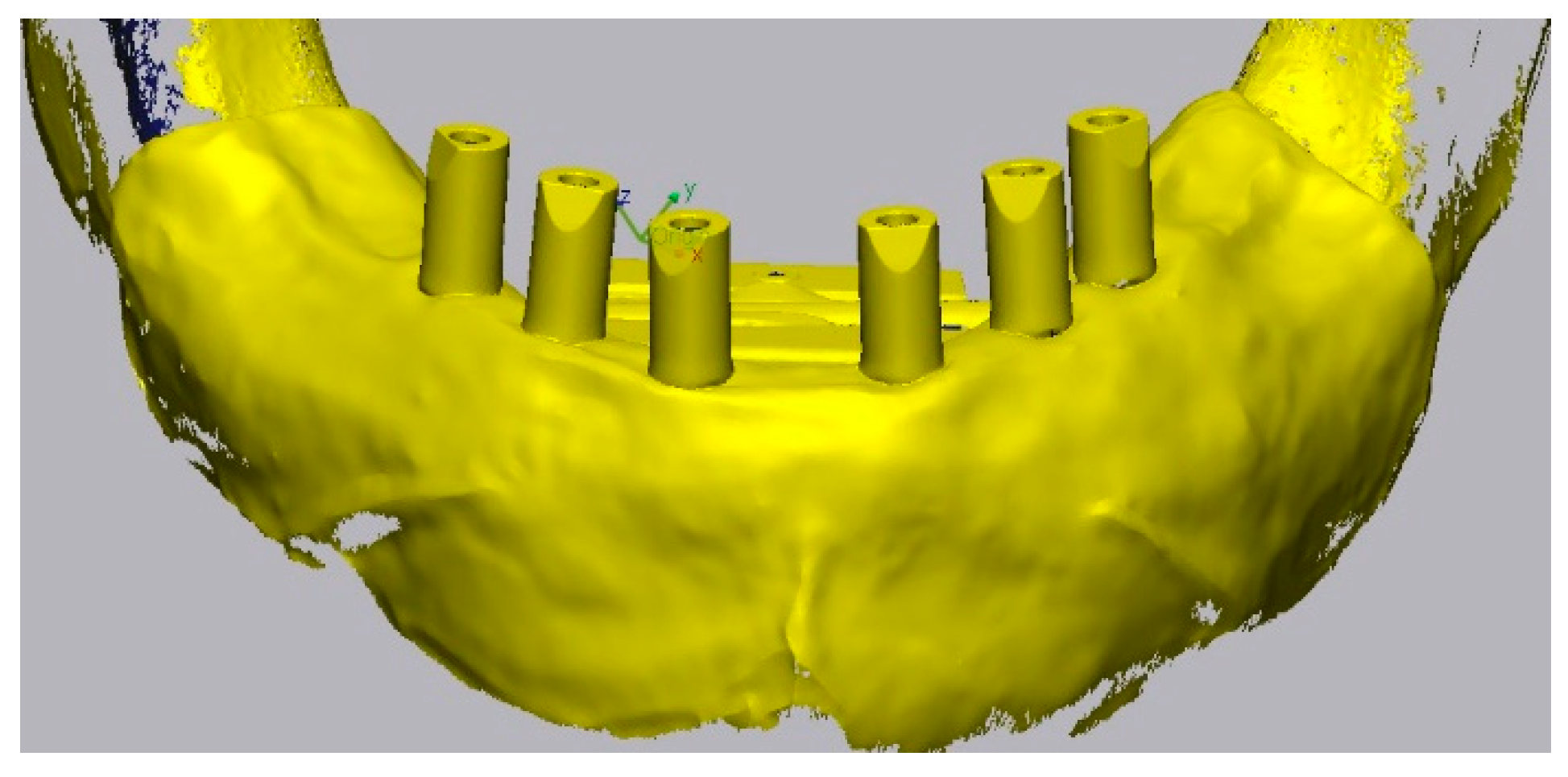
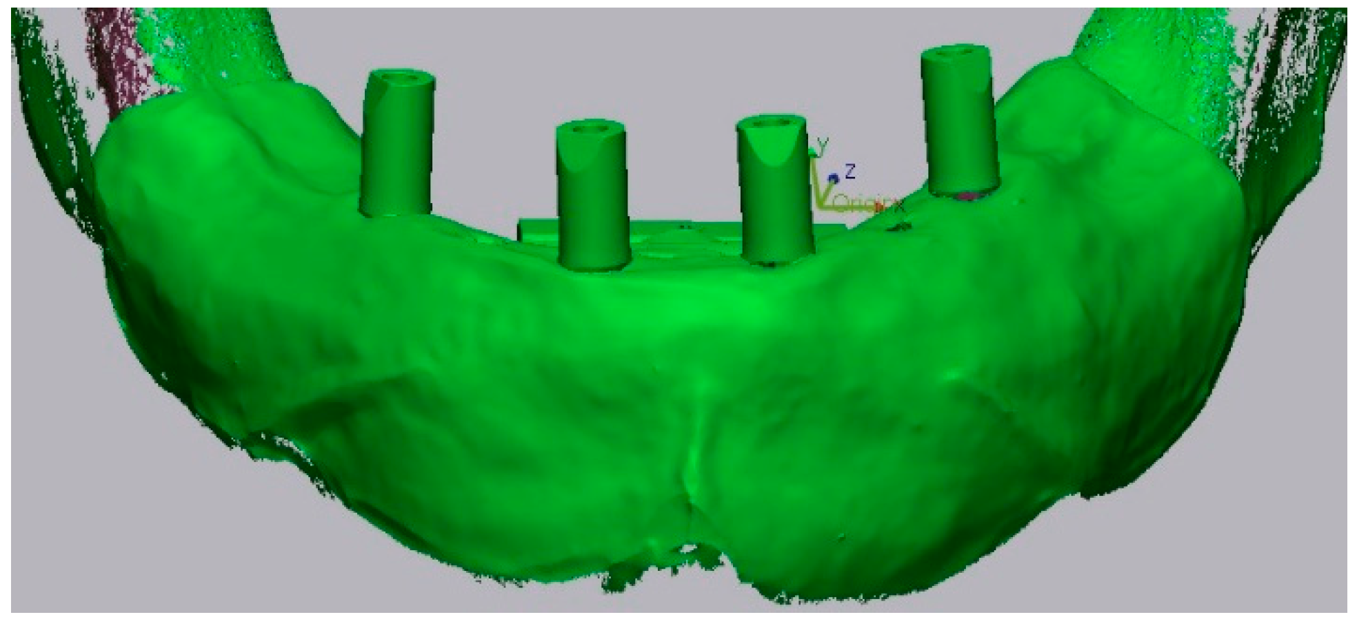
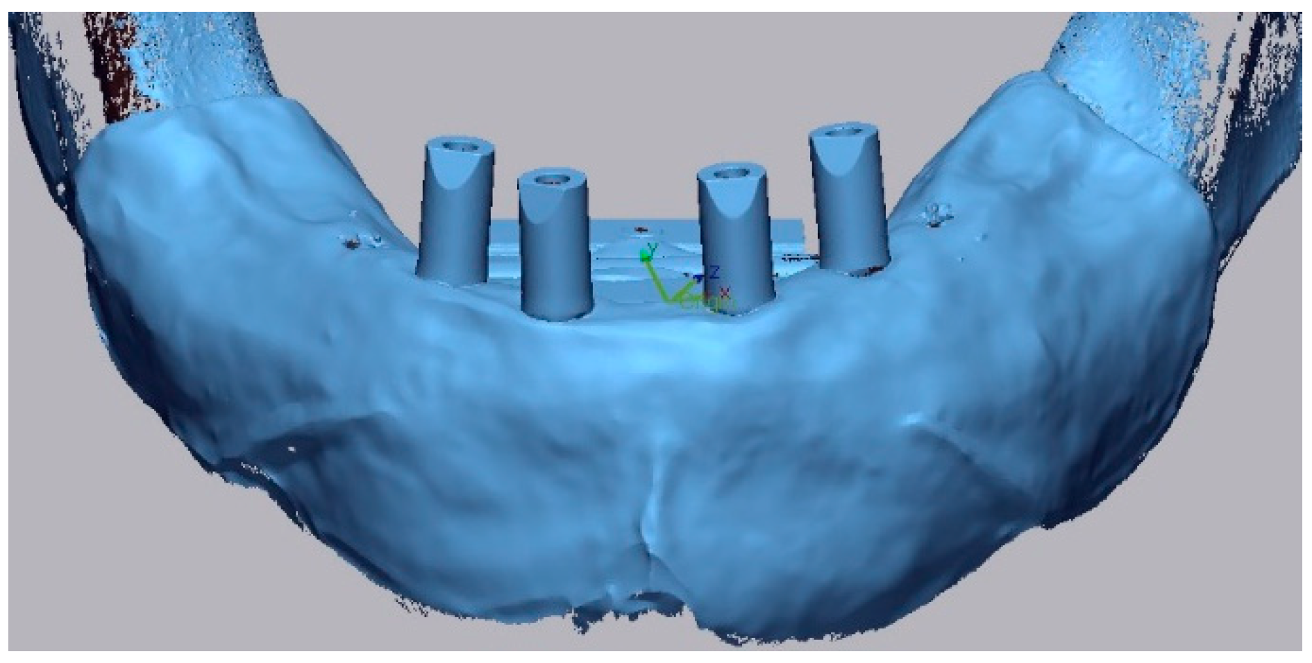
To determine implant position, the Caramês Classification (CC)—which considers the patient’s clinical information as the cornerstone of the therapeutic decision-making process—was applied. Based on the degree of resorption and the quantity of available bone (in terms of height and width) in both jaws, the CC defines five classes of bone atrophy, each associated with three therapeutic options. In this study, the models were fabricated according to class I of the CC (CCI), which represents the least severe bone resorption. Based on this classification, three physical models were created, each simulating one of the three therapeutic options proposed for this classification: Option A was 6 implants (2 anterior implants placed in the lateral incisor positions, 2 implants placed in the premolar region, anterior to the mental foramen, and 2 implants placed in the first molar positions); Option B was 4 implants (2 anterior implants placed in the canine positions, anterior to the mental foramen, and 2 posterior implants placed in the first molar positions); and Option C was 4 implants (2 anterior implants placed in the lateral incisor positions and 2 distal implants positioned in the premolar region, anterior to the mental foramen). Considering the posterior bone availability and the favorable anterior region with higher bone density, two fixed full-arch rehabilitation options (Options A and B) and one removable full-arch option (Option C) are proposed [31].
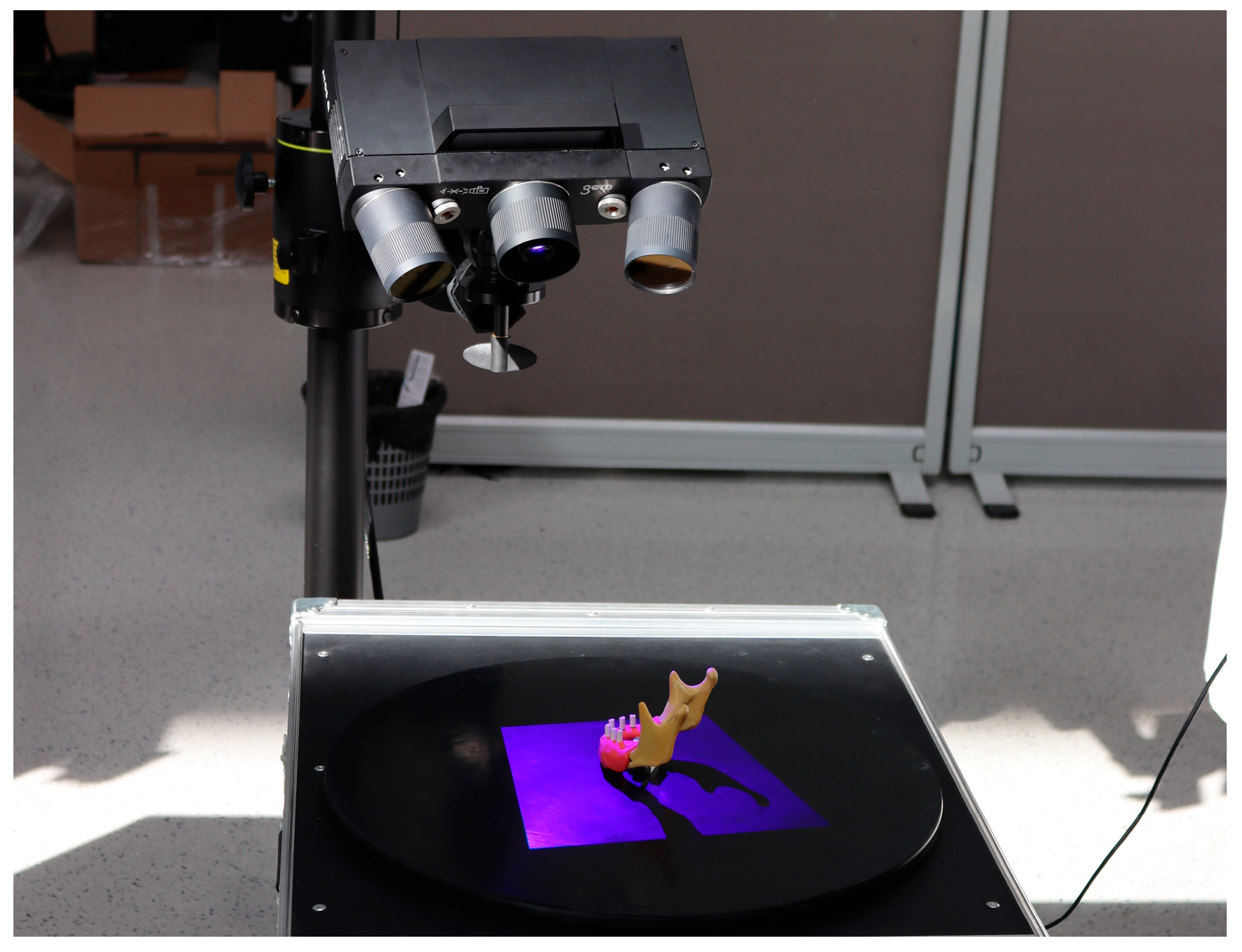
The three models were scanned using a high-precision industrial 12-megapixel scanner GOM Atos Compact Scan 12M (Zeiss, Oberkochen, Germany) (Figure 2), resulting in three corresponding digital reference models (Figure 3, Figure 4 and Figure 5).
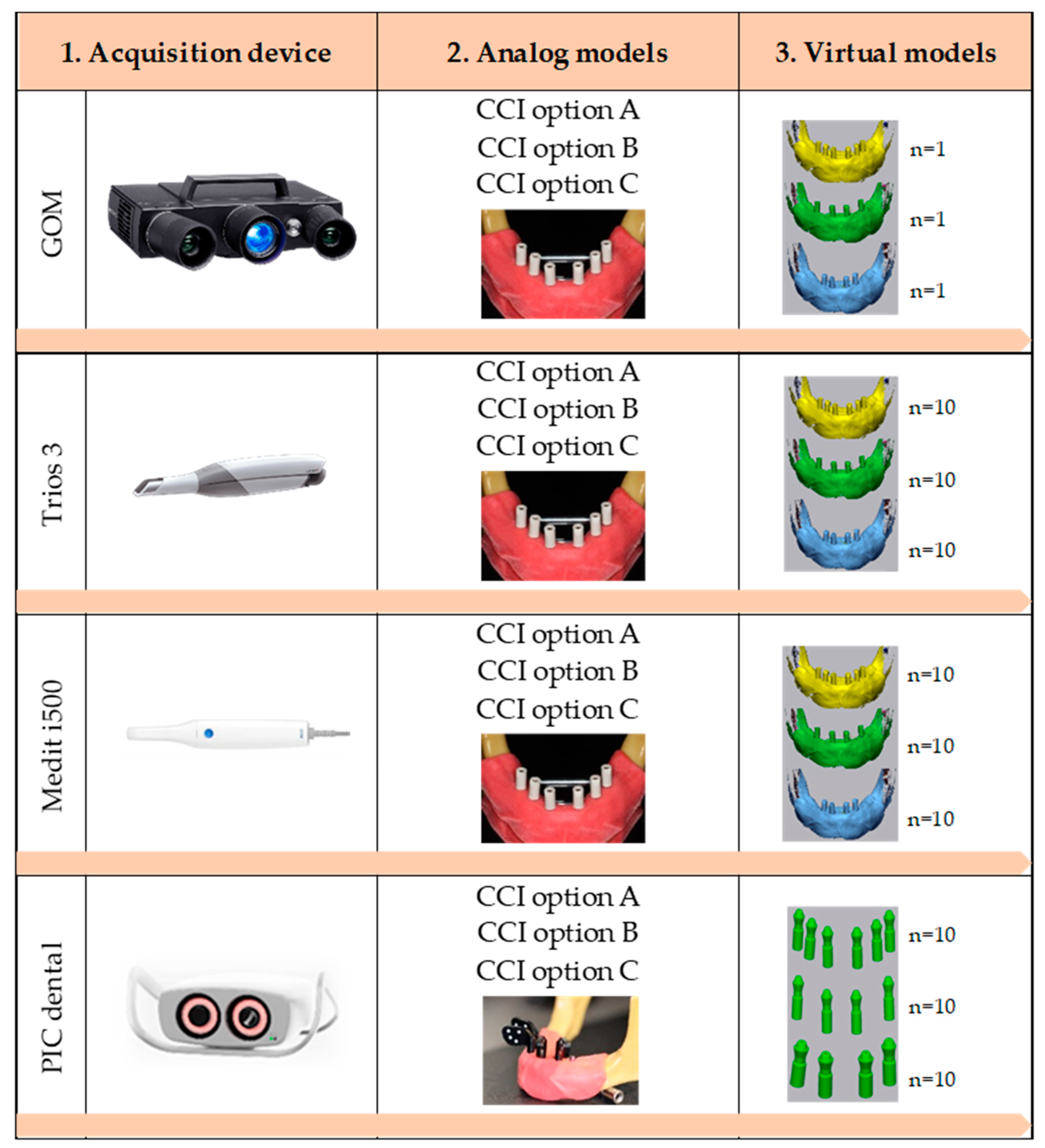
A total of 90 digital impressions were subsequently obtained, with 30 impressions for each of the three implant distribution types. Of these, 10 were captured using the IO Trios 3 scanner (3Shape, Copenhagen, Denmark), 10 with the IO Medit i500 scanner (Medit, Seoul, Republic of South Korea), and 10 using photogrammetry technology with the PICdental system (PIC dental, Spain) (Figure 6). For digital impressions using the industrial scanner and the IOSs, mono CARES ® Scanbodies were employed for 4.6 mm screw abutments (PEEK/TAN, Straumann, Basel, Switzerland), with a tightening torque of 10 Ncm. For digital impressions with photogrammetry, PIC transfers (PIC dental, Madrid, Spain) were utilized, also tightened to 10 Ncm (Figure 6).
All scans were performed in accordance with the manufacturers’ recommended protocols to ensure standardization between the two IOS systems. The scanning sequence included the occlusal surfaces first, followed by the lingual and buccal surfaces. For the photogrammetry system, scanning was also carried out following the manufacturer’s guidelines. All scanners were calibrated prior to use, and the master model was scanned 10 consecutive times with each scanner. To maintain consistency, temperature and humidity were controlled, and all scans were conducted under standardized laboratory conditions by a single experienced operator.
The digital impressions were then processed through a standard laboratory workflow and imported into Exocad software 3.1 (Exocad GmbH, Darmstadt, Germany) for the generation of digital implant replicas. After the identification of the Scanbodies using the integrated library, the replicas were automatically aligned and exported as STL files. These STL files, containing the spatial configuration of the replicas, were subsequently imported into Geomagic Control X 2022.0.1 (3D Systems, Rock Hill, SC, USA) software for three-dimensional analysis.
Trueness was assessed using the best-fit alignment algorithm by superimposing each of the 90 STL files onto their corresponding reference models generated from high-precision industrial optical, resulting in 10 comparisons per group (Table 1). Alignment was performed within a tolerance of +30 and −30 µm. Trueness was quantified using the root mean square (RMS) values derived from the 3D comparisons, expressed in micrometer (µm), and reported as median values along with interquartile ranges (IQRs).
Statistical analysis was performed using SPSS software (IBM Statistics SPSS, v26.0; IBM Corp.; Chicago, IL, USA). The Shapiro–Wilk test was applied to assess the normality of data distribution, while the Levene test was used to evaluate the homogeneity of variances. As the data did not meet parametric assumptions, the non-parametric Kruskal–Wallis test was employed to compare discrepancies among groups. Pairwise comparisons were conducted using Bonferroni correction to adjust significance values and control the Type I error rate. A significance threshold of p = 0.05 was adopted for all statistical tests.

3. Results

Trueness was evaluated based on 90 digital measurements obtained using three different scanning systems: 3Shape Trios 3 (n = 30), Medit i500 (n = 30), and PIC Dental Photogrammetry (n = 30).
As Table 2 depicts, statistically significant differences (p < 0.001) were found across all scanners when comparing the three implant distribution configurations.
For the 3Shape scanner, the CCI C distribution yielded the highest trueness values, which corresponds to the lowest RMS scores (as trueness increases, RMS values decrease). No significant differences were found between CCI Options A and B (p = 0.142).
Regarding the Medit scanner, the CCI A distribution presented the lowest trueness. However, no significant differences were identified between CCI Options B and C (p = 0.119).
In the case of the PIC Dental system, statistically significant differences were observed among the three implant distributions evaluated. Nevertheless, post hoc pairwise comparisons revealed no statistically significant differences between CCI Options A and B (p = 0.058), nor between Options B and C (p = 0.105).
When comparing the three implant distributions (Table 3), statistically significant differences were observed (p < 0.001). Among them, the CCI C distribution demonstrated the highest trueness values while no statistically significant differences were found between the CCI Options A and B (p = 0.722).
A comparison of the scanning systems (Table 4) revealed significant differences in trueness among the three devices assessed (p < 0.001). The Medit IOS outperformed the others in terms of trueness, whereas the PIC Dental system demonstrated the least favorable results.

4. Discussion

The first null hypothesis (H01), which proposed that scanner type has no influence on trueness, was rejected, due to the statistically significant differences identified among the scanners. Similarly, the second null hypothesis (H02), which stated that implant distribution does not affect trueness was also rejected. The findings demonstrate that CCI C—featuring fewer implants in closer proximity—produced the best results in terms of trueness. Conversely, CCI Option B yielded the least favorable outcomes.
This in vitro study aimed to assess and compare the trueness of various digital impression methods for full-arch implant restorations. The investigation focused on two IOSs and one photogrammetry-based system. The intraoral scanners—3Shape TRIOS 3, which operates with confocal principles, and Medit i500, which uses real-time 3D video capture—acquire data from a relatively narrow scanning window. In contrast, the PIC Dental system uses photogrammetric principles to determine implant positions and angulations, by analyzing coordinates from calibrated reference markers [5,6,20,29,32].
The selection of these devices was based on two key considerations: first, the IOSs use fundamentally different image acquisition technologies, and the PIC Dental unit provides a distinct photogrammetric approach, allowing for a broad methodological comparison; second, these systems are frequently employed in clinical workflows, enhancing the practical relevance of the study findings.
The Caramês Classification for implant distribution was chosen because it provides a standardized approach to implant placement within the jawbone and is well established in the field of implant dentistry [31].
The selection of the GOM-Atos Compact Scan 12M industrial scanner (Zeiss, Germany), with a precision deviation ranging from 3 to 12 μm, as the reference system is supported by its high accuracy [18,33,34,35].
Achieving accurate intraoral scans for full-arch implant-supported restorations presents considerable challenges. Inaccuracies in the definitive cast may lead to a misfit that cannot be compensated for by the absence of periodontal ligaments, potentially resulting in complications involving the implants or prosthesis, including screw loosening, ceramic fractures, implant failure, or peri-implant bone loss [36]. Potential sources of error include the design and material of the Scanbodies, implant angulation and position, a lack of anatomical landmarks, mobility of the surrounding mucosa, uniform tissue morphology, increased inter-implant distances, and operator experience [21,36]. Additionally, cumulative errors, particularly in the mandible, can occur during image acquisition over larger scanning areas, further compromising accuracy [37].
To overcome some of these limitations, clinical reports have described the use of screwed optical markers in combination with photogrammetry to record the position of dental implants for fabricating implant-supported prostheses [26,30,38]. However, it is important to note that the photogrammetry exclusively captures the spatial positioning of implants. As a result, supplementary procedures, such as digital impressions with IOSs, are necessary to accurately record soft tissue morphology, requiring the integration of both datasets through superimposition [30]. Although photogrammetry is faster than both IOSs evaluated, the requirement for an additional step to digitize the soft tissue ultimately extends the overall workflow duration.
Numerous techniques have been reported in the literature to evaluate the accuracy of scanning devices, including mean 3D deviations, linear and angular measurements without model superimposition, absolute distance evaluations, and axis-specific (X, Y, and Z) deviations based on best-fit alignment algorithms [39]. In the present study, data analysis was performed using a best-fit alignment approach, wherein trueness was assessed by superimposing the scan data onto a reference model and calculating of the root mean square (RMS) error value [24]. The RMS method, a continuous metric, was chosen for several reasons: (a) it quantifies deviations across all the dataset, offering a comprehensive trueness assessment; (b) it facilitates direct comparisons among various scanning technologies; (c) it incorporates all data points, thereby minimizing bias associated with the selection of specific landmarks or planes and reducing operator dependency compared to linear or angular methods; and (d) it is widely adopted in the literature, particularly in studies within this field [40]. Despite its advantages, the RMS approach has limitations: it does not elucidate the type or spatial distribution of errors, and its outcomes are highly dependent on the alignment algorithm used [6]. This methodological dependence can introduce variability and may hinder the detection of localized inaccuracies in the scanned dataset. No additional localization algorithm was applied in the present study. Instead, to complement the global RMS analysis, alignment was performed using a best-fit method based on the iterative closest point algorithm, ensuring consistent alignment across datasets.
It is essential to note that the accuracy of an impression method is defined by two fundamental parameters: trueness, which refers to the closeness to a reference standard, and precision, which indicates the consistency of measurements across multiple repetitions. As previously described, and in accordance with ISO definitions, “accuracy” represents the combination of “trueness” and “precision”, where “trueness” refers to the ability of an impression to reproduce the dental arch as reliably as possible and without distortions, whereas “precision” (repeatability) refers to the degree of agreement among repeated impressions obtained under similar conditions [21,23,30,41,42]. In the present investigation, only “trueness” was assessed, defined as the closeness of agreement between the measurements obtained using the various devices and a reference standard, in this case, the GOM results. The parameter “precision”, defined as the reproducibility of digital impression measurements, has been examined in other studies, such as that by Faria et al., where it was evaluated through successive superimposition of scans acquired via IOSs and photogrammetry. The STL files from each group were compared using RMS values [29]. While the findings of this study offer meaningful insights, they are specific to the systems evaluated and should be extrapolated to other devices with caution. Digital impressions were obtained by a single experienced operator to minimize inter-operator variability across the groups [21,29]. The operator possessed over 7 years of clinical experience in digital dentistry and implant prosthodontics, ensuring competent use of the digital systems involved. Future research incorporating multiple operators could reveal potential differences attributable to variations in clinical expertise. To enhance reproducibility and maintain consistency in data acquisition, the procedures were conducted under standardized environmental conditions, including controlled lighting, temperature, and relative humidity, as advised in the literature [29,35].
Statistically significant differences of trueness were observed across all implant distributions for each scanning system. For the 3Shape Trios 3 intraoral scanner, no significant differences were found between the CCI A and CCI B distributions. Similarly, the Medit i500 scanner showed no significant differences between the CCI B and CCI C configurations. Conversely, the photogrammetry device (PIC Dental) demonstrated significant differences only between the CCI A and CCI C distributions. These findings suggest that the trueness provided by the PIC Dental system is less affected by the number and spatial arrangement of dental implants, as CCI A involves six implants, whereas CCI C consists of four implants positioned in closer proximity. A comparative analysis between the reference digital models and the impressions obtained using each method across the various implant distributions revealed that all devices achieved lower RMS values in the CCI C distribution, characterized by fewer implants placed in closer proximity. This observation indicates that for the systems evaluated, a higher number of implants and increased inter-implant distances adversely impact trueness. Additionally, the 3Shape Trios 3 and Medit i500 intraoral scanners consistently exhibited lower RMS values across all configurations when compared to the PIC Dental system, except for the 3Shape Trios 3 in the CCI B distribution. This exception suggests that the 3Shape Trios 3 scanner may be more susceptible to diminished performance with increased inter-implant spacing. Based on the findings of this investigation, the real-time 3D video acquisition technology used in the Medit i500 scanner demonstrated superior performance over long spans compared to the confocal imaging principles employed by the 3Shape scanner.
When evaluating trueness across all scanners, irrespective of implant distribution, the Medit i500 intraoral scanner demonstrated the highest performance, with statistically significant results. It was followed by the 3Shape Trios 3 intraoral scanner and the PIC Dental photogrammetry system, the latter of which recorded the greatest mean RMS values. This pattern is consistent with the findings of Revilla-León et al., who, in an in vitro investigation compared the accuracy of a conventional analogue impression, two intraoral scanners, and a photogrammetry-based system, finding that the photogrammetry device yielded the lowest accuracy and the largest discrepancies [30]. It is worth noting, however, that the literature presents conflicting results, with some studies reporting lower discrepancy values for impressions obtained using photogrammetry-based systems [4,6,43].
Several references in the literature propose accuracy thresholds aimed at minimizing clinical complications related to misfit in implant-supported prostheses, such as screw loosening or even the fracture of abutments and screws [14,44,45]. Among the reported values, a threshold of 30 μm is widely recognized and is considered the lowest and most stringent safety limit identified in the reviewed literature; therefore, it was adopted as the reference value in the present study [18,20]. Although differences were observed among the tested scanners, the distance deviation values for all devices (ranging from 13.65 to 58.80 μm) remained below the maximum clinically acceptable misfit threshold of reported in the literature [11,46].
In terms of the influence of implant distribution on trueness, the findings of this study indicate that fewer implants placed in closer proximity resulted in improved results. Specifically, the CCI C distribution, involving closely spaced implants, demonstrated the lowest RMS values in the trueness evaluation. This aligns with previous research, which has reported statistically significant differences among impression techniques when scanning four or six implants [47]. In the current study, both the number and spatial arrangement of implants were analyzed. The results showed that the four implants located in the anterior region (CCI C) produced more accurate outcomes than the same number of implants placed farther apart (CCI B). These findings support the premise that a reduced number of implants with minimal inter-implant distance contributes to improved passive fit. Consistent with the existing literature, the absence of mucosal landmarks is associated with reduced accuracy in digital impressions [18,48,49,50]. Despite methodological differences, studies by Çakmak G et al. and Mizumoto R et al. also concluded that implant positioning significantly affects trueness [51,52].
Although informative, this study is not without limitations. One of them is the reliance on RMS values obtained from 3D comparisons to assess trueness. As above mentioned, these values represent the mean of overall deviations and do not allow for implant-specific analysis of trueness [40]. Another limitation is the use of in vitro conditions for the impression procedures, which may not fully replicate clinical scenarios. This could result in an underestimation of deviations typically introduced by patient-related variables such as blood, saliva, tongue movement, soft tissue dynamics, and ambient lighting conditions [53]. It is essential to highlight that intraoral scanners in this study were used under controlled, optimal conditions, free from the presence of the aforementioned variables. As such, their clinical performance may not fully reflect the trueness observed in vitro. On the other hand, photogrammetry-based systems are expected to maintain a comparable level of accuracy in real-world settings. These systems have gained attention as a viable alternative for full-arch implant impressions, as they may help overcome certain limitations associated with intraoral scanning [4]. In clinical contexts where mucosal movement and absence of stable reference structures pose challenges, the unique design and acquisition method of photogrammetry devices, may offer greater reliability and ease of use compared to IOSs [27,30]. At present, photogrammetry systems are expensive and are primarily intended to be used in implant impressions. As these systems are limited to capturing the spatial positions of implant abutments within the oral cavity, the supplementary use of conventional intraoral scanners remains necessary to obtain detailed soft tissue information, as previously discussed [6]. Nonetheless, recent reports in the literature have highlighted the use of open-source software in photogrammetry as a cost-effective approach for creating digital models. This strategy presents a viable option for practitioners at all levels of experience, particularly for dental students and early-career clinicians aiming to incorporate digital workflows into their clinical practice [54].
Future in vitro investigations could further examine how implant-related variables—such as angulation, insertion depth, alternative spatial configurations, and Scanbodies’ supramucosal height and morphology—affect the trueness of digital impression techniques [39]. Additionally, research into the time efficiency of the photogrammetry process could offer clinically relevant insights for implant prosthodontics. The integration of photogrammetry with emerging technologies, including smartphone-based applications and artificial intelligence, may also enhance our understanding of its reliability and suitability for routine clinical workflows [55,56]. Investigations based on larger sample sizes could help to validate and expand upon the findings of the present investigation. Furthermore, incorporating conventional impression techniques in future research would enable a more detailed comparative analysis of trueness. It should be recognized, however, that in vitro studies cannot fully replicate the complexity and dynamic conditions of the intraoral environment. Thus, in vivo studies are recommended to assess the performance of scanning systems under clinical conditions, including variables such as bone resorption, salivary presence, soft tissue mobility, and ambient lighting. Collectively, the present findings and future investigations may contribute to identifying the digital scanning approach that offers the highest level of trueness for full-arch implant digital impressions in clinical practice.

5. Conclusions

Within the limitations of this in vitro study, the tested intraoral scanners demonstrated comparable trueness, whereas the photogrammetry-based system exhibited lower trueness values.
The results also suggest that a reduced number of implants and shorter inter-implant distances contribute to improving the trueness of digital impressions for full-arch implant rehabilitation, regardless of the scanning system used.
These conclusions are specific to the devices and experimental protocols employed in this study and should not be generalized without further validation. Future randomized clinical trials are recommended to assess the in vivo performance of these and other impression techniques.

Author Contributions

Conceptualization, J.C.F.; methodology, J.C.F., M.A.S.-F. and R.M.; software, J.C.F. and R.M.; validation, M.A.S.-F. and M.H.F.; formal analysis, J.C.F.; investigation, J.C.F. and J.C.S.-F.; resources, R.M.; data curation, S.J.O.; writing—original draft preparation, J.C.F.; writing—review and editing, M.A.S.-F. and S.J.O.; visualization, M.H.F.; supervision, J.C.S.-F.; project administration, J.C.S.-F. and M.H.F. All authors have read and agreed to the published version of the manuscript.

Funding

This research received no external funding.

Institutional Review Board Statement

Not applicable.

Informed Consent Statement

Not applicable.

Data Availability Statement

The original contributions presented in this study are included in the article. Further inquiries can be directed to the corresponding authors.

Conflicts of Interest

The authors declare no conflicts of interest.

References

  1. Amin, S.; Weber, H.P.; Finkelman, M.; El Rafie, K.; Kudara, Y.; Papaspyridakos, P. Digital vs. Conventional Full-Arch Implant Impressions: A Comparative Study. Clin. Oral Implants Res. 2017, 28, 1360–1367. [Google Scholar] [CrossRef] [PubMed]
  2. Huang, R.; Liu, Y.; Huang, B.; Zhang, C.; Chen, Z.; Li, Z. Improved Scanning Accuracy with Newly Designed Scan Bodies: An in Vitro Study Comparing Digital versus Conventional Impression Techniques for Complete-arch Implant Rehabilitation. Clin. Oral Implants Res. 2020, 31, 625–633. [Google Scholar] [CrossRef] [PubMed]
  3. D’Ambrosio, F.; Giordano, F.; Sangiovanni, G.; Di Palo, M.P.; Amato, M. Conventional versus Digital Dental Impression Techniques: What Is the Future? An Umbrella Review. Prosthesis 2023, 5, 851–875. [Google Scholar] [CrossRef]
  4. Pozzi, A.; Agliardi, E.; Lio, F.; Nagy, K.; Nardi, A.; Arcuri, L. Accuracy of Intraoral Optical Scan versus Stereophotogrammetry for Complete-Arch Digital Implant Impression: An in Vitro Study. J. Prosthodont. Res. 2023, 68, 172–180. [Google Scholar] [CrossRef] [PubMed]
  5. Jivănescu, A.; Bara, A.; Faur, A.-B.; Rotar, R.N. Is There a Significant Difference in Accuracy of Four Intraoral Scanners for Short-Span Fixed Dental Prosthesis? A Comparative In Vitro Study. Appl. Sci. 2021, 11, 8280. [Google Scholar] [CrossRef]
  6. Ma, B.; Yue, X.; Sun, Y.; Peng, L.; Geng, W. Accuracy of Photogrammetry, Intraoral Scanning, and Conventional Impression Techniques for Complete-Arch Implant Rehabilitation: An in Vitro Comparative Study. BMC Oral Health 2021, 21, 636. [Google Scholar] [CrossRef] [PubMed]
  7. Aswani, K.; Wankhade, S.; Khalikar, A.; Deogade, S. Accuracy of an Intraoral Digital Impression: A Review. J. Indian Prosthodont. Soc. 2020, 20, 27. [Google Scholar] [CrossRef] [PubMed]
  8. Imburgia, M.; Logozzo, S.; Hauschild, U.; Veronesi, G.; Mangano, C.; Mangano, F.G. Accuracy of Four Intraoral Scanners in Oral Implantology: A Comparative in Vitro Study. BMC Oral Health 2017, 17, 92. [Google Scholar] [CrossRef] [PubMed]
  9. Mangano, F.; Gandolfi, A.; Luongo, G.; Logozzo, S. Intraoral Scanners in Dentistry: A Review of the Current Literature. BMC Oral Health 2017, 17, 149. [Google Scholar] [CrossRef] [PubMed]
  10. Papaspyridakos, P.; Chen, C.-J.; Gallucci, G.O.; Doukoudakis, A.; Weber, H.-P.; Chronopoulos, V. Accuracy of Implant Impressions for Partially and Completely Edentulous Patients: A Systematic Review. Int. J. Oral Maxillofac. Implants 2014, 29, 836–845. [Google Scholar] [CrossRef] [PubMed]
  11. Jemt, T.; Lie, A. Accuracy of Implant-Supported Prostheses in the Edentulous Jaw: Analysis of Precision of Fit between Cast Gold-Alloy Frameworks and Master Casts by Means of a Three-Dimensional Photogrammetric Technique. Clin. Oral Implants Res. 1995, 6, 172–180. [Google Scholar] [CrossRef] [PubMed]
  12. Jemt, T.; Hjalmarsson, L. In Vitro Measurements of Precision of Fit of Implant-Supported Frameworks. A Comparison between “Virtual” and “Physical” Assessments of Fit Using Two Different Techniques of Measurements. Clin. Implant Dent. Relat. Res. 2012, 14, e175–e182. [Google Scholar] [CrossRef] [PubMed]
  13. Michelinakis, G.; Apostolakis, D.; Kamposiora, P.; Papavasiliou, G.; Özcan, M. The Direct Digital Workflow in Fixed Implant Prosthodontics: A Narrative Review. BMC Oral Health 2021, 21, 37. [Google Scholar] [CrossRef] [PubMed]
  14. Sahin, S.; Cehreli, M.C. The Significance of Passive Framework Fit in Implant Prosthodontics: Current Status. Implant Dent. 2001, 10, 85–92. [Google Scholar] [CrossRef] [PubMed]
  15. Kim, Y.; Oh, T.-J.; Misch, C.E.; Wang, H.-L. Occlusal Considerations in Implant Therapy: Clinical Guidelines with Biomechanical Rationale. Clin. Oral Implants Res. 2005, 16, 26–35. [Google Scholar] [CrossRef] [PubMed]
  16. Knechtle, N.; Wiedemeier, D.; Mehl, A.; Ender, A. Accuracy of Digital Complete-Arch, Multi-Implant Scans Made in the Edentulous Jaw with Gingival Movement Simulation: An in Vitro Study. J. Prosthet. Dent. 2022, 128, 468–478. [Google Scholar] [CrossRef] [PubMed]
  17. Di Fiore, A.; Meneghello, R.; Graiff, L.; Savio, G.; Vigolo, P.; Monaco, C.; Stellini, E. Full Arch Digital Scanning Systems Performances for Implant-Supported Fixed Dental Prostheses: A Comparative Study of 8 Intraoral Scanners. J. Prosthodont. Res. 2019, 63, 396–403. [Google Scholar] [CrossRef] [PubMed]
  18. Revell, G.; Simon, B.; Mennito, A.; Evans, Z.P.; Renne, W.; Ludlow, M.; Vág, J. Evaluation of Complete-Arch Implant Scanning with 5 Different Intraoral Scanners in Terms of Trueness and Operator Experience. J. Prosthet. Dent. 2022, 128, 632–638. [Google Scholar] [CrossRef] [PubMed]
  19. Rutkūnas, V.; Gečiauskaitė, A.; Jegelevičius, D.; Vaitiekūnas, M. Accuracy of Digital Implant Impressions with Intraoral Scanners. A Systematic Review. Eur. J. Oral Implantol. 2017, 10 (Suppl. 1), 101–120. [Google Scholar] [PubMed]
  20. Imburgia, M.; Kois, J.; Marino, E.; Lerner, H.; Mangano, F.G. Continuous Scan Strategy (CSS): A Novel Technique to Improve the Accuracy of Intraoral Digital Impressions. Eur. J. Prosthodont. Restor. Dent. 2020, 28, 128–141. [Google Scholar] [CrossRef] [PubMed]
  21. Arcuri, L.; Pozzi, A.; Lio, F.; Rompen, E.; Zechner, W.; Nardi, A. Influence of Implant Scanbody Material, Position and Operator on the Accuracy of Digital Impression for Complete-Arch: A Randomized in Vitro Trial. J. Prosthodont. Res. 2020, 64, 128–136. [Google Scholar] [CrossRef] [PubMed]
  22. ISO 5725-1:2023; Accuracy (Trueness and Precision) of Measurement Methods and results—Part 1: General Principles and Definitions. ISO: Geneva, Switzerland, 2023.
  23. Sanda, M.; Miyoshi, K.; Baba, K. Trueness and Precision of Digital Implant Impressions by Intraoral Scanners: A Literature Review. Int. J. Implant Dent. 2021, 7, 97. [Google Scholar] [CrossRef] [PubMed]
  24. Sampaio-Fernandes, M.A.; Pinto, R.; Almeida, P.R.; Sampaio-Fernandes, M.M.; Marques, D.; Figueiral, M.H. Accuracy of Extraoral Digital Impressions with Multi-Unit Implants. Appl. Sci. 2023, 13, 8769. [Google Scholar] [CrossRef]
  25. Kihara, H.; Hatakeyama, W.; Komine, F.; Takafuji, K.; Takahashi, T.; Yokota, J.; Oriso, K.; Kondo, H. Accuracy and Practicality of Intraoral Scanner in Dentistry: A Literature Review. J. Prosthodont. Res. 2020, 64, 109–113. [Google Scholar] [CrossRef] [PubMed]
  26. Gómez-Polo, M.; Gómez-Polo, C.; Del Río, J.; Ortega, R. Stereophotogrammetric Impression Making for Polyoxymethylene, Milled Immediate Partial Fixed Dental Prostheses. J. Prosthet. Dent. 2018, 119, 506–510. [Google Scholar] [CrossRef] [PubMed]
  27. Hussein, M.O. Photogrammetry Technology in Implant Dentistry: A Systematic Review. J. Prosthet. Dent. 2023, 130, 318–326. [Google Scholar] [CrossRef] [PubMed]
  28. Yan, Y.; Lin, X.; Yue, X.; Geng, W. Accuracy of 2 Direct Digital Scanning Techniques—Intraoral Scanning and Stereophotogrammetry—For Complete Arch Implant-Supported Fixed Prostheses: A Prospective Study. J. Prosthet. Dent. 2023, 130, 564–572. [Google Scholar] [CrossRef] [PubMed]
  29. Fernandes, J.C.; Sampaio-Fernandes, M.A.; Oliveira, S.J.; Malheiro, R.; Sampaio-Fernandes, J.C.; Figueiral, M.H. Precision of Photogrammetry and Intraoral Scanning in Full-Arch Implant Rehabilitation: An In Vitro Comparative Study. Appl. Sci. 2025, 15, 1388. [Google Scholar] [CrossRef]
  30. Revilla-León, M.; Att, W.; Özcan, M.; Rubenstein, J. Comparison of Conventional, Photogrammetry, and Intraoral Scanning Accuracy of Complete-Arch Implant Impression Procedures Evaluated with a Coordinate Measuring Machine. J. Prosthet. Dent. 2021, 125, 470–478. [Google Scholar] [CrossRef]
  31. Caramês, J. A Comprehensive Classification to Full Arch Implant Rehabilitation. Rev. Port. Estomatol. Med. Dentária Cir. Maxilofac. 2019, 60, 175–188. [Google Scholar] [CrossRef]
  32. Amornvit, P.; Rokaya, D.; Peampring, C.; Sanohkan, S. Confocal 3D Optical Intraoral Scanners and Comparison of Image Capturing Accuracy. Comput. Mater. Contin. 2020, 66, 303–314. [Google Scholar] [CrossRef]
  33. Cakmak, G.; Marques, V.R.; Donmez, M.B.; Lu, W.-E.; Abou-Ayash, S.; Yilmaz, B. Comparison of Measured Deviations in Digital Implant Scans Depending on Software and Operator. J. Dent. 2022, 122, 104154. [Google Scholar] [CrossRef] [PubMed]
  34. Bud, E.S.; Bocanet, V.I.; Muntean, M.H.; Vlasa, A.; Păcurar, M.; Zetu, I.N.; Soporan, B.I.; Bud, A. Extra-Oral Three-Dimensional (3D) Scanning Evaluation of Three Different Impression Materials—An In Vitro Study. Polymers 2022, 14, 3678. [Google Scholar] [CrossRef] [PubMed]
  35. Sampaio-Fernandes, M.A.F.; Pinto, R.; Sampaio-Fernandes, M.M.; Carlos, J.; Marques, D.; Figueiral, M.H. Accuracy of Silicone Impressions and Stone Models Using Two Laboratory Scanners: A 3D Evaluation. Int. J. Prosthodont. 2024, 37, e10–e24. [Google Scholar] [CrossRef] [PubMed]
  36. Wulfman, C.; Naveau, A.; Rignon-Bret, C. Digital Scanning for Complete-Arch Implant-Supported Restorations: A Systematic Review. J. Prosthet. Dent. 2020, 124, 161–167. [Google Scholar] [CrossRef] [PubMed]
  37. Kanjanasavitree, P.; Thammajaruk, P.; Guazzato, M. Comparison of Different Artificial Landmarks and Scanning Patterns on the Complete-Arch Implant Intraoral Digital Scans. J. Dent. 2022, 125, 104266. [Google Scholar] [CrossRef] [PubMed]
  38. Rivara, F.; Lumetti, S.; Calciolari, E.; Toffoli, A.; Forlani, G.; Manfredi, E. Photogrammetric Method to Measure the Discrepancy between Clinical and Software-Designed Positions of Implants. J. Prosthet. Dent. 2016, 115, 703–711. [Google Scholar] [CrossRef] [PubMed]
  39. Sicilia, E.; Lagreca, G.; Papaspyridakos, P.; Finkelman, M.; Cobo, J.; Att, W.; Revilla-León, M. Effect of Supramucosal Height of a Scan Body and Implant Angulation on the Accuracy of Intraoral Scanning: An In Vitro Study. J. Prosthet. Dent. 2024, 131, 1126–1134. [Google Scholar] [CrossRef] [PubMed]
  40. Lyu, M.; Di, P.; Lin, Y.; Jiang, X. Accuracy of Impressions for Multiple Implants: A Comparative Study of Digital and Conventional Techniques. J. Prosthet. Dent. 2022, 128, 1017–1023. [Google Scholar] [CrossRef] [PubMed]
  41. Ender, A.; Mehl, A. Accuracy of Complete-Arch Dental Impressions: A New Method of Measuring Trueness and Precision. J. Prosthet. Dent. 2013, 109, 121–128. [Google Scholar] [CrossRef] [PubMed]
  42. Flügge, T.V.; Schlager, S.; Nelson, K.; Nahles, S.; Metzger, M.C. Precision of Intraoral Digital Dental Impressions with ITero and Extraoral Digitization with the ITero and a Model Scanner. Am. J. Orthod. Dentofac. Orthop. 2013, 144, 471–478. [Google Scholar] [CrossRef] [PubMed]
  43. Tohme, H.; Lawand, G.; Chmielewska, M.; Makhzoume, J. Comparison between Stereophotogrammetric, Digital, and Conventional Impression Techniques in Implant-Supported Fixed Complete Arch Prostheses: An in Vitro Study. J. Prosthet. Dent. 2023, 129, 354–362. [Google Scholar] [CrossRef] [PubMed]
  44. Taylor, T.D. Prosthodontic Problems and Limitations Associated with Osseointegration. J. Prosthet. Dent. 1998, 79, 74–78. [Google Scholar] [CrossRef] [PubMed]
  45. Kallus, T.; Bessing, C. Loose Gold Screws Frequently Occur in Full-Arch Fixed Prostheses Supported by Osseointegrated Implants after 5 Years. Int. J. Oral Maxillofac. Implants 1994, 9, 169–178. [Google Scholar] [PubMed]
  46. Papaspyridakos, P.; Hirayama, H.; Chen, C.; Ho, C.; Chronopoulos, V.; Weber, H. Full-arch Implant Fixed Prostheses: A Comparative Study on the Effect of Connection Type and Impression Technique on Accuracy of Fit. Clin. Oral Implants Res. 2016, 27, 1099–1105. [Google Scholar] [CrossRef] [PubMed]
  47. Chochlidakis, K.; Papaspyridakos, P.; Tsigarida, A.; Romeo, D.; Chen, Y.; Natto, Z.; Ercoli, C. Digital Versus Conventional Full-Arch Implant Impressions: A Prospective Study on 16 Edentulous Maxillae. J. Prosthodont. 2020, 29, 281–286. [Google Scholar] [CrossRef] [PubMed]
  48. Mizumoto, R.M.; Yilmaz, B. Intraoral Scan Bodies in Implant Dentistry: A Systematic Review. J. Prosthet. Dent. 2018, 120, 343–352. [Google Scholar] [CrossRef] [PubMed]
  49. Çakmak, G.; Yilmaz, H.; Treviño, A.; Kökat, A.M.; Yilmaz, B. The Effect of Scanner Type and Scan Body Position on the Accuracy of Complete-arch Digital Implant Scans. Clin. Implant Dent. Relat. Res. 2020, 22, 533–541. [Google Scholar] [CrossRef] [PubMed]
  50. Bohner, L.; Gamba, D.D.; Hanisch, M.; Marcio, B.S.; Tortamano Neto, P.; Laganá, D.C.; Sesma, N. Accuracy of Digital Technologies for the Scanning of Facial, Skeletal, and Intraoral Tissues: A Systematic Review. J. Prosthet. Dent. 2019, 121, 246–251. [Google Scholar] [CrossRef] [PubMed]
  51. Çakmak, G.; Yilmaz, H.; Treviño Santos, A.; Kökat, A.M.; Yilmaz, B. Effect of Scanner Type and Scan Body Location on the Accuracy of Mandibular Complete-Arch Digital Implant Scans: An In Vitro Study. J. Prosthodont. 2022, 31, 419–426. [Google Scholar] [CrossRef] [PubMed]
  52. Mizumoto, R.M.; Alp, G.; Özcan, M.; Yilmaz, B. The Effect of Scanning the Palate and Scan Body Position on the Accuracy of Complete-arch Implant Scans. Clin. Implant Dent. Relat. Res. 2019, 21, 987–994. [Google Scholar] [CrossRef] [PubMed]
  53. Lim, J.-H.; Park, J.-M.; Kim, M.; Heo, S.-J.; Myung, J.-Y. Comparison of Digital Intraoral Scanner Reproducibility and Image Trueness Considering Repetitive Experience. J. Prosthet. Dent. 2018, 119, 225–232. [Google Scholar] [CrossRef] [PubMed]
  54. Zotti, F.; Rosolin, L.; Bersani, M.; Poscolere, A.; Pappalardo, D.; Zerman, N. Digital Dental Models: Is Photogrammetry an Alternative to Dental Extraoral and Intraoral Scanners? Dent. J. 2022, 10, 24. [Google Scholar] [CrossRef] [PubMed]
  55. Pascadopoli, M.; Zampetti, P.; Nardi, M.G.; Pellegrini, M.; Scribante, A. Smartphone Applications in Dentistry: A Scoping Review. Dent. J. 2023, 11, 243. [Google Scholar] [CrossRef] [PubMed]
  56. Wang, C.; Yang, J.Y.; Liu, H.K.; Yu, P.; Jiang, X.J.; Liu, R.J. Co-Mask R-CNN: Collaborative Learning-Based Method for Tooth Instance Segmentation. J. Clin. Pediatr. Dent. 2024, 48, 161. [Google Scholar] [CrossRef] [PubMed]
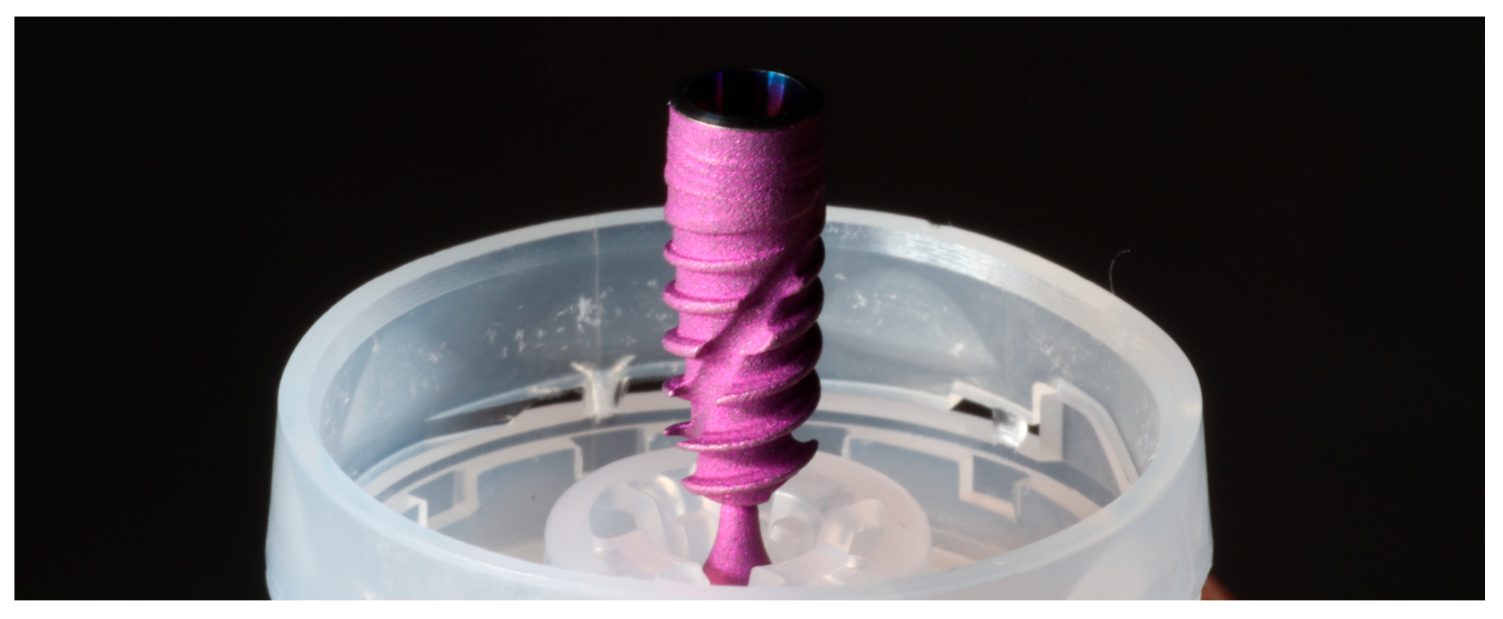
Figure 1. Straumann BLX 4.0 mm × 12 mm implant (Straumann, Basel, Switzerland).
Applsci 15 08016 g001
Figure 2. Acquisition of a digital reference model with the GOM-Atos Compact Scan 12M (Zeiss, Germany).
Applsci 15 08016 g002
Figure 3. Digital reference model for CCI Option A.
Applsci 15 08016 g003
Figure 4. Digital reference model for CCI Option B.
Applsci 15 08016 g004
Figure 5. Digital reference model for CCI Option C.
Applsci 15 08016 g005
Figure 6. Workflow overview. 1. Acquisition devices: reference model acquired with GOM; study models acquired with Trios 3, Medit i500, and PIC dental. 2. Different implant distributions studied, illustrated with an analogue model. 3. Mesh of virtual models corresponding to different implant distributions.
Applsci 15 08016 g006
Table 1. Description of the study groups according to image acquisition device and implant distribution.
ScannerNumber and Position of ImplantsGroup
3Shape Trios 3
IOS
6 (CCI Option A)TA
4 (CCI Option B)TB
4 (CCI Option C)TC
Medit i5006 (CCI Option A)MA
4 (CCI Option B)MB
4 (CCI Option C)MC
PIC Dental
Photogrammetry
6 (CCI Option A)PA
4 (CCI Option B)PB
4 (CCI Option C)PC
CCI Option A with 6 implants: 2 implants in the position of the lateral incisors, 2 anterior to the mental foramen and 2 in the region of the first molars; CCI Option B with 4 implants: 2 implants in the canine position and 2 implants in the first molar region; CCI Option C with 4 implants: 2 implants in the position of the lateral incisors and 2 implants anterior to the mental foramen.
Table 2. Descriptive statistics and comparison of intra-group trueness values (µm) by implant distribution for each scanning system.
ScannerNumber and Position of ImplantsGroupRMS (µm)
Median
IQRNIntra-Group
p-Values
IO 3Shape Trios 36 (CCI A)TA29.5527.85; 29.7310a<0.001
4 (CCI B)TB58.8045.78; 63.3810a
4 (CCI C)TC18.7517.95; 20.7510
IO Medit i5006 (CCI A)MA33.9532.10; 37.2010 <0.001
4 (CCI B)MB24.4520.48; 25.7010b
4 (CCI C)MC13.6511.90; 15.6010b
Photogrammetry PIC Dental6 (CCI A)PA53.5553.03; 53.9510c<0.001
4 (CCI B)PB49.4545.38; 51.3810c,d
4 (CCI C)PC43.7043.38; 45.8810d
Kruskal–Wallis rank sum test. RMS, root mean square; IQR, interquartile range. No significant intra-group differences: a p = 0.142; b p = 0.119; c p = 0.058; d p = 0.105.
Table 3. Summary statistics and comparison of trueness values (µm) across the different implant distribution groups.
Number and Position of ImplantsRMS (µm)
Median
IQRNp
6 (CCI A)34.2029.70; 53.1530a<0.001
4 (CCI B)45.7524.58; 57.0530a
4 (CCI C)19.0015.58; 43.4030
Kruskal–Wallis rank sum test. RMS, root mean square; IQR, interquartile range; a, no significant differences (p = 0.722).
Table 4. Descriptive statistics and comparison of trueness values (µm) across different scanning devices.
DeviceRMS (µm)
Median
IQRNp
IO 3Shape Trios 329.2020.13; 51.5030<0.001
IO Medit i50024.4514.45; 32.6330
Photogrammetry PIC Dental49.4544.13; 53.3030
Kruskal–Wallis rank sum test. RMS, root mean square; IQR, interquartile range.
Disclaimer/Publisher’s Note: The statements, opinions and data contained in all publications are solely those of the individual author(s) and contributor(s) and not of MDPI and/or the editor(s). MDPI and/or the editor(s) disclaim responsibility for any injury to people or property resulting from any ideas, methods, instructions or products referred to in the content.