Article

222Rn Exhalation Rate of Building Materials: Comparison of Standard Experimental Protocols and Radiological Health Hazard Assessment

1
Dipartimento di Scienze Matematiche e Informatiche, Scienze Fisiche e Scienze della Terra, Università degli Studi di Messina, Viale F. Stagno d’Alcontres, 31, 98166 Messina, Italy
2
Dipartimento di Ingegneria dell’Informazione ed Elettrica e Matematica Applicata (DIEM), Università degli Studi di Salerno, Via Giovanni Paolo II, 132, 84084 Fisciano, Italy
*
Author to whom correspondence should be addressed.
Appl. Sci. 2025, 15(14), 8015; https://doi.org/10.3390/app15148015
Submission received: 25 June 2025 / Revised: 15 July 2025 / Accepted: 16 July 2025 / Published: 18 July 2025
(This article belongs to the Section Environmental Sciences)

Abstract

This study evaluates the accuracy of 222Rn exhalation rates from building materials using two standard experimental protocols, thus addressing the increasing importance of rapid radon assessment due to health concerns and regulatory limits. In detail, six types of natural stones frequently employed for the construction of buildings of historical-artistic relevance were analyzed using the closed chamber method (CCM) combined with the Durridge Rad7 system, by using two experimental protocols that differed in the measurement duration: 10 days (Method 1) versus 24 h (Method 2). Obtained results revealed that the radon exhalation rates ranged from 0.004 to 0.072 Bq h−1, which are moderate to low if compared to studies in other regions. Statistical comparison using the u-test confirmed equivalence between protocols (u-test ≤ 2), thus supporting the validity of the faster Method 2 for practical applications. Furthermore, to estimate the potential indoor radon levels and determine the associated radiological risks to human health, for the investigated natural stones, the Markkanen room model was employed. As a result, simulated indoor radon concentrations remained well below regulatory thresholds (maximum value: 37.3 Bq m−3), thus excluding any significant health concerns under typical indoor conditions.

1. Introduction

Human exposure to ionizing radiation comes from both natural and artificial sources. According to the 2008 UNSCEAR report, the global average annual radiation dose is about 2.4 mSv from natural sources and 0.6 mSv from man-made activities [1].
Natural radiation includes external exposure from cosmic rays and terrestrial radionuclides present in soil and construction materials, as well as internal exposure from inhaling or ingesting naturally occurring radionuclides found in air, water, and food [2,3,4,5,6]. Artificial sources are mainly related to medical procedures that use ionizing radiation for diagnostics and treatment, such as radiology and radiotherapy [7,8,9,10,11,12,13]. Nuclear accidents, such as those at Chernobyl and Fukushima, have also released significant quantities of radionuclides into the environment, including 131I, 137Cs, 134Cs, and 90Sr [14,15,16,17,18,19].
Among natural sources, radon—specifically 222Rn, due to its greater isotopic abundance—is of particular concern, as it is the main contributor to internal exposure from natural radiation [1]. This noble gas is colorless, odorless, chemically inert, and continuously produced in the decay chain of 238U [20,21,22,23,24,25]. It can diffuse through soil, rocks, and water [26,27,28]. The ground and its underlying geological formations account for about 69% of radon emissions [29,30]. Geological features and weathering processes strongly influence the geogenic radon potential (GRP), i.e., the natural ability of the ground to generate and release radon gas into the environment, primarily influenced by the uranium content of rocks and soils, their permeability, and geological structure [31,32]. Understanding these factors is essential for predicting radon release and planning land use or building design strategies that reduce indoor accumulation, thereby enhancing safety [33]. In addition, construction materials sourced from quarries may emit radon, posing potential health risks to workers and building occupants. Many historic structures use locally quarried stones, yet little research exists on their radon emissions, in particular for Southern Italian heritage structures [34,35,36,37,38,39,40].
Notably, radon is classified as a carcinogen, and long-term exposure to elevated concentrations has been clinically linked to an increased risk of lung cancer. The World Health Organization identifies radon as the second leading cause of lung cancer after smoking [41]. This hazard arises mainly from its radioactive decay products—218Po and 214Po—which emit alpha particles. While radon itself is inert, its progeny is reactive and can adhere to airborne dust, allowing them to be inhaled and settle in the lungs. Once deposited in lung tissues, they can cause cellular damage that significantly heightens cancer risk [42,43]. Recent epidemiological studies have shown that even low-level radon exposure can significantly increase lung cancer risk [44]. Given these health implications, effective monitoring and mitigation of radon levels are critical components in view of the definition of efficient radiation protection strategies. In light of this, the European Union adopted Directive 2013/59/EURATOM, which was subsequently implemented in Italy through Legislative Decree D.Lgs. 101/2020 [45]. The directive emphasizes the importance of thoroughly investigating all potential indoor radon sources, including those arising from building materials.
Within this context, the present study investigates the accuracy of two different analytical approaches for measuring radon exhalation rates from building materials. The research focuses on six types of stone widely used in Southern Italian heritage architecture—namely Lecce stone, Modica stone, Ignimbrite Campana, Comiso stone, Mendicino stone, and Viterbo tuff. Measurements were conducted using the closed chamber method (CCM), i.e., an experimental technique used to measure the radon exhalation rate from a material by placing it inside a sealed container (chamber), in combination with the Durridge Rad7 instrument [46,47,48]. Specifically, the first analytical approach utilizes data from a 10-day measurement period, whereas the second relies on results from just the first 24 h [49]. A statistical comparison of the two methods was carried out using the u-test. To further assess the implications for indoor air quality, the resulting exhalation rates were applied to the Markkanen room model, i.e., a mathematical model used to estimate indoor radon concentration based on the radon exhalation rate from surfaces (such as walls and floors), the room’s volume, the ventilation rate, and the decay constant of radon, thus enabling an estimation of potential indoor radon concentrations.
Unlike prior studies, this paper (i) directly compares two standard analytical protocols using the same instrumentation; (ii) introduces a fast yet validated method for time-efficient assessments; and (iii) incorporates results into a radiological hazard framework using the Markkanen room model. The novelty also lies in focusing on heritage stones from southern Italy, rarely analyzed in terms of radon emissions.

2. Geological Framework of the Investigated Samples

During the Baroque period, Lecce stone became one of the most used building materials in southern Italy. Its popularity is largely attributed to its softness, which made it easy to work with for both structural and ornamental purposes. The Lecce Stone Formation is dated to the Upper Burdigalian–early Messinian interval, with a depositional duration of approximately 11 million years. It spans the Globigerinoides trilobus–Globorotalia miotumida planktonic foraminiferal zones and the Helicosphaera ampliaperta–Amaurolithus delicatus–A. amplificus calcareous nannofossil zones, respectively [50]. In the Salento region, where a Cretaceous basement composed of pure and dolomitic limestones is overlain by Neogene–Pleistocene sediments, sedimentation occurred in four main cycles between the late Burdigalian and the late Calabrian (Sicilian). These cycles resulted in six lithostratigraphic units—one of which was only recently defined—grouped into five formations: the Lecce Stone Formation (Lower–Upper Miocene), the Calcareniti di Andrano (Upper Miocene), the Lèuca Formation with its distinct Palmariggi Member (Lower Pliocene), the Uggiano la Chiesa Formation (Middle Pliocene–Lower Pleistocene), and the Calcareniti del Salento (Lower Pleistocene). The first of these sedimentary cycles comprises the Lecce Stone Formation, which is unconformably overlain by the Calcareniti di Andrano [51].
Going on, the town of Modica, like Ragusa and Comiso and other ones, was included in the UNESCO World Heritage list known as “Late Baroque Towns of the Val di Noto”, in which the main monuments’ building stones are calcarenites, mainly made of calcite and quarried in the cities themselves or in their surroundings [52]. The monuments in Modica’s historic city center have Baroque facades made of stone, typically creamy yellow in color, called Modica stone. The latter shows slight variations due to the use of pigments such as clayey and gypseous earths. Modica stone outcrops in the Hyblean Plateau, Southeastern Sicily, that represents a forebulge, result from bending the African continental foreland lithosphere below the advancing Maghrebian thrust-fold belt. The Hyblean Plateau was generated by a complex interplay of tectonic and sedimentary processes over a long period of time. The progressive downfaulting of the Hyblean foreland by a system of Cenozoic NE–SW-trending faults led to the formation of an asymmetric trough, the Caltanissetta Trough, characterized by a thick fill of Miocene to Quaternary sediments that give rise to marked negative gravity values. The Cretaceous–Eocene tectonic phase, that was responsible for the palaeogeographic pattern which existed during most of the Cenozoic, led to the formation of carbonate reefs and volcanic seamounts developed in the Upper Cretaceous, forming platforms fault-bounded to the west and southwest, from which were derived the detrital sediments found in the basin [53].
With reference to Ignimbrite Campana, it is worth noting that Caldera-forming eruptions are among the most destructive natural phenomena on Earth, and a prominent example is the Ignimbrite Campana itself, which resulted from a massive explosive eruption approximately 39,000 years ago. This event generated an extensive pyroclastic sequence composed of trachytic to phonolitic magma, blanketing several thousand square kilometers across the Campanian Plain in south-central Italy [54]. The Campanian Plain’s tectonic framework is defined by Upper Miocene thrust structures associated with the southern Apennines, which have been subsequently disrupted by various Quaternary fault systems. These faults reflect the final tectonic phases linked to the opening of the Tyrrhenian Sea. Structural analysis reveals that NW–SE-trending normal faults, active during the Lower Pleistocene, predate NE–SW-oriented faulting that developed after 700,000 years ago. The current fault configuration in the Bay of Naples includes E–W-trending strike-slip (left-lateral) faults, NE-trending extensional faults, and NW-oriented transtensional structures—likely the result of block rotation within a transtensional environment along an E–W fault zone [55].
With regard to Comiso stone, it originates from the lower portion of the Leonardo Member (Upper Oligocene) within the Ragusa Formation, which spans from the Upper Oligocene to the Miocene in the Hyblean Plateau. This formation is characterized by alternating calcareous and marly layers. The stone itself is a well-lithified, fine-grained calcarenite, predominantly composed of calcite. It exhibits a white to cream color, a compact and tenacious texture, and a uniform structure. Quarried in southeastern Sicily, Comiso stone was extensively used in the construction of monuments in the Baroque towns of the “Val di Noto”, which are now recognized as UNESCO World Heritage sites [52]. These cities were largely rebuilt using this stone after being devastated by the catastrophic 1693 earthquake. The outcrop of Comiso stone is located near the Comiso-Chiaramonte fault system, which extends toward the village of Monterosso Almo. This system delineates a raised southeastern sector characterized by the presence of Oligo-Miocene limestone formations. The structural framework is defined by three main SW–NE-trending, left-stepping en-echelon dextral strike-slip faults—namely, the Comiso, Chiaramonte, and Monterosso faults. These faults together form a shear zone oriented SSW–NNE [56].
Going on, the Mendicino stone is part of the Tortonian–Messinian sedimentary sequence that filled the basins formed during the early Tortonian in the western portion of the Calabrian Arc. This unit consists of a clastic marine sequence that lies unconformably on top of the Paleozoic basement and dips outward relative to the massif [57]. These deposits are subsequently overlain, again unconformably, by marine sediments deposited during the Upper Pliocene to Lower Pleistocene transgression. In the western sector of the Calabrian Arc—within the forearc region associated with the active subduction of the Ionian Basin—these Tortonian-aged basins were infilled with sediments. In half-graben structures such as the Crati Valley and along the western slopes of the Coastal Range (Catena Costiera), remnants of the Tortonian–Messinian succession are preserved and, in some locations, uplifted to elevations reaching 900 m above sea level. The age of the Mendicino stone has been determined using planktonic foraminiferal assemblages found in the overlying and underlying clay-rich layers. Four principal lithostratigraphic units have been identified within the succession, which can be laterally correlated across a broad area [58].
Finally, the “red tuff and black scoria” deposit—commonly referred to as Viterbo tuff—is a zeolite-rich volcanic rock belonging to the Sutri Formation. This unit was generated by eruptions from the Vico Volcano, located within the Roman Comagmatic Province (RCP), a north–west to south–east trending belt of ultrapotassic, mostly explosive volcanic districts and minor eruptive centers along the Tyrrhenian margin of central Italy [59]. Geological interpretations increasingly associate the volcanism in this region, particularly in the northern central Tyrrhenian Basin, with back-arc extension processes. According to this view, the Quaternary volcanism along the Tyrrhenian margin resulted from northeast–southwest extensional tectonics that occurred behind the Apennine orogenic front. This extensional regime is thought to have been driven by the northeastward rollback of a subducting slab, which also explains the eastward migration of volcanic activity in the region between approximately 8 and 0.8 million years ago. Within the RCP, the main volcanic centers include Vulsini, Vico, the Sabatini Mountains, and Colli Albani [60].

3. Materials and Methods

Six different stone samples, i.e., Lecce stone, Modica stone, Ignimbrite Campana, Comiso stone, Mendicino stone, and Viterbo tuff, were prepared in the laboratory by cutting large blocks into smaller cubes with sides of ~5 cm, using a circular saw.
Prior to testing, samples were oven-dried at 110 °C for 24 h to eliminate moisture, and surfaces were left untreated to represent real construction conditions [49].
The prepared specimens were subsequently analyzed to assess the radon exhalation rate, through the closed chamber method (CCM) [31]. In detail, the experimental setup consisted of a sealed cylindrical steel chamber (2.75 L), connected in a closed-loop configuration with a Rad7 (Durridge, Billerica, MA, USA) detector. The loop included CaSO4 desiccant, vinyl tubing, a particle filter to remove moisture, and charged radon progeny (see Figure 1) [49,61,62].
An internal pump circulates air continuously. As radon decays within the chamber, alpha particles from radon progeny (218Po, 214Po, 216Po) are detected by using a silicon solid-state detector. The alpha energies (from 6 MeV to 8.8 MeV) allow quantification of radon through progeny equilibrium, typically reached within 15–20 min [63]. The Rad7 detector was calibrated before measurements using a standard radon source, as per the manufacturer’s protocol [61]. Daily background checks and zero calibrations were performed to confirm detector stability. Desiccant replacement and system integrity (leak testing) were ensured before each test to minimize humidity and back diffusion interference. The calibration uncertainty (±5%) was factored into the total uncertainty budget for exhalation rate calculations, following recommended practice [63]. These quality control steps ensured data accuracy and compliance with international standards.
Thus, experimental data were analyzed using two distinct protocols. The first, referred to as Method 1, involved tracking the radon accumulation curve over a 10-day period until an equilibrium condition was achieved. The radon exhalation rate (E, expressed in Bq h−1) was then calculated using the following equation [63,64,65]:
E = C C 0 e λ T 1 e λ T λ V
In the above equation, C and C0 account for the equilibrium and initial radon concentration (Bq m−3) values, respectively, while λ (h−1) refers to the effective decay constant, which encompasses the intrinsic radon decay, potential back diffusion, and any leakage from the chamber. V denotes the total volume of the measurement system (m3), and T corresponds to the duration of exposure, which in our case was set to 240 h.
The second approach, referred to as Method 2, involved tracking the linear increase in radon concentration over a shorter period, i.e., 24 h. From the obtained linear profiles, the specific exhalation rate of 222Rn (E, in Bq h−1) was calculated using the following relation [63]:
E = m + λ 222 C 0 V
with m (Bq m−3 h−1) representing the initial slope of the radon concentration increase over time. The constant λ222, approximately equal to 7.55 × 10⁻³ h−1, corresponds to the radioactive decay constant of 222Rn. As in the previous method, C0 represents the initial radon concentration (Bq m−3) and V represents the total volume of the experimental setup (m3).
Method 2 is designed for faster, practical assessments. Literature confirms that early radon growth follows a linear trend, allowing accurate estimations before saturation [63].
It is worth noting that environmental factors, particularly humidity and temperature, can affect radon exhalation from building materials [63]. Elevated humidity can inhibit radon diffusion by partially saturating pore spaces, whereas temperature changes can modify the radon emanation coefficient due to changes in mineral structure and air density [63]. Although our laboratory experiments were carried out under controlled conditions using oven-dried samples and a constant ambient temperature, field applications may show variability. Therefore, in situ measurements should be supported with environmental monitoring in order to adapt and interpret radon data properly.
To assess the accuracy between results derived from the two analytical approaches, the statistical u-test was used to compare the methods, being suitable for small datasets and when comparing paired values with known uncertainties. In particular, the u-test ( u t e s t ) statistical parameter was quantified considering, as imput values, the radon exhalation rates obtained by Method 1 and Method 2 for each analyzed stone sample [66]:
u t e s t = E M e t h o d 1 E m e t h o d 2 u E m e t h o d 1 2 + u E m e t h o d 2 2 2
In detail, uE method 1 and uE method 2 represent the uncertainties associated with measurements obtained using Method 1 and Method 2, respectively. If the computed u t e s t value is less than or equal to 2, the predefined significance threshold, the null hypothesis (H0) is confirmed—indicating no statistically significant difference between the two methods, and thus their substantial equivalence in determining the radon exhalation rates in stones [66].
Going on, to further assess the potential radiological risks associated with long-term exposure in indoor environments, a simulation was carried out using the Markkanen room model [67]. This theoretical framework estimates the steady-state concentration of radon within an indoor space, based on the exhalation rates measured experimentally. The model incorporates two primary components: the accumulation of radon exhaled from construction materials (walls, ceiling, and floor) into the room, and the effect of ventilation, which influences the removal and dilution of radon-laden air. Key input parameters for the model include the volume of the room, the surface area of the building materials in contact with the indoor air, and the ventilation rate, defined as the volume of outdoor air exchanged with indoor air per hour. The time-dependent radon concentration C(t) within the room is described using a mass balance equation that incorporates the radon exhalation rate, its radioactive decay, and the dilution effect from ventilation [67]:
d C ( t ) d t = J · A V λ 222 + 1 τ C ( t )
The parameter J indicates the specific radon exhalation rate from building materials (Bq m−2 h−1), while A is the total surface area (m2) of the materials contributing to radon emission. V stands for the indoor volume (m3) of the room under analysis. The decay constant of 222Rn is denoted as λ222, and τ represents the air exchange time, i.e., the time required to replace the air within the room through ventilation (in hours). The analytical solution of the Equation (4) yields the following expression:
C ( t ) = C 1 e x p λ 222 + 1 τ t
The equilibrium radon concentration, denoted as C, is reached when the rate of radon released from building materials is exactly balanced by the combined effects of radioactive decay and ventilation, preventing any further increase in indoor concentration levels:
C = J · A V λ 222 + 1 τ
In detail, indoor radon concentrations were simulated assuming:
-
Standard room: 4 × 5 × 2.8 m.
-
Minimal ventilation rate: 0.2 h−1.
-
Uniform radon distribution.
-
No other sources of radon.
Simulations reflect conservative estimates under poor ventilation.
To facilitate reproducibility and methodological clarity, a schematic flowchart was included (see Figure 2). This diagram summarizes the key steps in the experimental process, including sample preparation, instrumentation setup, measurement protocols, data analysis, and simulation.

4. Results and Discussion

Figure 3 displays the most representative radon accumulation curves obtained during the 10-day measurement period using the Rad7 system for the following lithotypes: Lecce stone (a), Modica stone (b), Ignimbrite Campana (c), and Viterbo tuff (d). The corresponding radon growth trends for Comiso and Mendicino stones were previously presented in [29].
Additionally, the fitted effective decay constant is presented in Table 1, for all of the investigated samples.
Significantly, a comparison of fitted effective decay constant values, i.e., (0.023 ± 0.001) h−1, (0.027 ± 0.001) h−1, (0.017 ± 0.001) h−1, (0.050 ± 0.004) h−1, (0.081 ± 0.011) h−1, and (0.018 ± 0.001) h−1, for the Lecce, Modica, Ignimbrite Campana, Comiso, Mendicino, and Viterbo natural stones, respectively, with the radon decay constant, i.e., 0.008 h−1, highlights the significance of taking into account bound exhalation and leakage current. Indeed, their contributions, estimated at approximately 0.015 h−1, 0.019 h−1, 0.009 h−1, 0.042 h−1, 0.073 h−1, and 0.010 h−1 for the Lecce, Modica, Ignimbrite Campana, Comiso, Mendicino and Viterbo natural stones, respectively, cannot be neglected.
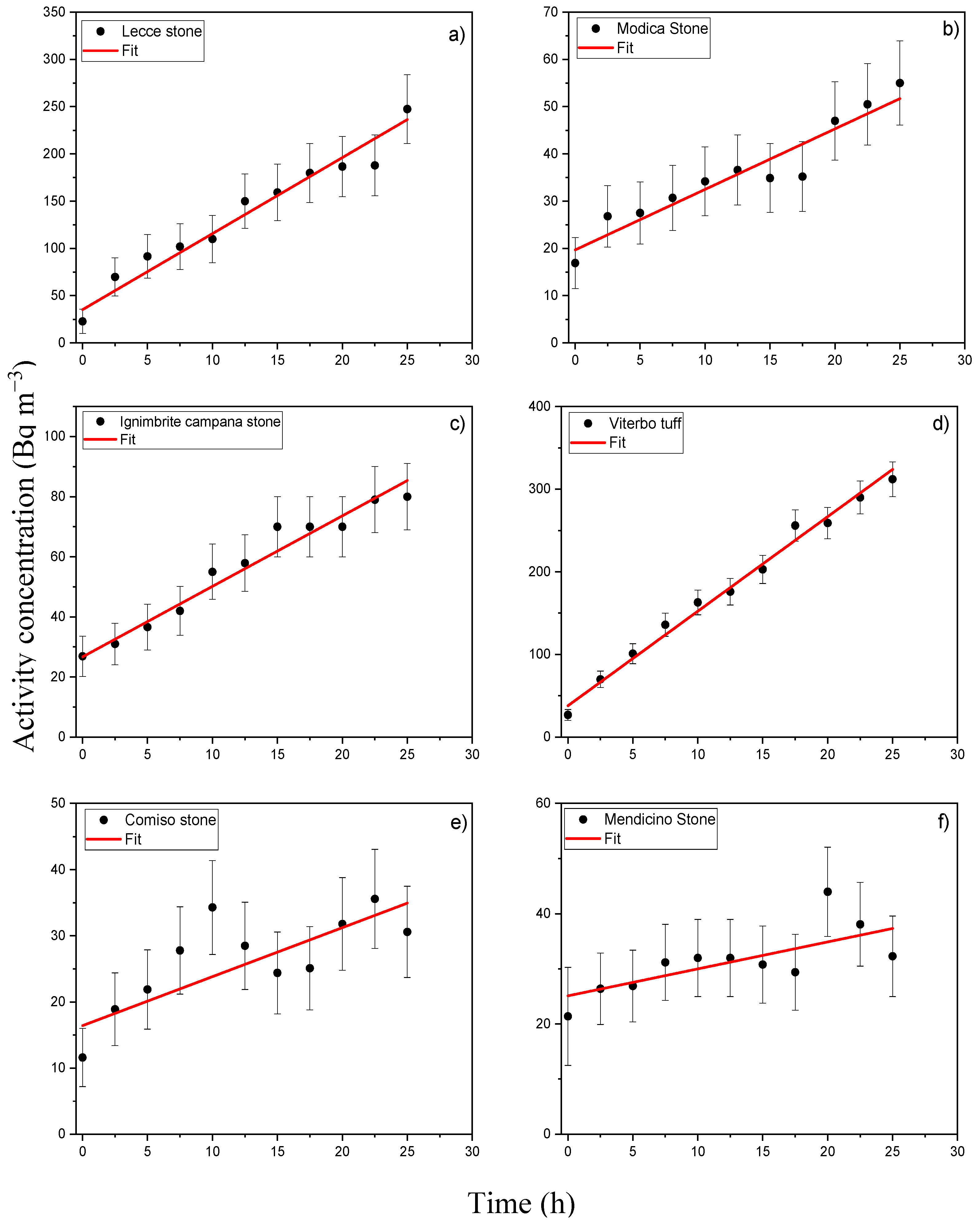
Figure 4 reports the most representative radon concentration curves characterized by a linear increase during the initial 24 h measurement period for all the investigated lithotypes.
Table 2 summarizes the calculated values of the specific radon exhalation rate, together with its uncertainty, for each of the six natural stone samples, as well as the corresponding utest values.
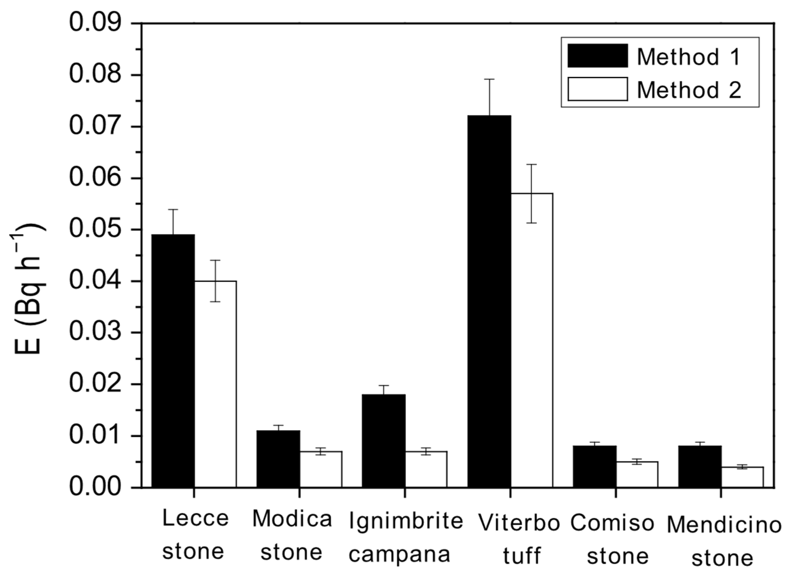
Significantly, exhalation rates varied among stone types due to mineral composition, porosity, and geological origin [68]. For example, the high exhalation from Viterbo tuff reflects its volcanic nature [59]. These variations are relevant for heritage sites where such stones are prevalent. Furthermore, compared to studies in other regions [37,38,68], the radon exhalation rates from the investigated stones are moderate to low. For example, granite samples can reach values over 1 Bq h−1, whereas our highest value (Viterbo tuff) is 0.072 Bq h−1. Finally, the u-test results, reported in Table 2, consistently support the statistical equivalence of the two data collection protocols, with all values being lower or equal to the critical threshold of 2.
Figure 5 reports a comparative bar chart to visually contrast the exhalation rates measured by both protocols.
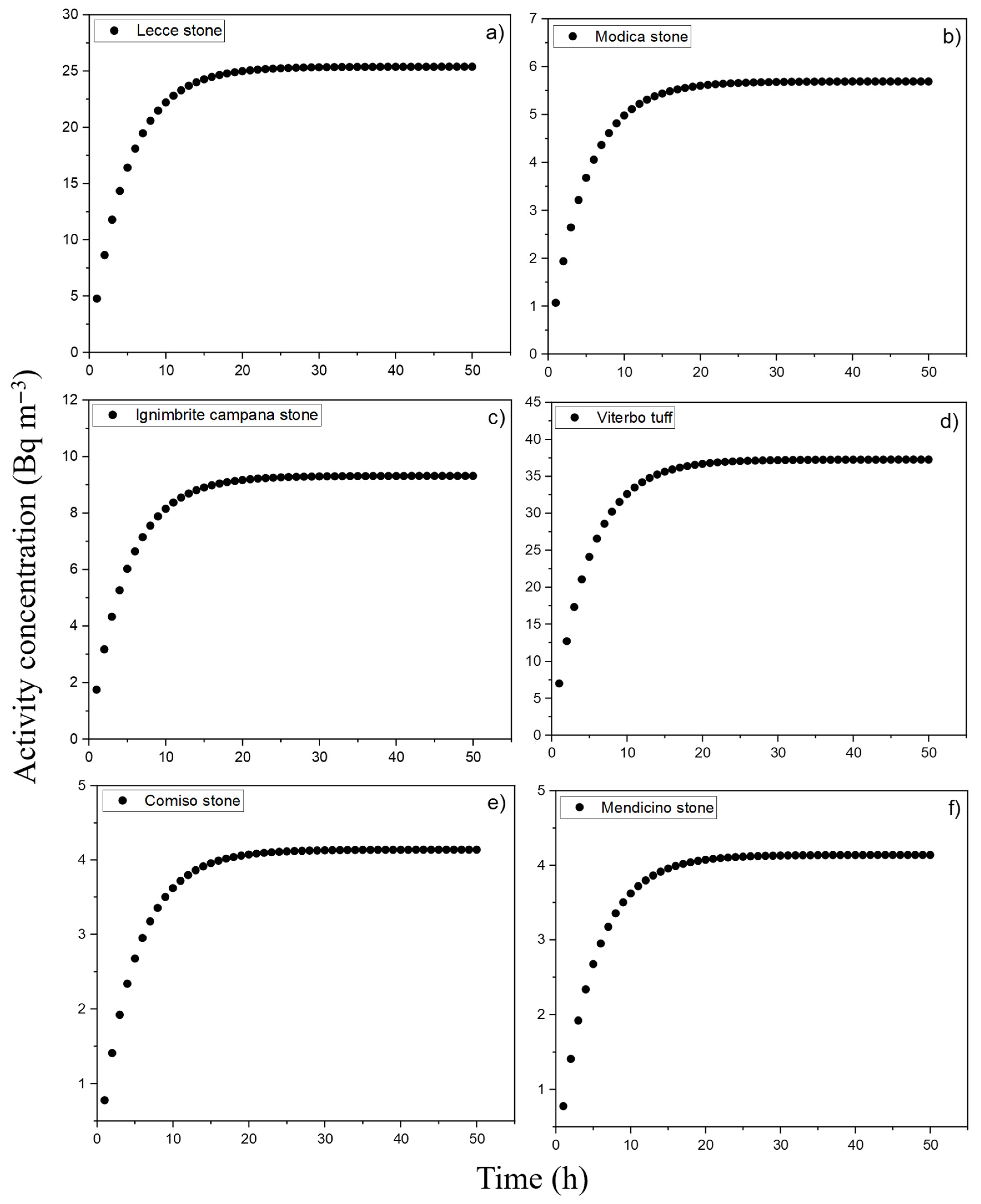
Going on, Figure 6 illustrates the simulated radon accumulation curves derived from the Markkanen room model (based on Equations (5) and (6)).
The simulations were conducted under the conditions reported at the end of the previous section, and assuming the highest radon exhalation rates (as determined by Method 1), i.e., the most conservative scenario [68]. Obtained C values are reported in Table 3.
It is important to highlight that, for all the investigated samples, the simulated indoor radon activity concentrations, ranged from 4.1 to 37.3 Bq m−3, remain well below the threshold values established by Legislative Decree No. 101 of 31 July 2020. Specifically, the decree sets a limit of 300 Bq m−3 (annual average) for existing buildings and workplaces, and a stricter limit of 200 Bq m−3 for new residential constructions starting from 31 December 2024 [45]. Moreover, simulated indoor concentrations are all below the WHO recommended limit of 100 Bq m−3 and the EPA action level of 148 Bq m−3 [41,69].
Given these results, it can be reasonably concluded that, under the tested conditions, no significant radiological hazard from indoor radon exposure is expected for human health. Finally, it is worth noting that the obtained results take into account limitations such as the small number of stone samples per type and the laboratory conditions that may differ from real-world humidity and temperature; nevertheless, poor ventilation or unusual construction, as well as wall thickness and building age, could raise exposure. To this aim, future investigations will be carried out to include in situ validation and a broader range of materials.

5. Conclusions

In this study, the radon exhalation rate of several natural stones of historical and artistic relevance, i.e., Lecce stone, Modica stone, Ignimbrite Campana, Comiso stone, Mendicino stone, and Viterbo tuff, were evaluated using the closed chamber method (CCM) in combination with the Durridge Rad7 system for alpha spectrometry of short-lived radon progeny. Two different analytical approaches were applied: Method 1, based on an extended 10-day measurement period, and Method 2, relying on a more rapid 24 h data acquisition. To determine the accuracy of the two methods, a statistical u-test was performed. Additionally, the Markkanen room model was used to simulate the indoor radon concentration associated with each stone type, providing insights into potential radiological risks. Based on the obtained results, it can be found that:
  • utest values indicated good agreement between the two methods, supporting the validity of the shorter, more time-efficient method for estimating radon exhalation rates. Thus, the faster 24 h method has been validated for use in practical applications, offering time-efficient assessments for construction materials. This has implications for quick screening in construction and heritage preservation—areas where fast decision-making is crucial for worker and public safety and compliance with safety standards.
  • The predicted indoor radon concentrations, for all samples, remained well below the regulatory limits set by Legislative Decree No. 101/2020, as well as the WHO recommended limit and the EPA action level, thereby excluding any significant health concerns under typical indoor conditions.
These results underscore the value of investigating radon emissions from natural building materials, particularly those with cultural and architectural significance. Such assessments are crucial for enhancing radiological safety, informing construction practices, and ensuring compliance with regulatory frameworks. Moreover, the confirmation of a faster and reliable analytical protocol, along with the implementation of a validated room model, offers practical tools for future studies and risk assessments in the domain of environmental radiation protection.

Author Contributions

Conceptualization, F.C. and V.V.; methodology, F.C. and S.M.; validation, D.M. and M.G.; formal analysis, L.P. and F.M.; investigation, F.C., G.P. and V.V.; resources, F.C., D.M. and V.V.; data curation, F.C. and S.M.; writing—original draft preparation, F.C. and L.P.; supervision, D.M., M.G. and V.V. All authors have read and agreed to the published version of the manuscript.

Funding

This research received no external funding.

Institutional Review Board Statement

Not applicable.

Informed Consent Statement

Not applicable.

Data Availability Statement

The data in this study are available upon reasonable request to the corresponding author.

Conflicts of Interest

The authors declare no conflicts of interest.

References

  1. UNSCEAR. United Nations Scientific Committee on the Effects of Atomic Radiation, Vol. I, Annex B: Exposure of the Public and Workers from Various Sources of Radiation, United Nations, New York; UNSCEAR: New York, NY, USA, 2008. [Google Scholar]
  2. Hendry, J.H.; Simon, S.L.; Wojcik, A.; Sohrabi, M.; Burkart, W.; Cardis, E.; Laurier, D.; Tirmarche, M.; Hayata, I. Human exposure to high natural background radiation: What can it teach us about radiation risks? J. Radiol. Prot. 2009, 29, A29–A42. [Google Scholar] [CrossRef] [PubMed]
  3. United Nations Scientific Committee on the Effects of Atomic Radiation. Sources and Effects of Ionizing Radiation: Report to the General Assembly, with Scientific Annexes; UNSCEAR: New York, NY, USA, 2000; ISBN 92-1-142238-8. [Google Scholar]
  4. Wilson, R.R.; Twedt, D.J.; Elliott, A.B. Comparison of Line Transects and Point Counts for Monitoring Spring Migration in Forested Wetlands. J. Field Ornithol. 2000, 71, 345–355. [Google Scholar] [CrossRef]
  5. Shahbazi-Gahrouei, D. Natural Background Radiation Dosimetry in the Highest Altitude Region of Iran. J. Radiat. Res. 2003, 44, 285–287. [Google Scholar] [CrossRef] [PubMed]
  6. Caridi, F.; Messina, M.; Faggio, G.; Santangelo, S.; Messina, G.; Belmusto, G. Radioactivity, radiological risk and metal pollution assessment in marine sediments from Calabrian selected areas, Southern Italy. Eur. Phys. J. Plus 2018, 133, 65. [Google Scholar] [CrossRef]
  7. Pontoriero, A.; Critelli, P.; Conti, A.; Cardali, S.; Angileri, F.F.; Germanò, A.; Lillo, S.; Carretta, A.; Brogna, A.; Santacaterina, A.; et al. The “Combo” radiotherapy treatment for high-risk grade 2 meningiomas: Dose escalation and initial safety and efficacy analysis. J. Neurooncol. 2023, 161, 203–214. [Google Scholar] [CrossRef] [PubMed]
  8. Iatì, G.; Parisi, S.; Santacaterina, A.; Pontoriero, A.; Cacciola, A.; Brogna, A.; Platania, A.; Palazzolo, C.; Cambareri, D.; Davì, V.; et al. Simultaneous Integrated Boost Radiotherapy in Unresectable Stage IV (M0) Head and Neck Squamous Cell Cancer Patients: Daily Clinical Practice. Rep. Pract. Oncol. Radiother. 2020, 25, 399–404. [Google Scholar] [CrossRef] [PubMed]
  9. Cantarini, L.; Brogna, A.; Fioravanti, A.; Galeazzi, M. Henoch-Schönlein purpura associated with pidotimod therapy. Clin. Exp. Rheumatol. 2008, 26, S152. [Google Scholar] [PubMed]
  10. Gupta, T. Radiation, Ionization, and Detection in Nuclear Medicine; Springer: Heidelberg, Germany, 2013; pp. 135–185. ISBN 978-3-642-34075-8. [Google Scholar]
  11. Donya, M.; Radford, M.; Elguindy, A.; Firmin, D.; Yacoub, M. Radiation in medicine: Origins, risks and aspirations. Glob. Cardiol. Sci. Pract. 2014, 2014, 57. [Google Scholar] [CrossRef] [PubMed]
  12. Krumholz, H.; Wang, Y.; Ross, J.; Chen, J.; Ting, H.; Shah, N.; Nasir, K.; Einstein, A.; Nallamothu, B. Exposure to Low-Dose Ionizing Radiation from Medical Imaging Procedures. N. Engl. J. Med. 2009, 361, 849–857. [Google Scholar] [CrossRef]
  13. Parisi, S.; Ferini, G.; Lillo, S.; Brogna, A.; Chillari, F.; Ferrantelli, G.; Settineri, N.; Santacaterina, A.; Platania, A.; Leotta, S.; et al. Stereotactic boost on residual disease after external-beam irradiation in clinical stage III non-small cell lung cancer: Mature results of stereotactic body radiation therapy post radiation therapy (SBRTpostRT) study. Radiol. Medica 2023, 128, 877–885. [Google Scholar] [CrossRef] [PubMed]
  14. Steinhauser, G.; Brandl, A.; Johnson, T.E. Comparison of the Chernobyl and Fukushima nuclear accidents: A review of the environmental impacts. Sci. Total Environ. 2014, 470–471, 800–817. [Google Scholar] [CrossRef] [PubMed]
  15. Omar-Nazir, L.; Shi, X.; Moller, A.; Mousseau, T.; Byun, S.; Hancock, S.; Seymour, C.; Mothersill, C. Long-term effects of ionizing radiation after the Chernobyl accident: Possible contribution of historic dose. Environ. Res. 2018, 165, 55–62. [Google Scholar] [CrossRef] [PubMed]
  16. Moysich, K.B.; Menezes, R.J.; Michalek, A.M. Chernobyl-related ionising radiation exposure and cancer risk: An epidemiological review. Lancet Oncol. 2002, 3, 269–279. [Google Scholar] [CrossRef] [PubMed]
  17. Hasegawa, A.; Tanigawa, K.; Ohtsuru, A.; Yabe, H.; Maeda, M.; Shigemura, J.; Ohira, T.; Tominaga, T.; Akashi, M.; Hirohashi, N.; et al. Health effects of radiation and other health problems in the aftermath of nuclear accidents, with an emphasis on Fukushima. Lancet 2015, 386, 479–488. [Google Scholar] [CrossRef] [PubMed]
  18. Bard, D.; Verger, P.; Hubert, P. Chernobyl, 10 Years After: Health Consequences. Epidemiol. Rev. 1997, 19, 187–204. [Google Scholar] [CrossRef] [PubMed]
  19. Wai, K.-M.; Krstic, D.; Nikezic, D.; Lin, T.-H.; Yu, P. External Cesium-137 doses to humans from soil influenced by the Fukushima and Chernobyl nuclear power plants accidents: A comparative study. Sci. Rep. 2020, 10, 7902. [Google Scholar] [CrossRef] [PubMed]
  20. Caridi, F.; D’Agostino, M.; Belvedere, A.; Marguccio, S.; Belmusto, G. Radon radioactivity in groundwater from the Calabria region, south of Italy. J. Instrum. 2016, 11, P05012. [Google Scholar] [CrossRef]
  21. Caridi, F.; Belmusto, G. Assessment of the public effective dose due to the 222Rn radioactivity in drinking water: Results from the Calabria region, southern Italy. J. Instrum. 2021, 16, P02033. [Google Scholar] [CrossRef]
  22. Torrisi, L.; Caridi, F.; Margarone, D.; Borrielli, A. Characterization of laser-generated silicon plasma. Appl. Surf. Sci. 2008, 254, 2090–2095. [Google Scholar] [CrossRef]
  23. Torrisi, L.; Caridi, F.; Margarone, D.; Giuffrida, L. Nickel plasma produced by 532-nm and 1064-nm pulsed laser ablation. Plasma Phys. Rep. 2008, 34, 547–554. [Google Scholar] [CrossRef]
  24. Caridi, F.; Belmusto, G. Overview of the technologies for assessment of natural radioactivity in drinking water. J. Instrum. 2019, 14, T02002. [Google Scholar] [CrossRef]
  25. Cantor, K. Drinking water and cancer. Cancer Causes Control 1997, 8, 292–308. [Google Scholar] [CrossRef] [PubMed]
  26. Barbosa-Lorenzo, R.; Barros-Dios, J.M.; Ruano-Ravina, A. Radon and stomach cancer. Int. J. Epidemiol. 2017, 46, 767–768. [Google Scholar] [CrossRef] [PubMed]
  27. Auvinen, A.; Salonen, L.; Pekkanen, J.; Pukkala, E.; Ilus, T.; Kurttio, P. Radon and other natural radionuclides in drinking water and risk of stomach cancer: A case-cohort study in Finland. Int. J. Cancer 2005, 114, 109–113. [Google Scholar] [CrossRef] [PubMed]
  28. Mills, W.A. Risk Assessment and Control Management of Radon in Drinking Water; Lewis Publishers, Inc.: Chelsea, MI, USA, 2025. [Google Scholar]
  29. Caridi, F.; Chiriu, D.; Pelo, S.D.; Faggio, G.; Guida, M.; Messina, G.; Ponte, M.; Ruffolo, S.A.; Majolino, D.; Venuti, V. Radon Exhalation Rate, Radioactivity Content, and Mineralogy Assessment of Significant Historical and Artistic Interest Construction Materials. Appl. Sci. 2024, 14, 11359. [Google Scholar] [CrossRef]
  30. Baskaran, M. Radon: A Tracer for Geological, Geophysical and Geochemical Studies; Springer: Basel, Switzerland, 2016; ISBN 978-3-319-21328-6. [Google Scholar]
  31. Mousavi Aghdam, M.; Crowley, Q.; Rocha, C.; Dentoni, V.; Da Pelo, S.; Long, S.; Savatier, M. A Study of Natural Radioactivity Levels and Radon/Thoron Release Potential of Bedrock and Soil in Southeastern Ireland. Int. J. Environ. Res. Public Health 2021, 18, 2709. [Google Scholar] [CrossRef] [PubMed]
  32. Coletti, C.; Ciotoli, G.; Benà, E.; Brattich, E.; Cinelli, G.; Galgaro, A.; Massironi, M.; Mazzoli, C.; Mostacci, D.; Morozzi, P.; et al. The assessment of local geological factors for the construction of a Geogenic Radon Potential map using regression kriging. A case study from the Euganean Hills volcanic district (Italy). Sci. Total Environ. 2022, 808, 152064. [Google Scholar] [CrossRef] [PubMed]
  33. Da Pelo, S.; Mousavi Aghdam, M.; Dentoni, V.; Loi, A.; Randaccio, P.; Crowley, Q. Assessment of natural radioactivity and radon release potential of silurian black shales. Radiat. Phys. Chem. 2023, 215, 111347. [Google Scholar] [CrossRef]
  34. La Verde, G.; Raulo, A.; D’avino, V.; Paternoster, G.; Roca, V.; La Commara, M.; Pugliese, M. Pietra leccese and other natural stones in puglia region: A new category of building materials for radiation protection? Int. J. Environ. Res. Public Health 2021, 18, 11213. [Google Scholar] [CrossRef] [PubMed]
  35. Dentoni, V.; Da Pelo, S.; Mousavi Aghdam, M.; Randaccio, P.; Loi, A.; Careddu, N.; Bernardini, A. Natural radioactivity and radon exhalation rate of Sardinian dimension stones. Constr. Build. Mater. 2020, 247, 118377. [Google Scholar] [CrossRef]
  36. Mancini, S.; Guida, M.; Cuomo, A.; Guida, D.; Ismail, A.H. Modelling of indoor radon activity concentration dynamics and its validation through in-situ measurements on regional scale. In Proceedings of the AIP Conference Proceedings, Cambridge, UK, 16–18 February 2018; Volume 1982. [Google Scholar]
  37. Bulut, H.A.; Şahin, R. Radon, Concrete, Buildings and Human Health—A Review Study. Buildings 2024, 14, 510. [Google Scholar] [CrossRef]
  38. Lee, H.; Lee, J.; Yoon, S.; Lee, C. 222 rn exhalation rates from some granite and marble used in korea: Preliminary study. Atmosphere 2021, 12, 1057. [Google Scholar] [CrossRef]
  39. Chen, J.; Rahman, N.M.; Atiya, I.A. Radon exhalation from building materials for decorative use. J. Environ. Radioact. 2010, 101, 317–322. [Google Scholar] [CrossRef] [PubMed]
  40. Estokova, A.; Singovszka, E.; Vertal, M. Investigation of Building Materials’ Radioactivity in a Historical Building—A Case Study. Materials 2022, 15, 6876. [Google Scholar] [CrossRef] [PubMed]
  41. WHO. WHO Indoor Radon a Public Health Perspective; World Health Organization: Geneva, Switzerland, 2021; p. 110. [Google Scholar]
  42. Wang, J.; Wang, H.; Qian, H. Biological effects of radiation on cancer cells. Mil. Med. Res. 2018, 5, 20. [Google Scholar] [CrossRef] [PubMed]
  43. Reisz, J.A.; Bansal, N.; Qian, J.; Zhao, W.; Furdui, C.M. Effects of ionizing radiation on biological molecules—Mechanisms of damage and emerging methods of detection. Antioxid. Redox Signal. 2014, 21, 260–292. [Google Scholar] [CrossRef] [PubMed]
  44. Darby, S.; Hill, D.; Auvinen, A.; Barros-Dios, J.M.; Baysson, H.; Bochicchio, F.; Deo, H.; Falk, R.; Forastiere, F.; Hakama, M.; et al. Radon in homes and risk of lung cancer: Collaborative analysis of individual data from 13 European case-control studies. BMJ 2005, 330, 223. [Google Scholar] [CrossRef] [PubMed]
  45. Legislation Italian D.Lgs. 101/20. 2020. Available online: https://www.normattiva.it/uri-res/N2Ls?urn:nir:stato:decreto.legislativo:2020-07-31;101 (accessed on 20 June 2025).
  46. Zhang, L.; Lei, X.; Guo, Q.; Wang, S.; Ma, X.; Shi, Z. Accurate measurement of the radon exhalation rate of building materials using the closed chamber method. J. Radiol. Prot. 2012, 32, 315–323. [Google Scholar] [CrossRef] [PubMed]
  47. Tuccimei, P.; Mollo, S.; Soligo, M.; Scarlato, P.; Castelluccio, M. Real-time setup to measure radon emission during rock deformation: Implications for geochemical surveillance. Geosci. Instrum. Methods Data Syst. Discuss. 2015, 4, 111–119. [Google Scholar] [CrossRef]
  48. Yanchao, S.; Junlin, W.; Bing, S.; Hongxing, C.; Wu, Y. Study on a new charcoal closed chamber method for measuring radon exhalation rate of building materials. Radiat. Meas. 2020, 134, 106308. [Google Scholar] [CrossRef]
  49. Tuccimei, P.; Castelluccio, M.; Soligo, M.; Moroni, M. Radon exhalation rates of building materials: Experimental, analytical protocol and classification criteria. In Building Materials: Properties, Performance and Applications; Nova Science Publishers, Inc.: New York, NY, USA, 2009; pp. 259–273. [Google Scholar]
  50. Bossio, A.; Mazzei, R.; Monteforti, B.; Salvatorini, G. Stratigrafia del neogene e quaternario del salento sud-orientale (con rilevamento geologico alla scala 1:25.000). Geol. Rom. 2005, 38, 31–60. [Google Scholar]
  51. Mazzei, R.; Margiotta, S.; Foresi, L.M.; Riforgiato, F.; Gianfranco, S. Mazzei Biostratigraphy and chronostratigraphy of the Miocene Pietra Leccese in the type area of Lecce (Apulia, southern Italy). Boll. Della Soc. Paleontol. Ital. 2009, 48, 129–145. [Google Scholar]
  52. Punturo, R.; Russo, L.; Giudice, A.; Mazzoleni, P.; Pezzino, A. Building stone employed in the historical monuments of Eastern Sicily (Italy). An example: The ancient city centre of Catania. Environ. Geol. 2006, 50, 156–169. [Google Scholar] [CrossRef]
  53. La Russa, M.F.; Belfiore, C.M.; Fichera, G.V.; Maniscalco, R.; Calabrò, C.; Ruffolo, S.A.; Pezzino, A. The behaviour to weathering of the Hyblean limestone in the Baroque architecture of the Val di Noto (SE Sicily): An experimental study on the “calcare a lumachella” stone. Constr. Build. Mater. 2015, 77, 7–19. [Google Scholar] [CrossRef]
  54. Forni, F.; Bachman, O.; Mollo, S.; De Astis, G. The origin of a zoned ignimbrite: Insights into the Campanian Ignimbrite magma chamber (Campi Flegrei, Italy). Earth Planet. Sci. Lett. 2016, 449, 259–271. [Google Scholar] [CrossRef]
  55. Calcaterra, D.; Cappelletti, P.; Langella, A.; Colella, A.; Gennaro, M. The ornamental stones of Caserta province: The Campanian Ignimbrite in the medieval architecture of Casertavecchia. J. Cult. Herit. 2004, 5, 137–148. [Google Scholar] [CrossRef]
  56. Ruffolo, S.A.; La Russa, M.F.; Ricca, M.; Belfiore, C.M.; Macchia, A.; Comite, V.; Pezzino, A.; Crisci, G.M. New insights on the consolidation of salt weathered limestone: The case study of Modica stone. Bull. Eng. Geol. Environ. 2017, 76, 11–20. [Google Scholar] [CrossRef]
  57. Olivetti, V.; Cyr, A.J.; Molin, P.; Faccenna, C.; Granger, D.E. Uplift history of the Sila Massif, southern Italy, deciphered from cosmogenic 10Be erosion rates and river longitudinal profile analysis. Tectonics 2012, 31, TC3007. [Google Scholar] [CrossRef]
  58. Forestieri, G.; Alvarez de Buergo, M. Relationships Between Petrophysical and Mechanical Properties of Certain Calcarenites Used in Building. Geotech. Geol. Eng. 2021, 39, 5021–5040. [Google Scholar] [CrossRef]
  59. Giampaolo, C.; Mengarelli, L.; Torracca, E.; Spencer, C. Zeolite characterization of “Vico red tuff with black scoria” ignimbrite flow: The extractive district of Civita Castellana (Viterbo, Italy). Nuovo Cim. B 2008, 123, 1459–1476. [Google Scholar] [CrossRef]
  60. Marra, F.; Taddeucci, J.; Freda, C.; Marzocchi, W.; Scarlato, P. Recurrence of volcanic activity along the Roman Comagmatic Province (Tyrrhenian margin of Italy) and its tectonic significance. Tectonics 2004, 23, TC4013. [Google Scholar] [CrossRef]
  61. Todorovic, N.; Nikolov, J.; Forkapic, S.; Bikit, I.; Mrdja, D.; Krmar, M.; Veskovic, M. Public exposure to radon in drinking water in Serbia. Appl. Radiat. Isot. Incl. Data Instrum. Methods Use Agric. Ind. Med. 2012, 70, 543–549. [Google Scholar] [CrossRef] [PubMed]
  62. Hasan, A.; Subber, A.; Shaltakh, A. Measurement of Radon Concentration in Soil Gas using RAD7 in the Environs of Al-Najaf Al-Ashraf City-Iraq. Adv. Appl. Sci. Res. 2011, 2, 273–278. [Google Scholar]
  63. Tuccimei, P.; Moroni, M.; Norcia, D. Simultaneous determination of 222Rn and 220Rn exhalation rates from building materials used in Central Italy with accumulation chambers and a continuous solid state alpha detector: Influence of particle size, humidity and precursors concentration. Appl. Radiat. Isot. 2006, 64, 254–263. [Google Scholar] [CrossRef] [PubMed]
  64. Burnett, W.; Dulai, H. Estimating the dynamics of groundwater input into the coastal zone via continuous radon-222 measurements. J. Environ. Radioact. 2003, 69, 21–35. [Google Scholar] [CrossRef] [PubMed]
  65. Petropoulos, N.; Anagnostakis, M.; Simopoulos, S. Building materials radon exhalation rate: ERRICCA intercomparison exercise results. Sci. Total Environ. 2001, 272, 109–118. [Google Scholar] [CrossRef] [PubMed]
  66. Mann, H.B.; Whitney, D.R. On a Test of Whether one of Two Random Variables is Stochastically Larger than the Other. Ann. Math. Stat. 1947, 18, 50–60. [Google Scholar] [CrossRef]
  67. Markkanen, M. Radiation Dose Assessments for Materials with Elevated Natural Radioactivity; Finnish Centre for Radiation and Nuclear Safety: Helsinki, Finland, 1995; Volume 2, ISBN 9517120796.
  68. Righi, S.; Bruzzi, L. Natural radioactivity and radon exhalation in building materials used in Italian dwellings. J. Environ. Radioact. 2006, 88, 158–170. [Google Scholar] [CrossRef] [PubMed]
  69. US Environmental Protection Agency. Exposure Factors Handbook, 2011th ed.; US Environmental Protection Agency: Washington, DC, USA, 2011.
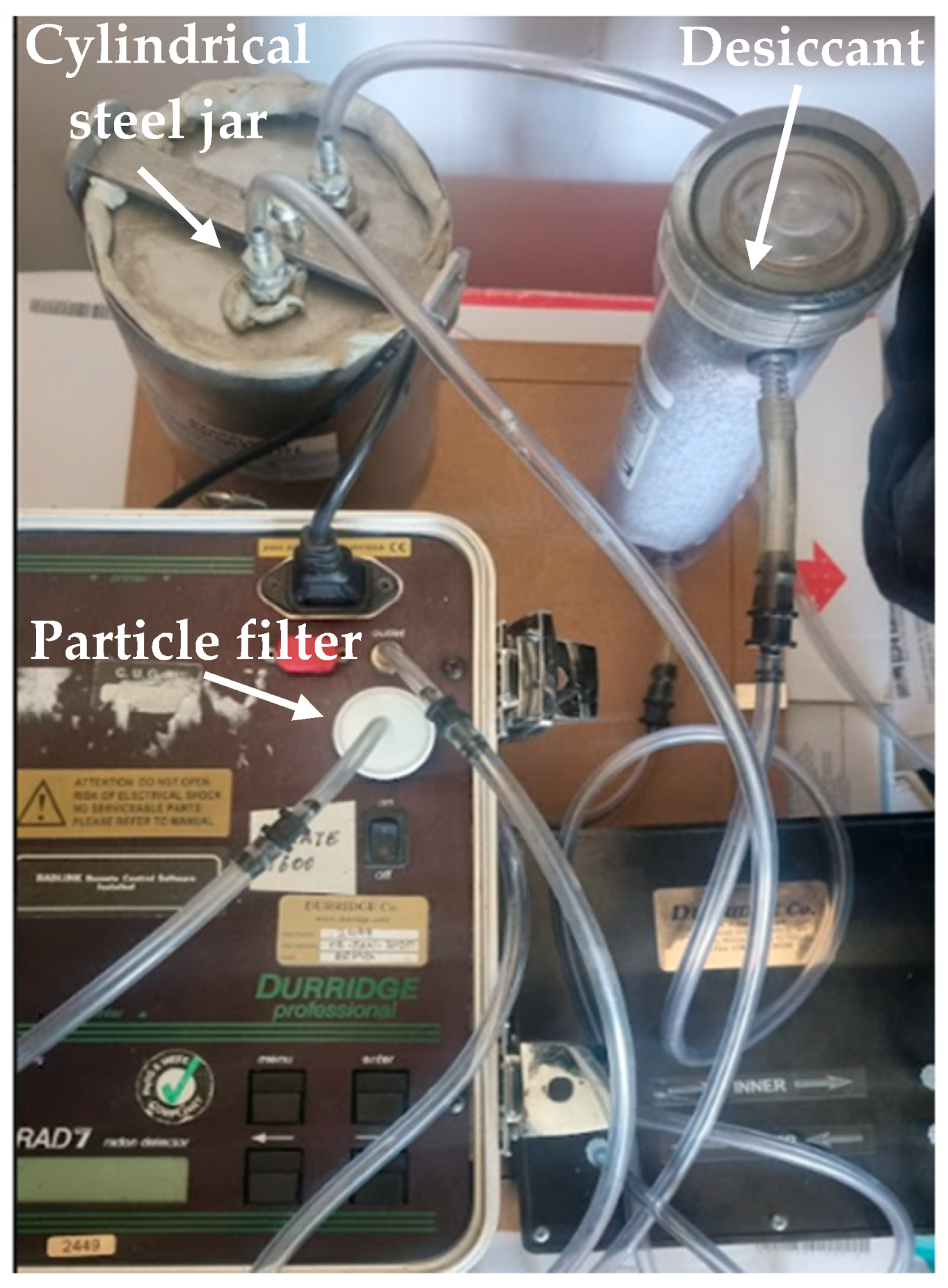
Figure 1. Experimental set-up of the Durridge Rad7 for the radon exhalation assessment.
Applsci 15 08015 g001
Figure 2. A schematic flowchart that summarizes the key steps in the experimental process.
Applsci 15 08015 g002
Figure 3. The most representative radon growth curves to equilibrium, through Method 1, for the Lecce stone (a), Modica stone (b), Ignimbrite campana (c), and Viterbo tuff (d).
Applsci 15 08015 g003
Figure 4. The most representative radon growth curves to equilibrium, obtained applying Method 2, for Lecce stone (a), Modica stone (b), Ignimbrite campana (c), Viterbo tuff (d), Comiso stone (e), and Mendicino stone (f).
Applsci 15 08015 g004
Figure 5. A comparative bar chart.
Applsci 15 08015 g005
Figure 6. Radon accumulation growth curve calculated by applying the Markkanen room model in the most precautionary scenario of highest radon exhalation rate values, (i.e., as assessed by using Method 1) standard-sized room (4 m × 5 m × 2.8 m), and minimum ventilation rate (0.2 h−1) for the six investigated natural stones.
Applsci 15 08015 g006
Table 1. The fitted effective decay constant for all investigated samples.
Sampleλ (h−1)
Lecce stone0.023 ± 0.001
Modica stone0.027 ± 0.001
Ignimbrite Campana0.017 ± 0.001
Comiso stone0.050 ± 0.004
Mendicino stone0.081 ± 0.011
Viterbo tuff0.018 ± 0.001
Table 2. Specific radon exhalation rate, together with its uncertainty, and corresponding u-test values for the analyzed samples.
SampleE (Bq h−1)
Method 1
E (Bq h−1)
Method 2
u-Test
Lecce stone0.049 ± 0.0120.040 ± 0.0100.6
Modica stone0.011 ± 0.0030.007 ± 0.0021.1
Ignimbrite Campana0.018 ± 0.0050.007 ± 0.0022.0
Viterbo tuff0.072 ± 0.0180.057 ± 0.0140.7
Comiso stone0.008 ± 0.0020.005 ± 0.0011.3
Mendicino stone0.008 ± 0.0020.004 ± 0.0011.8
Table 3. Indoor activity concentration values simulated using the Markkanen room model.
SampleSimulated Activity Concentration
(Bq m−3)
Lecce stone25.4
Modica stone5.7
Ignimbrite campana9.3
Viterbo tuff37.3
Comiso stone4.1
Mendicino stone4.1
Disclaimer/Publisher’s Note: The statements, opinions and data contained in all publications are solely those of the individual author(s) and contributor(s) and not of MDPI and/or the editor(s). MDPI and/or the editor(s) disclaim responsibility for any injury to people or property resulting from any ideas, methods, instructions or products referred to in the content.