Article

Quantitative Evaluation of Marginal and Internal Fit of CAD/CAM Ceramic Crown Restorations Obtained by Model Scanner, Intraoral Scanner, and Different CBCT Scans

1
Department of Prosthodontics, Faculty of Dentistry, Ankara University, Ankara 06560, Turkey
2
Department of Basic Medicine Science, Anatomy, Faculty of Dentistry, Ankara University, Ankara 06560, Turkey
3
Department of Oral and Maxillofacial Radiology, Faculty of Dentistry, Ankara University, Ankara 06560, Turkey
4
Department of Oral Diagnostics, Faculty of Dentistry, Semmelweis University, 1085 Budapest, Hungary
*
Author to whom correspondence should be addressed.
Appl. Sci. 2025, 15(14), 8017; https://doi.org/10.3390/app15148017
Submission received: 1 June 2025 / Revised: 13 July 2025 / Accepted: 14 July 2025 / Published: 18 July 2025

Abstract

(1) Background: This study aimed to evaluate the marginal and internal fit of ceramic crowns produced by various digital methods using microcomputed tomography (MCT) imaging. (2) Methods: The ceramic crown preparation was performed on typodont maxillary first premolar. The crown preparation was scanned with an intraoral scanner and a model scanner, and cone-beam computed tomography (CBCT) scans were performed with three different voxel sizes (0.075 mm, 0.1 mm, and 0.15 mm). The space between the crown and prepared teeth was measured at nine different points in both coronal and sagittal sections. Three different digital model acquisition techniques, namely, intraoral scanning, model scanning, and CBCT-based standard tessellation language (STL) reconstruction, were compared in terms of marginal and internal fit. (3) Results: Quantitative analyses revealed that model scanners exhibited the lowest marginal and internal gap values, indicating superior fit compared to intraoral scanners and CBCT-based models. The highest gap values were observed in the CBCT group with a voxel size of 0.15 mm. Overall, crowns obtained from model scanners demonstrated the highest success rates in both marginal and internal fit. (4) Conclusions: In conclusion, this study highlights the critical role of digital scanning accuracy in achieving clinically acceptable prosthetic fits and emphasizes the need for continued technological advancement.

1. Introduction

The marginal and internal fit of fixed prosthetic restorations applied in dentistry significantly affects the success and lifespan of fixed prosthetic restorations. In prostheses where successful marginal and internal fit of the crown cannot be achieved, plaque accumulation may lead to microleakage, resulting in issues such as caries, pulpal inflammation, periodontal disease, and a decrease in the retention of the restoration [1,2,3,4]. For this reason, obtaining an accurate impression that clearly reveals the marginal and internal fit is the first step in producing a successful prosthesis. However, traditional intraoral impressions can be challenging for both clinicians and patients due to factors such as gag reflex, lack of comfort, or the risk of soft tissue damage. Additionally, environmental conditions like temperature and humidity changes during the transfer of the impression to the laboratory may cause dimensional changes in the impression material. These factors can negatively impact the marginal and internal fit of the prosthesis [5,6,7].
With the advancement of digital dentistry, computer-aided design (CAD) and computer-aided manufacturing (CAM) technologies have replaced traditional impressions, which have some disadvantages [8]. This process involves scanning the prepared tooth surface and then designing the crowns. This workflow consists of sequential stages including data acquisition, design, and manufacturing. In the data acquisition phase, digital impressions are obtained either directly using intraoral scanners or indirectly via model scanners, resulting in the creation of a 3D model in standard tessellation language (STL) format. During the design phase, specialized software is used to digitally design the restoration. In the final stage, the designed restoration is fabricated using a CAM device. CAD/CAM technology is increasingly used in dental prosthetic restorations because it eliminates the drawbacks of traditional impression methods. It reduces laboratory time and material costs while increasing productivity. With the development of different manufacturing systems, a wide range of dental materials, such as ceramics, acrylic resins, waxes, and metal alloys, are now available in prefabricated block form for prosthetic restorations. In this context, the acquisition of digital data and models has become of great importance [9,10].
These models can be obtained directly with intraoral scanners or indirectly with laboratory scanners. The indirect method begins with a traditional impression, followed by the creation of a model, which is then typically digitized using three-dimensional (3D) laboratory scanners. As a third option, cone-beam computed tomography (CBCT) images can be converted into the STL using CAD software (Cerec CAD System, Sirona Dental Systems GmbH, Bensheim, Germany) and used in the production of prosthetic restorations without the need for traditional impression-taking [10,11,12,13].
Many studies have been conducted on the marginal fit of crowns produced using CAD-CAM technology and those obtained by traditional methods. The evaluation of marginal and internal fit is of critical importance. Furthermore, various factors such as scanning, design, and milling processes can affect the accuracy of the fit of restorations. The success of the restoration is directly related to the marginal and internal fit of the crown, and ensuring this integrity is determined by fundamental principles [13,14,15,16,17]. An in vitro alternative that is widely used to evaluate the marginal and internal fit success of crowns is microcomputed tomography (MCT). MCT is a non-destructive digital method for the 2D or 3D radiographic analysis of internal and marginal gaps in restorations. This method utilizes a cone-beam X-ray source and specialized reconstruction software to generate cross-sectional images for a comprehensive assessment of marginal and internal gaps [18]. Although no clear definition of acceptable marginal fit has been made, McLean et al. emphasized in a study involving 1000 restorations that the marginal discrepancy should be less than 120 µm [19]. Some researchers, however, state that the marginal discrepancy in CAD-CAM restorations should not exceed 100 µm. In these studies, factors such as cement space, cementation method, length of the prosthesis, material properties of the prosthesis, CAD-CAM production, and/or the method of obtaining the virtual model have been evaluated for their impact on the edge integrity. However, studies on the marginal integrity of crowns produced using CBCT scans are quite limited [15,16,17,18,19,20,21,22,23].
The aim of this study is to evaluate the marginal and internal fit of full ceramic crowns produced by different digital methods through micro-CT, and to compare the success of CAD-CAM systems and CBCT scans.

2. Materials and Methods

The ceramic crown preparation was performed on typodont maxillary first premolar. Tooth preparation was made by an experienced prosthodontist (B.A.) for complete-coverage zirconia incorporating 2 mm occlusal reduction, 1 mm axial reduction, and 360° 1 mm deep chamfer margin with rounded internal line angles. The restoration was fabricated with milling single-feldspathic ceramic blocks (CEREC Blocks; Sirona Dental Systems GmbH, Bensheim, Germany).
The crown preparation was scanned with an intraoral scanner (CEREC Omnicam system, Sirona Dental Systems GmbH, Bensheim, Germany), a model scanner (inEOS X5, Sirona Dental Systems GmbH, Bensheim, Germany), and CBCT (Planmeca Promax 3D Max Machine, Helsinki, Finland).
CBCT scans were performed with 3 different voxel sizes (0.075 mm, 0.1 mm, and 0.15 mm), the smallest distinguishable box-shaped unit of a three-dimensional digital image. These voxel sizes were chosen to reflect commonly used clinical resolutions in dental CBCT imaging, balancing diagnostic detail and radiation exposure, as supported by prior studies [24,25]. To standardize the imaging technique, all scans were acquired at 90 kVp, 12 mA, and a field of view (FOV) size of 50 × 40 mm. The DICOM data obtained from CBCT scans (Planmeca Promax 3D Max) were converted into STL format using the Planmeca Romexis software (version 6.2, Helsinki, Finland), which includes automated segmentation and mesh generation capabilities. To reduce voxel-induced surface noise and artifacts, the software’s built-in surface processing functions were used, including smoothing and contour optimization algorithms. All conversions were performed using consistent segmentation thresholds and under identical lighting and model orientation conditions. While these filtering steps aim to enhance surface continuity and suppress digital noise, the potential influence of voxel resolution and segmentation limitations on the final geometry must be acknowledged. No manual editing was applied to the mesh data to preserve comparability across groups. These steps were taken to ensure that STL models accurately reflect the reconstructed geometries derived from each CBCT voxel size and remain suitable for downstream CAD design and fit analysis.
Crown restorations for the upper first premolar were designed by transferring the 3D STL images obtained from the intraoral scanner, laboratory scanner, and CBCT scans to a CAD software (Cerec CAD System Sirona Dental Systems GmbH, Bensheim, Germany). The cement gap was set as 120 μm in the marginal area and 200 μm in the remaining areas inside the crown restoration. After the design of the total crown restoration was completed, the crown restorations were milled using feldspathic blocks (CEREC Blocs; Sirona Dental Systems GmbH, Bensheim, Germany) on a milling machine (Cerec In Lab MC XL, Sirona Dental Systems).
In this study, marginal fit was defined as the perpendicular distance between the crown margin and the preparation finish line. Internal fit refers to the space between the inner surface of the crown and the axial or occlusal surfaces of the tooth. The cement gap is the designed internal spacing within the CAD software intended to accommodate the luting agent, which was set at 120 µm near the margins and 200 µm internally. These definitions were used consistently throughout the study.
Marginal gap and internal fit values were measured using MCT. The high-resolution scanning capable Skyscan 1275 (Skycan, Kontich, Belgium) was used for MCT scans. Scanning parameters were set as follows: 125 kVp, 80 mA, 20 μm/pixel, and a rotation step of 0.2. A 1 mm thick aluminum filter was used to prevent artifacts during the scans. Subsequently, each scanned sample was reconstructed individually using Skycan’s NRecon (version 1.7.4.6 Skycan, Kontich, Belgium) software.

2.1. Linear Measurements

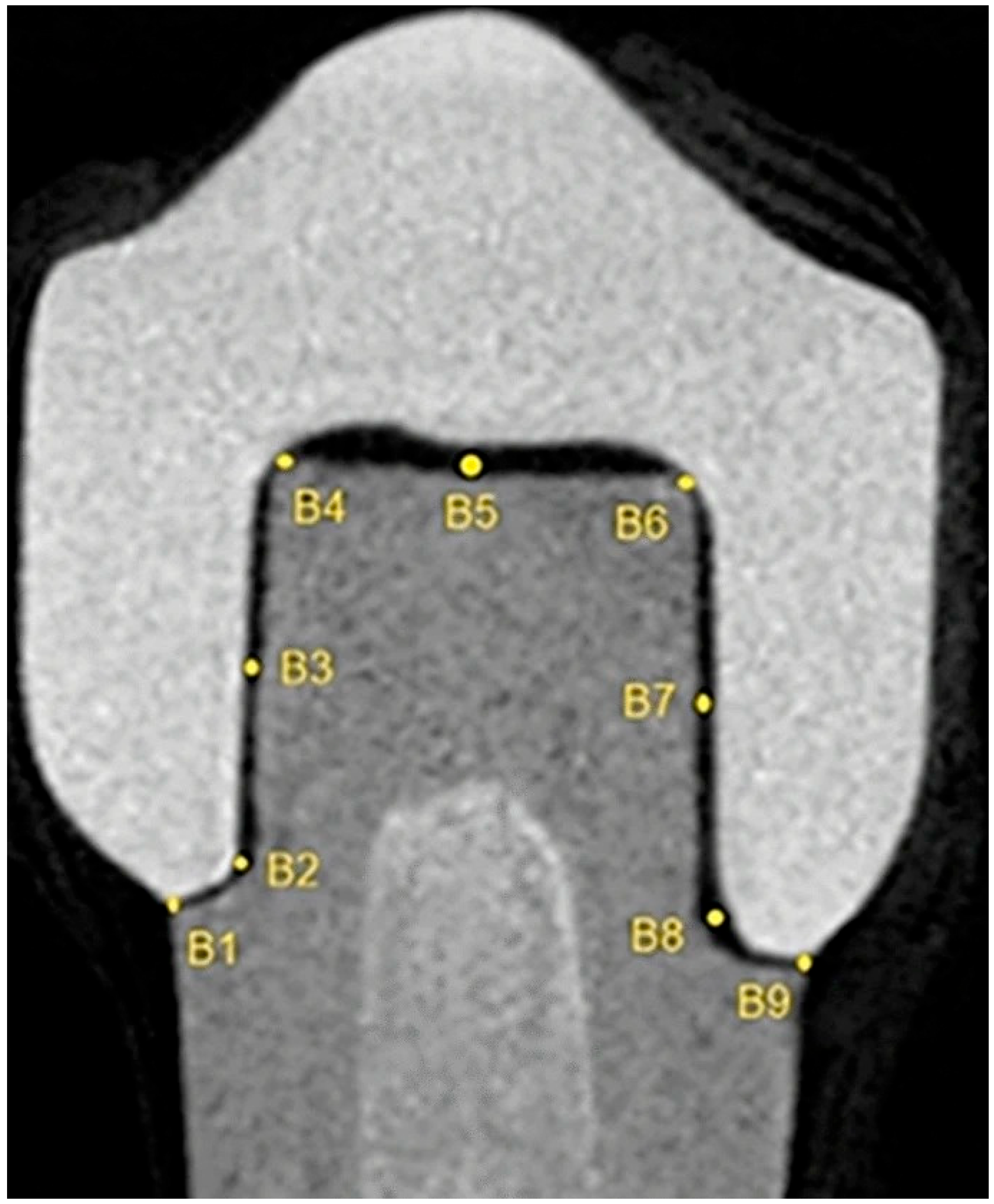
In the sagittal section, the space between the crown and the prepared tooth was measured from 9 different points (A1, A2… A9), starting from the buccal surface (Figure 1). In the coronal section, the space between the crown and the prepared tooth was measured from 9 different points (B1, B2… B9), starting from the mesial surface (Figure 2).

2.2. Statistical Analysis

In the statistical evaluation, firstly, descriptive statistics were presented by calculating mean ± standard deviation, minimum–maximum, and 95% confidence intervals for all continuous data. The arithmetic mean was obtained for each specimen from its nine point-specific gap values (A1–A9 or B1–B9), and the standard deviation was computed as the sample SD with Bessel’s correction, ensuring an unbiased estimate of population variance. Then, distribution characteristics were examined for each group with the Shapiro–Wilk test; parametric methods were preferred since p > 0.05 values supported the normal distribution assumption. Equality of variance between groups was tested with Levene’s test; this analysis confirmed homogeneity (p = 0.066) in internal-fit variable, while it revealed heterogeneity (p = 0.012) in marginal-fit variable. Levene’s statistic was calculated from absolute deviations (Zᵢⱼ = |Yᵢⱼ – Ῡᵢ·|) and yielded F(4, 55) = 2.19, p = 0.066 for internal fit versus F(4, 55) = 3.71, p = 0.012 for marginal fit. In this direction, classical one-way ANOVA was applied for measuring internal fit with homogeneous variances, and Welch ANOVA was applied for measuring marginal fit with heterogeneous variances (α = 0.05). Multiple comparisons in variables with significant general differences were made with Tukey’s HSD for internal fit with homogeneity of variances, and with Welch’s t-test between each paired group for non-homogeneous marginal fit; the obtained p values were adjusted with Holm correction. The sample size in each group was defined as 12 (n = 12).

3. Results

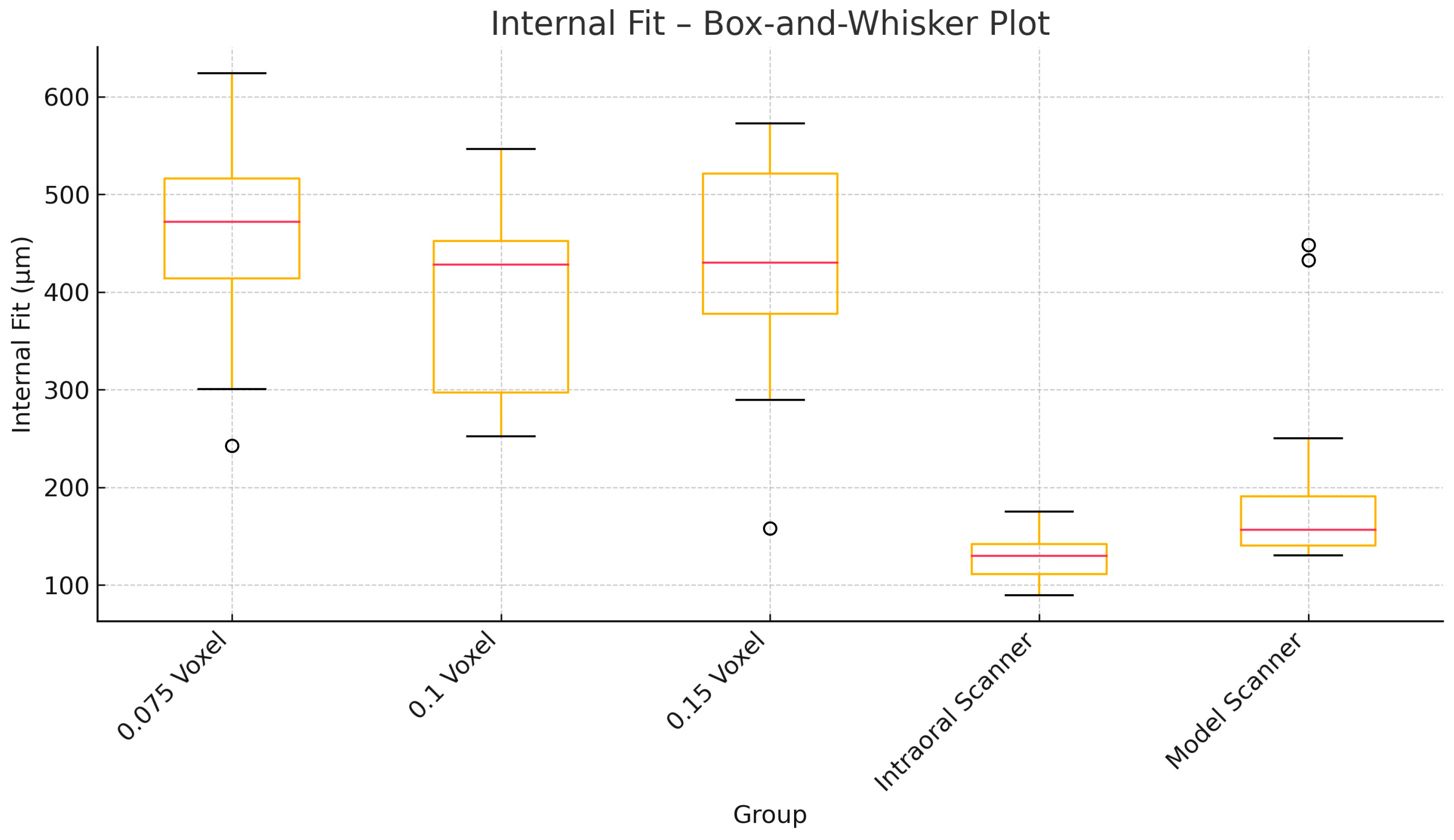
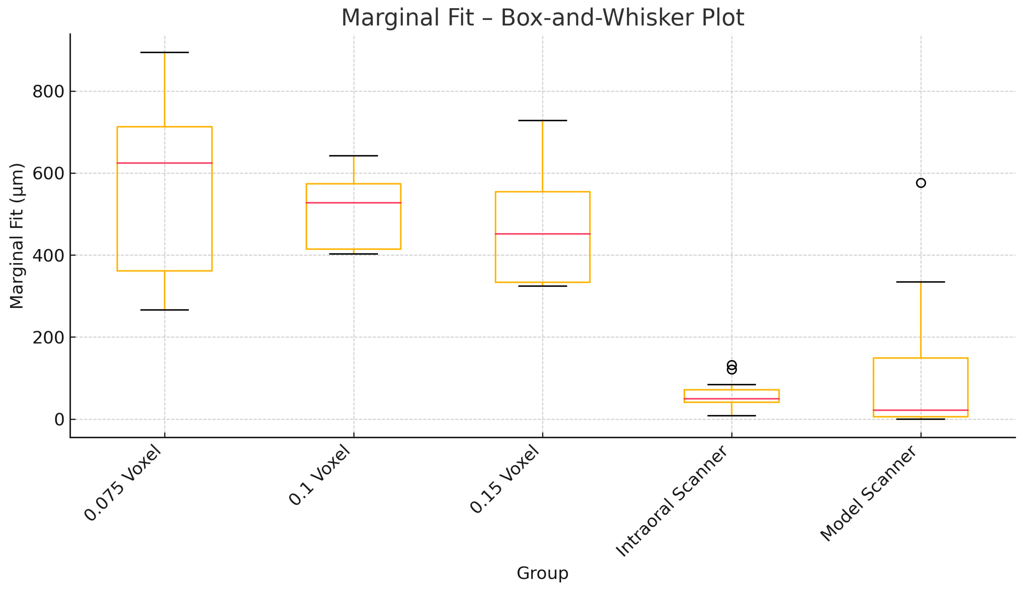
As a result of the linear measurements, compatibility was detected between the coronal and sagittal section data. When the measurements performed with different tomography voxel sizes (0.075 mm, 0.1 mm, and 0.15 mm) were compared among themselves, the 0.15 voxel group showed significantly higher marginal and internal fit discrepancies than the other voxel groups (p < 0.05). Similarly, the 0.15 voxel group showed significantly higher gap values (marginal gap: 580 ± 204 µm, internal gap: 502 ± 106 µm) compared to the intraoral and model scanner groups (p < 0.05). (Figure 3 and Figure 4). The difference between the 0.1 voxel and 0.075 voxel groups and each other was not statistically significant (p > 0.05).
Both intraoral and model scanners showed lower gap values than the values at 0.75 voxel, which is the lowest marginal and internal gap value. That is, the marginal and internal fits of the crowns created with scanners were found to be significantly higher than the fits of the crowns created by tomography (p < 0.05). Among the scanners, the model scanner showed significantly lower gap values than the intraoral scanner in terms of both internal and marginal fit (p < 0.05) (Table 1 and Table 2).

4. Discussion

There are many studies in the literature evaluating the marginal and internal fit of prosthetic dental restorations using different methods such as scanners, CBCT, and STL models [16,17,18,19,26,27]. In this study, three different methods of digital model acquisition were evaluated: intraoral scanning, model scanning, and CBCT-based STL reconstruction using three different FOVs. The aim was to compare the marginal and internal fit of the prosthetic restorations generated from each method. Quantitative analyses revealed that the model scanners had the lowest marginal gap of 77 ± 36 µm and the lowest internal gap of 117 ± 58 µm. These values emphasize that the model scanners are the most suitable models in terms of marginal and internal fit. All tomography-based models showed lower internal and marginal fit compared to scanner-based models, but the highest gap value was found in the model obtained with 0.15 voxel size tomography with 580 ± 204 µm and 502 ± 106 µm at the marginal and internal, respectively. This situation was attributed to the loss of resolution as the voxel size increases.
Specifically, the external marginal fit of restorations derived from intraoral and model scanning was found to be superior to that of CBCT-derived models. The scanner-based datasets demonstrated more ideal cement space dimensions, particularly in the marginal regions. This finding is clinically significant, as adequate marginal fit is a key determinant of long-term success in fixed prosthodontics. Marginal gaps exceeding 120 µm have been consistently associated with increased risk of periodontal disease and secondary caries formation. When cement spaces surpass this critical threshold, they require a greater volume of luting agent, which can result in higher plaque accumulation, marginal microleakage, and the development of microcracks within the restorative material. Such biologic and mechanical complications have been identified as primary contributors to early prosthetic failure. Therefore, ensuring optimal marginal fit particularly through high-resolution scanning techniques plays a crucial role in improving the prognosis and longevity of fixed dental restorations [19,28,29,30].
In a study conducted by Kale et al., a maxillary first molar tooth was used to create three different digital models: a control model obtained by scanning the extracted tooth with a 3D laboratory scanner; a virtual 3D model generated directly from CBCT data (CBCTscan group); and another virtual model (CBCT-CAD group) produced by designing a polyurethane model using CBCT data and CAD software, which was then rescanned. The vertical marginal discrepancy (VMD) values of monolithic zirconia crowns fabricated on these models were measured under a microscope. The mean VMD was reported as 41 µm in the control group, 44 µm in the CBCTscan group, and 60 µm in the CBCT-CAD group. No statistically significant difference was found between the control and CBCTscan groups (p = 0.274). However, there was a statistically significant difference between the CBCT-CAD group and both the control and CBCTscan groups (p < 0.001). These findings indicated that the 3D laboratory scanner group demonstrated superior marginal fit compared to the other groups. This result is in line with the findings of our study. However, in contrast to our methodology, only the laboratory scanner was evaluated in the mentioned study, and intraoral scanners were not utilized. Additionally, unlike our study, a prosthetic crown restoration model was not obtained directly from the CBCT data; instead, a digital model was created by rescanning a physical model derived from the CBCT dataset [31].
In our study, the crowns obtained using both intraoral and model scanners demonstrated statistically significantly better fit. It was hypothesized that smaller FOV settings, due to sharper beam collimation in upper and lower volume areas and an increased contrast-to-noise ratio, could provide greater accuracy. Therefore, models were generated at three different FOV settings. Nevertheless, even at the smallest FOV value of 0.75 mm, marginal and internal fit were still found to be lower than those of the other two models. In relation to this topic, Belgin et al. reported a significant difference in cement gap values between 3D model and CBCT groups in their study (p < 0.001). Similar to our findings, the cement gaps of fixed prostheses produced using a 3D laboratory scanner were significantly smaller than those obtained from CBCT data. Similarly, in line with our study, Şeker et al. evaluated the marginal fit of CBCT models obtained with three different voxel sizes and extraoral laser scanner models. They found that the marginal gap values of crowns fabricated using the extraoral laser scanner were significantly lower than those of crowns fabricated from CBCT images with 0.3 mm, 0.2 mm, and 0.125 mm voxel sizes—a result that is consistent with our findings (p < 0.001). Şeker et al. emphasized in their study that better results were obtained with lower voxel sizes. This situation is similarly supported in our study by the fact that the highest gap values were found in the 0.15 voxel group, which is our highest voxel group. However, as seen in our study, even the model obtained from the lowest voxel group could not reach the success rate of scanner-based models. These results suggest that in the context of fixed prosthetic restorations, CBCT-STL data still require further technological advancements and time to reach the level of accuracy provided by models acquired via laboratory scanners. However, it is important to clarify that the marginal gap values obtained from CBCT-based models in our study (ranging from 464 µm to 580 µm) significantly exceed the widely cited clinical threshold of 120 µm, as reported by McLean and von Fraunhofer. These values are not clinically acceptable for definitive restorations and highlight the current limitations of CBCT-STL workflows for prosthodontic applications [19,32,33,34].
In a study by Shenoy et al., similar to our research, the marginal fit of intraoral scanners, model scanners, and CBCT data was evaluated. However, unlike our study, they did not find any statistically significant differences between the groups. Nevertheless, clinically acceptable levels of marginal discrepancy were observed in all three groups [35].
In this study, marginal and internal fit of crown restorations were evaluated by micro-CT method. This non-invasive method is considered as one of the most suitable methods for the evaluation of marginal and internal fit because it allows repeated measurements [36,37]. Also in our study, to ensure standardization, we directly scanned the prepared tooth using a model scanner. This approach was chosen because involving the conventional model fabrication process would introduce dimensional changes in the dental stone, potentially compromising the standardization of the study. In future studies, the use of conventional impression techniques followed by standardized model fabrication, as suggested, should be considered.
As a result of repeated measurements, a statistically significant significance was observed between the coronal and sagittal sections. The lowest cement gap values were obtained with the model scanner, with particularly minimal gap values at the gingival margin.
One of the main limitations of the present study is the absence of a true reference (ground-truth) model meant that absolute deviations in fit could not be quantified. As such, our findings are constrained to relative comparisons between scanning methods, which limits the strength of conclusions regarding which modality most closely reproduces the actual tooth geometry. Future studies using high-resolution industrial CT or coordinate measuring systems as a reference could overcome this limitation. Additionally, since no physical replica of the crown-preparation geometry was available for direct measurement, potential distortions during virtual segmentation and STL generation—especially from CBCT—may have contributed to observed inaccuracies. Since all datasets represent reconstructed geometries derived from different scanning modalities, the findings are inherently limited to relative comparisons. While the inclusion of a true reference model would have enabled a more definitive evaluation of dimensional accuracy, our comparative approach still provides valuable insights into the performance differences between CBCT-based and optical scanning methods under clinically relevant conditions. Another of the technical limitations of this study relates to the use of the STL file format for 3D model generation and analysis. While STL is widely supported by most dental CAD/CAM systems and adequate for surface-based geometric evaluation, it only encodes external surface geometry without preserving color, texture, or internal structural data. This may limit the fidelity of complex anatomical surfaces and potentially affect the precision of modeling workflows in more advanced applications. Future studies employing alternative file formats such as Polygon File Format (PLY) or Non-Uniform Rational B-Splines (NURBS), which retain richer geometric and surface detail, may offer improved accuracy for in silico modeling and digital dentistry simulations [38,39].

5. Conclusions

As a result, the success rate in terms of marginal and internal fit was found to be higher in fixed prosthetic restorations obtained using both intraoral and model scanners compared to those obtained from CBCT-STL data, regardless of voxel size. Among these, restorations obtained with the model scanner demonstrated a higher success rate in marginal and internal fit than those obtained with the intraoral scanner. In light of all this information, although further technological advancements are still needed, only crowns obtained using intraoral and model scanners exhibited marginal and internal fit values within or near the clinically acceptable range. CBCT-derived models with marginal gaps significantly exceeded this benchmark and are not currently suitable for definitive prosthetic use.

Author Contributions

Conceptualization, B.A. and K.O.; methodology, B.A., A.Ş. and M.O.; software, M.A.K.; validation, F.G., B.A. and K.O.; formal analysis, M.A.K., A.Ş. and M.O.; investigation M.Ö., F.G. and B.A.; resources, B.A., A.Ş. and M.O.; data curation, M.A.K., F.G. and M.Ö.; writing—original draft preparation, M.Ö., B.A., A.Ş., M.A.K. and M.O.; writing—review and editing, M.Ö., K.O. and F.G.; visualization, M.O., B.A. and A.Ş.; supervision, M.A.K. and K.O.; project administration, B.A., K.O. and F.G. All authors have read and agreed to the published version of the manuscript.

Funding

This research received no external funding.

Institutional Review Board Statement

Not applicable.

Informed Consent Statement

Not applicable.

Data Availability Statement

Data will be shared if necessary.

Acknowledgments

All authors have reviewed and edited the output and take full responsibility for the content of this publication.

Conflicts of Interest

The authors declare no conflicts of interest.

Abbreviations

The following abbreviations are used in this manuscript:
CADComputer-aided design
CAMComputer-aided manufacturing
2DTwo dimensional
3DThree-dimensional
CBCTCone beam computed tomography
STLStandard tessellation language
MCTMicrocomputed tomography
VMDVertical marginal discrepancy
FOVField of view

References

  1. Karlsson, S. The fit of Procera titanium crowns. An in vitro and clinical study. Acta Odontol. Scand. 1993, 51, 129–134. [Google Scholar] [CrossRef]
  2. Contrepois, M.; Soenen, A.; Bartala, M.; Laviole, O. Marginal adap tation of ceramic crowns: A systematic review. J. Prosthet. Dent. 2013, 110, 447–454. [Google Scholar] [CrossRef]
  3. Hung, S.H.; Hung, K.S.; Eick, J.D.; Chappell, R.P. Marginal fit of por celain-fused-to-metal and two types of ceramic crown. J. Prosthet. Dent. 1990, 63, 26–31. [Google Scholar] [CrossRef] [PubMed]
  4. Tuntiprawon, M.; Wilson, P.R. The effect of cement thickness on the fracture strength of all-ceramic crowns. Aust. Dent. J. 1995, 40, 17–21. [Google Scholar] [CrossRef] [PubMed]
  5. Burton, J.F.; Hood, J.A.; Plunkett, D.J.; Johnson, S. The effects of disposable and custom-made impression trays on the accuracy of impressions. J. Dent. 1989, 17, 121–123. [Google Scholar] [CrossRef] [PubMed]
  6. Thongthammachat, S.; Moore, B.K.; Barco, M.T.; Hovijitra, S.; Brown, D.T.; Andres, C.J. Dimensional accuracy of dental casts: Influence of tray material, impression material, and time. J. Prosthodont. 2002, 11, 98–108. [Google Scholar] [CrossRef]
  7. Corso, M.; Abanomy, A.; Di Canzio, J.; Zurakowski, D.; Morgano, S.M. The effect of temperature changes on the dimensional stability of polyvinyl siloxane and polyether impression materials. J. Prosthet. Dent. 1998, 79, 626–631. [Google Scholar] [CrossRef]
  8. Robaian, A.; Maawadh, A.; Alghomlas, Z.I.; Alqahtani, A.M.; Alothman, T.A.; Alhajri, F.F.; Albar, N. Evaluation of the marginal microleakage of CAD CAM compared with conventional interim crowns luted with different types of cement: An in vitro study. Niger. J. Clin. Pract. 2021, 24, 828–832. [Google Scholar] [CrossRef]
  9. Miyazaki, T.; Hotta, Y. CAD/CAM systems available for the fabrication of crown and bridge restorations. Aust. Dent. J. 2011, 56, 97–106. [Google Scholar] [CrossRef]
  10. Beuer, F.; Schweiger, J.; Edelhoff, D. Digital dentistry: An overview of recent developments for CAD/CAM generated restorations. Br. Dent. J. 2008, 204, 505–511. [Google Scholar] [CrossRef]
  11. Güth, J.F.; Keul, C.; Stimmelmayr, M.; Beuer, F.; Edelhoff, D. Accuracy of digital models obtained by direct and indirect data capturing. Clin. Oral. Investig. 2013, 17, 1201–1208. [Google Scholar] [CrossRef]
  12. Wismeijer, D.; Mans, R.; van Genuchten, M.; Reijers, H.A. Patients’ preferences when comparing analogue implant impressions using a polyether impression material versus digital impressions (Intraoral Scan) of dental implants. Clin. Oral. Implant. Res. 2014, 25, 1113–1118. [Google Scholar] [CrossRef]
  13. Joda, T.; Brägger, U. Digital vs. conventional implant prosthetic workflows: A cost/time analysis. Clin. Oral. Implant. Res. 2015, 26, 1430–1435. [Google Scholar] [CrossRef] [PubMed]
  14. Abduo, J.; Lyons, K.; Swain, M. Fit of zirconia fixed partial denture: A systematic review. J. Oral. Rehabil. 2010, 37, 866–876. [Google Scholar] [CrossRef] [PubMed]
  15. Hunter, A.J.; Hunter, A.R. Gingival margins for crowns: A review and discussion. Part II: Discrepancies and configurations. J. Prosthet. Dent. 1990, 64, 636–642. [Google Scholar] [CrossRef] [PubMed]
  16. Batson, E.R.; Cooper, L.F.; Duqum, I.; Mendonça, G. Clinical outcomes of three different crown systems with CAD/CAM technology. J. Prosthet. Dent. 2014, 112, 770–777. [Google Scholar] [CrossRef]
  17. Baig, M.R.; Tan, K.B.; Nicholls, J.I. Evaluation of the marginal fit of a zirconia ceramic computer-aided machined (CAM) crown system. J. Prosthet. Dent. 2010, 104, 216–227. [Google Scholar] [CrossRef]
  18. Dauti, R.; Cvikl, B.; Lilaj, B.; Heimel, P.; Moritz, A.; Schedle, A. Micro-CT evaluation of marginal and internal fit of cemented polymer infiltrated ceramic network material crowns manufactured after conventional and digital impressions. J. Prosthodont. Res. 2019, 63, 40–46. [Google Scholar] [CrossRef]
  19. McLean, J.W.; von Fraunhofer, J.A. The estimation of cement film thickness by an in vivo technique. Br. Dent. J. 1971, 131, 107–111. [Google Scholar] [CrossRef]
  20. Hamza, T.A.; Ezzat, H.A.; El-Hossary, M.M.; Katamish, H.A.; Shokry, T.E.; Rosenstiel, S.F. Accuracy of ceramic restorations made with two CAD/CAM systems. J. Prosthet. Dent. 2013, 109, 83–87. [Google Scholar] [CrossRef]
  21. Luthardt, R.; Sandkuhl, O.; Herold, V.; Walter, M. Accuracy of mechanical digitizing with a CAD/CAM system for fixed restorations. Int. J. Prosthodont. 2001, 14, 146–151. [Google Scholar]
  22. Boening, K.W.; Wolf, B.H.; Schmidt, A.E.; Kastner, K.; Walter, M.H. Clinical fit of Procera AllCeram crowns. J. Prosthet. Dent. 2000, 84, 419–424. [Google Scholar] [CrossRef]
  23. Renne, W.; McGill, S.T.; Forshee, K.V.; DeeFee, M.R.; Mennito, A.S. Predicting marginalfit of CAD/CAM crowns based on the presence or absence of common preparation errors. J. Prosthet. Dent. 2012, 108, 310–315. [Google Scholar] [CrossRef]
  24. Pauwels, R.; Jacobs, R.; Singer, S.R.; Mupparapu, M. CBCT-based bone quality assessment: Are Hounsfield units applicable? Dentomaxillofacial Radiol. 2015, 44, 20140238. [Google Scholar] [CrossRef] [PubMed]
  25. Liang, X.; Jacobs, R.; Hassan, B.; Li, L.; Pauwels, R.; Corpas, L. A comparative evaluation of cone beam computed tomography (CBCT) and multi-slice CT (MSCT): Part II. On 3D model accuracy. Eur. J. Radiol. 2018, 102, 202–208. [Google Scholar] [CrossRef] [PubMed]
  26. Holden, J.E.; Goldstein, G.R.; Hittelman, E.L.; Clark, E.A. Comparison of the marginal f it of pressable ceramic to metal ceramic restorations. J. Prosthodont. 2009, 18, 645–648. [Google Scholar] [CrossRef] [PubMed]
  27. Martínez-Rus, F.; Suárez, M.J.; Rivera, B.; Pradíes, G. Evaluation of the absolute marginal discrepancy of zirconia-based ceramic copings. J. Prosthet. Dent. 2011, 105, 108–114. [Google Scholar] [CrossRef]
  28. Burns, D.R.; Beck, D.A.; Nelson, S.K. A review of selected dental literature on contemporary provisional fixed prosthodontic treatment: Report of the committee on research in fixed prosthodontics of the academy of fixed prosthodontics. J. Prosthet. Dent. 2003, 90, 474–497. [Google Scholar] [CrossRef]
  29. Kim, D.Y.; Kim, J.H.; Kim, H.Y.; Kim, W.C. Comparison and evaluation of marginal and internal gaps in cobalt-chromium alloy copings fabricated using subtractive and additive manufacturing. J. Prosthodont. Res. 2018, 62, 56–64. [Google Scholar] [CrossRef]
  30. Boitelle, P.; Mawussi, B.; Tapie, L.; Fromentin, O. A systematic review of CAD/CAM fit restoration evaluations. J. Oral. Rehabil. 2014, 41, 853–874. [Google Scholar] [CrossRef]
  31. Kale, E.; Cilli, M.; Özçelik, T.B.; Yilmaz, B. Marginal fit of CAD-CAM monolithic zirconia crowns fabricated by using cone beam computed tomography scans. J. Prosthet. Dent. 2020, 123, 731–737. [Google Scholar] [CrossRef] [PubMed]
  32. Belgin, H.B.; Kale, E.; Özçelik, T.B.; Yilmaz, B. Marginal fit of 3-unit CAD-CAM zirconia frameworks fabricated using cone beam computed tomography scans: An experimental study. Odontology 2022, 110, 339–348. [Google Scholar] [CrossRef] [PubMed]
  33. Hassan, B.; Couto Souza, P.; Jacobs, R.; de Azambuja Berti, S.; van der Stelt, P. Influence of scanning and reconstruction parameters on quality of three dimensional surface models of the dental arches from cone beam computed tomography. Clin. Oral. Investig. 2010, 14, 303–310. [Google Scholar] [CrossRef]
  34. Şeker, E.; Ozcelik, T.B.; Rathi, N.; Yilmaz, B. Evaluation of marginal fit of CAD/CAM restorations fabricated through cone beam computerized tomography and laboratory scanner data. J. Prosthet. Dent. 2016, 115, 47–51. [Google Scholar] [CrossRef] [PubMed]
  35. Shenoy, A.; Maiti, S.; Nallaswamy, D.; Keskar, V. An in vitro comparison of the marginal fit of provisional crowns using the virtual tooth preparation workflow against the traditional technique. J. Indian. Prosthodont. Soc. 2023, 23, 391–397. [Google Scholar] [CrossRef] [PubMed] [PubMed Central]
  36. Neves, F.D.; Prado, C.J.; Prudente, M.S.; Carneıro, T.A.; Zancopé, K.; Davı, L.R. Microcomputed tomography evaluation of marginal fit of lithium disilicate crowns fabricated by using chairside CAD/CAM systems or the heat-pressing technique. J. Prosthet. Dent. 2014, 112, 1134–1140. [Google Scholar] [CrossRef] [PubMed]
  37. Bayrak, A.; Akat, B.; Ocak, M.; Kılıçarslan, M.; Özcan, M. Micro-computed tomography analysis of fit of ceramic inlays produced with different CAD software programs. Eur. J. Prosthodont. Restor. Dent. 2020, 28, 1–6. [Google Scholar]
  38. Revilla-León, M.; Özcan, M. Additive manufacturing technologies used for processing polymers: Current status and potential application in prosthetic dentistry. J. Prosthodont. 2017, 28, 146–158. [Google Scholar] [CrossRef]
  39. Dawood, A.; Marti Marti, B.; Sauret-Jackson, V.; Darwood, A. 3D printing in dentistry. Br. Dent. J. 2015, 219, 521–529. [Google Scholar] [CrossRef]
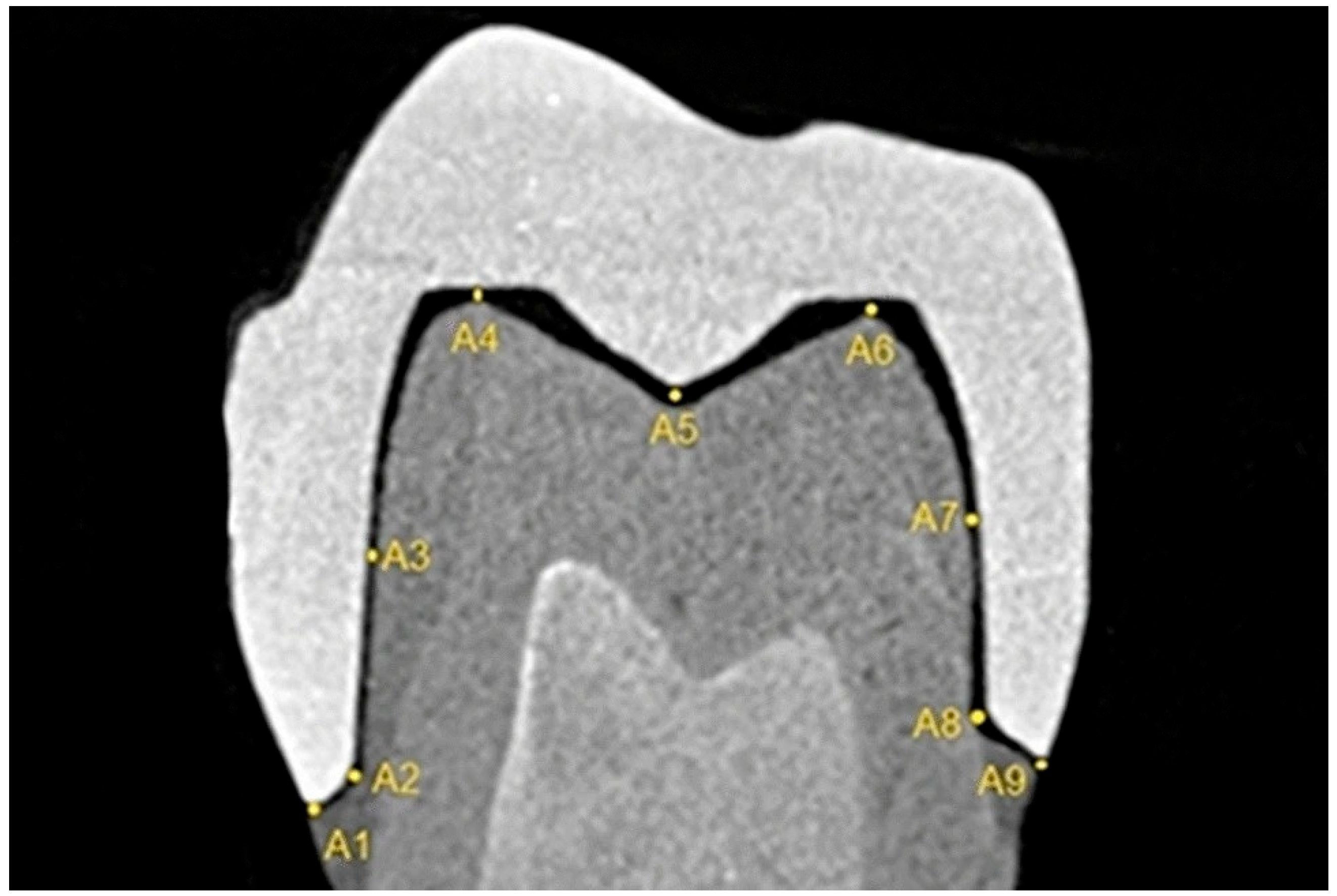
Figure 1. In the sagittal section, the space between the crown and the prepared tooth at 9 different points.
Applsci 15 08017 g001
Figure 2. In the coronal section, the space between the crown and the prepared tooth at 9 different points.
Applsci 15 08017 g002
Figure 3. Box-and-whisker plot illustrating the distribution of internal fit values (in µm) for each group. The plot displays the minimum, maximum, median (red line), first quartile (Q1), and third quartile (Q3). Outliers are represented as individual points.
Applsci 15 08017 g003
Figure 4. Box-and-whisker plot illustrating the distribution of marginal fit values (in µm) for each group. The plot displays the minimum, maximum, median (red line), first quartile (Q1), and third quartile (Q3). Outliers are shown as individual points.
Applsci 15 08017 g004
Table 1. Marginal and internal space values of models obtained with tomography and scanners. Values presented as mean ± standard deviation (SD), µm.
GroupMarginal Fit (Mean ± SD, µm)Internal Fit (Mean ± SD, µm)
0.075 voxel464 ± 139424 ± 122
0.1 voxel513 ± 86396 ± 94
0.15 voxel580 ± 204502 ± 106
Intraoral scanner176 ± 61178 ± 71
Model scanner77 ± 36117 ± 58
Table 2. Mean and standard deviation values of models obtained with tomography and scanners.
GroupVariableNMean (µm)Standard Deviation95% CI Lower95% CI UpperShapiro p
0.075 voxelMarginal fit12464.06138.54376.04552.090.1053 [Normal]
0.075 voxelInternal fit12424.18121.72346.84501.510.3547 [Normal]
0.1 voxelMarginal fit12513.4486.24458.64568.230.1402 [Normal]
0.1 voxelInternal fit12395.8394.06336.07455.580.2779 [Normal]
0.15 voxelMarginal fit12579.78204.40449.91709.650.4305 [Normal]
0.15 voxelInternal fit12456.62114.83383.66529.580.5639 [Normal]
IntraoralMarginal fit1258.7638.6434.2183.310.3198 [Normal]
IntraoralInternal fit12127.4924.90111.67143.310.8254 [Normal]
ModelMarginal fit12121.28186.872.55240.010.0009 [Not]
ModelInternal fit12205.91114.01133.47278.340.0004 [Not]
Disclaimer/Publisher’s Note: The statements, opinions and data contained in all publications are solely those of the individual author(s) and contributor(s) and not of MDPI and/or the editor(s). MDPI and/or the editor(s) disclaim responsibility for any injury to people or property resulting from any ideas, methods, instructions or products referred to in the content.