Article

Assessment of Alveolar Bone Dimensions in Immediate Versus Staged Reconstruction in Sites with Implant Failure

1
Department of Periodontics, College of Medicine, The Catholic University of Korea, Seoul 06591, Republic of Korea
2
Department of Medicine, Graduate School, The Catholic University of Korea, Seoul 06591, Republic of Korea
3
Dental Implantology, Graduate School of Clinical Dental Science, The Catholic University of Korea, Seoul 06591, Republic of Korea
*
Author to whom correspondence should be addressed.
These authors contributed equally to this work.
Appl. Sci. 2025, 15(14), 7934; https://doi.org/10.3390/app15147934
Submission received: 2 May 2025 / Revised: 8 July 2025 / Accepted: 11 July 2025 / Published: 16 July 2025

Abstract

Evaluating the implant site immediately after implant removal is crucial for assessing its condition and ensuring morphological stability. Immediate reconstruction at the time of implant removal has been proposed as a strategy to preserve alveolar ridge width. This study aims to evaluate whether immediate alveolar bone reconstruction at the time of implant removal provides comparable or superior dimensional stability of the alveolar ridge compared to staged reconstruction approaches. The null hypothesis of this study is that there is no significant difference in alveolar bone dimensions between immediate and staged reconstructions following implant removal. This retrospective study included seven participants, consisting of six males and one female. The participants were categorized into three groups based on the treatment approach following implant removal. In Group 1, no bone grafting was performed after implant removal. In Group 2, bone grafting was conducted following implant removal, with an adequate healing period before implant placement. In Group 3, bone grafting was performed simultaneously with implant removal. Cone-beam computed tomography (CBCT) imaging was conducted before implant removal (T0), after implant removal or bone grafting (T1), and after implant placement (T2). All removed implants were successfully replaced with new ones, regardless of bone grafting. In terms of alveolar ridge width at 1 mm below the crest, Group 1 exhibited the greatest reduction (ΔT1 − T0 = −5.1 ± 3.7 mm), while Group 2 showed a mild increase (+1.1 ± 2.6 mm), and Group 3 had a moderate decrease (−1.3 ± 1.0 mm). This suggests that delayed bone grafting can better preserve or enhance bone volume during healing. A reduction in buccal ridge height between T1 and T0 (ΔT1 − T0) was observed, particularly in Group 1. In contrast, an increase in buccal ridge height was most pronounced in Group 2. Although immediate reconstruction (Group 3) did not result in statistically significant gains, it achieved successful implant placement without complications and reduced the total treatment duration, which might be beneficial from a clinical efficiency and patient satisfaction standpoint. Therefore, staged bone grafting (Group 2) appears to offer greater dimensional stability, particularly in maintaining ridge height, whereas immediate reconstruction (Group 3) remains a clinically viable alternative for stable healing in select cases, especially when shorter treatment timelines are prioritized.

1. Introduction

Dental implants have become a widely accepted and effective solution for tooth replacement, demonstrating high long-term survival rates [1]. A variety of dental implant types currently is available on the medical market, and selection depends on clinical and anatomical considerations, patient health status, and prosthetic requirements. Implants can be classified based on shape, material, and surface treatment. In terms of shape, cylindrical and tapered implants are available, with tapered implants often preferred in areas with limited bone width or high esthetic demand due to their superior primary stability [2]. Regarding material, titanium implants are the gold standard due to their excellent biocompatibility and capacity for osseointegration, whereas zirconia implants offer better esthetics and reduced plaque accumulation [3,4]. Rough surfaces can enhance osseointegration, while smooth surfaces lower the risk of peri-implantitis but tend to integrate more slowly [5,6].
Despite the high success rate of dental implants, failures can occur due to a range of contributing factors such as peri-implantitis, implant fracture, micromovement during healing, and mechanical complications like screw loosening [7]. Implant failure can result in both functional and esthetic concerns, often necessitating reimplantation [8]. Reimplantation, when performed with thorough evaluation, meticulous planning, proper surgical techniques, and appropriate postoperative care, can serve as a viable option for patients with failed implants. However, it remains a complex procedure requiring careful consideration of multiple factors, including the underlying cause of implant failure, the quality of the surrounding bone and soft tissue, and the patient’s overall health status [9]. The success rate of dental implant reimplantation has been reported to range from 68% to 98.9% [10]. Studies indicate that reimplantation outcomes are more favorable for implants that failed due to mechanical complications, such as screw loosening or fracture, compared to those that failed due to peri-implantitis [5]. Several factors influence the success rate of reimplantation, including the extent of bone loss, the type of implant system used, the interval between implant removal and reimplantation, and the presence of systemic conditions or other risk factors for implant failure [11]. Furthermore, reimplantation tends to have a lower success rate than initial implant placement due to bone loss, defect formation, and scar tissue development, which can hinder osseointegration [12]. Evaluating the implant site immediately after removal is crucial for assessing its condition and ensuring morphological stability [13].
Immediate reconstruction at the time of implant removal has been proposed as a method to preserve alveolar ridge width. However, despite the increasing popularity of reimplantation, there is limited study on the morphological stability and potential complications associated with immediate reconstruction [14]. A comprehensive assessment of dental implant placement in previously failed sites, including preoperative evaluation, postoperative healing, and morphological stability, may provide valuable clinical insights [15]. A staged approach can be applied, where bone grafting is performed after a healing period, especially in sites compromised by infection or extensive bone loss [15,16]. The selection of graft materials, including both bone substitutes and barrier membranes, significantly influences the outcomes of alveolar ridge augmentation and subsequent dental implant success [17]. Various bone graft materials—such as autografts, allografts, xenografts, and synthetic substitutes—exhibit distinct properties that affect bone regeneration [18]. For instance, autografts are often considered the gold standard due to their osteogenic potential, while allografts and xenografts primarily provide osteoconductive and osteoinductive scaffolding [19,20]. Synthetic materials, like calcium phosphate ceramics, can be tailored for specific resorption rates and mechanical properties. Similarly, the choice of barrier membrane plays a crucial role in guided bone regeneration [21]. Resorbable membranes, typically made from collagen, offer ease of use and eliminate the need for a second surgery for removal [22]. Non-resorbable membranes may provide more rigid structural support but require additional procedures for retrieval [23]. The selection between resorbable and non-resorbable membranes should be based on the specific clinical scenario, considering factors like defect size and the need for space maintenance. Therefore, careful selection of appropriate graft materials and membranes, tailored to the patient’s specific clinical needs, is essential for optimizing the success of dental implant procedures. In this study, xenograft was used along with collagen membrane for alveolar bone reconstruction. Although simultaneous reconstruction at the time of implant removal has been suggested, the long-term dimensional stability and comparative effectiveness of immediate versus delayed approaches remain unclear. A direct comparison regarding ridge width and height after implant removal between immediate and staged reconstruction strategies is warranted. This study aimed to evaluate whether immediate alveolar bone reconstruction at the time of implant removal provides comparable or superior dimensional stability of the alveolar ridge compared to staged reconstruction approaches. This study was conducted to determine whether or not immediate alveolar bone reconstruction can provide sufficient bone augmentation to support subsequent implant placement while maintaining dimensional stability and minimizing complications by analyzing cone-beam computed tomography (CBCT)-based morphological outcomes. The null hypothesis of this study is that there is no significant difference in alveolar bone dimensions between immediate and staged reconstructions following implant removal.

2. Materials and Methods

2.1. Study Design

This research protocol was reviewed and approved by the Institutional Review Board of Seoul St. Mary’s Hospital, College of Medicine, The Catholic University of Korea (KC23RASI0246, approved 24 March 2023). This was a retrospective, non-randomized observational study. This study included seven participants (six males and one female) with a mean age of 52.3 ± 8.0 years. All participants were treated at Seoul St. Mary’s Hospital for the rehabilitation of sites with implant failure.
Participants were allocated into three groups based on the clinical treatment they had already received, determined by the treating clinician’s judgment at the time of care. Decisions regarding bone grafting and its timing were made individually, considering clinical factors such as bone loss extent, infection status, and patient-specific anatomical conditions. The participants were categorized into three groups based on the treatment approach following implant removal. In Group 1, no bone grafting was performed after implant removal (n = 3). In Group 2, bone grafting was performed after implant removal, allowing for an adequate healing period before implant placement (n = 2). In Group 3, bone grafting was performed simultaneously with implant removal (n = 2).
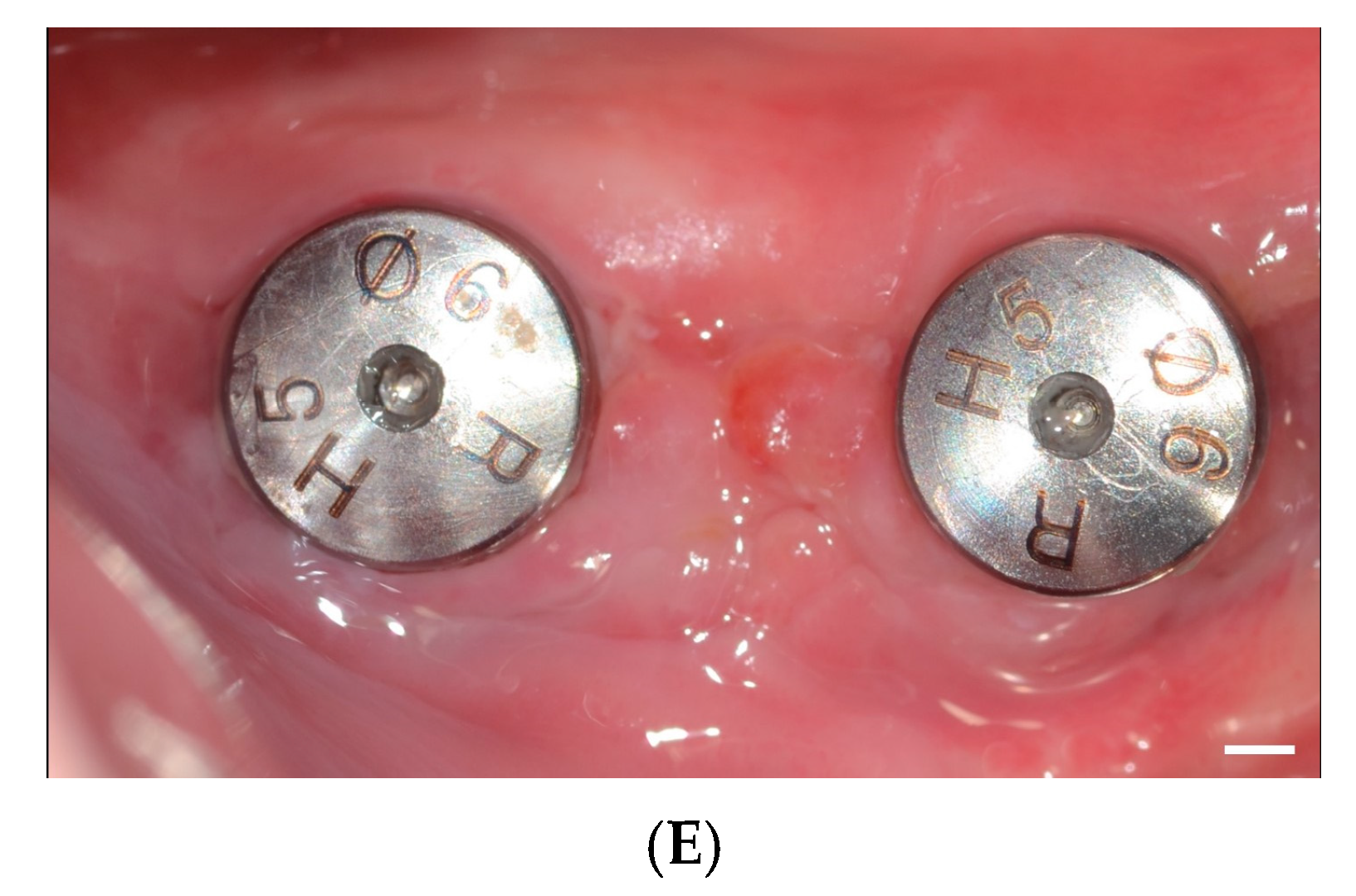
Implants were removed using forceps or an implant removal kit, followed by thorough debridement of the site (Figure 1A). In Group 1, after a sufficient healing period, a new implant was placed without bone grafting. In Group 2, bone grafting was performed after a sufficient healing period following implant removal. The defect was filled with deproteinized bovine bone graft material (Bio-Oss®, Geistlich Pharma, Wolhusen, Switzerland), ensuring its secure placement. If necessary, additional graft material was applied to both the buccal and palatal aspects to expand the ridge width. A resorbable collagen membrane (BioGide®, Geistlich Pharma, or Ossix, Dentsply Sirona, Charlotte, NC, USA) was carefully positioned over the augmented site to protect and stabilize the graft material, promoting optimal healing. In Group 3, bone grafting was performed simultaneously with implant removal (Figure 1B,C). The site was subsequently restored with dental implants (Osstem Implant Co., Ltd.) (Figure 1D,E).
All study participants underwent cone-beam computed tomography (CBCT) imaging using InVivoDental software (Version 6.0.5, Anatomage, San Jose, CA, USA) (Figure 2). CBCT images were acquired and exported as DICOM files, which were then analyzed using InVivo software (Version 6.0.5) for precise measurements. The imaging parameters were configured as follows: a voxel size of 0.2 mm and a slice thickness of 0.2 mm. The field of view measured 16 × 14.5 cm, with exposure settings of 85 kVp and 8 mA. CBCT imaging protocol was as follows. In Group 1, CBCT imaging was performed before implant removal (T0), after implant removal (T1), and after implant reinstallation (T2). In Group 2, CBCT imaging was performed before implant removal (T0), after bone grafting (T1), and after implant reinstallation (T2). In Group 3, CBCT imaging was performed before implant removal (T0), after implant removal and bone grafting (T1), and after implant reinstallation (T2).

2.2. Statistical Analysis

The data were expressed as means and standard deviations for each experimental group. Tests for normality and homogeneity of variance were performed to ensure the data conformed to a normal distribution and exhibited equal variances. Differences among groups were analyzed using Kruskal–Wallis test (SPSS 12 for Windows, SPSS Inc., Chicago, IL, USA). A p-value of less than 0.05 was considered statistically significant.

3. Results

All removed implants were successfully replaced with new implants, regardless of the bone grafting procedure. Immediate reconstruction in Group 3 did not result in uneventful healing. The alveolar ridge width at 1 mm below the crest was 12.7 ± 3.3 mm in Group 1, 9.4 ± 1.8 mm in Group 2, and 7.9 ± 1.2 mm in Group 3 (p > 0.05), with no significant differences among the groups. Similarly, the alveolar ridge width at different depths below the crest showed no significant differences among the groups (p > 0.05). At 3 mm below the crest, the alveolar ridge width was 13.9 ± 3.7 mm in Group 1, 10.6 ± 2.9 mm in Group 2, and 9.0 ± 0.4 mm in Group 3. At 5 mm below the crest, the ridge width measured 14.6 ± 4.0 mm in Group 1, 12.3 ± 1.3 mm in Group 2, and 10.6 ± 1.5 mm in Group 3. At 7 mm below the crest, the values were 13.1 ± 4.0 mm in Group 1, 13.0 ± 1.4 mm in Group 2, and 11.0 ± 1.4 mm in Group 3. While no significant differences were observed in alveolar ridge width across the groups, differences were noted in ridge height. Buccal ridge height was 11.1 ± 4.8 mm (Group 1), 24.0 ± 0.1 mm (Group 2), and 22.4 ± 4.1 mm (Group 3) (p < 0.05), indicating a significant difference among groups. Palatal/lingual ridge height was 9.8 ± 8.6 mm (Group 1), 26.1 ± 2.5 mm (Group 2), and 24.1 ± 2.7 mm (Group 3) (p > 0.05), with no significant differences.
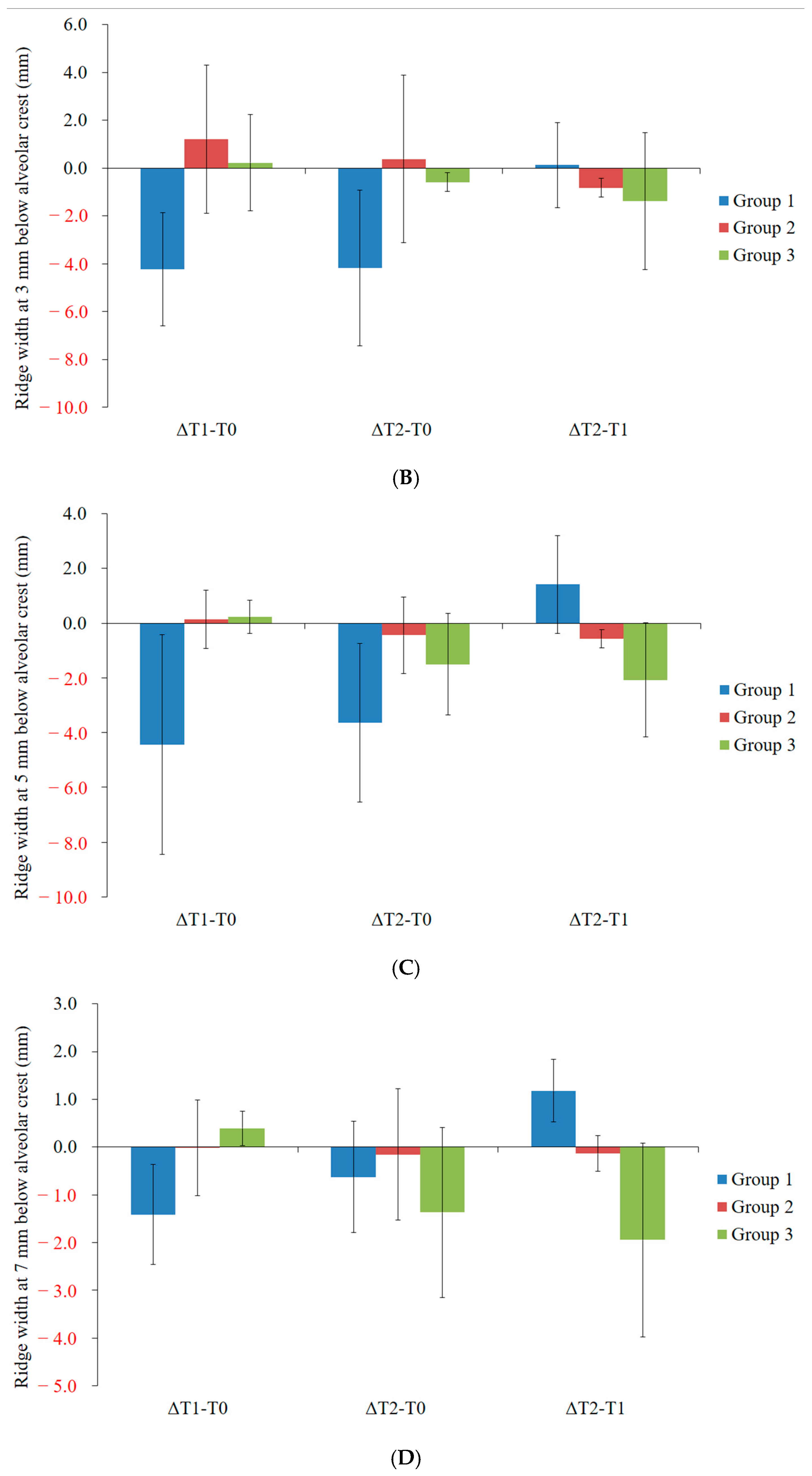
A reduction in alveolar ridge width at 1 mm below the crest was observed between T1 and T0 (ΔT1 − T0), particularly in Group 1 (Figure 3A). The mean change in alveolar ridge width at 1 mm below the crest (ΔT1 − T0) was −5.1 ± 3.7 mm in Group 1, 1.1 ± 2.6 mm in Group 2, and −1.3 ± 1.0 mm in Group 3. Between T2 and T1 (ΔT2 − T1), the changes in alveolar ridge width at 1 mm below the crest were 1.0 ± 2.0 mm (Group 1), −0.8 ± 0.7 mm (Group 2), and 0.3 ± 0.1 mm (Group 3). Similarly, a reduction in alveolar ridge width at 3 mm below the crest was noted between T1 and T0 (ΔT1 − T0), particularly in Group 1 (Figure 3B). The mean change in alveolar ridge width at 3 mm below the crest (ΔT1 − T0) was −4.2 ± 2.4 mm in Group 1, 1.2 ± 3.1 mm in Group 2, and 0.2 ± 2.0 mm in Group 3. Between T2 and T1 (ΔT2 − T1), the changes in alveolar ridge width at 3 mm below the crest were 0.1 ± 1.8 mm (Group 1), −0.8 ± 0.4 mm (Group 2), and −1.4 ± 2.9 mm (Group 3). Changes in alveolar ridge width at 5 mm and 7 mm below the crest are presented in Figure 3C,D.
Changes in buccal ridge height across time points are presented in Figure 3E. A reduction in buccal ridge height between T1 and T0 (ΔT1 − T0) was observed, particularly in Group 1. In contrast, an increase in buccal ridge height was most pronounced in Group 2. Changes in palatal/lingual ridge height across time points are shown in Figure 3F. A significant increase in palatal/lingual ridge height between T1 and T0 (ΔT1 − T0) was observed in Group 2.

4. Discussion

This study aims to evaluate the potential advantages and morphological stability of immediate reconstruction following implant removal. Immediate reconstruction may facilitate adequate bone augmentation to support subsequent dental implant placement while maintaining dimensional stability and minimizing complications.
A previous report showed that the alveolar dimensional changes after implant removal resulted in an 11.3% ridge width reduction at 1 mm below the crest and a 4.4% reduction at 3 mm below the crest [16]. Similarly, buccal and lingual ridge heights were significantly reduced by 2.2% and 6.3%, respectively. Group 1, where no grafting was performed, showed the greatest reductions in ridge width and height. This outcome is consistent with well-documented post-extraction bone remodeling processes, as mentioned earlier. In this present study, the alveolar dimension at 1 mm and 3 mm below the crest was reduced by 40.2 ± 29.5% and 30.5 ± 17.1%, respectively. Buccal and lingual ridge heights experienced a 20.4 ± 18.6% reduction and a 2.1 ± 3.5% reduction, respectively. The most notable dimensional stability in ridge height was observed in the staged grafting group (Group 2). This can be mechanistically attributed to the biologically favorable conditions created by delaying bone grafting until after initial healing. Delayed reconstruction may allow resolution of local inflammation and clearance of residual infection. A previous study suggested that performing alveolar bone reconstruction at the time of implant removal minimized dimensional changes and bone gain [14]. Group 3, which received immediate bone grafting at the time of implant removal, demonstrated acceptable ridge preservation without complications. Mechanistically, immediate grafting may preserve alveolar ridge contours by occupying the extraction socket and preventing collapse of the buccal and lingual plates.
The decision between no grafting and bone grafting probably should be based on clinician judgment, influenced by clinical experience and the specific clinical situation. Proper diagnosis and evaluation of the cause of implant failure, as well as a thorough assessment of the surgical site, are critical [24]. To obtain stable sites for reimplantation, bone grafting or other surgical interventions may be required as part of site preparation [25]. In cases where no significant bone loss is observed around the failed implant, additional reconstruction may not be necessary, particularly when failure results from mechanical complications, such as fixture fracture. Bone quality and quantity should be assessed both before and during implant removal [26].
In this study, immediate reconstruction did not demonstrate additional morphological stability or advantage. However, immediate reconstruction may reduce the overall treatment duration, potentially enhancing patient acceptance. Similarly, studies have shown that immediate implant placement and provisionalization can lead to high patient satisfaction and esthetic outcomes [27]. Reducing the number of surgeries through immediate reconstruction is a less invasive and more comfortable approach for the patient [28]. Additionally, minimizing treatment time may be associated with increased patient satisfaction [29].
The timing and approach to bone grafting are contingent upon individual patient factors and clinician assessment. Delayed bone grafting, performed after an adequate healing period, is advisable in the presence of infection or compromised healing at the extraction site [30]. This strategy allows resolution of inflammation and a healthier environment for graft placement and reduced risk of complications [31,32]. Conversely, immediate bone grafting at the time of implant removal is recommended when significant bone loss is anticipated or when the alveolar ridge needs to be preserved for future implant placement [33]. This approach can effectively maintain bone volume and prevent resorption, facilitating optimal conditions for subsequent implant procedures [34]. Ultimately, the decision between immediate and delayed bone grafting should be individualized, considering the patient’s clinical circumstances and practitioner expertise.
The following recommendations are proposed to optimize outcomes during implant removal and subsequent site management. Care must be taken to preserve the surrounding bone and soft tissue by employing minimally invasive techniques during implant extraction [26]. Utilizing commercially available implant removal kits can facilitate the removal of failed implants from various systems with minimal tissue injury [8]. Thorough debridement of the implant removal site is essential to eliminate residual debris or granulation tissue, reducing the risk of infection and promoting optimal healing [35]. Implementing these practices can enhance the preservation of alveolar bone and soft tissue integrity, improving the prognosis for future restorative procedures [36]. It should also be emphasized that successful immediate reconstruction may require a skilled operator.
The present study has certain limitations that warrant consideration. A notable limitation is the relatively small sample size, which may affect the generalizability of the results to a broader patient population [37]. Additionally, as a retrospective study focusing on short-term postoperative morphological assessments, it may not capture long-term outcomes [38]. In addition to the small sample size, the lack of randomization and the observational nature of group allocation introduce potential selection bias. Treatment decisions were based on clinical judgment and case-specific factors, which may have influenced outcomes independently of the grafting protocol. For example, patients with more favorable anatomical conditions may have been selected for immediate grafting, while more complex or infected cases may have been assigned to staged grafting. This inherent bias limits the ability to draw causal inferences from group comparisons. Patient characteristics such as age and sex may affect clinical outcomes. The choice of graft materials, including bone and membranes, can significantly influence postoperative results [39,40]. Another limitation of this study is the lack of detailed reporting on critical confounding variables such as smoking status, alcohol consumption, systemic health conditions, periodontal history and oral hygiene. The absence of this information limits the ability to adjust for or interpret their potential effects on the outcomes. Therefore, further research with larger sample sizes, extended follow-up periods, comprehensive baseline data collection, and stratified analysis is necessary to confirm these findings and to better understand how patient-level variables interact with treatment protocols and dimensional outcomes.
Treatment with delayed bone augmentation after implant removal showed the greatest dimensional stability during healing. Immediate reconstruction after implant removal proceeded without complications, potentially reducing the overall treatment duration. It is important to emphasize that the approach to dental implant reinstallation should be tailored to patient needs and the circumstances surrounding their implant failure. Factors such as extent of bone loss and patient overall health significantly influence the treatment strategy.

5. Conclusions

Within the limitations of this retrospective study, all removed implants were successfully replaced with new ones, regardless of bone grafting. Staged alveolar bone grafting was associated with the greatest dimensional stability, particularly in vertical ridge height, and may be preferred in sites with inflammation or extensive bone loss. Immediate grafting provided acceptable ridge preservation without complications and may be suitable in non-infected, mechanically failed sites, offering the benefit of reduced treatment time. No grafting resulted in the greatest dimensional loss, emphasizing the importance of intervention when preserving ridge morphology. In conclusion, this study indicates that staged bone grafting may offer enhanced stability, particularly concerning ridge height. Additionally, immediate reconstruction appears to be a viable method for achieving stable recovery without complications and a shorter treatment period. These findings highlight the need for a personalized treatment strategy based on clinical presentation and site-specific risk factors. While promising, the results are exploratory and require validation in larger, prospective, and controlled studies that evaluate long-term implant outcomes.

Author Contributions

Conceptualization, H.L., S.H., Y.K. and J.-B.P.; formal analysis, H.L., S.H., Y.K. and J.-B.P.; writing—original draft preparation, H.L., S.H., Y.K. and J.-B.P.; and writing—review and editing, H.L., S.H., Y.K. and J.-B.P. All authors have read and agreed to the published version of the manuscript.

Funding

The National Research Foundation of Korea (NRF) grant, funded by the Korean government (MSIT), provided funding for this study (No. RS-2023-00252568).

Institutional Review Board Statement

This research protocol was reviewed and approved by the Institutional Review Board of Seoul St Mary’s Hospital, College of Medicine, The Catholic University of Korea (KC23RASI0246, approved 24 March 2023).

Informed Consent Statement

Patient consent was waived due to the design of this study.

Data Availability Statement

The original contributions presented in this study are included in the article. Further inquiries can be directed to the corresponding author.

Acknowledgments

Parts of this paper were submitted as an abstract at the 111th Annual Meeting of the American Academy of Periodontology.

Conflicts of Interest

There are no conflicts of interest that the authors can identify with this work.

References

  1. Setzer, F.C.; Kim, S. Comparison of long-term survival of implants and endodontically treated teeth. J. Dent. Res. 2014, 93, 19–26. [Google Scholar] [CrossRef] [PubMed]
  2. Nandini, N.; Kunusoth, R.; Alwala, A.M.; Prakash, R.; Sampreethi, S.; Katkuri, S. Cylindrical Implant Versus Tapered Implant: A Comparative Study. Cureus 2022, 14, e29675. [Google Scholar] [CrossRef] [PubMed]
  3. Aldhuwayhi, S. Zirconia in Dental Implantology: A Review of the Literature with Recent Updates. Bioengineering 2025, 12, 543. [Google Scholar] [CrossRef] [PubMed]
  4. Park, J.B.; Lee, S.H.; Kim, N.; Park, S.; Jin, S.H.; Choi, B.K.; Kim, K.K.; Ko, Y. Instrumentation With Ultrasonic Scalers Facilitates Cleaning of the Sandblasted and Acid-Etched Titanium Implants. J. Oral Implant. 2015, 41, 419–428. [Google Scholar] [CrossRef] [PubMed]
  5. Tan, N.C.P.; Khan, A.; Antunes, E.; Miller, C.M.; Sharma, D. The effects of physical decontamination methods on zirconia implant surfaces: A systematic review. J. Periodontal Implant. Sci. 2021, 51, 298–315. [Google Scholar] [CrossRef] [PubMed]
  6. Chopra, M.; Vermani, M.; Swarup, N.; Pal, A.; Chowdhary, Z. Surface Treatment and Implant Bone Interface: A Systematic Literature Review. J. Long Term Eff. Med. Implant. 2020, 30, 283–294. [Google Scholar] [CrossRef] [PubMed]
  7. Kochar, S.P.; Reche, A.; Paul, P. The Etiology and Management of Dental Implant Failure: A Review. Cureus 2022, 14, e30455. [Google Scholar] [CrossRef] [PubMed]
  8. Solderer, A.; Al-Jazrawi, A.; Sahrmann, P.; Jung, R.; Attin, T.; Schmidlin, P.R. Removal of failed dental implants revisited: Questions and answers. Clin. Exp. Dent. Res. 2019, 5, 712–724. [Google Scholar] [CrossRef] [PubMed]
  9. Yang, J.; Siow, L.; Zhang, X.; Wang, Y.; Wang, H.; Wang, B. Dental Reimplantation Treatment and Clinical Care for Patients with Previous Implant Failure-A Retrospective Study. Int. J. Environ. Res. Public Health 2022, 19, 15939. [Google Scholar] [CrossRef] [PubMed]
  10. Anitua, E.; Montalvillo, A.; Eguia, A.; Alkhraisat, M.H. Clinical outcomes of dental implants placed in the same region where previous implants failed due to peri-implantitis: A retrospective study. Int. J. Implant. Dent. 2021, 7, 109. [Google Scholar] [CrossRef] [PubMed]
  11. Machtei, E.E.; Mahler, D.; Oettinger-Barak, O.; Zuabi, O.; Horwitz, J. Dental implants placed in previously failed sites: Survival rate and factors affecting the outcome. Clin. Oral Implant. Res. 2008, 19, 259–264. [Google Scholar] [CrossRef] [PubMed]
  12. Geraets, W.; Zhang, L.; Liu, Y.; Wismeijer, D. Annual bone loss and success rates of dental implants based on radiographic measurements. Dento Maxillo Facial Radiol. 2014, 43, 20140007. [Google Scholar] [CrossRef] [PubMed]
  13. Covani, U.; Marconcini, S.; Crespi, R.; Barone, A. Immediate implant placement after removal of a failed implant: A clinical and histological case report. J. Oral Implant. 2009, 35, 189–195. [Google Scholar] [CrossRef] [PubMed]
  14. Monje, A.; Soldini, M.C.; Rosen, P.S.; Tarnow, D.; Nart, J.; Pons, R. Alveolar Bone Reconstruction Simultaneous to Implant Removal due to Advanced Peri-Implantitis Defects: A Proof of Concept. J. Esthet. Restor. Dent. 2025, 37, 85–93. [Google Scholar] [CrossRef] [PubMed]
  15. Park, Y.S.; Lee, B.A.; Choi, S.H.; Kim, Y.T. Evaluation of failed implants and reimplantation at sites of previous dental implant failure: Survival rates and risk factors. J. Periodontal Implant. Sci. 2022, 52, 230–241. [Google Scholar] [CrossRef] [PubMed]
  16. Pons, R.; Carreño, M.; Amerio, E.; Gargallo-Albiol, J.; Nart, J.; Monje, A. Hard tissue dimensional changes following implant removal due to peri-implantitis: A retrospective study. Clin. Implant. Dent. Relat. Res. 2021, 23, 432–443. [Google Scholar] [CrossRef] [PubMed]
  17. Zhao, R.; Yang, R.; Cooper, P.R.; Khurshid, Z.; Shavandi, A.; Ratnayake, J. Bone Grafts and Substitutes in Dentistry: A Review of Current Trends and Developments. Molecules 2021, 26, 3007. [Google Scholar] [CrossRef] [PubMed]
  18. Ferraz, M.P. Bone Grafts in Dental Medicine: An Overview of Autografts, Allografts and Synthetic Materials. Materials 2023, 16, 4117. [Google Scholar] [CrossRef] [PubMed]
  19. Schmidt, A.H. Autologous bone graft: Is it still the gold standard? Injury 2021, 52 (Suppl. S2), S18–S22. [Google Scholar] [CrossRef] [PubMed]
  20. Steijvers, E.; Ghei, A.; Xia, Z. Manufacturing artificial bone allografts: A perspective. Biomater. Transl. 2022, 3, 65–80. [Google Scholar] [CrossRef] [PubMed]
  21. Yum, H.; Han, H.S.; Lee, J.T.; Cho, Y.D.; Kim, S. Bone regeneration using activin A/BMP2 chimera (AB204) with collagen membrane in rats with calvarial defects. J. Periodontal Implant. Sci. 2024, 54, 309–321. [Google Scholar] [CrossRef] [PubMed]
  22. Jang, Y.; Jang, J.; Kim, B.Y.; Song, Y.S.; Lee, D.Y. Effect of Gelatin Content on Degradation Behavior of PLLA/Gelatin Hybrid Membranes. Tissue Eng. Regen. Med. 2024, 21, 557–569. [Google Scholar] [CrossRef] [PubMed]
  23. Sbricoli, L.; Guazzo, R.; Annunziata, M.; Gobbato, L.; Bressan, E.; Nastri, L. Selection of Collagen Membranes for Bone Regeneration: A Literature Review. Materials 2020, 13, 786. [Google Scholar] [CrossRef] [PubMed]
  24. Gulati, M.; Govila, V.; Anand, V.; Anand, B. Implant Maintenance: A Clinical Update. Int. Sch. Res. Not. 2014, 2014, 908534. [Google Scholar] [CrossRef] [PubMed]
  25. Cha, H.S.; Kim, J.W.; Hwang, J.H.; Ahn, K.M. Frequency of bone graft in implant surgery. Maxillofac. Plast. Reconstr. Surg. 2016, 38, 19. [Google Scholar] [CrossRef] [PubMed]
  26. Masaki, C.; Kondo, Y.; Tomoeda, K.; Nodai, T.; Munemasa, T.; Mukaibo, T.; Hosokawa, R. Treatment strategies for dental implant removal: A literature review. Jpn. Dent. Sci. Rev. 2024, 60, 120–127. [Google Scholar] [CrossRef] [PubMed]
  27. Hartlev, J.; Kohberg, P.; Ahlmann, S.; Andersen, N.T.; Schou, S.; Isidor, F. Patient satisfaction and esthetic outcome after immediate placement and provisionalization of single-tooth implants involving a definitive individual abutment. Clin. Oral Implant. Res. 2014, 25, 1245–1250. [Google Scholar] [CrossRef] [PubMed]
  28. Kouveliotis, G.; Papapmanoli, E.; Tasopoulos, T.; Tampakos, I.; Michas, D.; Tzanakakis, E.-G.; Zoidis, P. Fully Digital Workflow for Immediate Loading Using a Minimally Invasive Surgical Approach: A Case Report. Prosthesis 2025, 7, 25. [Google Scholar] [CrossRef]
  29. Schropp, L.; Isidor, F.; Kostopoulos, L.; Wenzel, A. Patient experience of, and satisfaction with, delayed-immediate vs. delayed single-tooth implant placement. Clin. Oral Implant. Res. 2004, 15, 498–503. [Google Scholar] [CrossRef] [PubMed]
  30. Kim, Y.K.; Park, J.Y.; Kim, S.G.; Lee, H.J. Prognosis of the implants replaced after removal of failed dental implants. Oral Surg. Oral Med. Oral Pathol. Oral Radiol. Endodontol. 2010, 110, 281–286. [Google Scholar] [CrossRef] [PubMed]
  31. Thomas, M.V.; Puleo, D.A. Infection, inflammation, and bone regeneration: A paradoxical relationship. J. Dent. Res. 2011, 90, 1052–1061. [Google Scholar] [CrossRef] [PubMed]
  32. Juncar, M.; Bran, S.; Juncar, R.I.; Baciut, M.F.; Baciut, G.; Onisor-Gligor, F. Odontogenic cervical necrotizing fasciitis, etiological aspects. Niger. J. Clin. Pr. 2016, 19, 391–396. [Google Scholar] [CrossRef] [PubMed]
  33. Covani, U.; Barone, A.; Cornelini, R.; Crespi, R. Clinical outcome of implants placed immediately after implant removal. J. Periodontol. 2006, 77, 722–727. [Google Scholar] [CrossRef] [PubMed]
  34. Avila-Ortiz, G.; Elangovan, S.; Kramer, K.W.; Blanchette, D.; Dawson, D.V. Effect of alveolar ridge preservation after tooth extraction: A systematic review and meta-analysis. J. Dent. Res. 2014, 93, 950–958. [Google Scholar] [CrossRef] [PubMed]
  35. Bianconi, S.; Romanos, G.; Testori, T.; Del Fabbro, M. Management of Advanced Peri-Implantitis by Guided Bone Regeneration in Combination with Trabecular Metal Fixtures, Two Months after Removal of the Failed Implants: Two-Year Results of a Single-Cohort Clinical Study. J. Clin. Med. 2024, 13, 713. [Google Scholar] [CrossRef] [PubMed]
  36. Roy, M.; Loutan, L.; Garavaglia, G.; Hashim, D. Removal of osseointegrated dental implants: A systematic review of explantation techniques. Clin. Oral Investig. 2020, 24, 47–60. [Google Scholar] [CrossRef] [PubMed]
  37. Cho, S.; Min, E.J.; Hwa, S.; Lee, H.; Ko, Y.; Park, J.B. Crestal approach for maxillary sinus augmentation in individuals with limited alveolar bone height: An observational study. Medicine 2024, 103, e40331. [Google Scholar] [CrossRef] [PubMed]
  38. Lee, H.J.; Kim, Y.K.; Park, J.Y.; Kim, S.G.; Kim, M.J.; Yun, P.Y. Short-term clinical retrospective study of implants in geriatric patients older than 70 years. Oral Surg. Oral Med. Oral Pathol. Oral Radiol. Endodontol. 2010, 110, 442–446. [Google Scholar] [CrossRef] [PubMed]
  39. Cho, C.S.; Jo, I. Bone Morphogenic Protein-2-Conjugated Three-Dimensional-Printed Poly (L-Lactic Acid) (PLLA) Scaffold is likely Promising as an Effective Bone Substitute. Tissue Eng. Regen. Med. 2023, 20, 155–156. [Google Scholar] [CrossRef] [PubMed]
  40. Park, J.; Jung, N.; Lee, D.J.; Oh, S.; Kim, S.; Cho, S.W.; Kim, J.E.; Moon, H.S.; Park, Y.B. Enhanced Bone Formation by Rapidly Formed Bony Wall over the Bone Defect Using Dual Growth Factors. Tissue Eng. Regen. Med. 2023, 20, 767–778. [Google Scholar] [CrossRef] [PubMed]
Figure 1. Clinical procedures. (A) Occlusal view following the removal of the implant prosthesis. (B) Placement of deproteinized bovine bone graft material in the defect, covered by a resorbable collagen membrane. (C) Repositioning and suturing of the flap suture (nylon 5-0, Ethicon, Cincinnati, OH, USA). (D) Installation of a new implant (Osstem Implant Co., Ltd., Seoul, Republic of Korea) after an appropriate healing period. The scale bar in the image represents 1 mm. (E) Occlusal view following removal. The scale bar shown corresponds to 1 mm.
Applsci 15 07934 g001aApplsci 15 07934 g001b
Figure 2. CBCT-based alveolar ridge measurements. (A) Measurement of the alveolar ridge dimension before implant removal (T0). (B) The alveolar ridge dimension using CBCT after bone grafting (T1). (C) Measurement of the alveolar ridge dimension using CBCT after implant reinstallation (T2). (D) Superimposition of the T0 and T1 images. (E) Superimposition of the T1 and T2 images.
Applsci 15 07934 g002aApplsci 15 07934 g002b
Figure 3. Evaluation of dimensional changes. (A) Change in alveolar ridge width 1 mm below the crest. (B) Change in alveolar ridge width 3 mm below the crest. (C) Change in alveolar ridge width 5 mm below the crest. (D) Change in alveolar ridge width 7 mm below the crest. (E) Change in buccal ridge height. (F) Change in palatal/lingual ridge height. * p < 0.05 compared to Group 1.
Applsci 15 07934 g003aApplsci 15 07934 g003bApplsci 15 07934 g003c
Disclaimer/Publisher’s Note: The statements, opinions and data contained in all publications are solely those of the individual author(s) and contributor(s) and not of MDPI and/or the editor(s). MDPI and/or the editor(s) disclaim responsibility for any injury to people or property resulting from any ideas, methods, instructions or products referred to in the content.