Article

Association Between Vertebral Artery Stiffness and Idiopathic Subjective Tinnitus: A Prospective Study

1
Department of Otorhinolaryngology, School of Medicine, Gaziantep University, 27310 Gaziantep, Turkey
2
Department of Otorhinolaryngology, School of Medicine, TOBB-ET University, 06510 Ankara, Turkey
3
Department of Anesthesiology and Reanimation, School of Medicine, Gaziantep University Gaziantep, 27410 Gaziantep, Turkey
4
Department of Neurology, School of Medicine, Gaziantep University, 27410 Gaziantep, Turkey
5
Department of Otorhinolaryngology, School of Medicine, Sanko University, 27090 Gaziantep, Turkey
*
Author to whom correspondence should be addressed.
Appl. Sci. 2025, 15(14), 7890; https://doi.org/10.3390/app15147890 (registering DOI)
Submission received: 11 June 2025 / Revised: 9 July 2025 / Accepted: 11 July 2025 / Published: 15 July 2025

Abstract

Objectives: Tinnitus, defined as the perception of sound without an external stimulus, is a complex condition with unclear etiology. Emerging evidence suggests a link between vascular dysfunction, particularly arterial stiffness, and tinnitus. This study aimed to evaluate vertebral artery stiffness in patients with idiopathic subjective tinnitus and assess the utility of Doppler ultrasonography as a non-invasive diagnostic tool. Materials and Methods: In this prospective study, 31 patients with idiopathic tinnitus (11 males, 20 females; mean age: 50.42 ± 9.64 years) and 24 healthy controls (12 males, 12 females; mean age: 39.67 ± 10.63 years) underwent comprehensive clinical evaluations, including pure tone audiometry, blood tests, and vertebrobasilar Doppler ultrasonography. Vertebral artery stiffness index (VAS), resistive index (RI), and pulsatility index (PI) were measured bilaterally. Results: A total of 31 patients with idiopathic subjective tinnitus and 24 healthy controls were evaluated. The mean age was significantly higher in the tinnitus group compared to controls (50.42 ± 9.64 vs. 39.67 ± 10.63 years, p < 0.001). Lipid profile analysis revealed significantly higher levels of total cholesterol (193.6 ± 47.28 vs. 167.5 ± 28.99 mg/dL, p = 0.021), LDL (149.4 ± 37.9 vs. 106.1 ± 10.7 mg/dL, p < 0.005), and triglycerides (202.2 ± 83.5 vs. 148.6 ± 26.4 mg/dL, p < 0.005) in tinnitus patients. Doppler ultrasonography demonstrated significantly higher vertebral artery stiffness values in the tinnitus group (left: 2.87 ± 0.72 vs. 2.12 ± 0.22; right: 2.99 ± 0.77 vs. 2.14 ± 0.5; both p < 0.005). Similarly, pulsatility index (PI) was significantly elevated in patients compared to controls (left: 2.45 ± 1.2 vs. 1.2 ± 0.43; right: 2.49 ± 1.02 vs. 1.19 ± 0.42; both p < 0.005). No significant differences were observed in resistive index (RI) or vertebral artery diameters between groups. Among tinnitus patients, PI and VAS were significantly higher on the side corresponding to reported tinnitus symptoms (p < 0.05), suggesting a lateralized vascular contribution. Conclusions: The findings suggested a potential relationship between idiopathic subjective tinnitus and vertebral artery stiffness. We demonstrated the utility of Doppler ultrasonography, a cost-effective and non-invasive imaging modality, for evaluating vascular parameters in tinnitus patients, paving the way for broader clinical applications. By uncovering a significant association between vertebral artery stiffness and tinnitus, our findings suggest that vascular health assessments could enhance diagnostic and therapeutic strategies for tinnitus management.

1. Introduction

Tinnitus is a complex and often debilitating auditory condition defined by the perception of sound in the absence of external auditory stimuli. Globally, it affects approximately 10–15% of the adult population, with a significant proportion experiencing chronic and distressing symptoms that negatively impact their quality of life, sleep, emotional well-being, and cognitive performance [1,2,3,4]. Despite its high prevalence, the underlying pathophysiological mechanisms of tinnitus remain incompletely understood and are believed to be multifactorial, involving both peripheral and central auditory pathways as well as non-auditory brain networks [5,6,7].
Several theoretical models have been proposed to explain the onset and persistence of tinnitus. One prevailing hypothesis suggests that cochlear damage due to noise exposure, ototoxic drugs, or age-related degeneration leads to diminished peripheral input, which triggers maladaptive neuroplastic changes in the central auditory cortex. This maladaptive cortical reorganization may result in aberrant spontaneous neural activity, perceived as phantom sounds [8]. In addition, growing evidence has pointed to the involvement of oxidative stress, neuroinflammation, and impaired vascular regulation in the pathogenesis of tinnitus [9,10,11,12].
Vascular dysfunction, particularly microvascular insufficiency, has received increasing attention as a potential contributing factor in tinnitus development. Microvascular impairment may lead to hypoxia and ischemia in cochlear structures, notably affecting the stria vascularis, outer hair cells, and spiral ganglion neurons, which are critical for normal auditory function [13]. Among the systemic vascular contributors, arterial stiffness has emerged as a promising biomarker due to its association with endothelial dysfunction, oxidative stress, and atherosclerosis, all of which may compromise cochlear perfusion [14].
Arterial stiffness, typically characterized by reduced vascular compliance and elasticity, leads to elevated pulse pressure and increased pulsatile flow, which may exacerbate microvascular injury [15]. Most previous studies evaluating arterial stiffness in tinnitus patients have focused on central elastic arteries such as the carotid artery or assessed systemic parameters using pulse wave velocity (PWV) and carotid intima-media thickness (CIMT) [16,17]. While informative, these measurements do not provide direct information on the vertebrobasilar system, which plays a crucial role in posterior cerebral and inner ear perfusion.
The vertebral arteries, which supply blood to the posterior circulation including the cochlea, brainstem, and cerebellum, represent a highly relevant but understudied vascular region in tinnitus research. Recent studies have suggested that vertebrobasilar insufficiency may contribute to both auditory and vestibular symptoms, including tinnitus, vertigo, and sensorineural hearing loss [18,19]. Furthermore, abnormalities in vertebral artery hemodynamics, including elevated pulsatility index (PI), resistive index (RI), and stiffness index (SI), may reflect microvascular compromise relevant to tinnitus pathophysiology [20,21].
Doppler ultrasonography has become an increasingly utilized non-invasive technique for the assessment of arterial stiffness, as it allows real-time measurement of vessel diameter changes in relation to systolic and diastolic blood pressure, thus providing localized vascular parameters [22]. Unlike systemic indices, Doppler-derived stiffness measurements offer a region-specific evaluation that may better correlate with organ-specific microvascular dysfunction, such as cochlear ischemia in tinnitus patients.
Given these considerations, the current study aimed to investigate the potential relationship between vertebral artery stiffness and idiopathic subjective tinnitus by utilizing Doppler ultrasonography to obtain detailed vascular parameters. We hypothesized that vertebral artery stiffness and related indices would be significantly elevated in patients with idiopathic tinnitus compared to healthy controls, and that these changes may provide novel insights into the vascular contributions to tinnitus pathogenesis.

2. Materials and Methods

2.1. Study Design and Study Population

The study was approved by the Ethics Committee of the Faculty of Medicine of Gaziantep University (Approval No: 2023/12, Date: 8 December 2023). Informed consent forms were obtained from all patients and/or their first-degree relatives included in the study. The study was conducted according to the guidelines of the Declaration of Helsinki. From March 2018 to January 2020, a total of 31 patients with complaints of tinnitus (11 males and 20 females, aged 26 to 73 years; mean age: 50.42 ± 9.64 years) and 24 healthy individuals as controls (12 males and 12 females, aged 24 to 58 years; mean age: 39.67 ± 10.63 years) were included in this study.
Patients with tinnitus were monitored for three months in an outpatient clinic. The control group was matched by age and sex and consisted of individuals with normal hearing and unremarkable otorhinolaryngologic examinations. Both groups underwent comprehensive evaluations, including routine physical examinations, pure tone audiometry (PTA), blood tests (lipid profile, fasting glucose levels), and systolic and diastolic blood pressure (SBP, DBP) measurements. Vertebrobasilar Doppler ultrasonography was also performed to assess vascular parameters. Blood pressure readings were taken on three separate days, and the mean values were calculated for each participant.
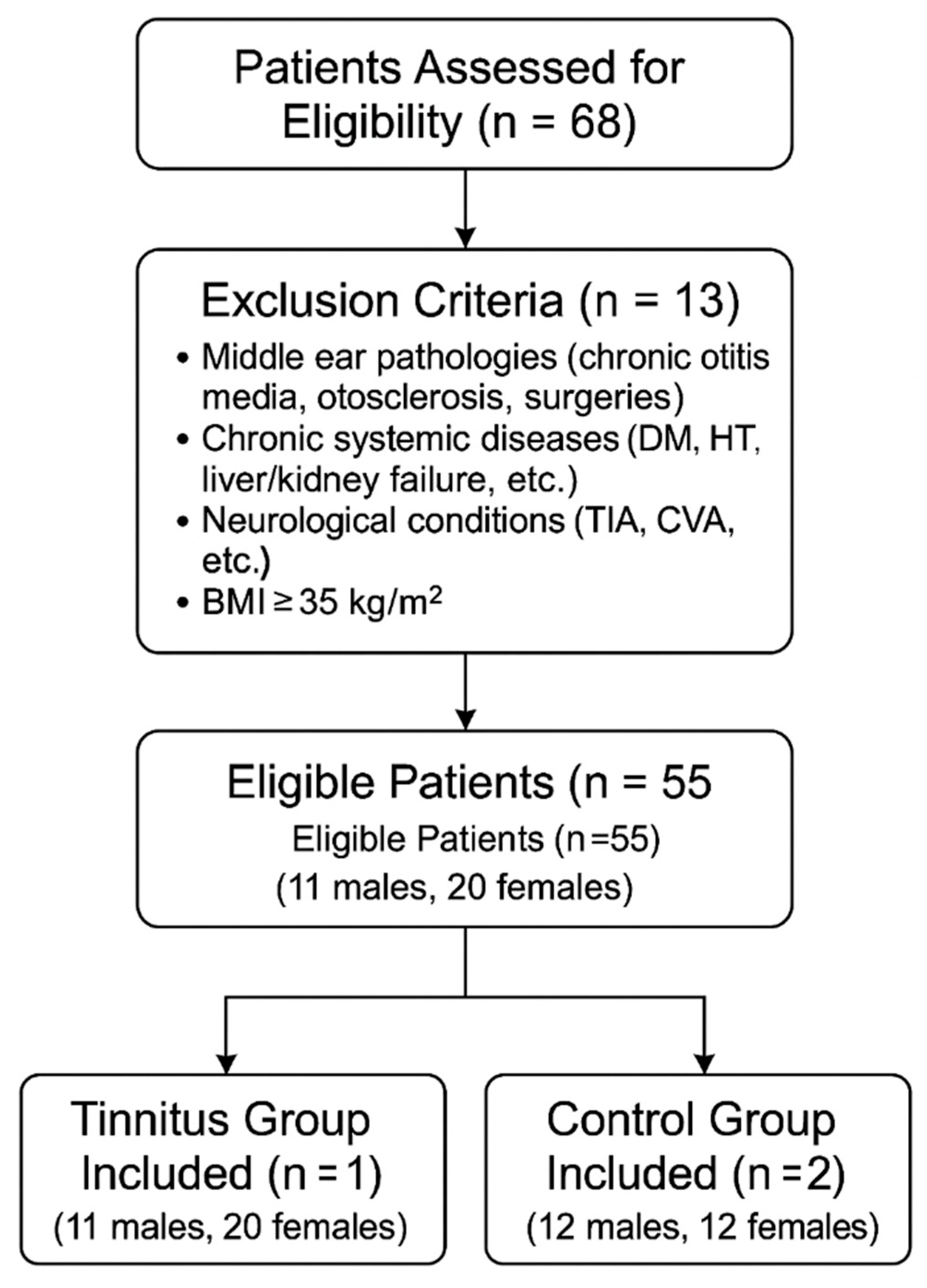
Exclusion criteria included middle ear pathologies affecting ossicular integrity or sound transmission, such as chronic otitis media, otosclerosis, or previous middle ear surgeries. Patients with chronic systemic conditions, including diabetes mellitus, hypertension, chronic liver or kidney failure, pulmonary insufficiencies, or neurologic conditions affecting cerebrovascular flow (e.g., transient ischemic attacks or cerebrovascular accidents), as well as individuals with obesity (body mass index ≥ 35 kg/m2), were also excluded (Figure 1).
Pure tone audiometry was conducted in a soundproof room at four frequencies (500, 1000, 2000, and 4000 Hz) using an AC40 Interacoustics device (Middelfart, Denmark). The testing was performed by an experienced audiologist, with each frequency assessed for one minute. Participants reporting whistling, buzzing, or other forms of sound perception were classified as having tinnitus. Additionally, the side of the tinnitus (unilateral or bilateral) was documented for each patient.

2.2. Radiological Evaluation

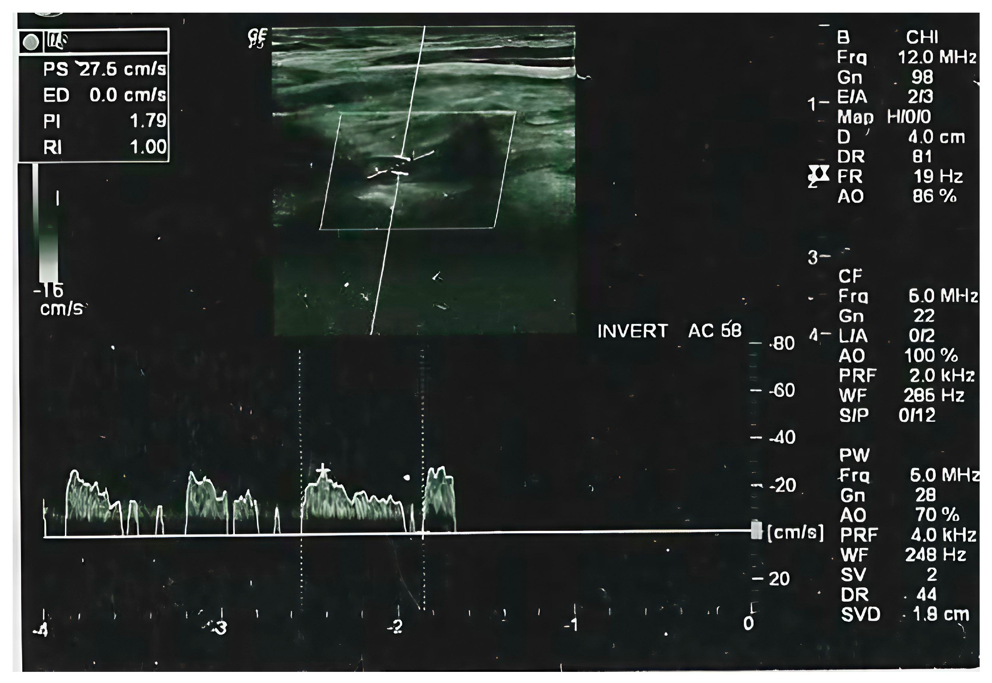
The same radiologists have performed the Doppler ultrasound examination using an extracranial color-coded duplex sonography portable ultrasound equipment LOGIQ P5 (GE Healthcare, Buckinghamshire, UK) at a scanning frequency of 5–12 MHz. Subjects were evaluated in a supine position with an extended neck position in a period of five heartbeats on average on both sides. Measurements were made between VA segments between vertebral bodies C4 and C6 (Figure 2). Data on both the dominant and nondominant sides of the vertebral arteries were evaluated together.
Vertebral artery diameters, arterial wall thicknesses, intimal surfaces, resistive index (RI), pulsatility index (PI) and mean velocity (MV) were measured ultrasonographic parameters. Resistive index (RI) and pulsatility index (PI) were two sonographic parameters that are calculated using specific formulas. Recent studies have demonstrated that RI and PI are associated with abnormalities of the vascular lumen [19]. The stiffness index was an additional parameter evaluated by calculating systolic and diastolic systemic blood pressure and arterial diameters between pressure changes [15]. Stiffness index = ln (SBP/DBP) × Dmin/(DD). SBP is the systolic blood pressure, DBP is the diastolic blood pressure, DD is the diastolic diameter, Dmax and Dmin are the maximum and minimum arterial diameters for pressure changes [16].

2.3. Statistical Analysis

All statistical analyses were performed using SPSS software (Statistical Package for the Social Sciences Version 21, Chicago, IL, USA). The normality of the variable distribution was performed using the Shapiro–Wilk test. The independent sample t test and Mann Whitney U tests were applied to compare continuous variables. The categorical variables were compared using chi-square and Fischer’s exact tests. A p-value of <0.05 was considered statistically significant.

3. Results

Thirty-one patients with complaints of subjective tinnitus and twenty-four healthy control individuals matched for age and sex were enrolled in this prospective study. The case group consisted of 11 males and 20 females, aged 26 to 73 years (mean: 50.42 ± 9.64 years), while the control group included 12 males and 12 females, aged 24 to 58 years (mean: 39.67 ± 10.63 years). There was no statistically significant difference between the groups in terms of gender distribution (p > 0.05).
Baseline characteristics, including mean age, sex, pure tone audiometry (PTA), lipid profile, fasting glucose levels, systolic blood pressure (SBP), and diastolic blood pressure (DBP), were measured in both groups. Lipid profiles revealed statistically significant differences in cholesterol, triglyceride, and low-density lipoprotein (LDL) levels between the case and control groups (p < 0.05). However, SBP and DBP values were similar between the groups, with no statistically significant differences observed (case group: SBP 116.48 ± 8.28, DBP 76.12 ± 3.58; control group: SBP 112.56 ± 5.8, DBP 75.15 ± 4.72; p > 0.05) (Table 1).
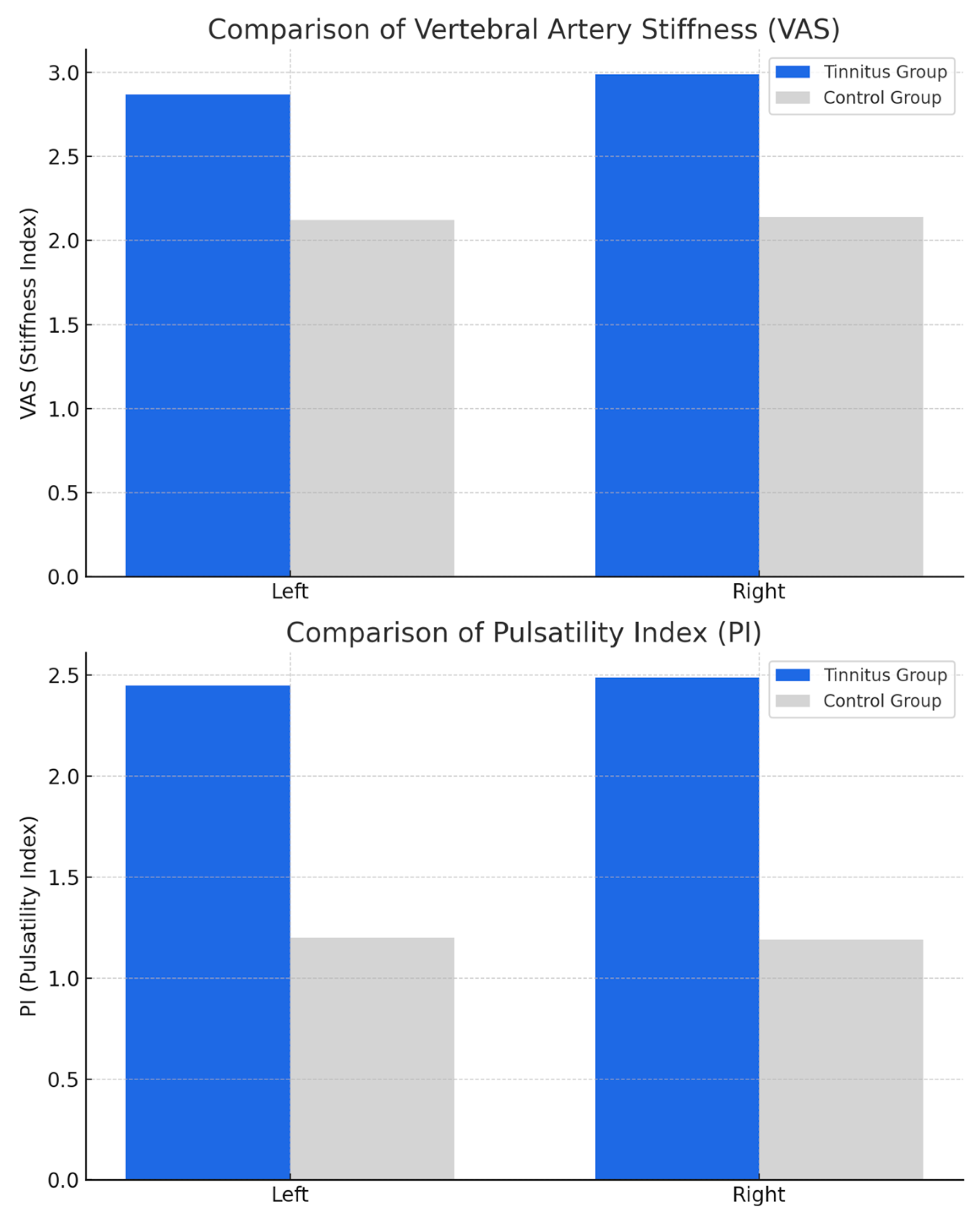
Vascular parameters assessed via vertebrobasilar Doppler ultrasonography, including vertebral artery stiffness (VAS), pulsatility index (PI), and resistive index (RI), are summarized in Table 2. Stiffness levels and PI values were significantly higher in the case group compared to the control group (p < 0.05). Specifically, VAS values for the left and right vertebral arteries were 2.87 ± 0.72 and 2.99 ± 0.77, respectively, in the case group, versus 2.12 ± 0.22 and 2.14 ± 0.5 in the control group. Similarly, PI values for the left and right vertebral arteries were 2.45 ± 1.2 and 2.49 ± 1.02, respectively, in the case group, compared to 1.2 ± 0.43 and 1.19 ± 0.42 in the control group. There were no significant differences in vertebral artery diameters between the groups (case group: left 0.39 ± 0.08, right 0.38 ± 0.085; control group: left 0.38 ± 0.08, right 0.38 ± 0.03) (Table 2).
Vertebral artery vascular parameters and the side of tinnitus were shown in Table 3. Of the tinnitus patients, 11 (35%) reported bilateral tinnitus, 11 (35%) reported tinnitus localized to the left side, and 9 (30%) reported right-sided tinnitus. Tinnitus was more frequently observed on the side with reduced blood supply from the vertebral artery. Table 3 details the VAS, PI, and RI values according to the side of tinnitus. VAS levels were higher on the left side in patients with left-sided tinnitus and higher on the right side in patients with right-sided tinnitus. Additionally, PI levels were significantly elevated on the side of the reported tinnitus (Table 3, Figure 3).

4. Discussion

In this prospective study, we demonstrated a significant association between vertebral artery stiffness and idiopathic subjective tinnitus, providing new evidence for a potential vascular contribution to tinnitus pathophysiology. Both vertebral artery stiffness index (VAS) and pulsatility index (PI) were markedly elevated in tinnitus patients compared to healthy controls. Importantly, we observed that these vascular abnormalities were lateralized and correlated with the side of tinnitus symptoms, suggesting a localized hemodynamic component.
Our findings are consistent with previous studies suggesting the role of vascular dysfunction and microcirculatory impairment in tinnitus development. Bayraktar et al. demonstrated increased carotid artery stiffness in patients with idiopathic tinnitus, emphasizing the role of systemic arterial stiffness in auditory dysfunction [16]. However, our study is among the first to specifically evaluate the vertebral arteries, which have a more direct contribution to cochlear and brainstem perfusion.
In addition, Gedikli et al. reported a positive relationship between systemic arterial stiffness parameters, including pulse wave velocity, and the presence of tinnitus [17]. While pulse wave velocity provides valuable systemic information, it does not fully capture localized vascular abnormalities in the vertebrobasilar system that directly influence cochlear perfusion. In contrast, our use of Doppler ultrasonography enabled regional assessment of vertebral artery stiffness, offering greater specificity for tinnitus-related microvascular changes.
Several mechanistic explanations may account for the observed association between increased vertebral artery stiffness and tinnitus. Increased arterial stiffness leads to elevated pulsatile pressure, impaired vascular compliance, and reduced capacity for flow regulation. In the cochlea, such hemodynamic disturbances may result in hypoxia, oxidative stress, and direct injury to the highly vascularized stria vascularis, spiral ganglion cells, and outer hair cells, thereby contributing to tinnitus generation. Badji et al. have previously emphasized that arterial stiffness is closely linked to microvascular brain injury through similar mechanisms [20].
The cochlea was known to be highly sensitive to changes in microvascular perfusion, and such alterations had been proposed as contributing factors in the development of tinnitus [23,24,25]. Atherosclerosis was previously identified as one of the key contributors to cochlear microcirculatory impairment, particularly in patients presenting with tinnitus [26,27]. Nakagawa et al. and Sasani et al. described the progression of atherosclerotic lesions in hyperlipidemic animal models and highlighted their impact on various microvascular beds, including those relevant for hearing [28,29], as well as a higher prevalence of tinnitus among individuals with ischemic cerebrovascular conditions [30]. Our findings of significantly elevated total cholesterol, LDL, and triglyceride levels in the tinnitus group were consistent with earlier reports highlighting the role of dyslipidemia and systemic vascular dysfunction in auditory pathologies. Furthermore, tinnitus symptoms were more frequently observed on the side with higher VAS and PI values, suggesting a possible hemodynamic mismatch and localized perfusion deficit.
Furthermore, the side-specific findings in our study may indicate localized vascular involvement. Although not all parameters showed statistically significant differences, we observed a trend toward higher vascular indices, such as vertebral artery stiffness and PI, on the same side as the reported tinnitus symptoms. For instance, right-sided stiffness and PI values were significantly higher in patients with right-sided tinnitus. Nakata et al. observed that abnormal cervical vascular findings were associated with worse outcomes in patients with idiopathic sudden sensorineural hearing loss, underscoring the relevance of vertebral artery function in auditory disorders [19]. These findings suggested a possible lateralized vascular contribution, although the interpretation should remain cautious given the limited sample size and the lack of statistical significance for some comparisons. Larger studies are needed to confirm these trends and clarify the clinical implications of such lateralization.
The vascular hypothesis of tinnitus is further supported by studies such as Del Vecchio et al. who linked vascular risk factors with sensorineural hearing loss and cognitive decline, highlighting the broader systemic implications of vascular dysfunction on the auditory system [18]. Likewise, Reeve et al. emphasized that cerebral and cochlear perfusion are closely linked, with vascular stiffness contributing to hypoperfusion in both central and peripheral auditory pathways [21].
Compared to previously published studies, our investigation differed in several important aspects. Earlier research examining arterial stiffness in tinnitus patients focused primarily on large central arteries, particularly the carotid artery, and utilized systemic parameters such as pulse wave velocity (PWV) or carotid intima-media thickness (CIMT) [16,17,22]. While these metrics are well-established markers of cardiovascular risk, they do not offer region-specific insight into the vertebrobasilar system, which directly supplies the cochlear and brainstem auditory pathways [18,21]. Unlike studies employing Young’s Elastic Modulus (YEM), which assumes homogeneity in arterial wall composition [31], our approach utilized a localized Doppler-derived stiffness index based on real-time arterial diameter and blood pressure measurements [15]. Compared to earlier studies evaluating CIMT or systemic arterial stiffness indices such as YEM, our Doppler-based methodology allows for dynamic, segmental assessment of the vertebral arteries, reflecting real-time systolic–diastolic diameter changes under physiological pressure conditions. This localized approach may offer better clinical relevance in disorders such as tinnitus where regional microvascular health is critical [30,31,32]. Additionally, we were the first to demonstrate a side-specific relationship between vertebral artery stiffness and the reported side of tinnitus, suggesting that focal hemodynamic alterations may be clinically relevant. These methodological innovations and anatomical focus allowed us to contribute new evidence supporting a vascular component in tinnitus pathophysiology.
Overall, our findings suggest that vertebral artery stiffness is an important vascular biomarker in tinnitus patients, and its assessment using Doppler ultrasonography may offer a valuable, non-invasive diagnostic tool for clinical practice. Although the differences in VAS and PI between tinnitus patients and controls were statistically significant, their clinical implications warrant further consideration. Elevated VAS and PI may reflect reduced vascular compliance and impaired microcirculatory regulation, which could contribute to cochlear hypoperfusion and inner ear dysfunction. In clinical practice, these parameters might serve as adjunctive biomarkers to identify tinnitus patients with a possible vascular etiology, especially when conventional audiometric assessments are inconclusive. However, there are currently no established cutoff values for VAS or PI in the context of tinnitus diagnosis or management. Therefore, the clinical utility of these measurements remains exploratory and requires further validation in larger, longitudinal studies to determine their predictive or therapeutic value.

Limitations of the Study

This study has several limitations that should be acknowledged. First, the tinnitus group was significantly older than the control group, which may have influenced vascular findings such as vertebral artery stiffness and pulsatility index. Arterial stiffness is known to increase with age due to reduced vascular compliance and endothelial dysfunction. Although our results remained statistically significant, we recognize that age could have contributed to the observed differences. Future studies should consider age-matched cohorts or include age-adjusted analyses to validate these findings more robustly.
Second, we did not use a tinnitus handicap inventory, which could have provided additional insights into the severity and impact of tinnitus on patients’ quality of life. However, such inventories are primarily used for assessing tinnitus severity rather than diagnostic purposes.
Third, the relatively small sample size may limit the generalizability of the findings and highlights the need for further studies with larger cohorts.
Finally, we were unable to employ advanced imaging modalities, such as magnetic resonance imaging (MRI), to evaluate cranial vascular structures due to financial constraints, which limited our ability to explore certain vascular parameters in more detail.
Despite these limitations, our study has notable strengths. It is one of the few clinical investigations exploring the relationship between vertebral artery stiffness and idiopathic subjective tinnitus. The use of Doppler ultrasonography, a non-invasive, accessible, and cost-effective imaging modality, allowed for a detailed evaluation of vertebral artery parameters, including stiffness and pulsatility index, providing valuable insights into the vascular contributions to tinnitus. Furthermore, the inclusion of a healthy control group ensured robust comparisons between case and control groups. Our findings open new avenues for understanding tinnitus as a condition potentially influenced by vascular health and highlight the clinical utility of non-invasive vascular assessments.

5. Conclusions

In conclusion, this study provided novel evidence that vertebral artery stiffness and pulsatility index are significantly elevated in patients with idiopathic subjective tinnitus. The observed lateralization of vascular abnormalities corresponding to tinnitus symptoms further supports the hypothesis of localized microvascular impairment contributing to tinnitus pathogenesis.
Our findings suggested that Doppler ultrasonography may serve as a useful adjunctive tool in the clinical evaluation of tinnitus by enabling the identification of vascular abnormalities that are not detectable through conventional audiological assessments. Given the non-invasive, accessible, and cost-effective nature of Doppler ultrasonography, its incorporation into the diagnostic algorithm for selected tinnitus patients may improve diagnostic accuracy and guide individualized management strategies.
Future studies with larger sample sizes and longitudinal follow-up are warranted to validate our findings, explore causality, and determine whether therapeutic modulation of vascular health may offer new treatment avenues for patients suffering from tinnitus.

Author Contributions

Conceptualization, I.A. and Y.I.; Methodology, I.A., A.Y., O.T., R.G. and K.T.; Software, I.A., A.Y., O.T., R.G., Y.I. and K.T.; Validation, I.A., A.Y., O.T., R.G., Y.I. and K.T.; Formal analysis, I.A., O.T., R.G., Y.I. and K.T.; Investigation, A.Y., R.G. and K.T.; Resources, I.A., A.Y., O.T., R.G. and Y.I.; Data curation, I.A., A.Y., O.T., R.G., Y.I. and K.T.; Writing—original draft, I.A.; Writing—review & editing, A.Y., O.T., R.G., Y.I. and K.T.; Visualization, O.T.; Supervision, A.Y. and O.T.; Project administration, I.A. and K.T.; Funding acquisition, I.A. All authors have read and agreed to the published version of the manuscript.

Funding

This research received no external funding.

Institutional Review Board Statement

The study was approved by the Ethics Committee of the Faculty of Medicine of Gaziantep University. Informed consent forms were obtained from all patients and/or their first-degree relatives included in the study. The study was conducted according to the guidelines of the Declaration of Helsinki.

Informed Consent Statement

Informed consent was obtained from all subjects involved in the study.

Data Availability Statement

Data is available upon request to the corresponding author.

Conflicts of Interest

The authors declare no conflicts of interest.

References

  1. GBD 2021 Risk Factors Collaborators. Global burden and strength of evidence for 88 risk factors in 204 countries and 811 subnational locations, 1990–2021: A systematic analysis for the Global Burden of Disease Study 2021. Lancet 2024, 403, 2162–2203. [Google Scholar] [CrossRef]
  2. GBD 2021 Europe Life Expectancy Collaborators. Changing life expectancy in European countries 1990–2021: A subanalysis of causes and risk factors from the Global Burden of Disease Study 2021. Lancet Public Health 2025, 10, e172–e188. [Google Scholar] [CrossRef]
  3. de Gruy, J.A.; Laurenzo, W.W.; Vu, T.H.; Paul, O.; Lee, C.; Spankovich, C. Prevalence and predictors of problematic tinnitus. Int. J. Audiol. 2025, 64, 307–313. [Google Scholar] [CrossRef]
  4. Batts, S.; Stankovic, K.M. Tinnitus prevalence, associated characteristics, and related healthcare use in the United States: A population-level analysis. Lancet Reg. Health Am. 2024, 29, 100659. [Google Scholar] [CrossRef] [PubMed]
  5. Richardson, M.L.; Luo, J.; Zeng, F.G. Attention-Modulated Cortical Responses as a Biomarker for Tinnitus. Brain Sci. 2024, 14, 421. [Google Scholar] [CrossRef] [PubMed]
  6. Sadekova, N.; Iulita, M.F.; Vallerand, D.; Muhire, G.; Bourmoum, M.; Claing, A.; Girouard, H. Arterial stiffness induced by carotid calcification leads to cerebral gliosis mediated by oxidative stress. J. Hypertens. 2018, 36, 286–298. [Google Scholar] [CrossRef]
  7. Vijayakumar, K.A.; Cho, G.W.; Maharajan, N.; Jang, C.H. A Review on Peripheral Tinnitus, Causes, and Treatments from the Perspective of Autophagy. Exp. Neurobiol. 2022, 31, 232–242. [Google Scholar] [CrossRef] [PubMed]
  8. Brinkmann, P.; Kotz, S.A.; Smit, J.V.; Janssen, M.L.F.; Schwartze, M. Auditory thalamus dysfunction and pathophysiology in tinnitus: A predictive network hypothesis. Brain Struct. Funct. 2021, 226, 1659–1676. [Google Scholar] [CrossRef]
  9. Haider, H.F.; Bojic, T.; Ribeiro, S.F.; Paco, J.; Hall, D.A.; Szczepek, A.J. Pathophysiology of Subjective Tinnitus: Triggers and Maintenance. Front. Neurosci. 2018, 12, 866. [Google Scholar] [CrossRef]
  10. Tsuzuki, N.; Wasano, K. Idiopathic sudden sensorineural hearing loss: A review focused on the contribution of vascular pathologies. Auris Nasus Larynx 2024, 51, 747–754. [Google Scholar] [CrossRef]
  11. Abouelleil, M.; Aljiboori, K.; Lyons, L.; Restrepo, A.; Singer, J. Commentary: Venous Causes of Pulsatile Tinnitus: Clinical Presentation, Clinical and Radiographic Evaluation, Pathogenesis, and Endovascular Treatments: A Literature Review. Neurosurgery 2022, 90, e65–e66. [Google Scholar] [CrossRef]
  12. Yao, Q.; Xu, C.; Wang, H.; Shi, H.; Yu, D. Video head impulse test results suggest that different pathomechanisms underlie sudden sensorineural hearing loss with vertigo and vestibular neuritis: Our experience in fifty-two patients. Clin. Otolaryngol. 2018, 43, 1621–1624. [Google Scholar] [CrossRef]
  13. Jiang, K.; Spira, A.P.; Reed, N.S.; Lin, F.R.; Deal, J.A. Sleep Characteristics and Hearing Loss in Older Adults: The National Health and Nutrition Examination Survey 2005–2006. J. Gerontol. A Biol. Sci. Med. Sci. 2022, 77, 632–639. [Google Scholar] [CrossRef] [PubMed]
  14. Xing, C.; Xie, X.; Wu, Y.; Xu, L.; Guan, X.; Li, F.; Zhan, X.; Yang, H.; Li, J.; Zhou, Q.; et al. Reference values of carotid intima-media thickness and arterial stiffness in Chinese adults based on ultrasound radio frequency signal: A nationwide, multicenter study. Chin. Med. J. 2024, 137, 1802–1810. [Google Scholar] [CrossRef] [PubMed]
  15. Boutouyrie, P.; Chowienczyk, P.; Humphrey, J.D.; Mitchell, G.F. Arterial Stiffness and Cardiovascular Risk in Hypertension. Circ. Res. 2021, 128, 864–886. [Google Scholar] [CrossRef]
  16. Bayraktar, C.; Tasolar, S. Relationship between increased carotid artery stiffness and idiopathic subjective tinnitus. Eur. Arch. Otorhinolaryngol. 2017, 274, 2125–2130. [Google Scholar] [CrossRef] [PubMed]
  17. Gedikli, O.; Kemal, O.; Yildirim, U.; Cecen, A.B.; Karabulut, H.; Akcay, M.; Terzi, O. Is there an association between the parameters of arterial stiffness and tinnitus? Acta Otolaryngol. 2020, 140, 128–132. [Google Scholar] [CrossRef]
  18. Del Vecchio, V.; Tricarico, L.; Pisani, A.; Serra, N.; D’Errico, D.; De Corso, E.; Rea, T.; Picciotti, P.M.; Laria, C.; Manna, G.; et al. Vascular Factors in Patients with Midlife Sensorineural Hearing Loss and the Progression to Mild Cognitive Impairment. Medicina 2023, 59, 481. [Google Scholar] [CrossRef]
  19. Nakata, T.; Okada, M.; Hanari, T.; Takagi, T.; Fujiwara, T.; Hato, N. Association of the prognosis and severity of idiopathic sudden sensorineural hearing loss with cervical ultrasonographic findings. Auris Nasus Larynx 2021, 48, 1074–1080. [Google Scholar] [CrossRef]
  20. Badji, A.; Sabra, D.; Bherer, L.; Cohen-Adad, J.; Girouard, H.; Gauthier, C.J. Arterial stiffness and brain integrity: A review of MRI findings. Ageing Res. Rev. 2019, 53, 100907. [Google Scholar] [CrossRef]
  21. Reeve, E.H.; Barnes, J.N.; Moir, M.E.; Walker, A.E. Impact of arterial stiffness on cerebrovascular function: A review of evidence from humans and preclincal models. Am. J. Physiol. Heart Circ. Physiol. 2024, 326, H689–H704. [Google Scholar] [CrossRef] [PubMed]
  22. Palombo, C.; Kozakova, M. Arterial stiffness, atherosclerosis and cardiovascular risk: Pathophysiologic mechanisms and emerging clinical indications. Vascul Pharmacol. 2016, 77, 1–7. [Google Scholar] [CrossRef] [PubMed]
  23. Lee, H.Y.; Shin, S.H.; Byun, S.W. Electrocochleography in Chronic Tinnitus: Correlations with Audiological Profiles and Psychological Distress. Am. J. Otolaryngol. 2024, 45, 104477. [Google Scholar] [CrossRef]
  24. Aazh, H.; Najjari, A.; Moore, B.C.J. A Preliminary Analysis of the Clinical Effectiveness of Audiologist-Delivered Cognitive Behavioral Therapy Delivered via Video Calls for Rehabilitation of Misophonia, Hyperacusis, and Tinnitus. Am. J. Audiol. 2024, 33, 559–574. [Google Scholar] [CrossRef]
  25. Bernal-Robledano, A.; Perez-Carpena, P.; Kikidis, D.; Mazurek, B.; Schoisswohl, S.; Staudinger, S.; Langguth, B.; Schlee, W.; Lopez-Escamez, J.A. Cognitive Screening and Hearing Assessment in Patients With Chronic Tinnitus. Clin. Exp. Otorhinolaryngol. 2024, 17, 15–25. [Google Scholar] [CrossRef]
  26. Wadhavkar, N.; Goldrich, D.Y.; Roychowdhury, S.; Kwong, K. Laceration of Aberrant Internal Carotid Artery Following Myringotomy: A Case Report and Review of Literature. Ann. Otol. Rhinol. Laryngol. 2022, 131, 555–561. [Google Scholar] [CrossRef] [PubMed]
  27. Zhou, D.; Ding, J.Y.; Ya, J.Y.; Pan, L.Q.; Yan, F.; Yang, Q.; Ding, Y.C.; Ji, X.M.; Meng, R. Understanding jugular venous outflow disturbance. CNS Neurosci. Ther. 2018, 24, 473–482. [Google Scholar] [CrossRef]
  28. Nakagawa, T.; Kikumori, A.; Kimura, N.; Shiomi, M. Distribution of atherosclerotic lesions in various arteries of WHHLMI rabbits, an animal model of familial hypercholesterolemia. Exp. Anim. 2019, 68, 293–300. [Google Scholar] [CrossRef]
  29. Sasani, M.; Aydin, A.L.; Aytan, N.; Yapicier, O.; Oktenoglu, T.; Ozer, N.K.; Ozer, A.F. Effect of a hypercholesterolemia as a starting factor on spinal degeneration in rabbits and role of Vitamin E (alpha-tocopherol). Surg. Neurol. Int. 2016, 7, 36. [Google Scholar] [CrossRef]
  30. Huang, Y.S.; Koo, M.; Chen, J.C.; Hwang, J.H. The association between tinnitus and the risk of ischemic cerebrovascular disease in young and middle-aged patients: A secondary case-control analysis of a nationwide, population-based health claims database. PLoS ONE 2017, 12, e0187474. [Google Scholar] [CrossRef]
  31. Petrova, M.; Gavino, A.; Li, Y.; McLachlan, C.S. Comparison of Parameters for Assessment of Carotid Stiffness and Their Association with Carotid Atherosclerosis in Rural Australian Adults: A Pilot Study. J. Clin. Med. 2023, 12, 2935. [Google Scholar] [CrossRef] [PubMed]
  32. Turk, M.; Zaletel, M.; Pretnar-Oblak, J. Ratio between carotid artery stiffness and blood flow—A new ultrasound index of ischemic leukoaraiosis. Clin. Interv. Aging 2016, 11, 65–71. [Google Scholar] [CrossRef] [PubMed]
Figure 1. Flowchart showing the patient inclusion and exclusion process for tinnitus and control groups.
Applsci 15 07890 g001
Figure 2. Doppler ultrasonography image showing measurement of vertebral artery parameters between C4–C6 levels.
Applsci 15 07890 g002
Figure 3. Comparison of vertebral artery stiffness (VAS) and pulsatility index (PI) between tinnitus and control groups for left and right vertebral arteries.
Applsci 15 07890 g003
Table 1. Demographic variables and blood sample data of the control and case groups.
Case (n = 31)Control (n = 24)p Value
Age50 ± 9.6446.6 ± 10.630.188
Gender (Female/Male)20/11 12/12 0.279
PTA (dB) Right22.16 ± 13.820.3 ± 7.30.585
   (dB) Left21.9 ± 9.420 ± 5.30.838
HDL (mg/dL)47.2 ± 11.649.3 ± 70.453
LDL (mg/dL)149.4 ± 37.9106.1 ± 10.7<0.005
TG (mg/dL)202.2 ± 83.5148.6 ± 26.4<0.005
Fasting Glucose (mg/dL)82.5 ± 7.480.5 ± 5.50.310
Cholesterol (mg/dL)193.6 ± 47.28167.5 ± 28.990.021
SBP (mmHg)116.48 ± 8.28112.56 ± 5.80.380
DBP (mmHg)76.12 ± 3.5875.15 ± 4.720.160
PTA: Pure tone audiometry, HDL: High-density lipoprotein, LDL: Low-density lipoprotein, TG: Triglycerides, SBP: Systolic blood pressure, DBP: Diastolic blood pressure.
Table 2. Comparison of vertebral artery vascular structural patterns between case and control groups.
Case (n = 31)Control (n = 24)p Value
Vertebral artery stiffness Left2.87 ± 0.722.12 ± 0.22<0.005
Vertebral artery stiffness Right2.99 ± 0.772.14 ± 0.5<0.005
Vertebral artery RI Left0.86 ± 0.320.94 ± 0.240.581
Vertebral artery RI Right0.84 ± 0.310.91 ± 0.220.509
Vertebral artery PI Left2.45 ± 1.21.2 ± 0.43<0.005
Vertebral artery PI Right2.49 ± 1.021.19 ± 0.42<0.005
RI: Resistive index, PI: Pulsatility index.
Table 3. Vertebral artery vascular parameters and the side of tinnitus.
LeftRightBilateralp Value
Vert. Art. Stiff Left3.27 ± 0.792.57 ± 0.762.2 ± 0.450.058
Vert. Art. Stiff Right2.7 ± 0.663.62 ± 0.872.76 ± 0.010.010
Vert Art. RI Left0.93 ± 0.150.97 ± 0.290.64 ± 0.320.543
Vert Art. RI Right0.84 ± 0.191.08 ± 0.190.69 ± 0.40.579
Vert Art. PI Left2.74 ± 1.582.41 ± 1.342.2 ± 0.450.024
Vert Art. PI Right2.6 ± 1.52.7 ± 0.952.21 ± 0.220.016
RI: Resistive index, PI: Pulsatility index.
Disclaimer/Publisher’s Note: The statements, opinions and data contained in all publications are solely those of the individual author(s) and contributor(s) and not of MDPI and/or the editor(s). MDPI and/or the editor(s) disclaim responsibility for any injury to people or property resulting from any ideas, methods, instructions or products referred to in the content.