Article

Efficacy of Oxygen Fluid (blue®m) on Human Gingival Fibroblast Viability, Proliferation and Inflammatory Cytokine Expression: An In Vitro Study

by
Rhodanne Nicole A. Lambarte
1,
Amani M. Basudan
2,*,
Marwa Y. Shaheen
2,
Terrence S. Sumague
1,
Fatemah M. AlAhmari
2,
Najla M. BinShwish
1,
Abeer S. Alzawawi
2,
Abdurahman A. Niazy
1,3,
Mohammad A. Alfhili
4 and
Hamdan S. Alghamdi
2
1
Molecular and Cell Biology Laboratory, Prince Naif bin AbdulAziz Health Research Center, King Saud University Medical City, Riyadh 11545, Saudi Arabia
2
Department of Periodontics and Community Dentistry, College of Dentistry, King Saud University, Riyadh 11451, Saudi Arabia
3
Department of Oral Medicine and Diagnostic Sciences, College of Dentistry, King Saud University, Riyadh 11545, Saudi Arabia
4
Department of Clinical Laboratory Sciences, College of Applied Medical Sciences, King Saud University, Riyadh 12372, Saudi Arabia
*
Author to whom correspondence should be addressed.
Appl. Sci. 2025, 15(13), 7459; https://doi.org/10.3390/app15137459
Submission received: 10 May 2025 / Revised: 30 June 2025 / Accepted: 1 July 2025 / Published: 3 July 2025

Abstract

Human gingival fibroblasts (HGnFs) play crucial roles in periodontal wound healing. This in vitro study examined the impact of varying concentrations of topical oxygen fluid (blue®m) on HGnF morphology, viability, proliferation, oxidative stress and pro-inflammatory cytokine production. The attempt was to underscore the potential of blue®m as a less cytotoxic alternative to chlorhexidine in the context of tissue-regeneration improvement. Primary HGnF cell cultures were subjected to oxygen fluid (blue®m) at concentrations of 0.6, 1.2 and 2.4% for a duration of 1 min. The positive control was 0.12% chlorhexidine. Cell morphology as well as actin cytoskeleton were assessed using microscopy and immunofluorescence staining. Cell viability and proliferation were assessed through AlamarBlue and trypan blue assays at 1, 2, 7, 10 and 14 days. Levels of reactive oxygen species (ROS) were quantified using DCFH-DA assay. Pro-inflammatory cytokines (IL-1β, IL-6, TNF-α, MMP-8 and TIMP-1) were assessed through ELISA. HGnF morphology and actin structure were preserved at all oxygen fluid concentrations. Cell viability and proliferation were significantly higher in the 0.6% and 1.2% groups than in the control and chlorhexidine groups (p ≤ 0.05). ROS levels were low at 0.6% and 1.2%, but increased at 2.4% and with chlorhexidine (p ≤ 0.05). Oxygen treatment reduced IL-1β, IL-6, TNF-α and TIMP-1 expression, while MMP-8 levels increased. Chlorhexidine significantly upregulated the expression of all proinflammatory cytokines (p ≤ 0.01). Oxygen fluid (blue®m) therapy improves the viability and proliferation of gingival fibroblasts and offers anti-inflammatory and preliminary antioxidative effects at the cellular level, especially at lower concentrations (0.6% and 1.2%), indicating potential application in periodontal wound management, subject to clinical validation.

1. Introduction

Wound healing is a complicated biological process consisting of a coordinated series of cellular and molecular activities to restore tissue integrity and function following an injury [1]. The wound-healing process is uniquely challenged in the oral cavity, particularly within gingival tissues, by constant microbial exposure and mechanical stress, presenting clinical hurdles for successful outcomes in periodontal surgery and implant therapy [2]. Human gingival fibroblasts (HGnFs) play a key role in this process by producing extracellular matrix, inflammation regulation and tissue remodeling [3,4].
A major challenge in the management of periodontal wounds is the equilibrium between infection control and tissue regeneration. Conventional antiseptics, such as chlorhexidine gluconate, are commonly utilized for their antibacterial qualities; nevertheless, their cytotoxic effects on fibroblasts and other oral cells might compromise fibroblast viability and potentially impede wound healing [5,6]. Consequently, there is an increasing demand for alternative strategies to manage infection and support cellular health and regenerative potential.
Recently, oxygen-based therapies have gained interest in regenerative medicine due to oxygen’s critical role in cellular metabolism and wound-healing phases, such as hemostasis, inflammation, proliferation and remodeling [7,8,9]. In the initial stages of wound healing, oxygen supports immune defense through oxidative bursts, while in later phases, it stimulates fibroblast proliferation, collagen synthesis, angiogenesis and matrix maturation [10,11]. Topical oxygen therapy, which refers to the direct application of oxygen onto the wound surface, may overcome local hypoxia associated with gingival inflammation and microvascular impairment [12,13,14]. Oxygen facilitates gingival repair by improving microcirculation and fostering tissue regeneration [14]. Furthermore, this method allows for controlled delivery with potentially fewer systemic side effects.
Oxygen fluid (blue®m), a stable oxygen-releasing liquid formulation, is one of the emerging topical oxygen therapy methods that has the potential to promote oral wound healing. This is due to its simplicity of application and the potential for consistent coverage [7,8,9]. However, data on its biological impact on gingival fibroblasts remain limited. Preliminary studies in other cell types and tissue models suggest dose-dependent effects, wherein sub-inhibitory doses may preserve or enhance wound-healing functions, underlining the importance of dose optimization for periodontal applications [10,15].
The wound-healing process is complicated by oxidative stress. Reactive oxygen species (ROS) can be elevated by excessive oxygen, which can lead to oxidative stress and the impedance of cellular processes [16]. Excessive ROS may induce apoptosis and chronic inflammation, whereas physiological ROS levels facilitate signaling for cell migration and proliferation [16,17].
In addition to oxidative stress, the healing process is significantly influenced by the modulation of inflammatory cytokines such as interleukin-1β (IL-1β), interleukin-6 (IL-6) and tumor necrosis factor-alpha (TNF-α), as well as enzymes such as matrix metalloproteinases (MMPs) and tissue inhibitor of metalloproteinases (TIMPs) [18,19,20,21]. Therefore, it is imperative to comprehend the influence of these factors on gingival fibroblasts in order to evaluate the clinical viability of topical oxygen fluid.
This study aimed to evaluate the impact of oxygen fluid (blue®m) at varying concentrations on the cellular expression of inflammatory mediators, oxidative stress, HGnF viability and proliferation. This study attempted to address a critical lacuna in periodontal regenerative research.

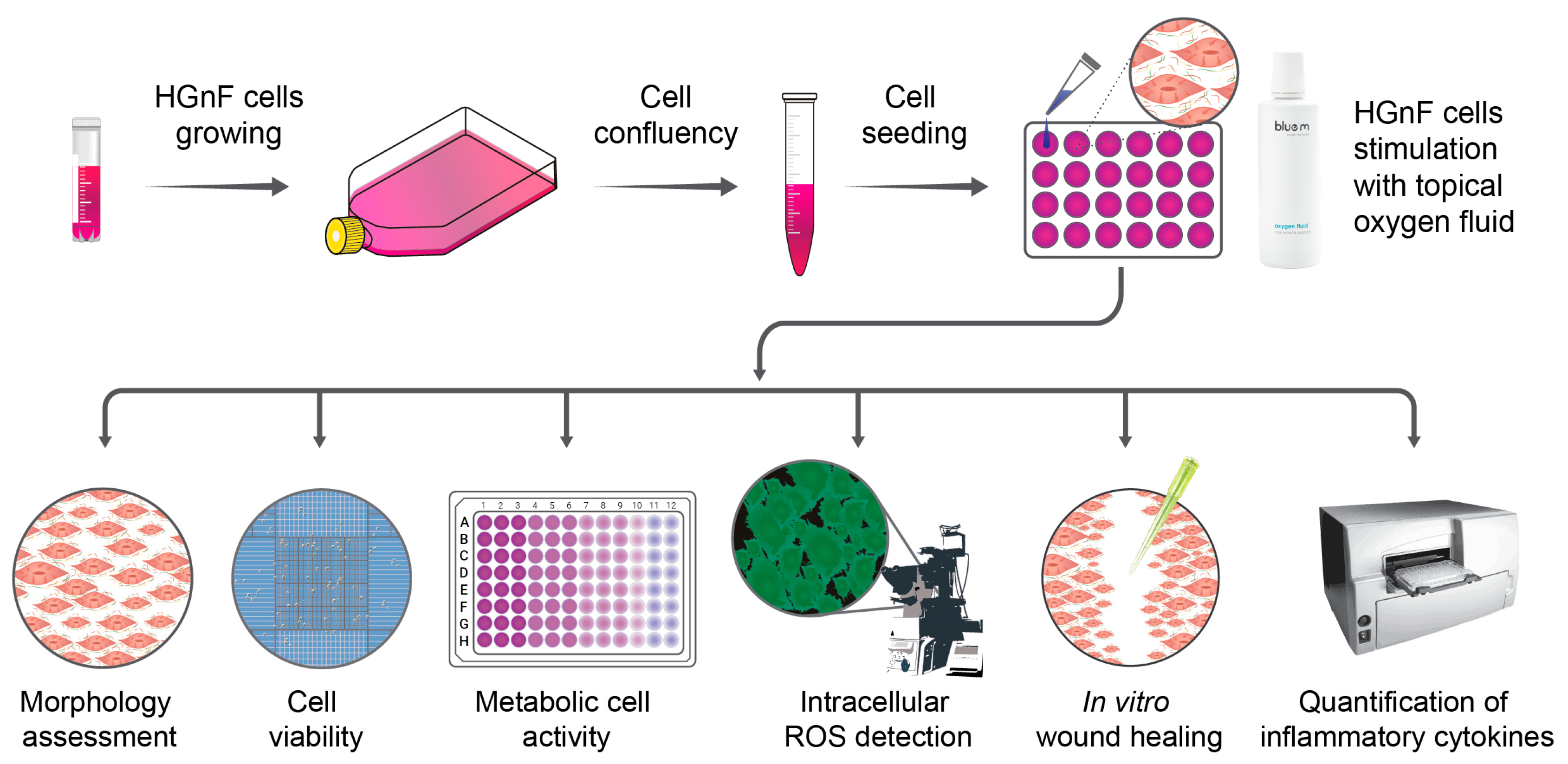
2. Materials and Methods

A schematic diagram of the study is shown in Figure 1. HGnF cells were grown, seeded and upon confluency cells were exposed in appropriate dilution of blue®m oxygen fluid (O2F).

2.1. Cell Culture

All experiments were conducted using a well-characterized human gingival fibroblast (HGnF) cell line derived from human gingiva (Cat. No. 2620; ScienCell™ Research Laboratories, Carlsbad, CA, USA). HGnFs were selected for this study due to their essential role in gingival connective tissue and wound healing, making them a relevant in vitro model for evaluating the biocompatibility of oral-care products [22,23]. Under aseptic conditions in a laminar flow hood, the cells were cultured in Dulbecco’s Modified Eagle’s Medium (DMEM) with 4500 mg/L glucose, 1 mM sodium pyruvate and 4 mM L-glutamine supplemented with 10% fetal bovine serum (FBS), 1% penicillin-streptomycin and 1% non-essential amino acids (all from Gibco, Invitrogen, New York, NY, USA). The cell culture conditions included incubation at 37 °C with 5% CO2 and 95% humidity, until 80% confluency was achieved, and all experiments were conducted between cell passages 3 and 6.

2.2. Stimulation of Gingival Fibroblasts with Topical blue®m Oxygen Fluid Therapy

To evaluate the effects of topical oxygen fluid exposure on HGnFs, the cells were grown in complete growth culture medium for 24 h, followed by addition of appropriate dilutions of the O2F (0.6, 1.2 and 2.4%). Commercially available blue®m oxygen fluid (BlueM Europe, Zwolle, The Netherlands) was dissolved in serum-free growth medium to achieve dilutions of 0.6, 1.2 and 2.4%, respectively. Test concentrations were selected based on preliminary pilot studies to identify non-cytotoxic, biologically active doses aligned with the composition found in the topical blue®m oxygen fluid [10]. Following the aforementioned growth period, the HGnF cells were stimulated for 1 min at 37 °C, by adding different concentrations of topical oxygen therapy solutions. This was followed by replenishment with regular growth medium and the cells were grown under standard conditions for varying periods and under differing micro-environmental conditions, based on the assays carried out. For all assays, HGnF cells exposed only to the growth culture medium were used as an experimental control, and exposure to 0.12% chlorhexidine (CHX), similar to oxygen fluid stimulation, was used as a standardized positive control.

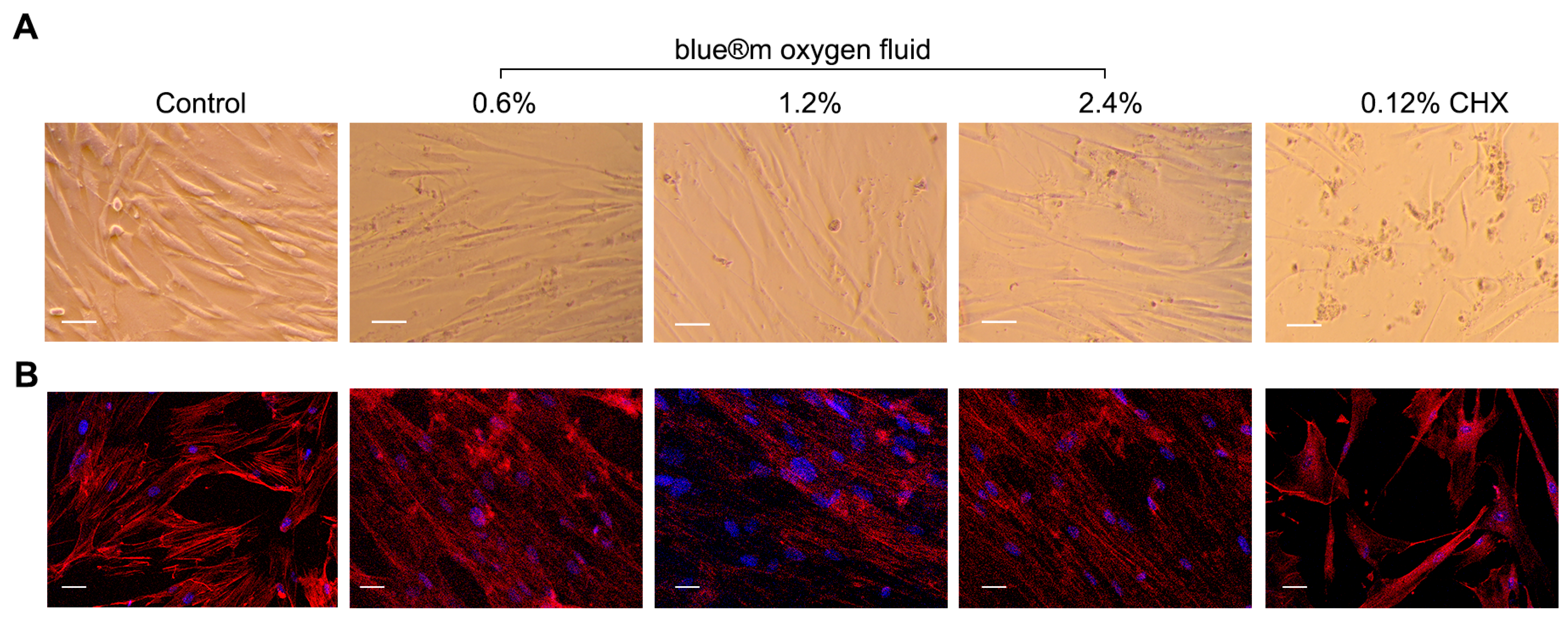
2.3. Cell Morphology

In order to determine the effects of oxygen fluid therapy on cell morphology, HGnFs were seeded at a density of 2 × 10⁴ cells/mL in 24-well culture plates (Greiner Bio-One GmbH, Frickenhausen, Germany) and maintained in DMEM growth media until confluency. The cells were exposed for 1 h at 37 °C with different concentrations of oxygen fluid (0.6, 1.2 and 2.4%) as previously mentioned. After 24 and 48 h, changes in the spindle-shaped morphology of the fibroblast cells were examined and imaged using inverted light microscope (Carl Zeiss Axiovert 40C, Göttingen, Germany) and recorded through a digital camera (Nikon, Dusseldorf, Germany).

2.4. Immunofluorescence Staining

For immunofluorescence staining, HGnF cells were seeded in 4-well dishes (Nunc, Thermo Fisher Scientific, Waltham, MA, USA) at a density of 5 × 10⁴ cells/well in growth culture media and expanded till confluency. This was followed by exposure to different concentrations of oxygen fluid (0.6, 1.2 and 2.4%) for 1 h at 37 °C. Afterwards, cells were cultured for 24 h in DMEM, gently washed with phosphate-buffered saline (PBS; Sigma-Aldrich, St. Louis, MO, USA) twice and fixed for 20 min at 4 °C with 4% paraformaldehyde in PBS. In order to enhance cell membrane permeability, a 15 min treatment with 0.1% Triton X-100 (Sigma-Aldrich) was carried out and subsequently a blocking treatment for non-specific binding sites was carried out for 1 h with 10% bovine serum albumin (BSA; Sigma-Aldrich) in PBS. In order to visualize cellular structure, F-actin filaments were stained using Alexa Fluor™ 594 Phalloidin (1:800, Invitrogen Molecular Probes, Eugene, OR, USA) for 20 min at room temperature. Nuclei of the viable cells were counterstained with 1 μM 4′,6-Diamidino-2-phenylindole dihydrochloride (DAPI; Invitrogen™ Molecular Probes) for 10 min. Samples were finally washed and stored in PBS, and examined under a Nikon C2 confocal laser scanning microscope (CLSM; Nikon Instruments Inc., Tokyo, Japan). Sample images were acquired using NIS-Elements Advanced Research software (version 4.0, Nikon, Japan) and Nikon C2 CLSM through 10×/1.84 NA air objective using (λexc) 405 nm/(λem) < 550 nm for DAPI and (λexc) 568 nm/(λem) > 600 nm for Phalloidin [24,25].

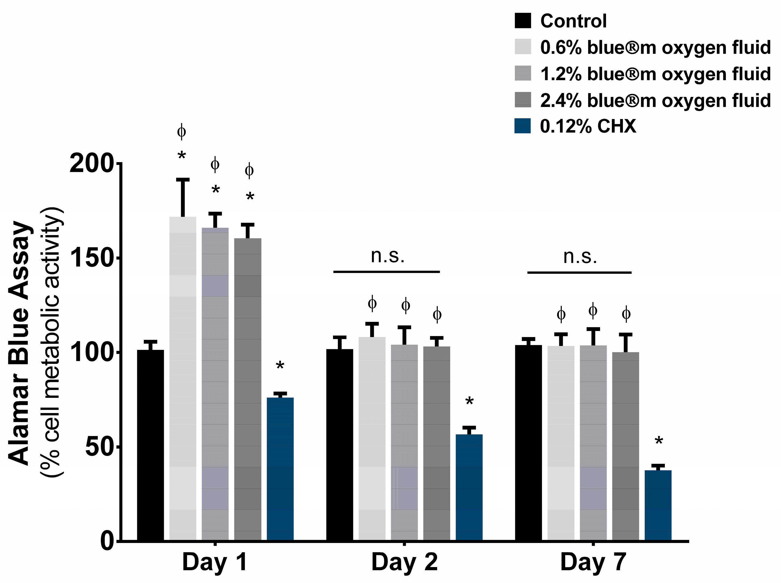
2.5. Cell Viability (AlamarBlue Assay)

Cell viability was determined using the AlamarBlue viability reagent (Invitrogen, Eugene, OR, USA) according to manufacturer’s instructions. In brief, HGnF cells were seeded in 96-well culture plates at an initial cell density of 1.5 × 10⁴ cells/well in 100 μL growth medium. At 24 h post-seeding, cells were treated with different concentrations of oxygen fluid (0.6, 1.2 and 2.4%) for 1 h at 37 °C and then rinsed in PBS. At the indicated time points, 10 μL of AlamarBlue substrate was added to each well and plates were incubated in the dark at 37 °C for 4 h. Background correction was performed using blank wells containing only culture medium and AlamarBlue reagent. Readings were taken at day 1, 2 and 7, using a BioTek Synergy HT microplate reader (Winooski, VT, USA) in fluorescent mode (λexc 530 nm/λem 590 nm) and expressed as a percentage of viable cell metabolic activity.

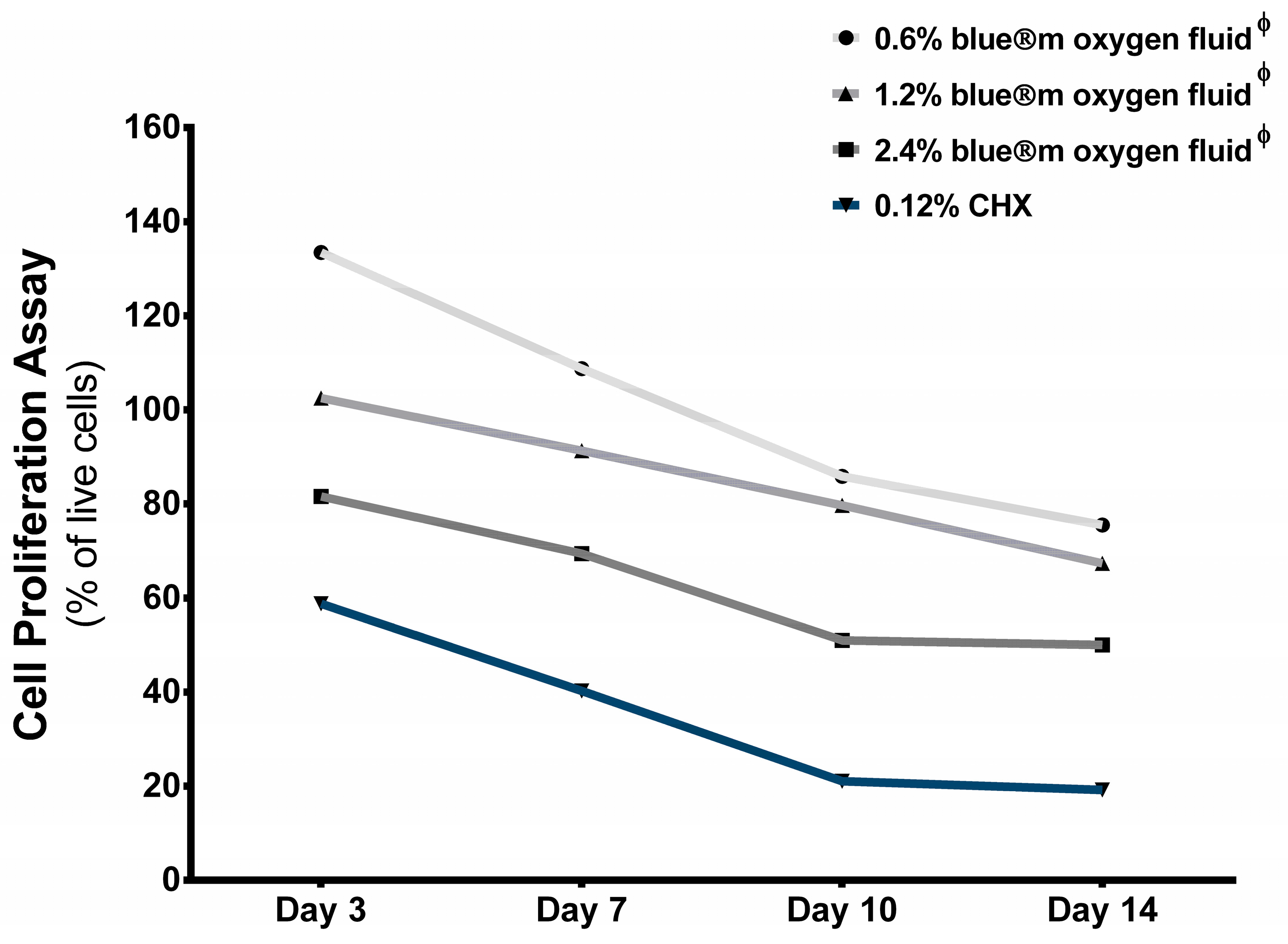
2.6. Cell Proliferation (Trypan Blue Assay)

Human gingival fibroblasts were seeded at 2 × 10⁴ cells/mL and cultured in DMEM culture media for 24 h. The cells were then exposed to different concentrations of oxygen fluid, as previously described. After 3, 7, 10 and 14 days, in order to ascertain cell proliferation and determine cell count, cells were detached with 0.25% trypsin/EDTA solution and were collected by centrifugation at 2000 rpm using a Sigma 2-6E centrifuge (Osterode am Harz, Germany). The cell pellet was resuspended in 1 mL of medium. An aliquot of cell suspension was mixed with an equal volume of 0.4% trypan blue and incubated for 5 min at room temperature. The number of viable cells were determined using a Countess Automated Cell Counter (Invitrogen, Eugene, OR, USA) and expressed as a percentage of live cells in the total cell suspension [26,27]. Each condition was measured in triplicate from at least three independent experiments to ensure reproducibility of results.

2.7. Wound-Healing Assay

HGnF cells were seeded into 12-well culture plates (Greiner Bio-One GmbH) at 5 × 10⁴ cells/mL and cultured until 90% confluence. A scratch was produced in the monolayer on each well using a 200 µL pipette tip (Thermo Scientific QSP, Waltham, MA, USA). Non-adherent cells were removed by washing once with PBS. The different concentrations of topical oxygen fluid were added to each well for 1 min and replaced with regular growth medium. At time points 24, 48 and 72 h, cell migration into the empty scratch surface under different conditions was monitored using phase-contrast microscopy and compared with cell migration under conditions of control media, then recorded by a digital camera. The area of wound healing was analyzed using the ImageJ software (version 1.5i, Bethesda, MD, USA) [28,29].

2.8. Reactive Oxygen Species Measurement

The HGnF cells were seeded in 4-well dishes at a density of 5 × 10⁴ cells/well and exposed for 24 h with different concentrations of blue®m oxygen fluid and CHX, as previously mentioned. The levels of cellular oxidative stress were measured after HGnF cells were stimulated using 2′,7′-dichlorodihydrofluorescein diacetate (DCFH-DA) assay kit (Sigma-Aldrich, St. Louis, MO, USA) according to manufacturer’s instructions [30]. Adherent cells were observed by Nikon C2 CLSM (Nikon Instruments Inc., Tokyo, Japan) with 10×/1.84 NA objective lens using (λexc) 488 nm/(λem) < 550 nm. The fields of view were captured randomly and the mean fluorescence intensity (MFI) was quantified using NIS-Elements Advanced Research Software (version 4.0, Nikon, Japan).

2.9. Detection of Pro-Inflammatory Cytokine Expression Using ELISA (Enzyme-Linked Immunosorbent Assay)

The conditioned media were collected from seeded HGnF cell cultures (5 × 10⁵ cells/well) exposed to different concentrations of blue®m oxygen fluid for a period of 24 h, to assess the production of pro-inflammatory cytokines, namely, interleukin-1β (IL-1β), interleukin-6 (IL-6), tumor necrosis factor-α (TNF-α), matrix metalloproteinase-8 (MMP-8) and tissue inhibitors of metalloproteinase-1 (TIMP-1) [31,32,33]. Specific ELISA kits were used for quantification, IL-1β, IL-6 and TNF-α were measured using kits from Solarbio® Life Science (Beijing, China), while MMP-8 and TIMP-1 were analyzed using kits from Elabscience® (Houston, TX, USA). For each ELISA kit, the in vitro assay was performed according to the manufacturers’ recommendations and analyzed using BioTek Synergy HT microplate reader (Winooski, VT, USA) at 450 nm.

2.10. Statistical Analysis

Results for all assays were expressed as mean ± SD of three independent experiments each carried out in triplicates. The experimental data were analyzed by one-way or two-way analysis of variance (ANOVA) followed by Tukey’s multiple comparisons test for post-hoc analysis. All statistical tests were performed using GraphPad Prism version 6.01 for Windows, and the statistical significance level was assumed when p-values were less than or equal to 0.05 (95% significance level).

3. Results

3.1. Cell Morphology and Immunofluorescence Staining

Microscopic examination of cell morphology indicated that exposure to different concentrations of blue®m oxygen fluid (0.6, 1.2 and 2.4%) did not drastically alter the spindle-shaped morphology of HGnF cells. This was further evidenced by immunofluorescence staining of the cells, which showed characteristic red and blue staining, indicative of actin filaments and viable nuclei, respectively. The presence of smooth muscle actin in the cultured cells was further indicative of myofibroblastic proliferation, a key characteristic responsible for the wound-healing process. Representative photomicrographs of the cultured HGnFs in the control, CHX and blue®m oxygen fluid groups are shown in Figure 2. While there was a visibly greater number of HGnF cells seen in the control group, it progressively decreased with increasing concentration of oxygen fluid and was the lowest when cultured with CHX. Within the cells cultured with blue®m oxygen fluid, the largest number of viable nuclei on immunofluorescence staining was observed at 1.2% concentration.

3.2. Cell Viability

Exposure of HGnF cells to varying concentrations of blue®m oxygen fluid, assessed using the AlamarBlue assay, resulted in higher cell viability compared to the control (Figure 3, Table 1). This increase was statistically significant (p < 0.01) on day 1, but not on days 2 or 7. On day 1 and 2, the highest mean percentage of cell viability was observed with 0.6% blue®m oxygen fluid, followed by 1.2% and 2.4%. However, by day 7, 1.2% blue®m oxygen fluid showed the highest viability, followed by 0.6% and 2.4% concentrations. Nevertheless, the differences in cell viability among the varying concentrations of blue®m oxygen fluid were not statistically significant. In contrast, exposure to 0.12% CHX significantly decreased the mean percentage cell viability (p < 0.05) at all time points compared to the control and blue®m oxygen fluid groups.

3.3. Cell Proliferation
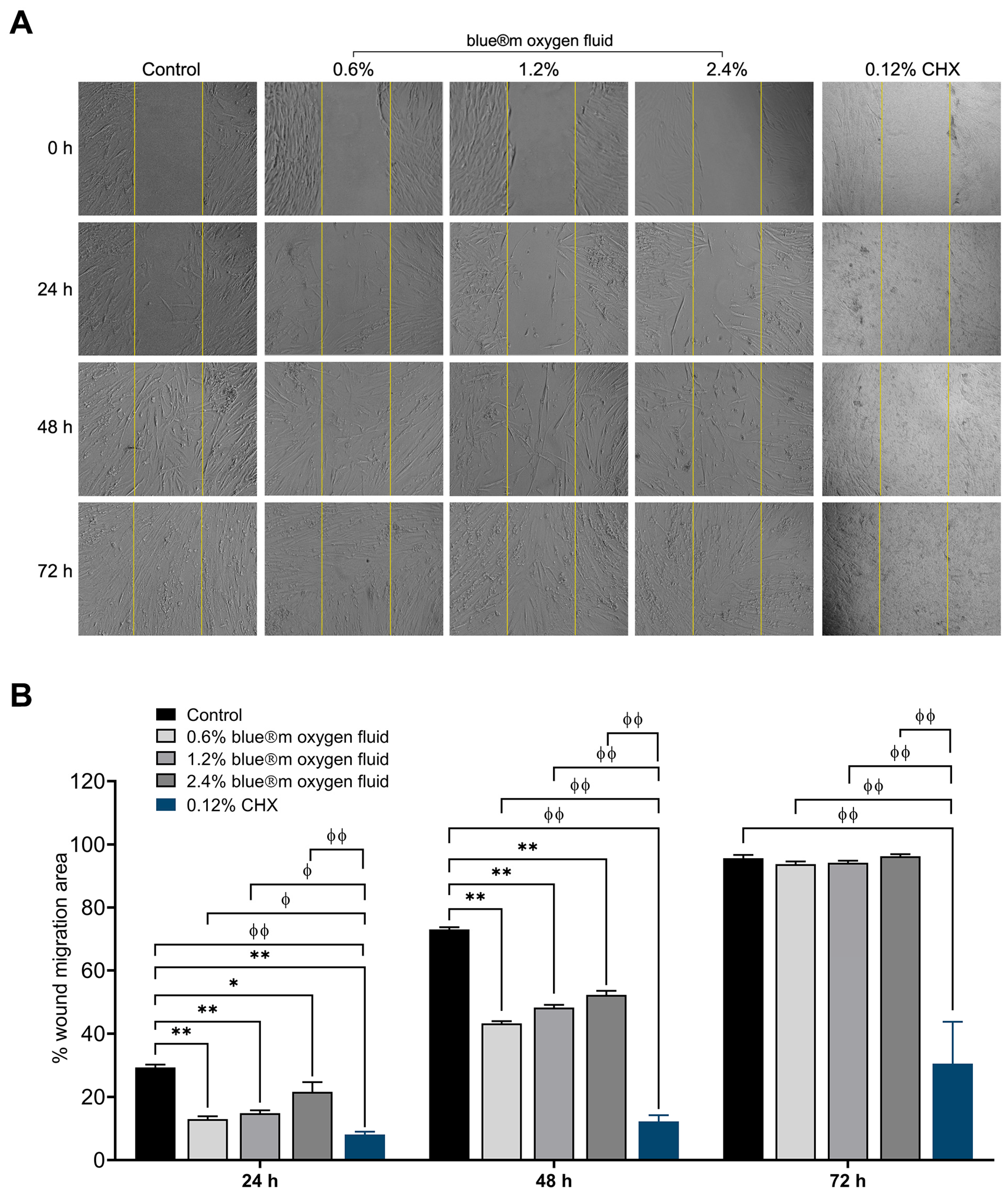
Results of the trypan blue assay for the estimation of cell proliferation showed the highest mean percentage of live cells in each group on day 3, followed by a consistent decrease on days 7, 10 and 14 (Figure 4, Table 1). Between the groups, and at all time periods of observation, the mean percentage of live cells was highest in HGnF cell cultures with 0.6%, followed by 1.2% and 2.4% blue®m oxygen fluid concentrations. The lowest mean values were observed in cultures treated with CHX. While the differences in mean cell-proliferation percentage between 0.6% and 1.2% blue®m oxygen fluid were statistically significant only by day 3, the further differences (days 7, 10 and 14) were not statistically significant. However, the mean values observed with 0.6% and 1.2% blue®m oxygen fluid were significantly higher than those with 2.4% blue®m oxygen fluid and CHX at all time periods. Similarly, the mean cell-proliferation percentage in HGnF cells cultured with CHX was significantly the lowest among all groups and at all times.

3.4. Wound Healing

Cell migration using the scratch wound assay was measured (Figure 5A) and the percentage of wound migration area at 24, 48 and 72 h is shown in Figure 5B. All blue®m oxygen fluid groups (0.6, 1.2 and 2.4%) showed significantly higher wound closure rates at 24 and 48 h than the 0.12% CHX group (p < 0.01). At every time point, the 0.6% blue®m oxygen fluid group showed the highest percentage of wound migration. By 72 h, no significant differences were found among the groups of blue®m oxygen fluid and the untreated control. In contrast, CHX exposure significantly impaired cell migration at all time points (24, 48 and 72 h) compared to both the blue®m oxygen fluid groups and the control (p < 0.01). These results implied that while blue®m oxygen fluid promoted migration in a concentration-dependent manner, exposure to CHX significantly suppressed the migratory response of HGnF cells.

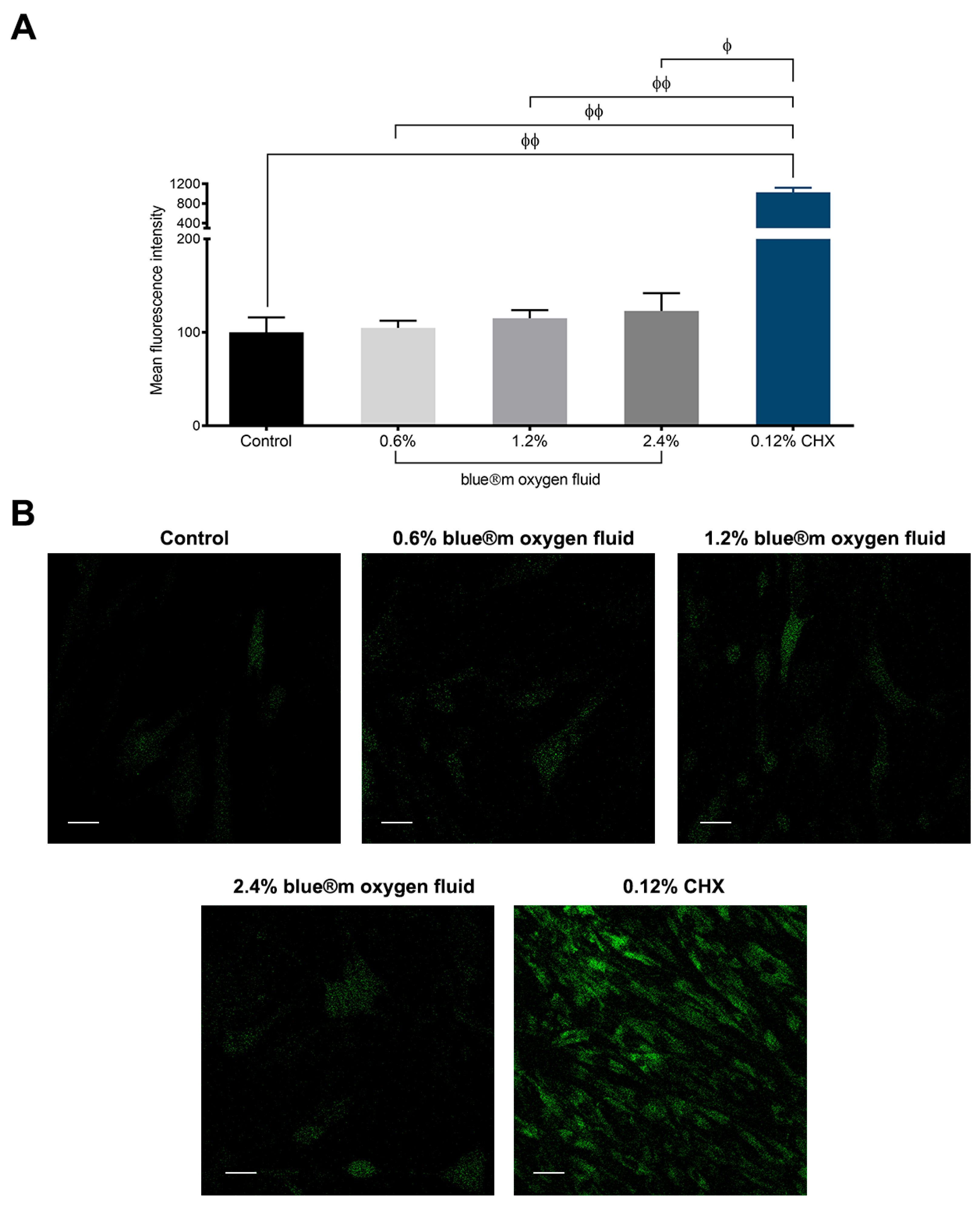
3.5. Results of Reactive Oxygen Species

MFI values, used to measure ROS as indicators of cellular oxidative stress in vitro for HGnF cells, indicated the lowest ROS levels in the control group. This was followed by increasing concentrations of blue®m oxygen fluid (Figure 6A, Table 1). No statistically significant differences were observed between the control group and the O2F concentrations. In contrast, the mean ROS levels in HGnF cells cultured with 0.12% chlorhexidine were significantly higher than those in the control and all blue®m oxygen fluid groups (p < 0.001). Photomicrographs of cultured HGnF cells after the assay for measuring ROS indicated a high level of oxidative stress, as demonstrated by the fluorescence intensity in cells treated with CHX (Figure 6B). ROS levels remained comparable to those of the control in HGnF cells exposed to blue®m oxygen fluid, suggesting no measurable oxidative stress at these concentrations. In contrast, 0.12% CHX was associated with significantly elevated ROS levels, indicating an increased cellular oxidative response.

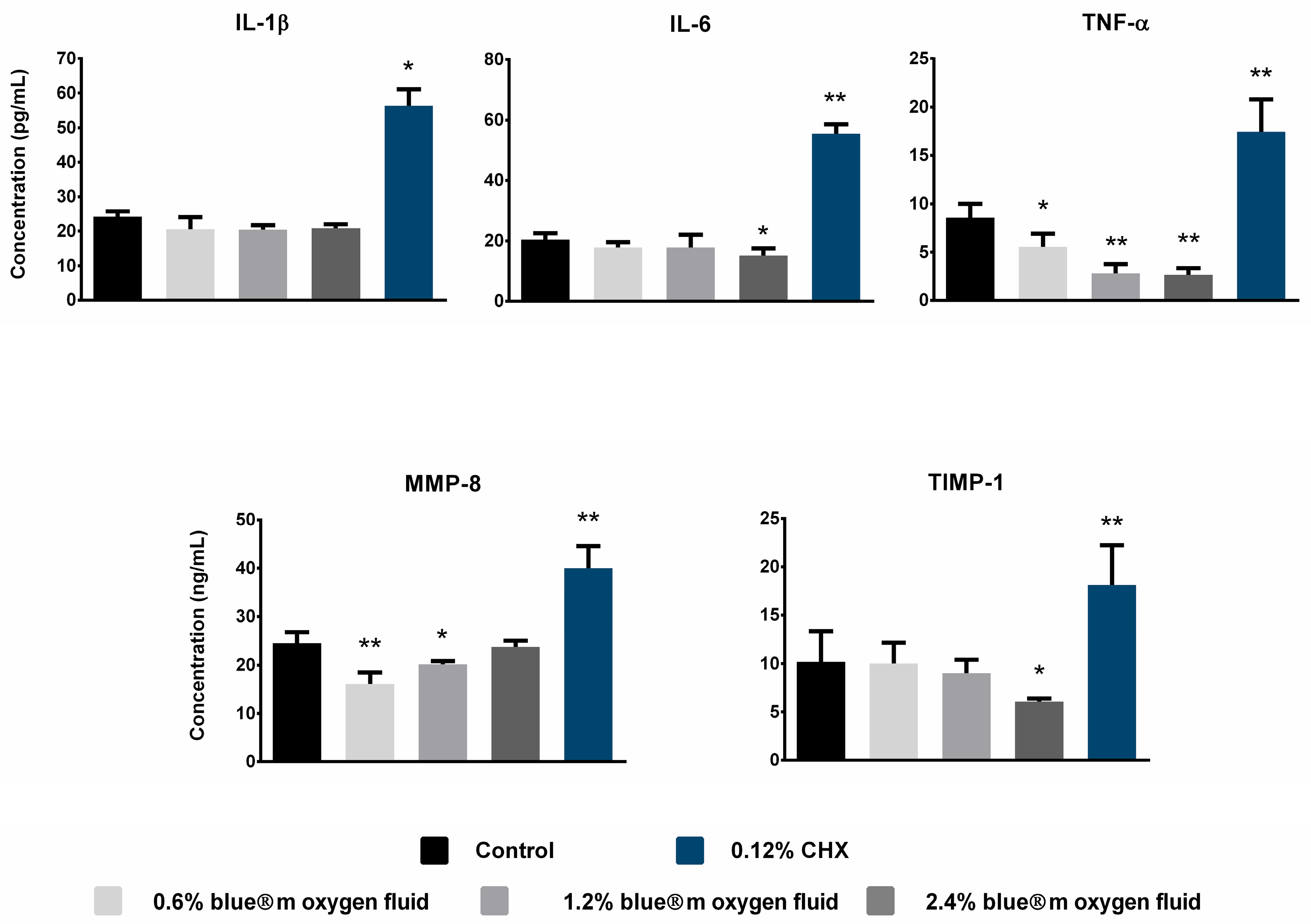
3.6. Detection of Pro-Inflammatory Cytokine Expression

The ELISA test was conducted to determine the expression levels of the pro-inflammatory cytokines (IL-1β, IL-6, TNF-α, MMP-8 and TIMP-1) in HGnF cells that were cultured under various treatment conditions (control, blue®m oxygen fluid at 0.6, 1.2 and 2.4%, and CHX). The control group exhibited a baseline level of cytokine expression (Figure 7, Table 1).
Pro-inflammatory cytokine production was suppressed in a concentration-dependent manner as a consequence of exposure to blue®m oxygen fluid. A moderate decrease in IL-1β, IL-6, TNF-α and TIMP-1 levels was observed at 0.6% blue®m oxygen fluid in comparison to the control. This suppression became more apparent at 1.2% blue®m oxygen fluid, and a further substantial decrease was observed at 2.4% blue®m oxygen fluid. TNF-α and TIMP-1 exhibited the most significant decrease among the cytokines assessed in response to rising blue®m oxygen fluid concentrations, with IL-1β and IL-6 following suit. Conversely, the concentration of blue®m oxygen fluid exposure resulted in an increase in MMP-8 levels.
On the other hand, CHX exposure significantly increased the expression levels of all measured cytokines relative to the control and that of the different blue®m oxygen fluid concentrations. The upregulation of cytokine production following CHX exposure was statistically significant (p < 0.01) across all markers, except IL-1β (p < 0.05). Overall, these findings demonstrate that oxygen fluid exerts a dose-dependent anti-inflammatory effect on HGnF cells, whereas chlorhexidine promotes a pro-inflammatory response.

4. Discussion

This study examined the effects of blue®m oxygen fluid on HGnF cultures at concentrations of 0.6%, 1.2% and 2.4% to assess their cellular response regarding biocompatibility and wound-healing capacity. These concentrations were chosen based on early findings indicating their potential therapeutic significance without inducing overt cytotoxicity [15,34,35].
In terms of morphology, HGnF cells maintained their characteristic spindle-shaped structure across all tested concentrations of blue®m oxygen fluid (0.6, 1.2 and 2.4%), as observed through light microscopy and confirmed by actin staining in immunofluorescence assays [3,4]. This preservation of morphology suggests that topical blue®m oxygen fluid, even at higher concentrations, does not induce significant cytotoxic alterations to the cytoskeleton or gross cell structure [5]. Notably, the presence of smooth muscle actin filaments indicated a degree of myofibroblastic proliferation, which is critical for the tissue remodeling processes. Myofibroblast proliferation is often necessary for effective wound healing; thus, the maintenance of actin architecture further supports the wound-healing potential of topical blue®m oxygen fluid [14,36].
The cell viability assay demonstrated that HGnF cells treated with blue®m oxygen fluid exhibited enhanced viability compared to untreated controls, particularly within the first 24 h [37]. The highest mean percentage of cell viability was observed at 0.6%, followed by 1.2% and 2.4%. Interestingly, although 2.4% blue®m oxygen fluid showed higher viability than the control, its effectiveness was lower compared to the other two concentrations. These results indicate that low to moderate levels of blue®m oxygen fluid enhance cellular metabolism by improving mitochondrial activity [11]. However, high concentrations may induce mild oxidative stress, thereby reducing these positive effects. In contrast, 0.12% CHX significantly decreased cell viability at all time intervals, confirming its cytotoxic effects on fibroblasts despite its antibacterial properties [38].
The proliferation assay confirmed these results, revealing significantly greater live cell counts in the 0.6% and 1.2% blue®m oxygen fluid groups compared to the 2.4% blue®m oxygen fluid and CHX groups. The percentage of viable proliferating cells reached a maximum on day three in all groups, and there was a gradual decline thereafter. However, HGnF cells treated with 0.6% and 1.2% blue®m oxygen fluid consistently exhibited superior proliferation in comparison to those treated with 2.4% blue®m oxygen fluid or CHX [7]. These findings indicate that HGnF proliferation is facilitated by lesser concentrations of blue®m oxygen fluid, whereas this benefit may be diminished by higher concentrations. Additionally, these findings are consistent with the hypothesis that moderate oxygen tension promotes fibroblast proliferation, while excessive oxygen may produce reactive oxygen species (ROS) that disrupt cell homeostasis [36]. In particular, the lowest proliferation rates were observed in cells that were exposed to 0.12% CHX, which is consistent with the prior literature that has emphasized its cytotoxicity toward gingival fibroblasts and other oral cells [3].
The present study’s findings in human gingival fibroblast cells that were stimulated with varying concentrations of blue®m oxygen fluid demonstrated a slower migration for 24 and 48 h, although there was no significant difference between the treated and untreated cells at 72 h. These findings suggest that blue®m oxygen fluid maintains the normal rate of cell migration and proliferation during the wound closure process subsequent to prolonged exposure. In contrast, the CHX group demonstrated a significant decrease in HGnF cell migration, which led to a protracted process of tissue repair. In a comparable investigation, it was observed that the migration of HGnFs was slowed after 24 h of stimulation with blue®m oxygen fluid [10].
The analysis of oxidative stress using the DCFH-DA assay further elucidated the dose-dependent effects of blue®m oxygen fluid [18]. ROS levels were lowest in the control group and remained relatively low in the 0.6% and 1.2% blue®m oxygen fluid groups [8]. A significant increase in ROS production was observed only at the highest tested concentration (2.4%), and was markedly elevated in the CHX group. These results suggest that lower concentrations of blue®m oxygen fluid do not induce excessive oxidative stress, thus allowing for favorable cellular responses [30]. However, at 2.4%, increased ROS levels might begin to counteract some of the benefits of oxygen therapy. High levels of ROS can damage cellular components such as lipids, proteins and DNA, leading to impaired cell viability and function [39]. Thus, the concentration of blue®m oxygen fluid used for topical therapy must be carefully optimized to harness its therapeutic benefits while avoiding oxidative damage. Importantly, the measurement of reactive ROS demonstrated that blue®m oxygen fluid exposure did not substantially elevate oxidative stress levels at 0.6% and 1.2% concentrations, whereas 2.4% blue®m oxygen fluid led to a significant increase. In contrast, chlorhexidine-treated cells exhibited markedly higher ROS levels, consistent with previous reports of chlorhexidine-induced oxidative damage [38].
The modulation of pro-inflammatory cytokine production by HGnF cells in response to blue®m oxygen fluid exposure was one of the primary findings of this study [40]. The non-inflammatory nature of the culture milieu was evident in the low baseline cytokine expression levels of untreated controls [41]. The secretion of IL-1β, IL-6, TNF-α and TIMP-1 was reduced in a concentration-dependent manner as a result of treatment with 0.6, 1.2 and 2.4% blue®m oxygen fluid. Despite the concurrent rise in ROS levels, the anti-inflammatory effect was most pronounced at a concentration of 2.4%. The oxygen fluid treatment exhibited the greatest sensitivity to TNF-α and TIMP-1, indicating that oxygen therapy may specifically downregulate key pathways implicated in extracellular matrix remodeling and inflammation [13]. It is intriguing that the levels of MMP-8 increased in response to oxygen fluid exposure. MMP-8’s upregulation may indicate improved matrix turnover and wound resolution, which are essential for effective tissue repair, as it is involved in collagen degradation and wound remodeling [33].
Conversely, CHX treatment significantly upregulated all measured pro-inflammatory cytokines [42]. The increase was especially pronounced for IL-6 and TNF-α, two central mediators of inflammatory responses. This observation corroborates previous findings that chlorhexidine, while effective as an antimicrobial agent, can induced inflammatory and cytotoxic responses in host cells [6,43]. The exaggerated inflammatory milieu induced by chlorhexidine may impair wound healing by promoting tissue damage and delaying fibroblast proliferation, underscoring its cytotoxic potential [5]. Although ELISA analysis of cytokine expression further reinforces the anti-inflammatory effects of oxygen therapy, this was observed at higher concentrations (2.4%), which negatively correlates with the other positive effects on HGnF observed at lower concentrations (0.6% and 1.2%) of blue®m oxygen fluid.
Pro-inflammatory cytokines such as IL-1β and TNF-α strongly stimulate IL-6 expression in human gingival fibroblasts by activating NF-κB [44]. Furthermore, in gingival fibroblasts, MMP activity upregulates the expression of TIMP-1 and IL-6 to modulate extracellular matrix remodeling. These findings highlight the complex regulatory network controlling cytokine and protease responses in gingival fibroblasts, which may significantly influence periodontal inflammation and tissue remodeling.
The data from this study emphasize several critical findings that are relevant to future research. Initially, topical oxygen fluid therapy, particularly at lower concentrations of 0.6% and 1.2%, increases the viability and proliferation of HGnFs while simultaneously reducing oxidative stress and inflammatory cytokine production [12]. In the context of periodontal wound healing, fibroblast function is essential for the deposition of extracellular matrix and the restoration of tissue integrity, which is why these cellular effects are desirable [45,46]. Secondly, the anti-inflammatory properties of blue®m oxygen fluid may provide therapeutic benefits over traditional antiseptics, such as chlorhexidine, which, despite their antimicrobial effects, may impede healing through cytotoxicity and inflammation [10,15]. The results of this study offer preliminary insights that may assist in the selection of therapeutic parameters for topical oxygen therapies, thereby bolstering their potential for promoting gingival tissue regeneration while minimizing cytotoxic effects.
Several limitations of this in vitro study must be acknowledged. The experimental model, while highly controlled, fails to completely replicate the intricate microenvironment of periodontal wounds, where interactions among immune cells, bacteria and mechanical forces are crucial. The long-term effects of topical oxygen fluid exposure were not evaluated beyond 14 days. Further research utilizing three-dimensional tissue models, co-culture systems incorporating immune cells and in vivo assessments is essential to validate these findings. Increased MMP-8 expression following oxygen fluid exposure may facilitate wound remodeling; however, excessive matrix metalloproteinase activity is also associated with tissue breakdown. Consequently, the equilibrium between matrix deposition and degradation requires careful consideration for prospective therapeutic applications. The observed rise in reactive oxygen species at elevated oxygen levels highlights the necessity of optimizing the therapeutic window for oxygen fluid application.

5. Conclusions

According to the results of the current in vitro study, it can be concluded that topical oxygen-releasing products such as blue®m oxygen fluid represent a promising method for enhancing the function of gingival fibroblasts. Topical blue®m oxygen fluid therapy is effective in improving human gingival fibroblast cell viability, facilitating proliferation, preserving cytoskeletal integrity and providing anti-inflammatory and antioxidative effects in a concentration-dependent manner. Lower concentrations of blue®m oxygen fluid (0.6% and 1.2%) showed notable efficacy, whereas higher concentrations (2.4%) exhibited reduced benefits. In contrast, treatment with 0.12% chlorhexidine was associated with cytotoxicity, increased oxidative stress and expression of pro-inflammatory cytokines.

Author Contributions

Conceptualization: R.N.A.L., A.M.B., A.A.N. and H.S.A.; Methodology: R.N.A.L., A.M.B. and H.S.A.; Software: T.S.S.; Validation: R.N.A.L., A.M.B., T.S.S. and N.M.B.; Formal analysis: R.N.A.L., T.S.S. and N.M.B.; Investigation: R.N.A.L., A.M.B., M.Y.S., T.S.S. and N.M.B.; Resources: M.Y.S., F.M.A., A.S.A., A.A.N. and M.A.A.; Data curation: R.N.A.L., A.M.B., T.S.S. and H.S.A.; Writing—original draft preparation: A.M.B., A.A.N. and H.S.A.; Writing—review and editing: R.N.A.L., A.M.B., M.Y.S., T.S.S., F.M.A., N.M.B., A.S.A., A.A.N., M.A.A. and H.S.A.; Visualization: R.N.A.L., F.M.A., A.S.A., T.S.S., M.A.A. and H.S.A.; Supervision: A.M.B., A.A.N. and H.S.A.; Project administration: R.N.A.L., A.A.N. and H.S.A.; Funding acquisition: A.M.B., A.A.N., M.A.A. and H.S.A. All authors have read and agreed to the published version of the manuscript.

Funding

This work was funded by the Ongoing Research Funding program (ORF_2025_036), King Saud University, Riyadh, Saudi Arabia.

Institutional Review Board Statement

Ethical review and approval were waived for this study due to the fact that all experiments were conducted using commercially sourced human gingival fibroblast (HGnF) cell lines (Cat. No. 2620; ScienCell™ Research Laboratories, Carlsbad, CA, USA).

Informed Consent Statement

Not applicable.

Data Availability Statement

The original contributions presented in this study are included in the article. Further inquiries can be directed to the corresponding author.

Conflicts of Interest

The authors declare no conflicts of interest.

Abbreviations

The following abbreviations are used in this manuscript:
HGnFHuman gingival fibroblast
ROSReactive oxygen species
ILInterleukin
TNFTumor necrosis factor
MMPMatrix metalloproteinases
TIMPTissue inhibitors of metalloproteinases
DMEMDulbecco’s modified eagle’s medium
PBSPhosphate-buffered saline
DAPI4′,6-diamidino-2-phenylindole dihydrochloride
CLSMConfocal laser scanning microscope
EDTAEthylenediaminetetraacetic acid
DCFH-DA2′,7′-Dichlorodihydrofluorescein diacetate
MFIMean fluorescence intensity
ELISAEnzyme-linked immunosorbent assay
ANOVAAnalysis of variance
CHXChlorhexidine

References

  1. Ngeow, W.C.; Tan, C.C.; Goh, Y.C.; Deliberador, T.M.; Cheah, C.W. A Narrative Review on Means to Promote Oxygenation and Angiogenesis in Oral Wound Healing. Bioengineering 2022, 9, 636. [Google Scholar] [CrossRef] [PubMed]
  2. Pippi, R. Post-Surgical Clinical Monitoring of Soft Tissue Wound Healing in Periodontal and Implant Surgery. Int. J. Med. Sci. 2017, 14, 721–728. [Google Scholar] [CrossRef] [PubMed]
  3. Guo, C.; Rizkalla, A.S.; Hamilton, D.W. FGF and TGF-beta growth factor isoform modulation of human gingival and periodontal ligament fibroblast wound healing phenotype. Matrix Biol. 2025, 136, 9–21. [Google Scholar] [CrossRef] [PubMed]
  4. Ahangar, P.; Mills, S.J.; Smith, L.E.; Gronthos, S.; Cowin, A.J. Human gingival fibroblast secretome accelerates wound healing through anti-inflammatory and pro-angiogenic mechanisms. NPJ Regen. Med. 2020, 5, 24. [Google Scholar] [CrossRef]
  5. Liu, J.X.; Werner, J.; Kirsch, T.; Zuckerman, J.D.; Virk, M.S. Cytotoxicity evaluation of chlorhexidine gluconate on human fibroblasts, myoblasts, and osteoblasts. J. Bone Jt. Infect. 2018, 3, 165–172. [Google Scholar] [CrossRef]
  6. Fiorillo, L.; D’Amico, C.; Mehta, V.; Cicciù, M.; Cervino, G. Chlorhexidine cytotoxicity on oral Behaviors: Last 20 Years systematic review. Oral Oncol. Rep. 2024, 9, 100245. [Google Scholar] [CrossRef]
  7. Sindhusha, V.B.; Rajasekar, A. Efficacy of Oxygen-Enriched Mouthwash as a Pre-procedural Mouth Rinse Against Oral Microbes Produced During Ultrasonic Scaling. Cureus 2023, 15, e49164. [Google Scholar] [CrossRef]
  8. Basudan, A.M.; Abas, I.; Shaheen, M.Y.; Alghamdi, H.S. Effectiveness of Topical Oxygen Therapy in Gingivitis and Periodontitis: Clinical Case Reports and Review of the Literature. J. Clin. Med. 2024, 13, 1451. [Google Scholar] [CrossRef]
  9. Shaheen, M.Y.; Abas, I.; Basudan, A.M.; Alghamdi, H.S. Local Oxygen-Based Therapy (blue®m) for Treatment of Peri-Implant Disease: Clinical Case Presentation and Review of Literature about Conventional Local Adjunct Therapies. Medicina 2024, 60, 447. [Google Scholar] [CrossRef]
  10. Leano, S.M.; De Souza, W.; De Vecchi, R.; Lopes, A.; Deliberador, T.; Granjeiro, J.M. A multimodal in vitro approach to assess the safety of oral care products using 2D and 3D cellular models. Front. Toxicol. 2024, 6, 1474583. [Google Scholar] [CrossRef]
  11. Garima; Agarwal, T.; Costantini, M.; Pal, S.; Kumar, A. Oxygenation therapies for improved wound healing: Current trends and technologies. J. Mater. Chem. B 2022, 10, 7905–7923. [Google Scholar] [CrossRef] [PubMed]
  12. Njokweni, M. Adjunctive topical oxygen therapy in the management of complex diabetes-related wounds: A South African case study series. Foot 2023, 57, 101961. [Google Scholar] [CrossRef] [PubMed]
  13. Frykberg, R.G. Topical Wound Oxygen Therapy in the Treatment of Chronic Diabetic Foot Ulcers. Medicina 2021, 57, 917. [Google Scholar] [CrossRef] [PubMed]
  14. Roi, C.; Gaje, P.N.; Ceausu, R.A.; Roi, A.; Rusu, L.C.; Boia, E.R.; Boia, S.; Luca, R.E.; Rivis, M. Heterogeneity of Blood Vessels and Assessment of Microvessel Density-MVD in Gingivitis. J. Clin. Med. 2022, 11, 2758. [Google Scholar] [CrossRef]
  15. Cunha, G.; D’Angieri Saugo, G.; Gabrielli, M.A.C.; Barbeiro, C.O.; de Almeida, L.Y.; Bufalino, A.; Pereira-Filho, V.A. Cytotoxicity evaluation of Chlorhexidine and Blue(R)M applied to a human gingival fibroblast (HGF-1) and keratinocytes (NOK-SI): In vitro study. J. Stomatol. Oral Maxillofac. Surg. 2024, 125, 101923. [Google Scholar] [CrossRef]
  16. Joorabloo, A.; Liu, T. Recent advances in reactive oxygen species scavenging nanomaterials for wound healing. Exploration 2024, 4, 20230066. [Google Scholar] [CrossRef]
  17. Grootveld, M.; Lynch, E.; Page, G.; Chan, W.; Percival, B.; Anagnostaki, E.; Mylona, V.; Bordin-Aykroyd, S.; Grootveld, K.L. Potential Advantages of Peroxoborates and Their Ester Adducts Over Hydrogen Peroxide as Therapeutic Agents in Oral Healthcare Products: Chemical/Biochemical Reactivity Considerations In Vitro, Ex Vivo and In Vivo. Dent. J. 2020, 8, 89. [Google Scholar] [CrossRef]
  18. Settem, R.P.; Sharma, A. Oral bacterium contributes to periodontal inflammation by forming advanced glycation end products. Infect. Immun. 2025, 93, e0056024. [Google Scholar] [CrossRef]
  19. Naruishi, K. Biological Roles of Fibroblasts in Periodontal Diseases. Cells 2022, 11, 3345. [Google Scholar] [CrossRef]
  20. Sadek, K.M.; El Moshy, S.; Radwan, I.A.; Rady, D.; Abbass, M.M.S.; El-Rashidy, A.A.; Dorfer, C.E.; Fawzy El-Sayed, K.M. Molecular Basis beyond Interrelated Bone Resorption/Regeneration in Periodontal Diseases: A Concise Review. Int. J. Mol. Sci. 2023, 24, 4599. [Google Scholar] [CrossRef]
  21. Radzki, D.; Negri, A.; Kusiak, A.; Obuchowski, M. Matrix Metalloproteinases in the Periodontium-Vital in Tissue Turnover and Unfortunate in Periodontitis. Int. J. Mol. Sci. 2024, 25, 2763. [Google Scholar] [CrossRef] [PubMed]
  22. Fadl, A.; Leask, A. Hiding in Plain Sight: Human Gingival Fibroblasts as an Essential, Yet Overlooked, Tool in Regenerative Medicine. Cells 2023, 12, 2021. [Google Scholar] [CrossRef] [PubMed]
  23. Haekkinen, L.; Uitto, V.-J.; Larjava, H. Cell biology of gingival wound healing. Periodontol. 2000 2000, 24, 127–152. [Google Scholar] [CrossRef]
  24. AlMobarak, S.; AlMadi, E.; Almohaimede, A.; Badran, M.; Lambarte, R.A. The Effect of Commiphora molmol Nanoparticles as an Endodontic Irrigant on the Morphology, Viability, Migration, and Proliferation of Human Bone Marrow Mesenchymal Stem Cells: An In Vitro Study. Int. J. Mol. Sci. 2025, 26, 1412. [Google Scholar] [CrossRef]
  25. Staehlke, S.; Brief, J.; Senz, V.; Eickner, T.; Nebe, J.B. Optimized Gingiva Cell Behavior on Dental Zirconia as a Result of Atmospheric Argon Plasma Activation. Materials 2023, 16, 4203. [Google Scholar] [CrossRef]
  26. Strober, W. Trypan Blue Exclusion Test of Cell Viability. Curr. Protoc. Immunol. 2015, 111, A3.B.1–A3.B.3. [Google Scholar] [CrossRef]
  27. Bhavikatti, S.K.; Karobari, M.I.; Zainuddin, S.L.A.; Marya, A.; Nadaf, S.J.; Sawant, V.J.; Patil, S.B.; Venugopal, A.; Messina, P.; Scardina, G.A. Investigating the Antioxidant and Cytocompatibility of Mimusops elengi Linn Extract over Human Gingival Fibroblast Cells. Int. J. Environ. Res. Public Health 2021, 18, 7162. [Google Scholar] [CrossRef]
  28. Aldoss, A.; Lambarte, R.; Alsalleeh, F. High-Glucose Media Reduced the Viability and Induced Differential Pro-Inflammatory Cytokines in Human Periodontal Ligament Fibroblasts. Biomolecules 2023, 13, 690. [Google Scholar] [CrossRef]
  29. Rasband, W.S. US National Institutes of Health: Bethesda, MD, USA. 2011. Available online: http://imagej.nih.gov/ij/ (accessed on 23 March 2025).
  30. Saramet, V.; Stan, M.S.; Ripszky Totan, A.; Tancu, A.M.C.; Voicu-Balasea, B.; Enasescu, D.S.; Rus-Hrincu, F.; Imre, M. Analysis of Gingival Fibroblasts Behaviour in the Presence of 3D-Printed versus Milled Methacrylate-Based Dental Resins-Do We Have a Winner? J. Funct. Biomater. 2024, 15, 147. [Google Scholar] [CrossRef]
  31. AlJunaydil, N.A.; Lambarte, R.N.A.; Sumague, T.S.; Alghamdi, O.G.; Niazy, A.A. Lovastatin and Resveratrol Synergistically Improve Wound Healing and Inhibit Bacterial Growth. Int. J. Mol. Sci. 2025, 26, 851. [Google Scholar] [CrossRef]
  32. Basso, F.G.; Pansani, T.N.; Turrioni, A.P.; Soares, D.G.; de Souza Costa, C.A.; Hebling, J. Tumor Necrosis Factor-alpha and Interleukin (IL)-1beta, IL-6, and IL-8 Impair In Vitro Migration and Induce Apoptosis of Gingival Fibroblasts and Epithelial Cells, Delaying Wound Healing. J. Periodontol. 2016, 87, 990–996. [Google Scholar] [CrossRef] [PubMed]
  33. Hu, J.J.; Yu, X.Z.; Zhang, S.Q.; Zhang, Y.X.; Chen, X.L.; Long, Z.J.; Hu, H.Z.; Xie, D.H.; Zhang, W.H.; Chen, J.X.; et al. Hydrogel with ROS scavenging effect encapsulates BR@Zn-BTB nanoparticles for accelerating diabetic mice wound healing via multimodal therapy. iScience 2023, 26, 106775. [Google Scholar] [CrossRef] [PubMed]
  34. Mattei, B.M.; Imanishi, S.A.W.; de Oliveira Ramos, G.; de Campos, P.S.; Weiss, S.G.; Deliberador, T.M. Mouthwash with Active Oxygen (blue®m) Reduces Postoperative Inflammation and Pain. Case. Rep. Dent. 2021, 2021, 5535807. [Google Scholar] [CrossRef] [PubMed]
  35. Kumar, M.; Meher, B.; Mishra, L.; Panda, S.; Sokolowski, K.; Lapinska, B. Evaluation of Blue® m Mouthwash vs. Chlorhexidine Mouthwash in Patients with Gingivitis—A Pilot Study. Appl. Sci. 2024, 14, 11862. [Google Scholar] [CrossRef]
  36. Furukawa, M.; JR, K.K.; Yamada, M.; Senda, A.; Manabe, A.; Miyazaki, A. Cytotoxic Effects of Hydrogen Peroxide on Human Gingival Fibroblasts In Vitro. Oper. Dent. 2015, 40, 430–439. [Google Scholar] [CrossRef]
  37. Berner, T.; Nakahara, K.; Kobayashi, E.; Tanaka, A.; Taniguchi, Y.; Iizuka, T.; Sawada, K. Investigating the effect of antiseptic solution on the release of interleukin-6 and transforming growth factor beta 1 from human gingival fibroblasts using wound healing assays. J. Oral Sci. 2020, 62, 293–297. [Google Scholar] [CrossRef]
  38. Pilloni, A.; Ceccarelli, S.; Bosco, D.; Gerini, G.; Marchese, C.; Marini, L.; Rojas, M.A. Effect of Chlorhexidine Digluconate in Early Wound Healing of Human Gingival Tissues. A Histological, Immunohistochemical and Biomolecular Analysis. Antibiotics 2021, 10, 1192. [Google Scholar] [CrossRef]
  39. Sterczala, B.; Grzech-Lesniak, K.; Michel, O.; Trzeciakowski, W.; Dominiak, M.; Jurczyszyn, K. Assessment of Human Gingival Fibroblast Proliferation after Laser Stimulation In Vitro Using Different Laser Types and Wavelengths (1064, 980, 635, 450, and 405 nm)-Preliminary Report. J. Pers. Med. 2021, 11, 98. [Google Scholar] [CrossRef]
  40. Li, X.; Wang, X.; Zheng, M.; Luan, Q.X. Mitochondrial reactive oxygen species mediate the lipopolysaccharide-induced pro-inflammatory response in human gingival fibroblasts. Exp. Cell Res. 2016, 347, 212–221. [Google Scholar] [CrossRef]
  41. Dong, Y.; Wang, Z. ROS-scavenging materials for skin wound healing: Advancements and applications. Front. Bioeng. Biotechnol. 2023, 11, 1304835. [Google Scholar] [CrossRef]
  42. Page-McCaw, A.; Ewald, A.J.; Werb, Z. Matrix metalloproteinases and the regulation of tissue remodelling. Nat. Rev. Mol. Cell Biol. 2007, 8, 221–233. [Google Scholar] [CrossRef] [PubMed]
  43. Stavroullakis, A.T.; Carrilho, M.R.; Levesque, C.M.; Prakki, A. Profiling cytokine levels in chlorhexidine and EGCG-treated odontoblast-like cells. Dent. Mater. 2018, 34, e107–e114. [Google Scholar] [CrossRef] [PubMed]
  44. Chatzigiannidou, I.; Teughels, W.; Van de Wiele, T.; Boon, N. Oral biofilms exposure to chlorhexidine results in altered microbial composition and metabolic profile. NPJ Biofilms Microbiomes 2020, 6, 13. [Google Scholar] [CrossRef] [PubMed]
  45. Cardoso, L.M.; Pansani, T.N.; de Souza Costa, C.A.; Basso, F.G. Regulation of interleukin-6 and matrix metalloproteinases syntheses by bioflavonoids and photobiomodulation in human gingival fibroblasts. Lasers Med. Sci. 2022, 37, 2973–2987. [Google Scholar] [CrossRef]
  46. Smith, P.C.; Martinez, C.; Martinez, J.; McCulloch, C.A. Role of Fibroblast Populations in Periodontal Wound Healing and Tissue Remodeling. Front. Physiol. 2019, 10, 270. [Google Scholar] [CrossRef]
Figure 1. Schematic diagram of the workflow of the in vitro study of HGnF cells stimulated with topical blue®m oxygen fluid.
Applsci 15 07459 g001
Figure 2. Representative photomicrographs of the HGnFs in culture in the control, blue®m oxygen fluid and CHX groups: (A) Spindle-shaped morphology of cells and (B) immunofluorescent staining of F-actin in cells (red color) and nuclei (blue color). (CHX: chlorhexidine).
Applsci 15 07459 g002
Figure 3. Cell viability of HGnFs following exposure to varying concentrations of topical blue®m oxygen fluid and CHX at different time points. Data are presented as mean percentage values of cell metabolic activity ± SD at each time point. Statistical comparisons were made between the control, CHX and different concentration of blue®m oxygen fluid using one-way ANOVA with Tukey’s post hoc test (p < 0.05; n = 9). CHX: chlorhexidine; * statistically significant difference from control; ϕ statistically significant difference from CHX; “n.s.” not significant.
Applsci 15 07459 g003
Figure 4. Cell proliferation of HGnF cells assessed by Trypan Blue exclusion assay following exposure to different concentrations of blue®m oxygen fluid and CHX over the observation period. Data are presented as mean percentage values ± SD (n = 9). Statistical analysis was performed using one-way ANOVA with Tukey’s post hoc test (p < 0.05). ϕ statistically significant difference from CHX. CHX: chlorhexidine.
Applsci 15 07459 g004
Figure 5. Wound-healing capacity of HGnF cells exposed to blue®m oxygen fluid using a scratch migration assay. (A) Representative phase-contrast images showing cell migration at 0, 24, 48 and 72 h; yellow vertical lines indicate the initial area covered by the cells after wounding. Magnification 10×. (B) Percentage of wound migration area measured using ImageJ software. Data are presented as mean ± SD, n = 8. * p < 0.05, ** p < 0.01 statistically significant differences from control; ϕ p < 0.05, ϕϕ p < 0.01 statistically significant differences from CHX.
Applsci 15 07459 g005
Figure 6. Cellular oxidative stress levels in gingival fibroblasts exposed with blue®m oxygen fluid at different concentrations for 24 h. (A) The mean fluorescence intensities of the control and treatment groups were analyzed using NIS-Elements AR software. Data are presented as the mean of 8 measurements ± SD. Phi (ϕ) indicates statistically significant difference at p < 0.05, and (ϕϕ) p < 0.001. (B) Representative images observed by CLSM. Scale bar: 50 µm; magnification: 20×.
Applsci 15 07459 g006
Figure 7. Expression levels of pro-inflammatory cytokines (IL-1β, IL-6, TNF-α, MMP-8 and TIMP-1) in HGnF cells following exposure to different concentrations of blue®m oxygen fluid and 0.12% chlorhexidine (CHX), compared to the control. Cytokine levels were measured using ELISA. Data are presented as the mean ± SD (n = 6). Statistical analysis was performed using one-way ANOVA. CHX: chlorhexidine; * p < 0.05, ** p < 0.01 compared to control.
Applsci 15 07459 g007
Table 1. Mean values for the in vitro assays, in the different experimental groups, expressed as mean ± standard deviation.
In Vitro AssaysExperimental Groups
Controlblue®m Oxygen Fluid0.12% CHX
0.6%1.2%2.4%
Cell viability
Day 1100.00 ± 4.27 171.82 ± 19.71166.08 ± 7.41160.54 ± 7.1576.14 ± 2.11
Day 2100.00 ± 6.19108.07 ± 7.23104.16 ± 9.27102.18 ± 4.5956.66 ± 3.58
Day 7100.00 ± 3.21102.48 ± 6.23103.72 ± 8.71100.14 ± 9.4437.63 ± 2.52
Cell proliferation
Day 3-133.47 ± 10.49102.54 ± 15.9080.86 ± 12.7158.62 ± 3.63
Day 7-108.75 ± 17.8892.03 ± 2.8475.43 ± 14.3140.24 ± 2.11
Day 10-86.74 ± 4.0580.70 ± 5.4657.01 ± 17.8020.31 ± 5.84
Day 14-75.53 ± 13.3572.38 ± 14.8750.03 ± 12.1419.54 ± 2.25
Wound healing
Day 129.28 ± 2.2812.97 ± 2.2514.82 ± 2.2221.58 ± 7.628.08 ± 0.92
Day 273.02 ± 1.7643.23 ± 1.7848.26 ± 2.1152.27 ± 3.2312.24 ± 1.95
Day 395.64 ± 2.5893.74 ± 2.1493.74 ± 2.1496.24 ± 1.7030.51 ± 13.26
Intracellular ROS detection98.75 ± 13.86104.83 ± 7.63115.09 ± 8.79122.87 ± 19.041027.01 ± 93.36
Quantification of inflammatory cytokines
IL-1β24.25 ± 1.5219.87 ± 2.4820.42 ± 1.3320.87 ± 1.1456.37 ± 4.75
IL-620.42 ± 2.1517.98 ± 2.5217.77 ± 2.8315.17 ± 2.3555.48 ± 3.15
TNF-α8.56 ± 1.465.55 ± 1.362.81 ± 0.822.67 ± 0.6917.45 ± 3.34
MMP-824.52 ± 2.2616.01 ± 2.4920.19 ± 0.6823.78 ± 1.2640.03 ± 4.54
TIMP-110.18 ± 3.0810.02 ± 1.419.03 ± 1.376.06 ± 0.3318.13 ± 4.09
Disclaimer/Publisher’s Note: The statements, opinions and data contained in all publications are solely those of the individual author(s) and contributor(s) and not of MDPI and/or the editor(s). MDPI and/or the editor(s) disclaim responsibility for any injury to people or property resulting from any ideas, methods, instructions or products referred to in the content.

