Article

Phenotypic Characterization and Whole-Genome Analysis Revealing the Promising Metabolic Potential of a Newly Isolated Streptomyces sp. CH6

Institute of Biological and Food Technology, Hanoi Open University, Hanoi 100000, Vietnam
*
Author to whom correspondence should be addressed.
Appl. Sci. 2025, 15(13), 7126; https://doi.org/10.3390/app15137126
Submission received: 22 May 2025 / Revised: 18 June 2025 / Accepted: 19 June 2025 / Published: 25 June 2025

Abstract

Streptomyces spp. are considered a prolific resource of bioactive and structurally diverse secondary metabolites for natural product drug discovery. In this study, 20 out of 56 actinomycetes from soils showed antibacterial activity against at least one tested bacterium. Among them, the CH6 isolate could be a potential source of antibacterial compounds, as indicated by inhibition zone diameters (11.1–32.0 mm) and MIC values (from 8 to 128 µg/mL) against microbial pathogens. The extract showed moderate antioxidant activity against DPPH, hydroxyl, and superoxide anion radicals. Notably, CH6 extract displayed strong inhibitory effects on cancer cells, including MCF-7, A549, HepG2, and HT29, with IC50 values ranging from 18.0 to 73.4 µg/mL, without cytotoxic activity against non-cancerous HEK-293 cells. The genome of CH6 consists of a 6,936,977 bp linear chromosome with a 73.0% GC content, 5831 protein-coding genes, and 13 biosynthetic gene clusters (BGCs). The highest dDDH and ANI values between CH6 and the most closely related type strain, Streptomyces evansiae DSM 41979T, were 45.8% and 92.6%, respectively, which suggests that CH6 is a novel species. Interestingly, cluster 2, with a size of 133,857 bp, comprised both guangnanmycin and scabichelin clusters, which have been reported for the first time. These findings showed that Streptomyces sp. CH6 could be a novel species and a producer of guangnanmycin and even new secondary metabolites, particularly those with antibacterial and anticancer activities.

1. Introduction

Bioactive microbial natural products are considered a prolific source of new drugs to combat infectious diseases and cancers. Starting with the discovery of penicillin from Penicillium notatum in 1928, over 23,000 microbial compounds with antibacterial, immunosuppressive, antifungal, and cytotoxic activities have been reported, of which 42% account for fungi and 32% for bacteria [1]. Around 78% of antibacterial compounds and 74% of anticancer agents are related to natural products [2]. Due to overexploitation, the number of compounds extracted from terrestrial-borne microbes has significantly reduced over the last three decades [3]. Notably, the emergence of antimicrobial resistance among pathogens and tumoral resistance to chemotherapy is a growing public health challenge [4]. Therefore, there is an urgent need to discover novel bioactive compounds to better meet clinical therapeutic needs.
Streptomyces spp. are filamentous, sporulating, Gram-positive, and the largest genus of phylum Actinobacteria, and have gained significant attention due to their functions and essential role in clinical treatments and agriculture [2]. As of 2025, 908 species with validly published names have been reported at the time of writing (https://lpsn.dsmz.de/genus/streptomyces, accessed on 21 May 2025) [5]. Four novel Streptomyces spp., including Streptomyces caledonius, Streptomyces machairae, Streptomyces pratisoli, and Streptomyces achmelvichensis, isolated from machair grassland soil, were recently proposed [6]. It is worth noting that the genus Streptomyces secretes the majority of secondary metabolites with antibacterial, anticancer, antioxidant, and antiviral activities [7]. Approximately 70% of antibiotics available in the market are synthesized by Streptomyces spp. [8], with some of them serving as anticancer, antioxidant, antiviral, and immunosuppressive agents [9]. Previously, doxorubicin, antimycin, and bagremycin act as anticancer compounds to treat different types of cancer cells, while streptomycin, chloramphenicol, and chlortetracycline are common antibiotics to treat infectious diseases [2,3]. Recently, guangnanmycin was shown to be a newly characterized member of the leinamycin family of sulfur-containing natural products, first identified in Streptomyces sp. CB01883, a strain isolated from soil in Guangnan County, China [10]. It also contains a quinone moiety, which is capable of undergoing redox cycling and generating reactive oxygen species (ROS). Several studies have shown that quinone-containing compounds can induce apoptosis in human cancer cells through ROS generation, making guangnanmycin a promising candidate for anticancer drug development [11,12,13]. Despite the high rediscovery rate of known molecules due to the traditional method used and low production in nature, the isolation and characterization of novel and efficient Streptomyces strains for medicinal and pharmaceutical sectors are still an area of active interest in the current scenario.
Recent advances in next-generation sequencing have allowed rapid and cost-effective Streptomyces genome sequencing, paving a new way for natural product research [14]. Different from other bacterial genera, the Streptomyces chromosome is linear with a size that ranges from 6 to 12 Mb, encoding 5300 to 11,000 coding sequences (CDSs) [15]. The large genome is attributed to horizontal gene transfer, gene duplications, and recombination [15,16]. Importantly, most compounds are synthesized by biosynthetic gene clusters (BGCs), which contain catalytic enzymes essential for the biosynthesis of secondary metabolites. Genome mining, as a new approach to identify previously unknown BGCs, demonstrated that at least 17 BGCs are present in Streptomyces chromosomes; most of the BGCs are totally silent or cryptic when cultivated under normal laboratory culture conditions [17]. Streptomyces argillaceus ATCC 12956, for instance, encodes 31 BGCs in the linear genome, among which argimycins P is synthesized by a 45.282 kb cluster consisting of 14 functional proteins [18]. Therefore, the genomic information of the useful Streptomyces species provides opportunities for the discovery of novel BGCs as well as secondary metabolites with novel bioactivities.
The aims of this study were to isolate Streptomyces strains from soils, characterize their bioactivities, and mine their genomic information. Screening the antibacterial activity of Streptomyces isolates revealed Streptomyces sp. CH6 to be the most potent candidate. Further antimicrobial, antioxidant, and cytotoxic activities were determined. Whole-genome sequencing and genome mining were performed to demonstrate the novelty of the taxonomic identity and BGCs of the strain.

2. Materials and Methods

2.1. Isolation of Actinomycetes and Screening for Their Antibacterial Activity

The soil samples were collected from vegetable fields in Hanoi, Vietnam, in February 2020. Soils were sampled with a sterilized spoon, placed in zip-lock plastic bags, and transported aseptically to the laboratory for additional processing. Isolation of the actinomycetes was performed by the serial dilution method on the International Streptomyces Project (ISP) 2, supplemented with nalidixic acid (25 μg/mL) and nystatin (50 μg/mL) to avoid unwanted microorganisms. All plates were incubated at 30 °C for 8–10 days. Then, the Streptomyces-like colonies that emerged were sub-cultured on ISP2 medium to purify and kept at 30 °C for 4 days. Suspected colonies were characterized morphologically and then preserved in 30% glycerol at −80 °C.
All isolates were fermented in a 1000 mL flask containing 300 mL YIM38 medium by pipetting 30 mL of seed culture, and then they were incubated on a rotary shaker incubator (180 rpm) at 30 °C for 4 days. Mycelia were removed from the culture broth by centrifugation at 8000 rpm for 10 min, and the cell-free supernatant was collected. For extracting secondary metabolites, 2:1 v/v ethyl acetate was added to the cell-free supernatant and shaken vigorously for 2 h to obtain the upper organic phase of ethyl acetate. All samples were evaporated to dryness using a rotary vacuum evaporator to completely eliminate the extracting solvent, which was subsequently dissolved in dimethyl sulfoxide (DMSO) in order to use for the agar well-diffusion method [19]. Gram-positive Staphylococcus aureus ATCC 29213, Salmonella enterica serovar Typhimurium ATCC 14028, and Mycobacterium smegmatis MC2 155, and Gram-negative Escherichia coli ATCC 11105 and Pseudomonas aeruginosa ATCC 9027 were used as test bacteria and spread on Luria–Bertani (LB) agar (HiMedia, Mumbai, India) plates. About 50 μL of each extract (100 μg/mL) was put into 6 mm wells, and the plates were then examined for the presence of a distinct zone of inhibition after 18–24 h at 37 °C.

2.2. Determination of Minimum Inhibitory Concentration (MIC)

The MIC values of potent candidates against 7 tested bacteria and yeast C. albicans ATCC 10231 were determined using the micro dilution method, as described previously [20]. The CH6 extract was dissolved in 1% DMSO (v/v) and serially diluted in Mueller–Hinton broth (HiMedia, Mumbai, India) in case of bacterial strains or in RPMI 1640 (HiMedia, Mumbai, India) medium for testing yeast to give stock concentrations. About 180 µL of fresh bacterial and yeast culture suspensions (2 × 10⁴ CFU/mL) were prepared in the 96-well plates, followed by the addition of 20 µL of extract. The plates were incubated at 37 °C for test bacteria and 30 °C for yeast for 24–48 h. The experiment was performed in triplicate, and the lowest concentration that inhibited the growth of the microorganisms was established as the MIC.

2.3. Growth Curve Assay

M. smegmatis MC2 155, as an overnight culture, was grown in LB medium supplemented with 0.05% Tween80 at 37 °C. The seed culture was transferred to Hartmans-de Bont minimal medium to an optical density at 500 nm (OD500) of 0.04 and then cultivated until an OD500 of 0.4 was reached [21]. The cells were treated with 10, 20, and 30 μg/mL extract, with one group of cells serving as a control without the treatment. The OD500 values were recorded over a period of 24 h. Regarding S. aureus ATCC 29213, the seed culture cultivated in RPMI medium was added to fresh LB medium to a final OD500 of 0.1 [22]. About 10, 20, and 30 μg/mL extract was added to the culture, an OD500 of 0.4, followed by cultivation at 37 °C and sampling every hour. The time points of CH6 exposure during the growth curves of M. smegmatis MC2 155 and S. aureus ATCC 29213 were set to ‘0’.

2.4. Antioxidant Assays

The ability of the CH6 extract to inhibit 1,1-diphenyl-2-picrylhydrazyl (DPPH) and hydroxyl radicals was evaluated according to a previous study with slight modifications [23]. In brief, different doses of CH6 extract were reacted with 0.1 mM DPPH solution to obtain a final volume of 200 µL. The 96-well plate was incubated in the dark at room temperature for 30 min, and the absorbance was measured at 517 nm against an equal amount of DPPH. To evaluate the antioxidant activity through hydroxyl radical scavenging capacity, a mixture of 0.25 mL of 0.435 mM brilliant green, 0.5 mL of 0.5 mM FeSO4, and 0.375 mL of 3% (v/v) H2O2 was prepared, and the reaction was initiated by the addition of 0.5 mL of crude extract. After inoculation at 37 °C for 30 min, the absorbance of the mixture was measured at 536 nm. Antioxidant activity against superoxide anion radicals was conducted by pipetting the mixture containing 900 μL of 0.05 M Tris–HCl (pH 8.2), 200 μL of crude extract, and 80 μL of 2.5 mM pyrogallol [24]. After incubation at room temperature for 5 min, measurement at 299 nm was performed. For the three antioxidant assays, ascorbic acid was used as a positive control, and the experiments were performed in triplicate.

2.5. Cytotoxic Activity

The cytotoxic effect of CH6 extract on the cell viability of human cancer cell lines, such as human lung cancer A549, hepatoma HepG2, breast cancer MCF-7, colon cancer HT-29, and non-cancerous HEK-293 cell lines, was evaluated using the 3-(4,5-dimethylthiazol-2-yl)-2, 5-diphenyltetrazolium bromide (MTT) assay, based on a previously described protocol [25]. After exposure to different doses of the CH6 extract, MTT was added to each well containing seeded cells, followed by incubation at 37 °C in a humid atmosphere with 5% CO2 and 95% air for 4 h. After that, the MTT solution was eliminated, and 150 μL of DMSO was added to dissolve the formazan crystals. The samples were spectrophotometrically measured at 570 nm using a microplate reader. The IC50 value was calculated using Prism 9, and the experiment was performed in triplicate.
Nuclear staining was detected using the Hoechst 33342 staining (Thermo Fisher Scientific, Waltham, MA, USA). HepG2 cells were seeded into 24-well plates (Biologix Europe GmbH, Niederzier, Germany) at a density of 1 × 10⁵ cells/well and incubated overnight. Then, cells were treated with the CH6 extract at varying concentrations (6, 12, and 24 µg/mL). After 24 h, HepG2 cells were fixed with cold 100% ethanol for 10 min at −20 °C, washed with PBS buffer, and stained with Hoechst 33342 for 10 min in the dark. Cells were then rinsed with PBS buffer, and nuclear morphology was visualized using a fluorescence microscope (Olympus IX73, Olympus Corporation, Tokyo, Japan). The samples treated with DMSO were used as positive controls.

2.6. Phenotypic Characterization

The colors of the aerial mycelium, substrate mycelium, and soluble pigment from the CH6 strain were observed in ISP1-ISP9 agar plates at 30 °C for 10 days [16]. Aerial morphology was visualized using a light microscope (Nikon, Tokyo, Japan). Salt resistance was evaluated using ISP2 supplemented with 0–8% NaCl (w/v) at 30 °C for 8–10 days. The temperature range and optimum temperature of CH6 were evaluated on ISP2 medium at 10, 15, 20, 28, 30, 37, 47, and 50 °C. The pH range and optimum pH were determined on ISP2 medium, adjusted to pH 4–12 using 0.5 M HCl and 0.5 M NaOH solutions. Production of extracellular enzymes, such as CMCase, protease, chitinase, and amylase, and the ability to utilize sole carbon sources were tested following the methodology described previously [7].

2.7. Genomic DNA Extraction, Whole-Genome Sequencing, and Assembly

The CH6 strain was grown in 20 mL ISP2 medium for 3 days at 30 °C in a 200 rpm orbital shaker. Cells were obtained, washed with 10 mM EDTA, and treated with 10 mg/mL lysozyme. Genomic DNA was extracted using a G-spinTM Genomic DNA Extraction Mini Kit (iNtRON, Seongnam-si, Republic of Korea) according to the manufacturer’s protocol. The quality and concentration of genomic DNA samples were determined using 1% (w/v) agarose gel electrophoresis and Nanodrop (Thermo Fisher Scientific, Waltham, MA, USA).
The whole-genome sequence of the CH6 strain was obtained by the Illumina sequencing platform. The raw reads were evaluated by FastQC v.0.11.9 and Fastp v0.23.1. Trimmomatic v0.39 with the default settings was utilized to obtain a sequence with low-quality scores (average quality threshold of 20). The trimmed sequences were assembled de novo using Unicycler v0.4.8 with default parameters, in which contigs shorter than 500 bp were excluded. Genome completeness was checked using CheckM [26].

2.8. Annotation and Comparative Genome Analysis

Functional annotation analyses were implemented by the NCBI Prokaryotic Genome Annotation Pipeline (PGAP) and analyzed by Rapid Annotation using the Subsystem Technology (RAST) with the following settings: default pipeline for RASTtk and domain bacteria, and automatically fixed error options turned on [27,28]. The presence of a plasmid was identified using plasmidSpades [29].
Digital DNA–DNA hybridization (dDDH) was estimated using the Type Strain Genome Server (TYGS) to compare the CH6 genome with the genomes of the type strains from the DSMZ database [30]. The 16S rRNA gene sequences of CH6 and Streptomyces evansiae DSM 41979T were retrieved from the draft genome sequences. For phylogenetic analysis, 16S rRNA sequences of CH6 and its closely related species were aligned and analyzed using the neighbor-joining method in MEGA 11.0 [31]. The average nucleotide identity (ANI) values were calculated for CH6 together with its phylogenetic neighbors using the OrthoANI [32]. Protein sequences annotated by PGAP were analyzed with the default settings—1 × 10⁻² E-value and inflation value of 1.5—on the OrthoVenn2 web server to compare orthologous clusters between the genomes of Streptomyces sp. CH6, S. evansiae DSM 41979T (JAVRET000000000), Streptomyces intermedius JCM 4483T (BAAASH000000000), and Streptomyces spheroides NCIMB 11891T (AWQW00000000). The biosynthetic gene clusters (BGCs) encoding secondary metabolites were predicted using antiSMASH 8.0 [33]. The genes of interest were found by BLAST search (https://blast.ncbi.nlm.nih.gov/Blast.cgi, accessed on 21 May 2025) (e-value cutoff < 10−10, identity > 30%) against the NCBI NR protein database.

3. Results

3.1. Isolation and Preliminary Screening of Antibacterial Metabolites Extracted from Actinomycete Cultures

In the present study, a total of 56 morphologically distinct presumptive actinobacterial isolates were successfully recovered from soil samples collected in Hanoi, Vietnam, by a series of repeated pure cultures on ISP2 agar plates. All isolates were characterized based on morphological colonies and diffusible pigments.
All actinobacterial isolates were then extracted with ethyl acetate and screened for their capability to secrete antibacterial compounds against five Gram-positive and -Gram-negative bacteria. Out of 56 isolates, only 20 isolates (35.7%) showed antibacterial activity against at least one tested pathogen. Among them, only two isolates (CH6 and CH8.1) were active against all Gram-negative and -Gram-positive bacteria (Figure 1). P. aeruginosa ATCC 9027 and S. aureus ATCC 29213 were quite sensitive to actinobacterial extracts, while M. smegmatis MC2 155 was quite resistant. Notably, the CH6 isolate was found to have promising broad-spectrum and strong activity against all indicator bacteria, including Salmonella Typhimurium ATCC 14028 (18.0 ± 0.5 mm), E. coli ATCC 11105 (22.1 ± 0.1 mm), M. smegmatis MC2 155 (11.1 ± 1.5 mm), P. aeruginosa ATCC 9027 (32.0 ± 0.1 mm), and S. aureus ATCC 29213 (25.1 ± 0.2 mm). Therefore, the CH6 isolate was selected for further experiments.

3.2. Antibacterial Activity of the CH6 Crude Extract

To verify antimicrobial activity, the MIC and growth assays of the crude CH6 extract against various indicator microorganisms were utilized, and they are presented in Table 1.
The lowest MIC value (8 µg/mL) was observed for S. aureus ATCC 29213, followed by P. aeruginosa ATCC 9027 (16 µg/mL) and B. cereus ATCC 11778 (16 µg/mL). The highest MIC of 64 µg/mL was observed for the CH6 extract against M. smegmatis MC2 155. In addition, the crude extract weakly inhibited the growth of yeast C. albicans ATCC 10231, with an MIC value of 128 µg/mL. In agreement with this result, CH6 exhibited no antifungal activity against phytopathogenic fungi such as Colletotrichum gloeosporioides and Fusarium solani, as shown in a dual culture assay (Figure S1).
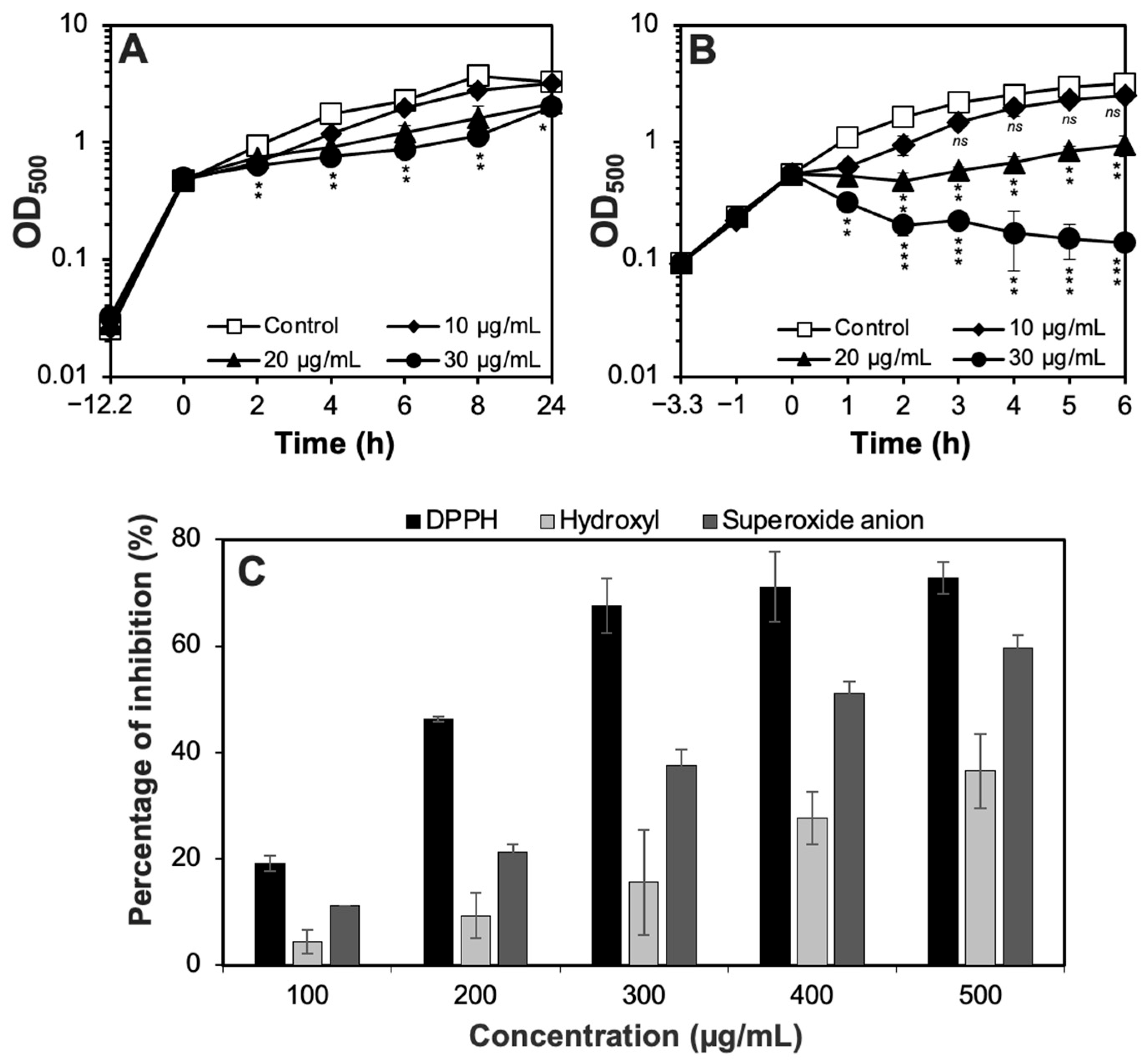
The antibacterial effect of CH6 extract on cell growth was further monitored against M. smegmatis MC2 155 and S. aureus ATCC 29213, as shown in Figure 2. It was observed that 10 µg/mL CH6 extract did not show growth delay (p > 0.05) in M. smegmatis MC2 155. Exposure to 30 µg/mL extract showed significant inhibitory effects on the bacterial growth from 2 to 8 h but not after 24 h (Figure 2A). In contrast, the CH6 extract strongly inhibited the growth of S. aureus ATCC 29213 along the growth curve. About 20 µg/mL of CH6 extract extended the growth lag phase and inhibited bacterial growth until 6 h. After exposure to 30 µg/mL, S. aureus cells were significantly reduced (p < 0.001) and unable to grow (Figure 2B).

3.3. Antioxidant Potential of the CH6 Extract

An assessment of the in vitro antioxidant potential of CH6 extract against free radicals proved that the extract had a moderate antioxidant activity in a concentration-dependent manner (Figure 2C). In case of DPPH radicals, the strongest radical scavenging activities of 67.6 ± 5.1%, 71.2 ± 6.6%, and 72.8 ± 3.0% were observed at 300, 400, and 500 µg/mL of the CH6 extract, respectively. This suggests that CH6 extract might have a hydrogen-donating ability. In addition, CH6 extract also showed moderate superoxide anion scavenging activity measured from 11.2 ± 0.2% to 59.8 ± 2.2% at 100 to 500 µg/mL (Figure 2C). As for hydroxyl radical scavenging activity, the antioxidant activity was low. Antioxidant activity reached only 36.5 ± 6.9% at 500 µg/mL extract, which was about 2-fold lower than that of DPPH scavenging activity.

3.4. Anticancer Potential of the CH6 Extract

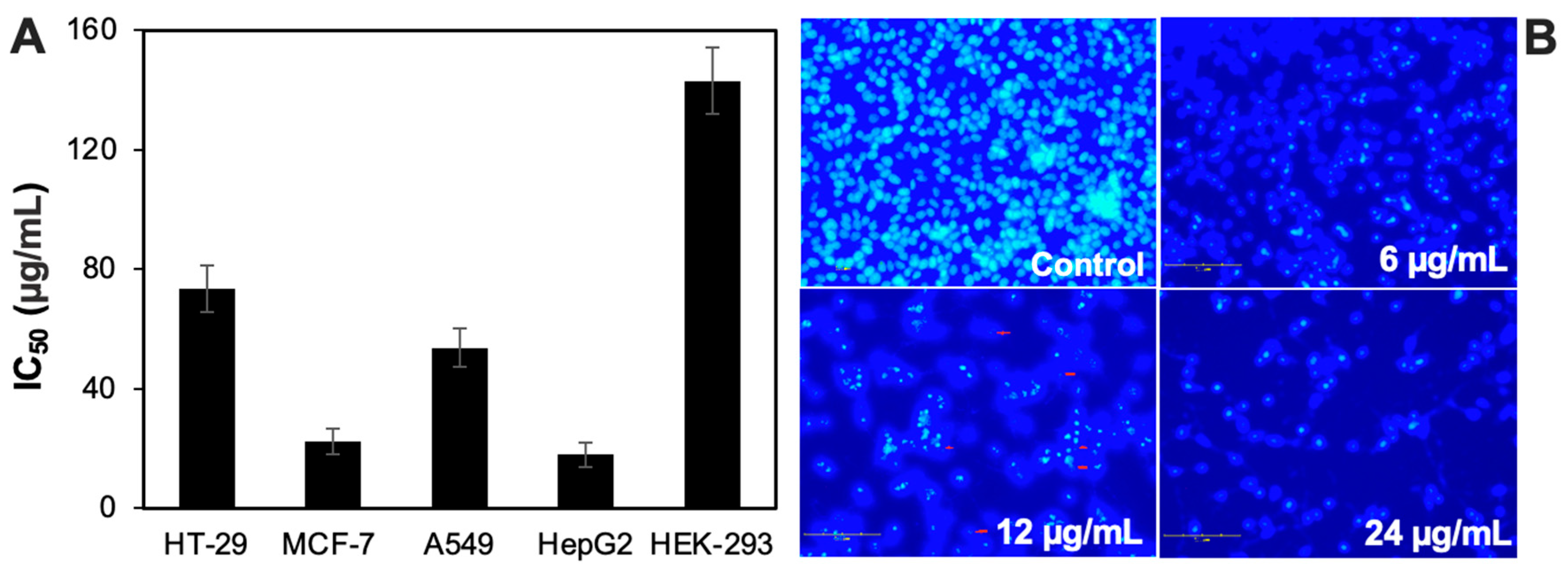
Cancer cells, including human lung cancer A549, hepatoma HepG2, breast cancer MCF-7, colon cancer HT-29, and non-cancerous HEK-293 cell lines, were utilized to assess the cytotoxic potential of the CH6 extract. HepG2 was the most sensitive to exposure to CH6, with an IC50 value of 18.0 ± 4.1 µg/mL, followed by MCF-7 (IC50 of 22.5 ± 4.3 µg/mL) and A549 (IC50 of 53.7 ± 6.4 µg/mL) (Figure 3A). HT-29 was less sensitive, as indicated by an IC50 value of 73.4 ± 7.8 µg/mL. Notably, the CH6 extract showed a low cytotoxicity against normal cells such as HEK-293, with an IC50 value of 143.0 ± 11.2 µg/mL, which was about 7.9-fold and 6.4-fold higher than that for HepG2 and MCF-7, respectively.
Upon treatment with 6–24 µg/mL extract, HepG2 morphologies changed. Nuclear staining with Hoechst 33342 showed distinct changes to morphology and cell numbers among the treatment groups. Control cells exhibited round, uniformly stained nuclei with a high cell density, consistent with normal cellular morphology. In contrast, cells treated with 6–24 µg/mL CH6 showed a gradual decrease in nuclear integrity, with signs of condensation, fragmentation, and reduced cell numbers at higher concentrations, indicating the induction of apoptosis. The results suggest that the CH6 extract induces apoptotic cell death in HepG2 cells.

3.5. Phenotypic Analysis of the CH6 Isolate

The CH6 strain was aerobic and Gram-positive and grew well on all ISP agar plates at 30 °C for nine days. The presence of aerial and substrate mycelia was also observed as white and pale-yellow colors, respectively (Figure S2). No diffusible pigment was observed after incubation at 30 °C for nine days. When cultured in ISP2 medium, a well-developed, filamentous, and branched mycelium was observed using light microscopy, indicating typical characteristics of the members of the Streptomyces genus (Figure S2). It grew at 15–47 °C (optimum 30 °C), at pH 3–10 (optimum pH 7.0), and in the presence of up to 6% (w/v) NaCl (Table S1). For the enzymatic tests, the CH6 strain was found to digest CMC, chitin, casein, and soluble starch, suggesting its ability to produce extracellular enzymes. In addition, this strain was able to ferment glucose, sucrose, fructose, raffinose, galactose, and mannitol as carbon sources.

3.6. Genomic Features and Taxonomy Study

The whole genome of CH6 was sequenced by the Illumina platform, yielding 495,603,483 clean Illumina data. After de novo genome assembly, the genome sequence of CH6 was shown to have 79 contigs and contain a 6,936,977 bp linear chromosome with a 73.0% GC content (Table 2). A total of 5902 genes, 5831 protein-coding sequences (CDSs), 65 tRNA genes, 3 rRNA genes, and 3 ncRNA genes were present in the CH6 genome. In addition, CRISPRs and plasmids were not found. This genome was smaller than other reported genomes of the Streptomyces genus, such as S. evansiae DSM 41979T, S. spheroides NCIMB 11891T, and S. chiangmaiensis TA4-1T (Table 2). The completeness was evaluated through a checkM analysis, leading to completeness of 100% and 0% duplication. Therefore, the genome sequence of Streptomyces sp. CH6 was deposited onto Genbank under accession number JBNOXK000000000.
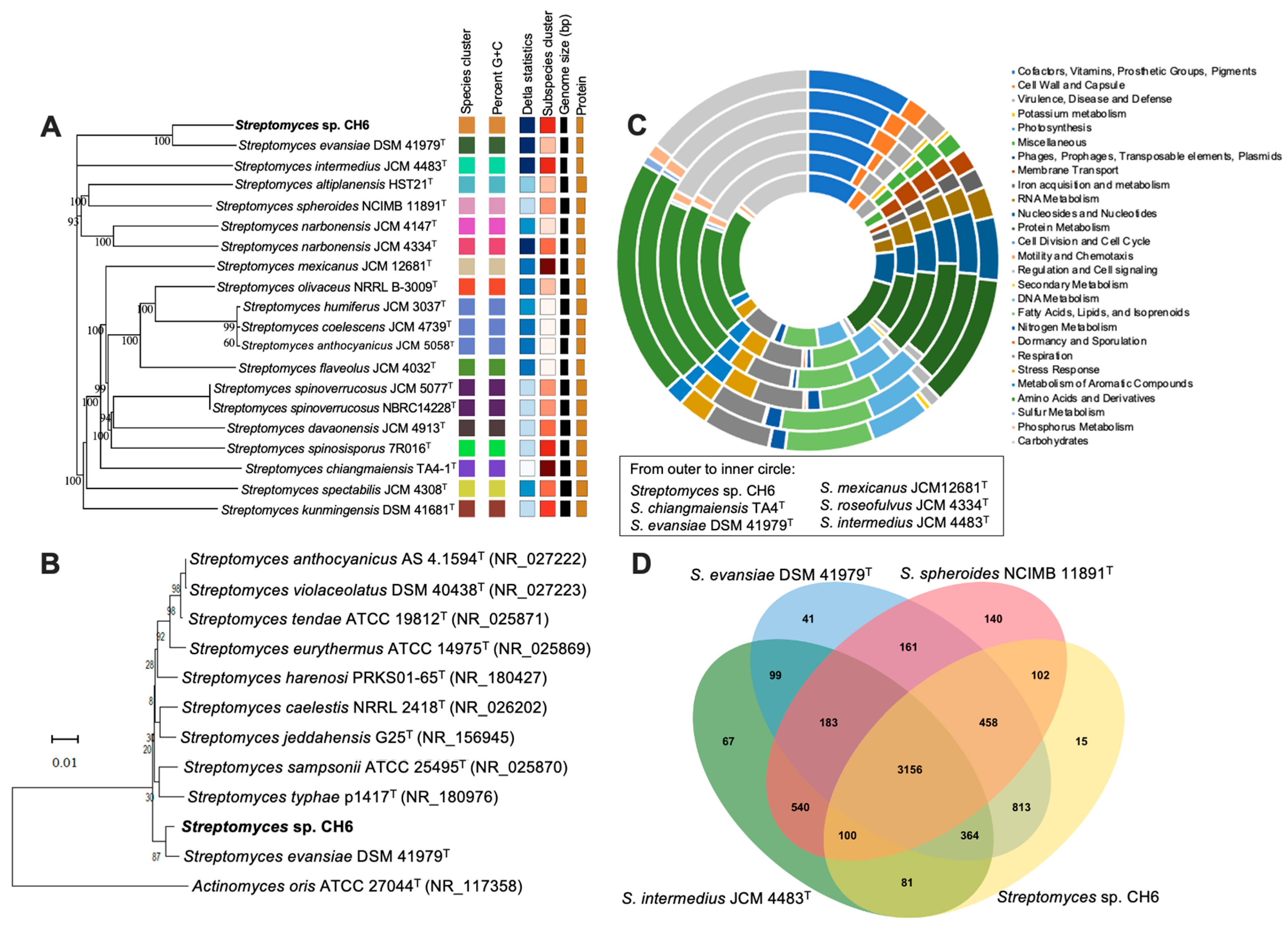
Phylogenomic analysis of the whole-genome sequences with the Type Strain Genome Server (TYGS) showed that Streptomyces sp. CH6 was closely related to S. evansiae DSM 41979T, S. kunmingensis DSM 41681T, S. roseofulvus JCM 4334, and S. olivaceus NRRL B-3009 with digital DNA–DNA hybridization (dDDH) values (formula d4) of 45.8%, 23.2%, 23.1%, and 23.0%, respectively (Figure 4A). This reveals that CH6 could be a new species of the genus Streptomyces. In support of this result, the CH6 genome was compared to its closest related S. evansiae DSM 41979T, Streptomyces intermedius JCM 4483T, and Streptomyces mexicanus JCM12681T, resulting in ANI values of 92.6%, 78.2%, and 77.8%, respectively. Using the taxonomic assignment of the genome by GTDB-Tk, the CH6 genome could not be affiliated with known members of the genus Streptomyces. An analysis of the 16S rRNA sequence showed that Streptomyces sp. CH6 displayed the highest similarity with the 16S rRNA sequence of S. evansiae DSM 41979T (98.9%), Streptomyces harenosi PRKS01-65T (98.1%), Streptomyces caelestis NRRL 2418T (98.0%), and Streptomyces sampsonii strain ATCC 25495T (98.0%). Phylogenetic analysis also demonstrated that CH6 and S. evansiae DSM 41979T were clustered together with a low bootstrap value of 87% (Figure 4B). These findings were consistent with the genomic analysis, indicating the isolated strain may be a novel species.

3.7. Functional Annotation and Comparative Genomes

The genomic analysis using RAST showed the presence of 1937 CDSs classified into 24 functional categories of the SEED database. The quality present in the CH6 genome was 18% in the subsystem and 82% in the non-subsystem inclusion quality. The most frequent categories were “Amino Acids and Derivatives” (330 CDSs), “Carbohydrates” (266 CDSs), Protein Metabolism (217 CDSs), and “Cofactors, Vitamins, Prosthetic Groups, Pigments” (189 CDSs) (Figure 4C). A number of genes related to “Stress Response” (53 CDSs), “Metabolism of Aromatic Compounds” (36 CDSs), and “Secondary Metabolism” were predicted, showcasing its ecological resilience, which was much lower than that of S. chiangmaiensis TA4T and S. mexicanus JCM12681T. Moreover, orthologous cluster analysis confirmed the presence of 6320 clusters of orthologous proteins and 2999 single-copy gene clusters for Streptomyces sp. CH6, S. evansiae DSM 41979T, S. intermedius JCM 4483T, and S. spheroides NCIMB 11891T. A total of 5089, 5275, 4590, and 4840 orthologous gene clusters were found in CH6, DSM 41979T, JCM 4483T, and NCIMB 11891T (Figure 4D). About 3156 orthologous gene clusters were common among the four Streptomyces species. Despite showing the closest relationship, 813 clusters were shared by Streptomyces sp. CH6 and S. evansiae DSM 41979T, which was 8.0-fold lower than that of Streptomyces sp. CH6 and S. spheroides NCIMB 11891T. About 15 clusters from the CH6 genome were unique.

3.8. Identification of Secondary Metabolite-Biosynthetic Gene Clusters

Prediction of secondary metabolites using antiSMASH revealed that Streptomyces sp. CH6 contained 22 BGCs encoding for non-ribosomal peptides (NRPS), polyketides (PKS), lanthipeptides, and terpenes. Among them, 13 clusters exhibited different similarities to BGCs with known function (Table 3). The BGCs of three NRPSs and three PKS/NRPS hybrids were observed, while PKS alone was not found. Meanwhile, four gene clusters displayed high similarity (> 70%) to the reported BGCs: ectoine (80.7%), guangnanmycin (76.0%), birimositide (73.0%), and SGR PTMs (71.0%). Among the remaining nine BGCs, six exhibited some level of similarity (30–70%) with known BGCs, such as AmfS (61.0%), geosmin (57.3%), isorenieratene (56.7%), totopotensamide (54.1%), hopene (53.9%), and detoxin S1 (30.6%) (Figure S3). The other three BGCs, such as detoxin S1, kinamycin, and julichrome Q3-3, showed low similarity values ranging from 13.2% to 19.8%.
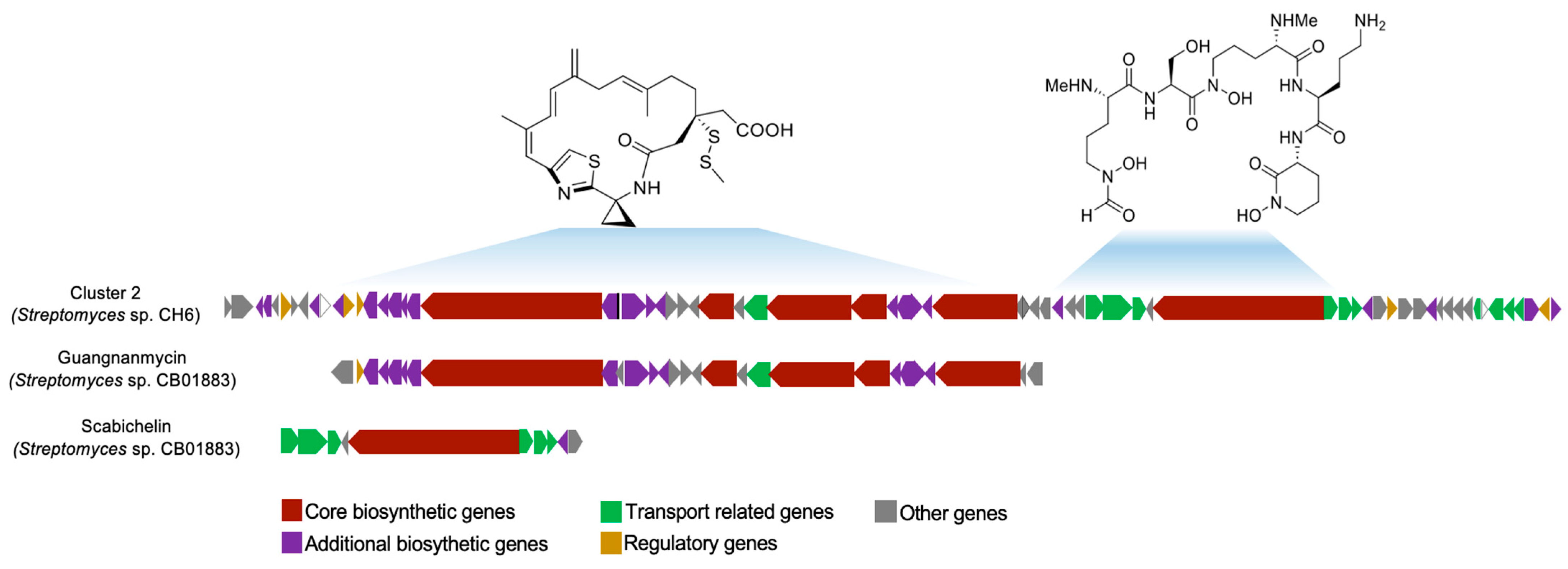
Notably, a large cluster 2 with a size of 133,857 bp exhibited 76% similarity with the known cluster, which was able to synthesize guangnanmycin, a congener of the well-known antitumor drug lead, leinamycin. It was about 1.8-fold larger than guangnanmycin produced by Streptomyces sp. CB01883 (Figure 5). Further investigations revealed that the 30,020 bp scabichelin cluster was also present in cluster 2. The large BGC from the CH6 is unique and has not been found in any reference gene clusters from the MIBiG database.

4. Discussion

The global increase in antibiotic-resistant bacteria poses a serious threat to public health, with approximately 1.27 million deaths attributed to antimicrobial resistance in 2019 [40]. While the development of synthetic antibiotics remains important, there is renewed interest in natural sources. Among these, the genus Streptomyces has contributed over two-thirds of clinically used antibiotics and continues to serve as a rich source of bioactive compounds [41]. Researchers are actively investigating new strains, particularly from understudied or extreme environments, to discover novel antimicrobial agents [42]. As part of these ongoing efforts, we explored microbial resources from Vietnam, a region known for its rich biodiversity.
We successfully isolated Streptomyces sp. CH6, which showed strong antibacterial activity against both Gram-positive and Gram-negative bacteria. In addition, the CH6 crude extract displayed antioxidant and anticancer activity. The 16S rRNA analysis revealed that the Streptomyces sp. CH6 strain is most closely related to S. evansiae DSM 41979T, S. harenosi PRKS01-65T, and S. caelestis NRRL 2418T with 98.9%, 98.1%, and 98.0% sequence similarity, respectively. Although these levels of similarity typically suggest a close taxonomic relationship, it is well recognized that 16S rRNA gene sequences can lack sufficient resolution to distinguish between closely related Streptomyces species. To clarify the genomic relationship and facilitate genome mining, we conducted a whole-genome comparison using ANI. The ANI between CH6 and S. evansiae DSM 41979 (GCA_031845585.1) was found to be 92.6%, which is below the generally accepted species boundary of 95–96% [43]. This result strongly suggests that Streptomyces sp. CH6 represents a genomically distinct species. In support of these results, the dDDH between Streptomyces sp. CH6 and S. evansiae DSM 41979T, S. kunmingensis DSM 41681T, and S. roseofulvus JCM 4334 are 45.8%, 23.2%, and 23.1%, respectively, which are well below the 70% threshold typically used to define bacterial species boundaries. Our next experiments will focus on comparing the phenotypic, biochemical, and metabolic characteristics of the CH6 strain with those of its closest relatives, such as S. evansiae DSM 41979ᵀ, in order to provide further evidence that the CH6 strain represents a novel Streptomyces species.
The whole-genome sequencing and bioinformatics analysis of Streptomyces sp. CH6 strain revealed 13 BGCs for secondary metabolite biosynthesis with similarities ranging from 13.2% to 80.7% to known clusters (Table 3). Among them, nine clusters showed moderate similarity (50–80%), suggesting the potential to produce known or related bioactive compounds. The totopotensamide cluster (54.1%) has been reported to exhibit cytotoxic activity, reinforcing the strain’s therapeutic potential [44]. The genome of the CH6 strain also contains the detoxin S1 biosynthetic cluster (30.6%), which is associated with a natural product originally identified in Streptomyces sp. NRRL S-325. However, the biological functions of detoxin S1 remain largely uncharacterized [45]. Supporting the extract’s antioxidant activity, the isorenieratene cluster (56.7%) encodes carotenoids known for free radical scavenging [46], while the ectoine cluster (80.7%) produces an osmoprotectant that stabilizes cells under oxidative stress [47]. In addition, some of the identified clusters, such as kinamycin (13.2%) and julichrome Q3-3 (16.5%), showed very low similarity to known reference clusters, suggesting that Streptomyces sp. CH6 may have the potential to produce novel secondary metabolites. Collectively, these clusters suggest a strong genetic basis for the production of bioactive metabolites in the CH6 strain.
To validate this biosynthetic potential, in vitro assays were conducted to evaluate the antioxidant and anticancer activities of the CH6 crude extract. The extract showed moderate antioxidant activity, with up to 72.8% DPPH radical scavenging and increased superoxide and hydroxyl radical scavenging at higher concentrations. It also showed selective anticancer effects, particularly against HepG2 and MCF-7 cell lines, with limited toxicity toward normal HEK-293 cells. These results confirm that the CH6 can produce bioactive compounds that reflect its genomic potential.
Of particular interest, we identified a biosynthetic gene cluster with high similarity to the guangnanmycin cluster (76%). It features a simpler structure than leinamycin, including a macrolactam ring, a methyldisulfide side chain, and a unique 1-aminocyclopropane-1-carboxylic acid (ACC) unit. ROS, produced by guangnanmycin, can trigger oxidative stress and induce apoptosis in cancer cells. This mechanism is common among quinone-based compounds and contributes to their cytotoxic effects. The biosynthetic pathway responsible for guangnanmycin production is encoded by the gnm gene cluster, a modular PKS/NRPS hybrid system. It includes several key enzymes, such as acyl-CoA synthetase GnmS for ACC activation, PLP-dependent cysteine lyase GnmY that produces hydropersulfide from L-thiocysteine, and hydropersulfide S-methyltransferase GnmP that methylates the -SSH group [48]. These enzymes are essential for constructing the compound’s sulfur-containing moiety, which contributes to its unique reactivity and cytotoxic potential of guangnanmycin [13]. The amino acid sequences of GnmP and GnmY from the CH6 strain showed 87.5% and 84.9% similarity (Figure S4) to GnmP and GnmY from the Streptomyces sp. CB01883 strain, respectively, suggesting functional conservation of these key biosynthetic enzymes in Streptomyces sp. CH6. Furthermore, inactivation of gnmB completely abolishes guangnanmycin production, confirming its essential role in the biosynthesis of guangnanmycin [10]. The presence of gnmS, gnmY, gnmP, and gnmB homologs in the CH6 genome suggests that this strain possesses the genetic capacity to biosynthesize guangnanmycin-like sulfur-containing compounds, which may contribute to the antioxidant and anticancer activities observed in the crude extract.
Although genome mining revealed a guangnanmycin-like biosynthetic gene cluster in Streptomyces CH6 with 76% similarity to a known reference cluster, the crude extract showed strong antimicrobial activity against both Gram-positive and Gram-negative bacteria. The actual production of guangnanmycin or a related metabolite has not yet been experimentally confirmed. In future studies, we will purify and identify the active compounds from the culture extract of Streptomyces sp. CH6 using chromatographic techniques (e.g., HPLC, flash chromatography), followed by structural elucidation via NMR and MS spectroscopy. Additionally, linking the compound to the gene cluster via gene knockout or heterologous expression will help establish a direct genotype–phenotype connection.

5. Conclusions

In summary, the present study describes Streptomyces sp. CH6 isolated from the soil in Vietnam as a novel species with promising antibacterial, antioxidant, and anticancer properties. The crude extract of Streptomyces sp. CH6 showed broad-spectrum antibacterial activity against both Gram-positive and Gram-negative bacteria, as well as antioxidant and selective anticancer properties in vitro. The genome of Streptomyces sp. CH6 consists of a 6,936,977 bp chromosome with a GC content of 73% and encodes 5831 proteins. Analysis revealed 13 putative biosynthetic gene clusters (BGCs) showing similarity (13.2–80.7%) to those responsible for the production of known antibacterial, antioxidant, and antitumor compounds. These findings suggest that Streptomyces sp. CH6 has the genetic potential to produce novel secondary metabolites. Overall, this study provides a comprehensive genomic overview of Streptomyces sp. CH6 and lays the groundwork for future experimental validation of guangnanmycin production or the discovery of other bioactive compounds with potential medical applications.

Supplementary Materials

The following supporting information can be downloaded at: https://www.mdpi.com/article/10.3390/app15137126/s1, Figure S1: Antifungal activity against Colletotrichum gloeosporioides (A) and Fusarium solani (B) of CH6; Figure S2: Colony morphology (A) and hyphal growth observed via light microscopy at 40× (B); Figure S3: Prediction of biosynthetic gene clusters (BGCs) present in Streptomyces sp. CH6. Genes are color-coded in antiSMASH by predicted function, as indicated in the bottom legends; Figure S4: Multiple sequence alignments of GnmP (A) and GnmY (B) from Streptomyces sp. CH6 with their homologs from Streptomyces sp. CB01883. GnmP and GnmY from the CH6 strain shared 87.5% and 84.9% sequence identity with those from the CB01883 strain, respectively. The multiple sequence alignment was performed using Clustal Omega (https://www.ebi.ac.uk/jdispatcher/msa/clustalo, accessed on 21 May 2025) and presented using Jalview (https://www.jalview.org/). Intensity of the blue color gradient is based on 50% identity; Table S1: Morphological, physiological, and biochemical characteristics of CH6 strain.

Author Contributions

Conceptualization, C.T.N. and T.T.T.T.; methodology, C.T.N., H.T.N., V.T.H.D., K.P.D. and T.T.T.T.; investigation, C.T.N. and H.T.N.; validation, C.T.N., H.T.N. and V.T.H.D.; formal analysis, C.T.N., T.T.T.T. and K.P.D.; writing—original draft preparation, C.T.N. and H.T.N.; writing—review and editing, T.T.T.T. All authors have read and agreed to the published version of the manuscript.

Funding

This research was funded by a grant from the Ministry of Education and Training, Vietnam, project number: B2023-MHN-02.

Institutional Review Board Statement

Not applicable.

Informed Consent Statement

Not applicable.

Data Availability Statement

The original contributions presented in this study are included in the article and Supplementary Materials. Further inquiries can be directed to the corresponding author.

Conflicts of Interest

The authors declare no conflicts of interest.

Abbreviations

The following abbreviations are used in this manuscript:
DPPH1,1-diphenyl-2-picrylhydrazyl
MICMinimum inhibitory concentration
dDDHdigital DNA–DNA hybridization
ANIAverage nucleotide identity
BGCsBiosynthetic gene clusters
IC50Half-maximal inhibitory concentration
CDSsProtein-coding sequences
ROSReactive oxygen species
NMRNuclear magnetic resonance
MSMass spectrometry

References

  1. Demain, A.L. Importance of microbial natural products and the need to revitalize their discovery. J. Ind. Microbiol. Biotechnol. 2014, 41, 185–201. [Google Scholar] [CrossRef] [PubMed]
  2. Pham, J.V.; Yilma, M.A.; Feliz, A.; Majid, M.T.; Maffetone, N.; Walker, J.R.; Kim, E.; Cho, H.J.; Reynolds, J.M.; Song, M.C.; et al. A review of the microbial production of bioactive natural products and biologics. Front. Microbiol. 2019, 10, 1404. [Google Scholar] [CrossRef]
  3. Xiong, Z.-Q.; Wang, J.-F.; Hao, Y.-Y.; Wang, Y. Recent advances in the discovery and development of marine microbial natural products. Mar. Drugs 2013, 11, 700–717. [Google Scholar] [CrossRef] [PubMed]
  4. Quach, N.T.; Nguyen, Q.H.; Vu, T.H.N.; Le, T.T.H.; Ta, T.T.T.; Nguyen, T.D.; Van Doan, T.; Van Nguyen, T.; Dang, T.T.; Nguyen, X.C.; et al. Plant-derived bioactive compounds produced by Streptomyces variabilis LCP18 associated with Litsea cubeba (Lour.) Pers as potential target to combat human pathogenic bacteria and human cancer cell lines. Braz. J. Microbiol. 2021, 52, 1215–1224. [Google Scholar] [CrossRef]
  5. Parte, A.C.; Sardà Carbasse, J.; Meier-Kolthoff, J.P.; Reimer, L.C.; Göker, M. List of Prokaryotic names with Standing in Nomenclature (LPSN) moves to the DSMZ. Int. J. Syst. Evol. Microbiol. 2020, 70, 5607–5612. [Google Scholar] [CrossRef] [PubMed]
  6. Prole, J.R.; Allenby, N.; Manning, D.A.C.; Goodfellow, M. Identification of four novel Streptomyces isolated from machair grassland soil using a culture-based bioprospecting strategy: Streptomyces caledonius sp. nov., Streptomyces machairae sp. nov., Streptomyces pratisoli sp. nov. and Streptomyces achmelvichensis sp. nov. Int. J. Syst. Evol. Microbiol. 2025, 75, 006736. [Google Scholar] [CrossRef]
  7. Quach, N.T.; Vu, T.H.N.; Nguyen, T.T.A.; Le, P.C.; Do, H.G.; Nguyen, T.D.; Thao, P.T.H.; Nguyen, T.T.L.; Chu, H.H.; Phi, Q.-T. Metabolic and genomic analysis deciphering biocontrol potential of endophytic Streptomyces albus RC2 against crop pathogenic fungi. Braz. J. Microbiol. 2023, 54, 2617–2626. [Google Scholar] [CrossRef]
  8. Mast, Y.; Stegmann, E. Actinomycetes: The antibiotics producers. Antibiotics 2019, 8, 105. [Google Scholar] [CrossRef]
  9. Li, L.-F.; Wu, Q.-X.; Wu, H.; Li, Y.; Peng, Q.; Han, R.-H.; Zhang, D.-H.; Yu, W.-D.; Xu, R.; Wang, J.; et al. Complete genome sequence of Streptomyces sp. HP-A2021, a promising bacterium for natural product discovery. Biochem. Genet. 2023, 61, 2042–2055. [Google Scholar] [CrossRef]
  10. Pan, G.; Xu, Z.; Guo, Z.; Hindra; Ma, M.; Yang, D.; Zhou, H.; Gansemans, Y.; Zhu, X.; Huang, Y.; et al. Discovery of the leinamycin family of natural products by mining actinobacterial genomes. Proc. Natl. Acad. Sci. USA 2017, 114, E11131–E11140. [Google Scholar] [CrossRef]
  11. Yahata, K.; Furstner, A. Total Synthesis of the Guangnanmycin A Alcohol. Angew. Chem. Int. Ed. Engl. 2024, 63, e202319070. [Google Scholar] [CrossRef]
  12. Brodska, B.; Holoubek, A. Generation of reactive oxygen species during apoptosis induced by DNA-damaging agents and/or histone deacetylase inhibitors. Oxid. Med. Cell. Longev. 2011, 2011, 253529. [Google Scholar] [CrossRef] [PubMed]
  13. Meng, S.; Steele, A.D.; Yan, W.; Pan, G.; Kalkreuter, E.; Liu, Y.-C.; Xu, Z.; Shen, B. Thiocysteine lyases as polyketide synthase domains installing hydropersulfide into natural products and a hydropersulfide methyltransferase. Nat. Commun. 2021, 12, 5672. [Google Scholar] [CrossRef]
  14. Nofiani, R.; Ardiningsih, P.; Rizky; Putri, C.D.; Rikayati, R.; Elfahmi; Syamsurizal; Sukito, A.; Juanssilfero, A.B.; Siregar, J.E.; et al. Complete genome sequence, biological activities, and metabolomic profiles of mangrove-derived Streptomyces sp. SM1P. Curr. Microbiol. 2025, 82, 193. [Google Scholar] [CrossRef] [PubMed]
  15. Nikolaidis, M.; Hesketh, A.; Frangou, N.; Mossialos, D.; Van de Peer, Y.; Oliver, S.G.; Amoutzias, G.D. A panoramic view of the genomic landscape of the genus Streptomyces. Microb. Genom. 2023, 9, mgen001028. [Google Scholar] [CrossRef]
  16. Quach, N.T.; Vu, T.H.N.; Bui, T.L.; Le, T.T.X.; Nguyen, T.T.A.; Ngo, C.C.; Phi, Q.-T. Genomic and physiological traits provide insights into ecological niche adaptations of mangrove endophytic Streptomyces parvulus VCCM 22513. Ann. Microbiol. 2022, 72, 27. [Google Scholar] [CrossRef]
  17. Kumar, H.; Vijayakumar, S.; Shintre, N.; Tamhane, V.; Deshpande, N.; Joshi, T.; Mathpal, S.; Anbarasu, A.; Ramaiah, S. In silico exploration of biosynthetic gene clusters in marine Streptomyces sp. and Nocardiopsis sp. from the western coast of India: Genome-based profiling using whole genome sequencing. J. Genet. Eng. Biotechnol. 2025, 23, 100483. [Google Scholar] [CrossRef]
  18. Ye, S.; Molloy, B.; Braña, A.F.; Zabala, D.; Olano, C.; Cortés, J.; Morís, F.; Salas, J.A.; Méndez, C. Identification by genome mining of a type I polyketide gene cluster from Streptomyces argillaceus involved in the biosynthesis of pyridine and piperidine alkaloids argimycins P. Front. Microbiol. 2017, 8, 194. [Google Scholar] [CrossRef]
  19. Balouiri, M.; Sadiki, M.; Ibnsouda, S.K. Methods for in vitro evaluating antimicrobial activity: A review. J. Pharm. Anal. 2016, 6, 71–79. [Google Scholar] [CrossRef]
  20. Al-Dhabi, N.A.; Esmail, G.A.; Ghilan, A.M.; Arasu, M.V.; Duraipandiyan, V.; Ponmurugan, K. Chemical constituents of Streptomyces sp. strain Al-Dhabi-97 isolated from the marine region of Saudi Arabia with antibacterial and anticancer properties. J. Infect. Public Health 2020, 13, 235–243. [Google Scholar] [CrossRef]
  21. Tung, Q.N.; Busche, T.; Van Loi, V.; Kalinowski, J.; Antelmann, H. The redox-sensing MarR-type repressor HypS controls hypochlorite and antimicrobial resistance in Mycobacterium smegmatis. Free Radic. Biol. Med. 2020, 147, 252–261. [Google Scholar] [CrossRef]
  22. Loi, V.V.; Huyen, N.T.T.; Busche, T.; Tung, Q.N.; Gruhlke, M.C.H.; Kalinowski, J.; Bernhardt, J.; Slusarenko, A.J.; Antelmann, H. Staphylococcus aureus responds to allicin by global S-thioallylation-Role of the Brx/BSH/YpdA pathway and the disulfide reductase MerA to overcome allicin stress. Free Radic. Biol. Med. 2019, 139, 55–69. [Google Scholar] [CrossRef]
  23. Quach, N.T.; Vu, T.H.N.; Nguyen, T.T.A.; Ha, H.; Ho, P.H.; Chu-Ky, S.; Nguyen, L.H.; Van Nguyen, H.; Thanh, T.T.T.; Nguyen, N.A.; et al. Structural and genetic insights into a poly-γ-glutamic acid with in vitro antioxidant activity of Bacillus velezensis VCN56. World J. Microbiol. Biotechnol. 2022, 38, 173. [Google Scholar] [CrossRef] [PubMed]
  24. Vu, T.H.N.; Pham, N.S.; Le, P.C.; Pham, Q.A.; Quach, N.T.; Nguyen, V.T.; Do, T.T.; Chu, H.H.; Phi, Q.T. Distribution, cytotoxicity, and antioxidant activity of fungal endophytes isolated from Tsuga chinensis (Franch.) Pritz. in Ha Giang province, Vietnam. Ann. Microbiol. 2022, 72, 36. [Google Scholar] [CrossRef]
  25. Mosmann, T. Rapid colorimetric assay for cellular growth and survival: Application to proliferation and cytotoxicity assays. J. Immunol. Methods. 1983, 65, 55–63. [Google Scholar] [CrossRef] [PubMed]
  26. Parks, D.H.; Imelfort, M.; Skennerton, C.T.; Hugenholtz, P.; Tyson, G.W. CheckM: Assessing the quality of microbial genomes recovered from isolates, single cells, and metagenomes. Genome Res. 2015, 25, 1043–1055. [Google Scholar] [CrossRef]
  27. Aziz, R.K.; Bartels, D.; Best, A.A.; DeJongh, M.; Disz, T.; Edwards, R.A.; Formsma, K.; Gerdes, S.; Glass, E.M.; Kubal, M.; et al. The RAST Server: Rapid Annotations using Subsystems Technology. BMC Genom. 2008, 9, 75. [Google Scholar] [CrossRef]
  28. Tatusova, T.; DiCuccio, M.; Badretdin, A.; Chetvernin, V.; Nawrocki, E.P.; Zaslavsky, L.; Lomsadze, A.; Pruitt, K.D.; Borodovsky, M.; Ostell, J. NCBI prokaryotic genome annotation pipeline. Nucleic Acids Res. 2016, 44, 6614–6624. [Google Scholar] [CrossRef]
  29. Antipov, D.; Hartwick, N.; Shen, M.; Raiko, M.; Lapidus, A.; Pevzner, P.A. plasmidSPAdes: Assembling plasmids from whole genome sequencing data. Bioinformatics 2016, 32, 3380–3387. [Google Scholar] [CrossRef]
  30. Meier-Kolthoff, J.P.; Göker, M. TYGS is an automated high-throughput platform for state-of-the-art genome-based taxonomy. Nat. Commun. 2019, 10, 2182. [Google Scholar] [CrossRef]
  31. Tamura, K.; Stecher, G.; Kumar, S. MEGA11: Molecular Evolutionary Genetics Analysis Version 11. Mol. Biol. Evol. 2021, 38, 3022–3027. [Google Scholar] [CrossRef] [PubMed]
  32. Lee, I.; Ouk Kim, Y.; Park, S.C.; Chun, J. OrthoANI: An improved algorithm and software for calculating average nucleotide identity. Int. J. Syst. Evol. Microbiol. 2016, 66, 1100–1103. [Google Scholar] [CrossRef] [PubMed]
  33. Blin, K.; Shaw, S.; Vader, L.; Szenei, J.; Reitz, Z.L.; Augustijn, H.E.; Cediel-Becerra, J.D.D.; Crécy-Lagard, V.D.; Koetsier, R.A.; Williams, S.E.; et al. antiSMASH 8.0: Extended gene cluster detection capabilities and analyses of chemistry, enzymology, and regulation. Nucleic Acids Res. 2025; online ahead of print. [Google Scholar] [CrossRef]
  34. Chen, Y.; Guo, M.; Yang, J.; Chen, J.; Xie, B.; Sun, Z. Potential TSPO ligand and photooxidation quencher isorenieratene from Arctic Ocean Rhodococcus sp. B7740. Mar. Drugs 2019, 17, 316. [Google Scholar] [CrossRef] [PubMed]
  35. Luo, Y.; Huang, H.; Liang, J.; Wang, M.; Lu, L.; Shao, Z.; Cobb, R.E.; Zhao, H. Activation and characterization of a cryptic polycyclic tetramate macrolactam biosynthetic gene cluster. Nat. Commun. 2013, 4, 2894. [Google Scholar] [CrossRef]
  36. O’Hara, K.A.; Wu, X.; Patel, D.; Liang, H.; Yalowich, J.C.; Chen, N.; Goodfellow, V.; Adedayo, O.; Dmitrienko, G.I.; Hasinoff, B.B. Mechanism of the cytotoxicity of the diazoparaquinone antitumor antibiotic kinamycin F. Free Radic. Biol. Med. 2007, 43, 1132–1144. [Google Scholar] [CrossRef]
  37. Quach, N.T.; Loan, T.T.; Nguyen, T.T.A.; Nguyen Vu, T.H.; Pham, Q.A.; Chu, H.H.; Phi, Q.-T.; Thuoc, D.V. Phenotypic and genomic characterization provide new insights into adaptation to environmental stressors and biotechnological relevance of mangrove Alcaligenes faecalis D334. Res. Microbiol. 2023, 174, 103994. [Google Scholar] [CrossRef]
  38. Dong, Y.; Ding, W.; Sun, C.; Ji, X.; Ling, C.; Zhou, Z.; Chen, Z.; Chen, X.; Ju, J. Julichrome monomers from marine Gastropod Mollusk-associated Streptomyces and stereochemical revision of julichromes Q(3 ⋅ 5) and Q(3 ⋅ 3). Chem. Biodivers. 2020, 17, e2000057. [Google Scholar] [CrossRef]
  39. Li, C.; Alam, K.; Zhao, Y.; Hao, J.; Yang, Q.; Zhang, Y.; Li, R.; Li, A. Mining and biosynthesis of bioactive lanthipeptides from microorganisms. Front. Bioeng. Biotechnol. 2021, 9, 692466. [Google Scholar] [CrossRef]
  40. Murray, C.J.; Ikuta, K.S.; Sharara, F.; Swetschinski, L.; Aguilar, G.R.; Gray, A.; Han, C.; Bisignano, C.; Rao, P.; Wool, E.; et al. Global burden of bacterial antimicrobial resistance in 2019: A systematic analysis. Lancet 2022, 399, 629–655. [Google Scholar] [CrossRef]
  41. Barka, E.A.; Vatsa, P.; Sanchez, L.; Gaveau-Vaillant, N.; Jacquard, C.; Meier-Kolthoff, J.P.; Klenk, H.P.; Clement, C.; Ouhdouch, Y.; van Wezel, G.P. Taxonomy, Physiology, and Natural Products of Actinobacteria. Microbiol. Mol. Biol. Rev. 2016, 80, 1–43. [Google Scholar] [CrossRef] [PubMed]
  42. Mohammadipanah, F.; Wink, J. Actinobacteria from Arid and Desert Habitats: Diversity and Biological Activity. Front. Microbiol. 2015, 6, 1541. [Google Scholar] [CrossRef] [PubMed]
  43. Chun, J.; Oren, A.; Ventosa, A.; Christensen, H.; Arahal, D.R.; da Costa, M.S.; Rooney, A.P.; Yi, H.; Xu, X.W.; De Meyer, S.; et al. Proposed minimal standards for the use of genome data for the taxonomy of prokaryotes. Int. J. Syst. Evol. Microbiol. 2018, 68, 461–466. [Google Scholar] [CrossRef] [PubMed]
  44. Lin, Z.; Flores, M.; Forteza, I.; Henriksen, N.M.; Concepcion, G.P.; Rosenberg, G.; Haygood, M.G.; Olivera, B.M.; Light, A.R.; Cheatham, T.E., 3rd; et al. Totopotensamides, polyketide-cyclic peptide hybrids from a mollusk-associated bacterium Streptomyces sp. J. Nat. Prod. 2012, 75, 644–649. [Google Scholar] [CrossRef]
  45. Navarro-Munoz, J.C.; Selem-Mojica, N.; Mullowney, M.W.; Kautsar, S.A.; Tryon, J.H.; Parkinson, E.I.; De Los Santos, E.L.C.; Yeong, M.; Cruz-Morales, P.; Abubucker, S.; et al. A computational framework to explore large-scale biosynthetic diversity. Nat. Chem. Biol. 2020, 16, 60–68. [Google Scholar] [CrossRef]
  46. Bhatt, T.; Patel, K. Carotenoids: Potent to Prevent Diseases Review. Nat. Prod. Bioprospect. 2020, 10, 109–117. [Google Scholar] [CrossRef]
  47. Pastor, J.M.; Salvador, M.; Argandona, M.; Bernal, V.; Reina-Bueno, M.; Csonka, L.N.; Iborra, J.L.; Vargas, C.; Nieto, J.J.; Canovas, M. Ectoines in cell stress protection: Uses and biotechnological production. Biotechnol. Adv. 2010, 28, 782–801. [Google Scholar] [CrossRef]
  48. Ma, M.; Lohman, J.R.; Liu, T.; Shen, B. C-S bond cleavage by a polyketide synthase domain. Proc. Natl. Acad. Sci. USA 2015, 112, 10359–10364. [Google Scholar] [CrossRef]
Figure 1. Heatmap showing antibacterial activity of metabolites extracted from bacterial isolates against at least one pathogen. ST: Salmonella enterica serovar Typhimurium ATCC 14028; EC: Escherichia coli ATCC 11105; SM: Mycobacterium smegmatis MC2 155; PA: Pseudomonas aeruginosa ATCC 9027; SA: Staphylococcus aureus ATCC 29213. (-): no activity. All sorts of color in figure represents the zone of inhibition (mm), red indicates the largest diameter of the inhibition zone, white represents no zone of inhibition.
Applsci 15 07126 g001
Figure 2. Antibacterial and antioxidant activities of CH6 extract. Growth curve analysis of M. smegmatis MC2 155 (A) and S. aureus ATCC 29213 (B) in response to the different concentrations of CH6 extract. (C) Antioxidant potential demonstrated by CH6 extract in the DPPH, hydroxyl, and superoxide anion scavenging assays. p-values were calculated via a Student’s unpaired two-tailed t-test in Prism 9 (ns p > 0.05; * p < 0.05; ** p < 0.01; *** p < 0.001).
Applsci 15 07126 g002
Figure 3. Anticancer activity of CH6 extract against human cancer and non-cancerous cell lines. (A) Cytotoxicity as IC50 values of CH6 extract on a panel of human cancer and non-cancerous cell lines. (B) Morphology of HepG2 treated and untreated by different concentrations of CH6 extract. Nucleus counterstained with Hoechst 33342 (blue), observed under a fluorescence microscope. Scale bar represents 100 µm.
Applsci 15 07126 g003
Figure 4. Phylogenomic analysis and comparative genomes revealing Streptomyces sp. CH6 as a novel species. (A) Phylogenomic tree based on TYGS, exhibiting the relationship between the CH6 strain with closely related type strains. The numbers above the branches are GBDP pseudo-bootstrap support values > 60% from 100 replications, with an average branch support of 89.9%. (B) Phylogenetic tree constructed from 16S rRNA gene sequences retrieved from the CH6 genomes. (C) RAST subsystem category distributions of CH6 assigned to genes in 6 different Streptomyces genomes. (D) Genome-wide comparison showing the number of shared and unique orthologous gene clusters between CH6 and other related Streptomyces species.
Applsci 15 07126 g004
Figure 5. Comparison of guangnanmycin and scabichelin BGCs between Streptomyces sp. CH6 and previously reported Streptomyces sp. CB01883.
Applsci 15 07126 g005
Table 1. MIC of CH6 crude extract against the tested bacteria and yeast.
Tested PathogenMIC (µg/mL)
S. Typhimurium ATCC 1402832
E. coli ATCC 1110532
M. smegmatis MC2 15564
P. aeruginosa ATCC 902716
S. aureus ATCC 292138
E. aerogenes ATCC 1304832
B. cereus ATCC 1177816
C. albicans ATCC 10231128
Table 2. Genome characteristics of CH6 and its related type strains.
AttributeStreptomyces sp. CH6S. evansiae DSM 41979TS. chiangmaiensis TA4-1TS. mexicanus JCM 12681TS. spheroides NCIMB 11891T
Genome size (bp)6,936,9777,502,3969,707,9848,193,1008,727,323
GC content (%)73.073.369.772.570.5
rRNA genes33331820
tRNA genes6565406766
ncRNA genes33334
CDSs58316577933074367799
Predicted genes59026678937675247886
GenBank accessionJBNOXK000000000JAVRET000000000JAYWVC000000000JBHTGD000000000AWQW00000000
Table 3. List of proposed secondary metabolite biosynthetic gene clusters present in Streptomyces sp. CH6 genome.
ClusterTypeLength (bp)Most Similar Known ClusterSimilarity (%)Potential Activity
1NRPS-like, NRPS, nucleoside114,856Detoxin S119.8-
2TransAT-PKS, NRPS, NRP-metallophore133,857Guangnanmycin76.0Anticancer [13]
3Terpene21,104Detoxin S130.6-
4Lanthipeptide-class-iii22,655AmfS61.0-
5HglE-KS, terpene55,247Isorenieratene56.7Antioxidant, anti-UVB radiation [34]
6Terpene26,704Hopene53.9-
7NRPS, T1PKS49,405SGR PTMs71.0Antifungal, antibiotic, antioxidant [35]
8NI-siderophore33,324Kinamycin13.2Antibacterial, anticancer [36]
9Ectoine10,399Ectoine80.7Antioxidant [37]
10NRPS, T1PKS, T3PKS, NRPS-like57,731Totopotensamide 54.1-
11Terpene21,173Julichrome Q3-316.5Antibacterial, anticancer [38]
12Terpene22,436Geosmin57.3-
13NRPS, lanthipeptide-class-ii32,791Birimositide73.0Antibacterial [39]
Disclaimer/Publisher’s Note: The statements, opinions and data contained in all publications are solely those of the individual author(s) and contributor(s) and not of MDPI and/or the editor(s). MDPI and/or the editor(s) disclaim responsibility for any injury to people or property resulting from any ideas, methods, instructions or products referred to in the content.