1. Introduction
The instep kick is one of the most fundamental and extensively studied movements in soccer, as it plays a decisive role in goal scoring. Biomechanically, it has been defined as an open-chain movement of the lower limb from the proximal to the distal part, involving continuous braking and acceleration [
1,
2]. This movement requires precise coordination among multiple joints and muscle groups across the pelvis, hip, knee, and ankle, spanning multiple planes of motion [
3]. Investigating its biomechanical characteristics is essential for understanding the mechanisms underlying kicking performance and optimizing training strategies.
A high ball velocity is often used as a parameter of kicking performance [
1,
2]. However, the success of a kick is determined not only by ball velocity but also by precision, and often, kicks have an inherent characteristic of ball spin included which may or may not increase precision. However, studies have shown that the ball spin is inversely related to the ball velocity [
4]. Most studies have focused on the joint angles, angular velocities, and force generation of the kicking leg of male players [
5,
6], showing that the coordination and efficient energy transfer in the kicking leg are important factors in achieving higher ball speeds [
7,
8]. However, the role of the support leg remains largely underexplored.
Existing research on the lower limb joints of the support leg has primarily focused on cross-sectional comparisons related to gender [
9,
10,
11], leg preference [
12,
13,
14,
15], sports injuries [
16,
17,
18], and approach strategies [
19,
20]. These studies have highlighted the role of the support leg in stabilizing the body and optimizing force transmission [
21], and a further investigation into its kinematic characteristics and their relationship with kicking performance is essential. A study reporting on the kinetics has shown that that the joint moments at the ankle, knee, and hip of the support leg primarily counteract ground reaction forces (GRF) thus facilitating impact absorption rather than generating positive energy [
22]. Another key mechanism through which the support leg contributes to kicking performance is its role in pelvic rotation. In the leg acceleration phase, when the knee of the kicking leg extends as the hip of the kicking leg flexes, the reaction force at the support leg’s hip joint facilitates counterclockwise pelvic rotation, which is essential for the proximal-to-distal energy transfer in the kicking motion. While some studies have analyzed this mechanism, they have not specifically examined how different kinematic parameters of the support leg contribute to kicking performance.
Previous research on the direct role of the support leg in determining ball velocity has been mixed: a transversal study found a low predictive effect of the support leg kinematics on ball velocity [
21]. However, this study incorporated both the kicking leg and the support leg into a single model, which may have masked the support leg’s true contribution. On the contrary, an intervention study demonstrated that technique refinement training significantly increased the knee extension velocity and vertical hip displacement of the support leg, directly enhancing the swing speed of the kicking leg and, as a consequence, ball velocity [
23]. This discrepancy suggests that the relationship between support leg kinematics and kicking performance is more complex than previously assumed and requires further investigation using more comprehensive analytical approaches.
Although research on the biomechanical properties of the support leg has made progress, there are still research gaps. First, existing studies mostly focus on cross-sectional comparisons of specific variables and do not include a complete kinematic analysis of the support leg during kicking. Second, almost all studies have dealt with amateur and/or university level players, and none have studied the technique in professional players. Third, a majority of the studies have a relatively small sample of players (under 25 players including for cross-sectional studies), which can limit the robustness of the findings. Finally, existing studies focus more on ball speed, while hardly any see its effect on ball spin [
4]. A more comprehensive analysis of the relationship between support leg variables and ball velocity and ball spin, using a large sample of players including professional players, can provide crucial insights for coaches and practitioners to improve kicking performance.
Unlike previous studies that predominantly focused on the kicking leg, this study uniquely emphasizes the role of support leg kinematics as a key determinant of both ball velocity and spin. By incorporating a large and diverse sample that includes professional male and female players, and applying advanced statistical models across leg preference and competition level, this research offers novel biomechanical insights with direct practical implications for performance enhancement and training design in football.
Thus, this study aims to explore the influence of the kinematic characteristics of the hip, knee, and ankle of the support leg on ball velocity and ball spin. The specific objectives are to compare and explore the differences between the hip, knee, and ankle for different competitive levels and leg preferences, as well as their influence on ball velocity and ball spin.
2. Materials and Methods
2.1. Design
A transversal study was conducted where kicking technique was compared between players belonging to four different levels (professional male (MalePro), professional female (FemalePro), elite youth (EliteU19), and non-elite youth (MaleU19)). The players kicked the ball with both their preferred and non-preferred legs and the support leg data was analyzed in these kicks.
2.2. Participants
A total of seventy-eight players, playing in different categories of Spanish football (
Table 1) voluntarily participated in this study. They did not have a lower limb injury in the three months preceding their participation in this study, enabling them to achieve maximum instep kicking in the current test. All players were informed about the testing protocol and signed an informed consent. The study design, processing, and analysis followed the guidelines of the Declaration of Helsinki and was approved by the ethics committee of the host university.
2.3. Data Capture
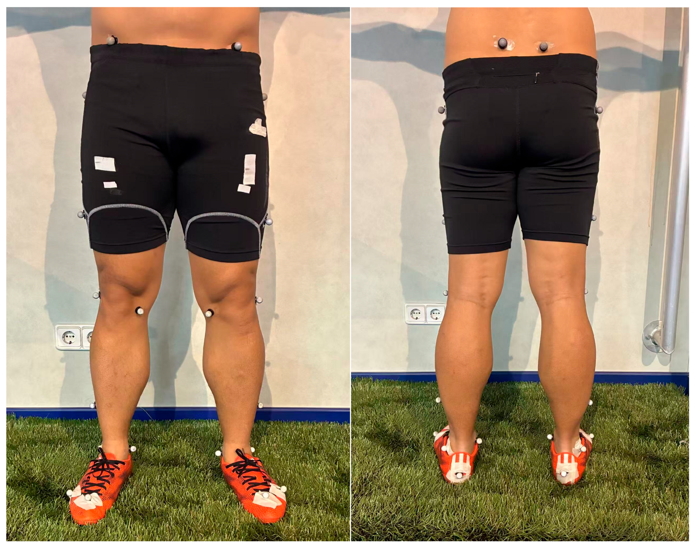
A six-camera Vicon Motion Capture System (Oxford Metrics Ltd., Oxford, UK) was used to capture kicking data at 250 Hz. The protocol required the players to perform the movement on FIFA-approved artificial turf and using a regulation football (complying with FIFA standards: diameter = 22 cm; pressure: 1 atmosphere). Participants wore football shoes appropriate for artificial turf, and prior to data capture, twenty-eight retro-reflective markers (diameter = 14 mm) were placed on specific landmarks on the players’ bodies (
Table 2) as per a previously validated model [
24,
25].
After placing the markers, a static capture of the players was performed, and the additional markers were removed in preparation for the dynamic trial (
Table 2 and
Figure 1). Following this, a standardized 10 min warm-up was performed under the supervision of the strength and conditioning coach of the team involving mobility and activation drills. The players performed practice kicks as the last part of the warm up in order to familiarize themselves with the experimental procedure. For the data capture, the players were free to choose their approach angle but were instructed to kick the ball with a three-step run-up prior to the kick. They were required to kick using the instep kicking technique and aim at a target 1 m × 1 m, placed at a 1 m height at a net 7 m in front. Kicks were repeated with both the preferred and non-preferred legs until three successful kicks were captured for both the preferred and non-preferred legs, respectively.
2.4. Data Processing
According to previous definitions of kicking, the movement was cropped from the toe-off of the kicking leg to the peak hip flexion of the kicking leg [
24,
25]. The static and six dynamic trials per trial were reconstructed following Vicon
® guidelines [
26]. Calibration parameters were adjusted per session, and marker trajectories were labeled and then processed by combining duplicates, resulting in 28 valid trajectories for the static trial and 20 valid trajectories for the dynamic trial (
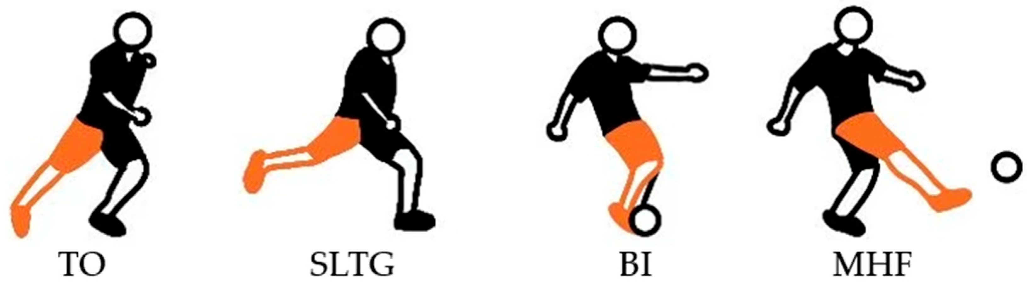
Table 2). The kicking phase was defined using the following key events: toe-off of the kicking leg (TO), defined as the moment the kicking foot leaves the ground; support leg touch-down (SLTG), defined as the first frame in which the support foot contacts the ground; ball impact (BI), defined as the frame when the kicking foot contacts the ball; and maximum hip flexion of the kicking leg (MHF), defined as the peak flexion angle of the hip of the kicking leg (
Figure 2). After identifying key events, marker trajectories were inspected frame by frame and missing data were interpolated using Vicon
®’s cubic spline algorithm (for a maximum of 10 frames).
Static trials were used to define the lower limb segments and joint centers based on markers placed on anatomical landmarks. The local coordinate systems for each segment were established as follows: the Z-axis was defined as the longitudinal axis, representing rotational movements such as internal and external rotation; the Y-axis was the anteroposterior axis, describing flexion and extension; and the X-axis was the mediolateral axis, describing adduction and abduction. In the dynamic trials, joint angles and angular velocities across the pelvis, hip, knee, and ankle were calculated. The movement of the soccer ball was tracked using four markers, with the ball center calculated based on its geometric relationships. Ball velocity (m/s) was determined by the displacement of the ball center between consecutive frames, while ball spin (rad/s) was derived from the rotational motion of the markers around the ball center. Both parameters were calculated automatically using custom-defined Vicon scripts [
25].
MATLAB R2024a (MathWorks, Natick, MA, USA) was used to filter and smooth the kinematic data to reduce high-frequency noise. During the pre-impact and post-impact phases, a 4th-order Butterworth low-pass filter was applied with cut-off frequencies of 15 Hz and 30 Hz, respectively. For angular velocity data, a 6th-order Butterworth filter with a 50 Hz cut-off frequency was used. During the impact phase, the Woltring smoothing algorithm with a “lowess” method and a 10-frame window was applied, effectively handling rapid signal changes by performing localized nonlinear regression. The data was then exported, and the following kinematic variables were extracted in MS Excel (Windows 365, Redmond, WA, USA). For the hip joint, the extracted variables included the adduction-abduction range of motion (ROM), peak adduction angular velocity, peak abduction angular velocity, flexion-extension ROM, peak flexion angular velocity, peak extension angular velocity, internal-external rotation ROM, peak internal rotation angular velocity, and peak external rotation angular velocity. For the knee joint, the extracted variables included the flexion-extension ROM, peak flexion angular velocity, and peak extension angular velocity. For the ankle joint, the extracted variables included the plantarflexion-dorsiflexion ROM, peak plantarflexion angular velocity, and peak dorsiflexion angular velocity. Both the ROM and peak angular velocity were calculated over the entire kicking phase. ROM is calculated by subtracting the minimum angle from the maximum angle during the kicking phase. Peak angular velocity was direction-specific and defined as the maximum angular velocity value for each movement direction.
2.5. Statistical Analysis
From the three trials exported, the trial with median ball velocity for both preferred and non-preferred leg kicks was selected for analysis [
24,
25]. Normality and variance homogeneity were evaluated using the Shapiro–Wilk and Levene’s tests, respectively. Kinematic differences of the support leg during kicking were assessed using two-way ANOVA or the Scheirer-Ray-Hare (SRH) test, with “Group” (competition level: MaleU19, EliteU19, MalePro, FemalePro, as defined in
Table 1) and “Leg Preference” (preferred vs. non-preferred leg) as independent variables. Effect size (partial eta squared, η
2) was used to interpret the impact of variables. An η
2 value of 0.01 indicates a small effect, 0.06 indicates a medium effect, and 0.14 or higher indicates a large effect [
27]. Post-hoc analyses with Bonferroni correction were applied to significant results. The SRH test was performed in Python 3.11.7 (Python Software Foundation, Wilmington, DE, USA), and other analyses were conducted using SPSS
® 27.0 (IBM Corp., Armonk, NY, USA).
A simplified a priori power analysis was also conducted using G*Power (version 3.1) for a two-way ANOVA with four group levels and two leg conditions (preferred vs. non-preferred). Assuming a medium effect size (f = 0.25), α = 0.05, and power = 0.80, the required sample size was estimated at 52. The actual sample size (N = 78) therefore provided sufficient power to detect both main and interaction effects.
A linear mixed-effects model (LMM) was used to assess the relationship between lower limb joint kinematics and kicking performance. Before running the models, the variance inflation factor (VIF) was used to check for multicollinearity, and all values were below 5, indicating no severe multicollinearity issues (
Table 3 and
Table 4). Ball velocity and ball spin were the dependent variables, while joint ROM and peak angular velocity metrics served as independent variables. “Group” was included as a fixed effect to control for baseline differences, and “player” was treated as a random effect to account for individual variability. Separate models were created for preferred and non-preferred leg kicks. The marginal R
2 (R
2m) ranged from 0.169 to 0.261, indicating that the fixed effects explained 16.9% to 26.1% of the variance. The conditional R
2 (R
2c) ranged from 0.295 to 0.725, showing that the full model, including random effects, explained 29.5% to 72.5% of the variance. Residuals vs. fitted value plots were examined to assess model assumptions (
Figure A1). No systematic patterns or severe heteroscedasticity were observed in most models. Slight increases in residual spread were noted at higher fitted values in some non-preferred leg models, but overall, model assumptions were considered adequately met. Regression coefficients and
p-values were calculated, with a 0.05 confidence interval. LMM was performed with Python 3.11.7.
3. Results
3.1. Analysis of Hip, Knee, and Ankle Range of Motion
The ROM of the lower limb joints is shown in
Figure 3. No significant effects were found in hip flexion-extension ROM or adduction-abduction ROM across groups or leg preferences. Internal-external rotation ROM showed a significant main effect of group with a large effect size (F (3,123) = 11.505,
p < 0.001, η
2 = 0.219). Post-hoc analysis revealed that for the preferred leg kicks, MalePro exhibited significantly lower rotation ROM compared to EliteU19 (
p = 0.022). For the non-preferred leg kicks, MalePro had the lowest ROM among all groups, while no significant differences were observed among the other three groups (all
p < 0.05).
Knee flexion-extension ROM was significantly influenced by leg preference (H = 15.900, p < 0.001, η2 = 0.105), with a medium effect size. Post-hoc analysis showed that among professional players, the preferred leg exhibited a significantly greater knee flexion-extension ROM compared to the non-preferred leg (all p < 0.01).
Ankle plantarflexion-dorsiflexion ROM showed no significant differences across groups or leg preferences in the two-way ANOVA.
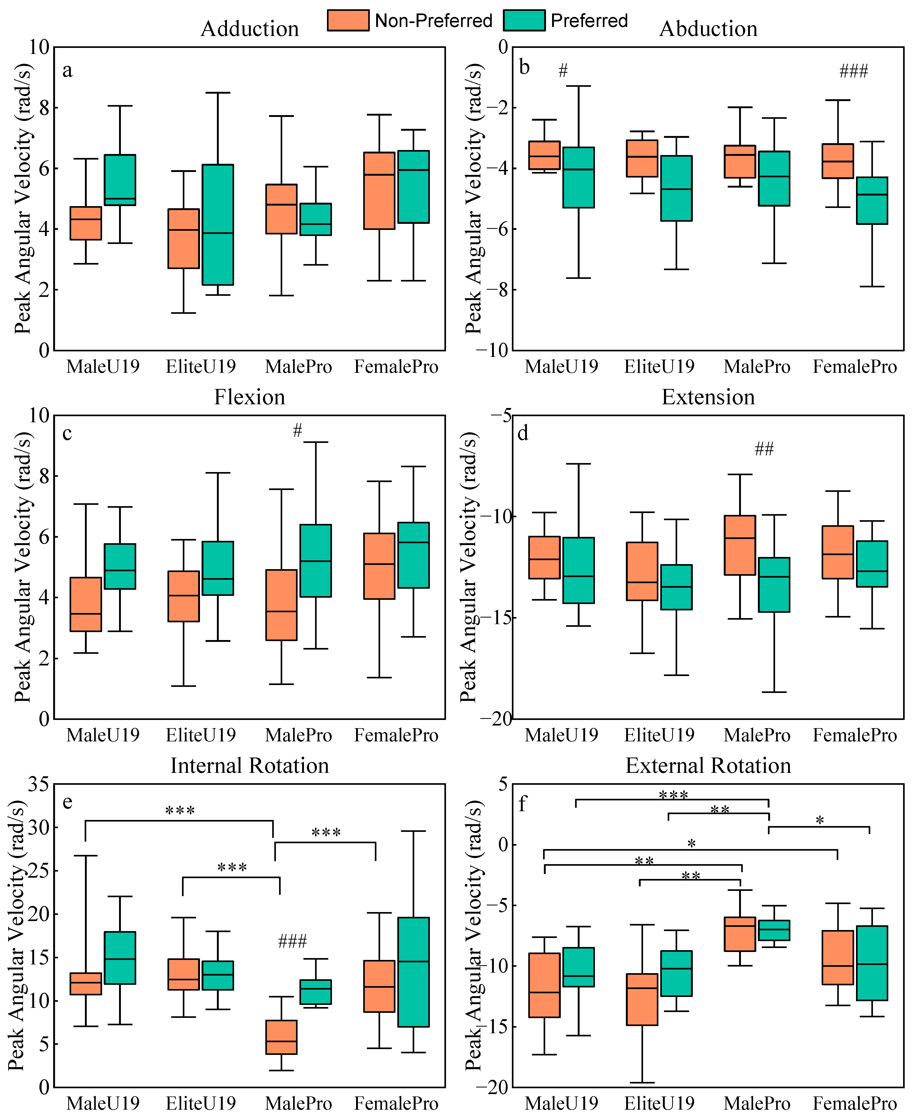
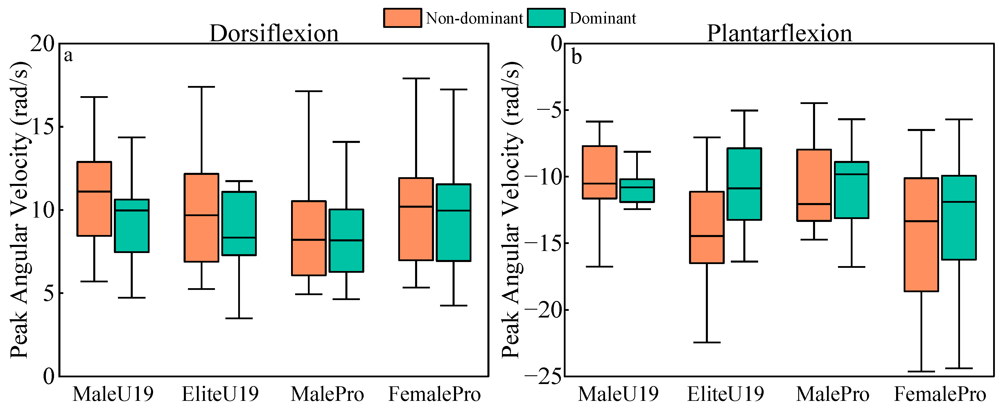
3.2. Analysis of Peak Angular Velocity of the Hip, Knee, and Ankle
The peak angular velocities of the lower limb joints are shown in
Figure 4,
Figure 5 and
Figure 6. Peak hip flexion and extension angular velocities were both significantly influenced by leg preference, with medium effect sizes (flexion: H = 11.019,
p = 0.001, η
2 = 0.077; extension: F = 7.933,
p = 0.006, η
2 = 0.061). Post-hoc analysis revealed that within the MalePro, the preferred leg kicks exhibited significantly higher peak angular velocities compared to the non-preferred leg kicks in both flexion (
p = 0.019) and extension (
p = 0.009). No significant effects were found for peak hip adduction angular velocity. Peak hip abduction angular velocity was significantly influenced by leg preference, with a medium effect size (H = 17.886,
p < 0.001, η
2 = 0.123). Post-hoc analysis revealed that in both the EliteU19 (
p = 0.048) and FemalePro (
p < 0.001), the preferred leg kicks exhibited significantly higher peak abduction angular velocity compared to the non-preferred leg kicks. Peak hip internal rotation angular velocity was significantly influenced by both leg preference and group, with a medium effect size for leg preference (H = 8.351,
p = 0.004, η
2 = 0.050) and a large effect size for group (H = 10.822,
p = 0.013, η
2 = 0.195). Post-hoc analysis showed no significant differences among groups for the preferred leg kicks. However, for the non-preferred leg kicks, the MalePro exhibited the lowest peak internal rotation angular velocity, with no significant differences among the other three groups. Additionally, within the MalePro, the preferred leg kicks demonstrated a significantly higher peak internal rotation angular velocity than the non-preferred leg kicks. Peak hip external rotation angular velocity was significantly influenced by group, with a large effect size (H = 14.977,
p = 0.002, η
2 = 0.265). Post-hoc analysis revealed that for the preferred leg kicks, the MalePro had the lowest peak external rotation angular velocity, while no significant differences were found among the other three groups (all
p < 0.05). For the non-preferred leg kicks, the MalePro exhibited significantly lower peak external rotation angular velocity than EliteU19 (
p < 0.001) and MaleU19 (
p = 0.004), but showed no significant difference compared to FemalePro. Additionally, the FemalePro group had significantly lower peak external rotation angular velocity than EliteU19 (
p = 0.033).
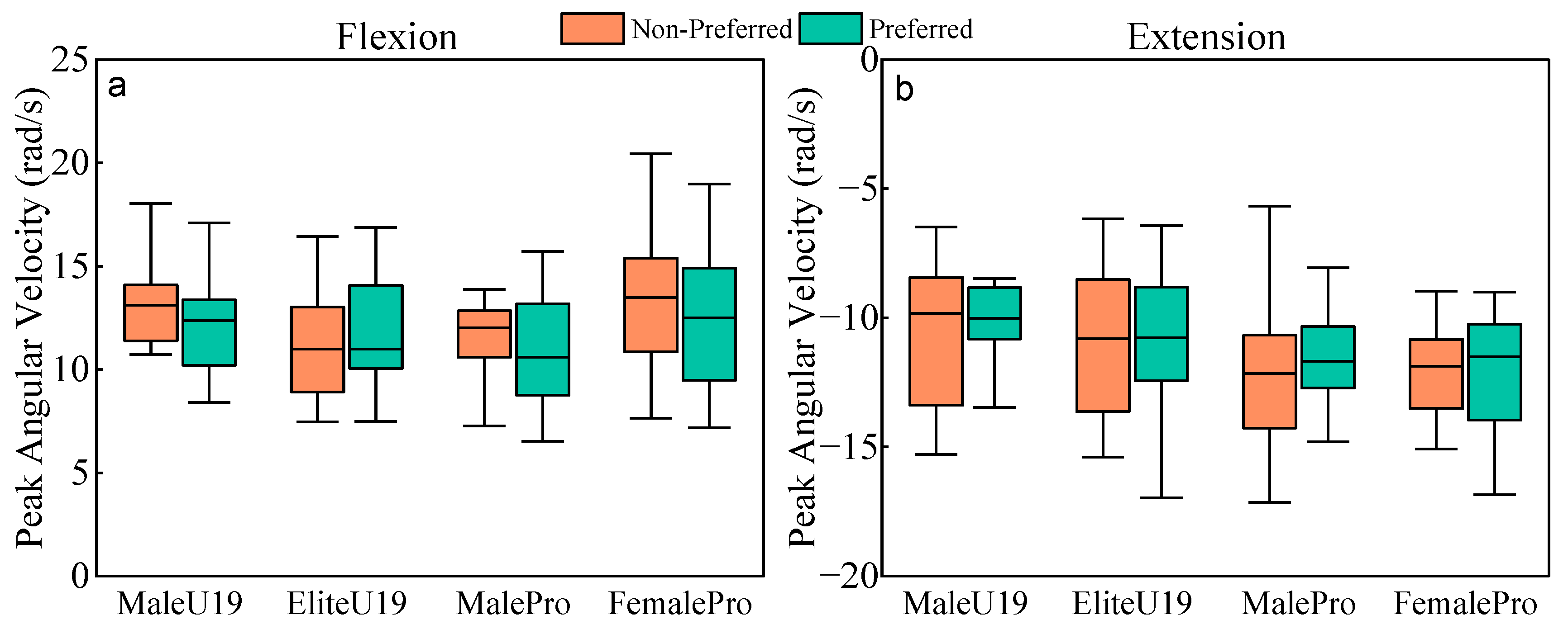
No significant effects of group or leg preference were found for peak knee flexion and extension angular velocities, nor for peak ankle dorsiflexion and plantarflexion angular velocities.
3.3. Impact of Lower Limb Joint Kinematics on Ball Velocity and Spin
All regression models were successfully fitted, and the results are shown in
Table 3 and
Table 4. For the preferred leg kicks, peak hip extension velocity (β = 0.230,
p = 0.034) and peak knee extension velocity (β = 0.193,
p = 0.047) had a positive impact on ball velocity, while hip adduction-abduction ROM (β = 0.235,
p = 0.047) and peak knee extension velocity (β = 1.057,
p = 0.028) had a positive impact on ball spin. However, knee flexion-extension ROM (β = −0.140,
p = 0.029) exhibited a negative impact on ball spin.
For the non-preferred leg kicks, knee flexion-extension ROM (β = 0.046, p = 0.020) was positively associated with ball velocity, whereas peak hip external rotation velocity (β = −0.151, p = 0.029) showed a negative association. Additionally, peak knee extension velocity (β = 1.640, p = 0.001) had a positive impact on ball spin, while knee flexion-extension ROM (β = −0.230, p = 0.003) had a negative impact on ball spin.
4. Discussion
This study aimed to examine the role of support leg kinematics in determining ball velocity and ball spin during instep kicking, considering different competitive levels and leg preference. Preferred and non-preferred limb kicks of 78 players from different competitive levels (female professional, male professional, male elite youth, and male non-elite youth) were analyzed using a motion capture system, and key kinematic parameters were extracted to assess their relationship with kicking performance. Regression analysis revealed that for preferred leg kicks, ball velocity increased with greater peak hip and knee extension angular velocities, while for non-preferred leg kicks, ball velocity decreased with increased peak hip external rotation angular velocity. Ball spin for both kicks increased with greater peak knee extension angular velocity and decreased with greater knee flexion-extension ROM, while specifically for preferred limb kicks, it also increased with an increase in hip adduction-abduction ROM. These findings highlight the critical yet often overlooked role of the support leg in optimizing kicking performance, and considering these movement patterns in the kicking motion should be incorporated while kicking for speed and accuracy.
4.1. The Role of Support Leg in Ball Velocity
Ball velocity increased with greater peak extension angular velocity of the support hip for preferred leg kicks. Following SLTG, the hip continued to extend, reaching its peak angular velocity prior to ball impact. Simultaneously, the kicking leg executed a forward-downward motion, characterized by hip flexion and knee extension [
13,
23,
28]. These findings align with Augustus et al., who conducted a technical refinement intervention [
23]. In their study, participants were instructed to increase both knee and hip extension in the support leg, with the objective of enhancing vertical displacement on the support side. This modification elevated pelvic positioning, allowing the kicking leg to initiate its downswing from a higher point [
23]. Consequently, the prolonged acceleration pathway of the kicking leg facilitated greater knee extension velocity prior to ball impact. Building upon this theoretical framework, this study extends these findings to professional-level players, across different competitive levels, in that an increased hip extension velocity in the support leg prior to ball impact contributes to enhanced ball velocity.
Ball velocity also increased with a higher peak extension angular velocity of the support knee for preferred leg kicks. Following toe-off, the extension angular velocity of the support knee rapidly reached its peak, then gradually decreased, maintaining a lower extension angular velocity and a weaker flexion moment prior to ground contact [
19,
22,
28]. At this stage, the primary function of the knee is to prepare for the absorption of impact forces upon landing. Currently, no studies have explicitly examined the relationship between peak knee extension angular velocity before ground contact and ball velocity. However, based on the observed kinematic patterns of the support knee, it is reasonable to hypothesize that for the preferred leg kicks, a higher knee extension angular velocity may facilitate a faster transition into an optimal support posture, ensuring greater stability and impact absorption upon ground contact. However, the findings also indicate that peak knee extension angular velocity has a positive effect on ball spin for both the preferred and non-preferred leg kicks, with this effect being more pronounced in the non-preferred leg kicks. This suggests that the peak knee extension angular velocity has an optimal range for both ball velocity and ball spin; thus, it can play a crucial role in kicking accuracy. Although no prior research has directly examined the relationship between peak knee extension angular velocity before ground contact and ball spin, existing studies have demonstrated that both the vastus medialis (VM) and vastus lateralis (VL) of the quadriceps, as well as the hamstrings, are actively engaged before landing [
29]. These muscle groups work in coordination to regulate knee extension, ensuring controlled ground contact. Consequently, excessively high knee extension velocity prior to landing may lead to overactivation of the knee extensors, disrupting the flexor-extensor coordination pattern, thereby increasing ball spin. This effect is particularly pronounced in less technically proficient players in the non-preferred leg kicks, indicating a possible area of improvement for their kicks.
A third finding of this study was that the ball velocity decreased with greater peak external rotation angular velocity of the support hip for non-preferred leg kicks. Given that players typically approach the ball at an angle, hip external rotation occurs in the support leg following kicking leg toe-off to adjust the foot placement angle prior to landing. Inoue et al. (2014) reported that the hip joint reaction force in the support leg induces a counter-clockwise rotation of the pelvis in the horizontal plane, a mechanism that plays a crucial role in pelvic stability [
22]. However, in the non-preferred leg kicks, an excessively high hip external rotation angular velocity before ground contact may compromise landing stability, leading to uncoordinated pelvic rotation. This instability may disrupt the acceleration pathway of the kicking leg, ultimately resulting in reduced ball velocity. Furthermore, the results of this study indicate that male professional players exhibited lower hip external rotation angular velocities in both the preferred and non-preferred leg kicks compared to the other three groups. This suggests that higher-level players tend to exert more precise control over hip external rotation in the support leg, thereby minimizing pelvic oscillation and optimizing energy transfer to the kicking leg. In contrast, lower-level players or those using their non-preferred leg kicks generally lack sufficient neuromuscular control over hip external rotation, which compromises pelvic stability and overall kicking performance. In addition, professional female players exhibited greater hip external rotation angular velocities in preferred leg kicks compared to male professionals and also showed significantly larger hip internal-external rotation ROM in non-preferred leg kicks. These differences may be partly attributed to anatomical characteristics, such as a wider pelvis and greater femoral neck-shaft angle, which can influence joint mobility and control strategies during dynamic tasks [
9]. Such findings highlight the need to consider anatomical and biomechanical factors when evaluating support leg function across sexes and may inform more individualized training interventions.
In practice, by using technical refinement, resistance training, and plyometric training to improve the strength of the hip and knee extensor muscles, the supporting leg can better resist the impact of landing and increase the body position before the ball impacts, which is beneficial for increasing ball velocity [
23,
30,
31,
32]. To improve rotational control, use of resisted exercises [
33], multi-planar drills [
34,
35], and technical drills targeting controlled rotation in both preferred and non-preferred kicks can be implemented [
36]. The findings support current training approaches that emphasize hip and knee extensor strength and neuromuscular control to improve power and single-leg stability during kicking [
37].
4.2. The Role of Support Leg in Ball Spin
Ball spin decreased with greater support knee flexion-extension ROM for both preferred and non-preferred leg kicks. Based on the previously defined ROM calculation method, the maximum knee angle occurred at kicking leg toe-off, while the minimum value was observed after SLTG [
15,
19,
28]. Therefore, the knee flexion-extension ROM primarily represents the knee joint’s adjustment capacity prior to SLTG. Previous studies have investigated the relationship between knee flexion-extension ROM during the support phase (from SLTG to ball impact) and ball velocity, reporting no significant correlation [
21]. Expanding upon these findings, this study further demonstrates that knee ROM before the support phase (i.e., pre-landing ROM) also does not affect ball velocity, but it has a negative effect on ball spin. This may be because a greater knee ROM before SLTG allows for more effective pre-landing adjustments, enhances support leg stability at ground contact, and facilitates a more efficient ball-foot impact during the instep kick, thus ultimately reducing ball spin. This effect was statistically significant in professional players (both male and female), where the preferred leg kicks exhibited greater knee flexion-extension ROM than the non-preferred leg kicks. This finding likely reflects an advantage of the preferred leg kicks in the support leg adjustment capacity, allowing for more precise control of support leg positioning and improved energy transfer efficiency within the kicking kinetic chain.
Ball spin increased with greater hip adduction-abduction ROM of the support leg for preferred leg kicks. The maximum angle occurred at ball impact, while the minimum was observed after SLTG [
13,
15]. Following ground contact, the support leg primarily undergoes hip adduction [
13,
22], and a greater hip adduction-abduction ROM indicates excessive hip adduction during the support phase, which may limit counterclockwise pelvic rotation and reduce the effectiveness of the proximal-to-distal force transmission chain, ultimately influencing the spin and stability of the ball upon impact.
Coaches can implement neuromuscular training to improve knee flexion-extension control, especially in the non-dominant leg [
37]. Exercises that enhance hip joint stability, promote greater support leg control, reduce excessive hip adduction, and optimize energy transfer throughout the kicking motion could help players improve their ball spin [
17,
38]. This will also help provide a more controlled and stable base for the kicking leg.
4.3. Limitations and Future Research Directions
Given the complexity of instep kicking, the use of discrete variables (e.g., peak angular velocity, ROM) limits the ability to fully capture the temporal dynamics of the support leg’s contribution. Since kicking is a highly coordinated motion involving sequential energy transfer, analyzing only peak values may overlook phase-specific interactions. In addition, individual approach strategies—such as run-up speed and angle—may introduce variability that affects support leg mechanics and overall kicking performance [
15,
16,
19,
20]. These factors were not controlled in this study to maintain ecological validity, but should be considered in future work. Subsequent research should incorporate time-series analysis and also quantify or control approach parameters to better understand their interaction with support leg kinematics and performance outcomes.
5. Conclusions
This study examined how support leg kinematics influence ball velocity and ball spin, considering competitive level and leg preference. The results revealed that greater knee and hip extension angular velocity contributed to higher ball velocity, while excessive hip external rotation angular velocity had a negative effect. Additionally, greater knee flexion-extension ROM was associated with reduced ball spin, whereas increased hip adduction-abduction ROM and higher peak knee extension angular velocity were linked to greater ball spin. These findings highlight the crucial role of the support leg in absorb impulse, stability, and spin regulation, offering insights for training programs aimed at improving kicking performance.
Coaches and therapists are encouraged to incorporate hip and knee extensor strengthening, neuromuscular training, and multi-directional stability exercises to optimize support leg function. These strategies may enhance both ball velocity and spin control by improving the quality of support during the kicking motion.
Importantly, the influence of the support leg on ball velocity appears to be indirect, primarily by providing a stable base that enables efficient kicking leg mechanics. Future research should further explore functional indicators of support leg stability, with the aim of developing quantifiable metrics to assess its contribution during kicking. Interventional and longitudinal studies are also needed to determine how targeted training may modulate support leg mechanics and improve kicking performance over time.


