Review

Diversity of Collagen Proteins and Their Biomedical Applications in Drug Delivery

1
School of Pharmaceutical Sciences and Food Engineering, Liaocheng University, Liaocheng 252000, China
2
State Key Laboratory of Macromolecular Drugs and Large-Scale Preparation, Liaocheng University, Liaocheng 252000, China
3
Shandong Provincial Key Laboratory of Applied Technology for Protein and Peptide Drugs, Liaocheng University, Liaocheng 252000, China
*
Author to whom correspondence should be addressed.
Appl. Sci. 2025, 15(12), 6472; https://doi.org/10.3390/app15126472
Submission received: 5 April 2025 / Revised: 31 May 2025 / Accepted: 4 June 2025 / Published: 9 June 2025

Abstract

:
Collagens and collagen-like proteins (CLPs) are the most abundant proteins distributed throughout the kingdom of organisms, including microorganisms, terrestrial animals, and marine animals. Due to their unique texture structure and prominent biocompatibility, collagens have been used as a crucial component of drug delivery systems. Herein, the composition and structure of collagens from different biological species were contrasted at first. Additionally, the biological function of collagen receptors was analyzed and summarized. The review focuses on the research progress of collagen as a biomaterial with excellent properties for delivering different drugs. As our understanding of the structure and function of collagen from different biological sources deepens, more new collagen materials and carriers with outstanding performance will continue to emerge and promote the application of collagen in the field of drug delivery.

1. Introduction

The physical integrity of cells is greatly impacted by the extracellular matrix (ECM), which involves proliferation, differentiation, migration, and adhesion. Collagen, which belongs to the fibrous protein family, is the oldest and most abundant ECM protein in all connective tissues such as skin, ligaments, tendons, muscles, blood vessels, bone, cartilage, or cornea. It has a molecular weight of around 300 kDa, a diameter of about 1.4 nm, and an approximate length of 280 nm [1]. Up to now, totals of 29 distinct types of collagens, named with Roman number I-XXIX in chronological order of discovery, have been identified which are coded by at least 45 different genes [2]. The classification of collagens is based on their domain and structural complexity. Each collagen contains its own unique alpha chain with its own domain structure. The most abundant among the 29 isoforms of collagen is type I (principally in the skin, tendons, muscles, and bone), type II (in cartilage), type III (often found alongside type I), and type IV (at the basement membrane). Collagen protein is composed of triple-helical polypeptide chains and contains the repeating sequence (Gly-X-Y)n with Pro often located at positions X and Y. Such triple-helical structures are important for the biological function of collagen, such as the elasticity and waterproofness of the skin, the structural stability of connective tissue cells, wound healing, and bone repair.
Generally, collagens prepared from different animals such as bovine, porcine, equine, and marine organisms have been widely used as biomaterials for hemostasis, stomatology, drug delivery, plastic surgery, bone defect, and tissue engineering, which is also named collagen-based biomaterials (CBBs). In addition, although collagen or collagen peptides have been produced recombinantly by transgenic organisms, such as E. coli, yeast, and mammalian cells, their wide application is hindered by their lower yields and higher cost. Furthermore, the heterologous expression cannot achieve the post-translational modifications of glycosylation and hydroxylation on proper Pro and Lys residues, as well as heterotrimeric compositions. Therefore, it is obvious that natural collagens will remain the major source of CBBs in the future [3]. In contrast to terrestrial mammalian collagens, marine collagens have obvious advantages concerning safe, easy obtain, no adverse inflammatory reactions, and no immunologic responses. As a result, marine collagens are a promising source for pharmaceutical applications. Herein, the primary focus of this article is on the wide variety of collagen found in prokaryotes, eukaryotes, marine, and terrestrial organisms, as well as its structural and functional diversification, and the genetic disorders that are connected to collagen. These diseases are closely linked to the signaling pathways involved in collagen receptors. The article further focuses on the use of collagen in drug delivery, including small molecule chemotherapeutics, protein and peptide drugs, therapeutic cellular and gene-based drugs, demonstrating the wide range of uses of collagen in the treatment of diseases.

2. Diversity of Collagens

Over the past few decades, the structure and biophysical properties of metazoan collagen have been extensively investigated. This scientific situation gives us an illusion that collagen only exists in mammalian organisms. However, more and more studies have suggested that there exists a collagen or CLP containing large regions of Gly-Xaa-Yaa (GXY) repeats in prokaryotic cells, even in viruses. The abundance of collagen and collagenous structures throughout the biological kingdom indicates their importance in biological viability. The molecular evolution of collagen is outlined below.

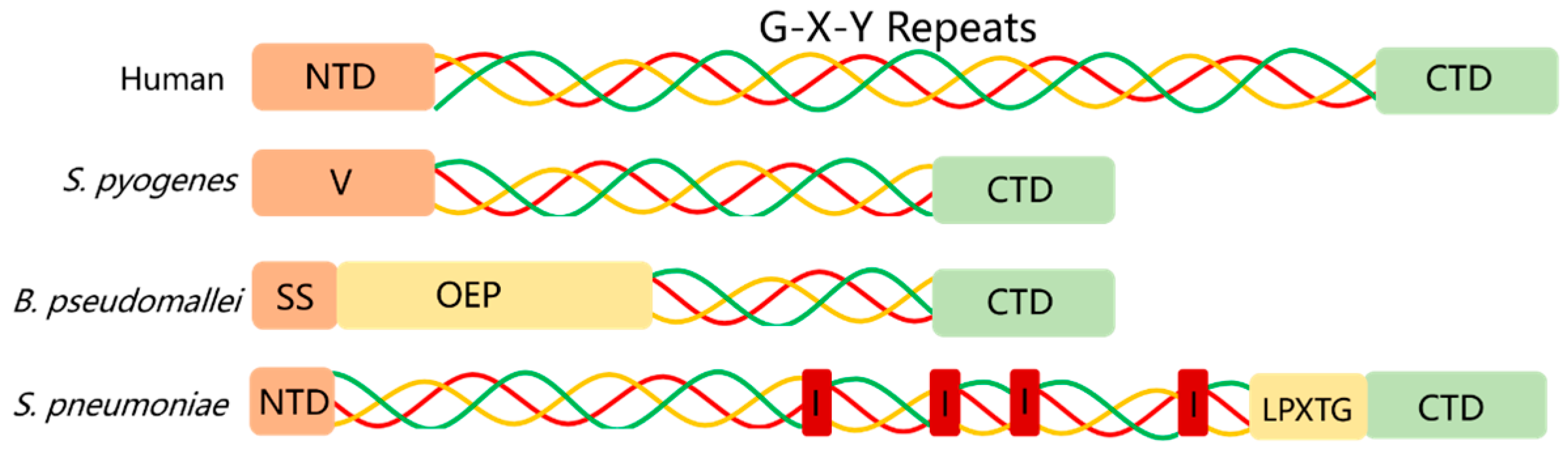
2.1. Prokaryotic Collagen

The earliest report on bacterial collagen was that a collagen-like domain containing several units of (Gly-Xaa-Pro)6 was identified in the amino acid sequence of pullulanase from Klebsiella pneumoniae [4]. At the turn of the twenty-first century, the first true prokaryotic collagens, forming the triple helices, were discovered in the Gram-positive bacteria Streptococcus pyogenes and named Streptococcal collagen-like protein 1 and 2 (Scl1 and Scl2 or SclA and SclB) [5,6] (Figure 1). Since then, different non-animal CLPs have been found in bacteria, such as Mcl1 protein from M. anisopliae [7], Bc1 proteins from B. anthracis [8], Pc1 from S. pneumoniae [9], Lc1 from L. pneumophila [10], Buc1 from Burkholderia spp. [11], and CLPs from Bacillus amyloliquefaciens [12]. In addition, there are some pieces of evidence suggesting that virophages have collagen-modifying enzymes and CLPs. For example, the genes coding glycosyltransferase were discovered in the Paramecium bursaria chlorella virus and the genome sequences of viruses such as lymphocystis disease virus, shrimp white spot syndrome virus, the Saimiriine herpesvirus 2, and bacteriophage T4 (Figure 1) suggested that these viruses possess their own CLPs [13,14,15,16]. The discovery of prokaryotic collagen has become a growing field with intense studies. As for the origins of collagen-like sequences in prokaryotes, a possible hypothesis is that the convergent evolution led to the rise in collagen-like repeats by spontaneous mutations, transposition of repeat sequences, and motif organization [17]. In addition, the genetic transfer facilitated by viruses or bacteriophages may be the cause of the diffusion of collagenous sequences between bacteria [18].
To comprehend the genetic distribution of bacteria CLPs, the Yeo group gathered 60,789 CLP sequences to analyze their distribution and characteristics. The results indicated that the vast majority of CLPs, approximately 98.9%, were classified into four phyla: Firmicutes (68.1%), Proteobacteria (22.2%), Actinobacteria (5.3%), and Bacteroides (3.2%). The number of GXY repeats, which is a typical characteristic of CLPs, was significantly different among the four main bacterial phyla. The most GXY repeats were present in CLPs from Bacillus subtilis, reaching 1034 [19].
Three characteristic domains are present in the prokaryotic CLPs, just like in eukaryotic proteins. For example, the Scl proteins have an N-terminal variable domain, a typical triple helix Gly-X-Y repeat domain, and a C-terminal non-collagenous domain required for cell-wall anchoring. The N-terminal variable domain differs among the CLPs (Figure 2). Additionally, the number of Gly-X-Y repeats varies [19,20,21]. In the G-X-Y repeats, prokaryotic collagen has a Y residue that is predominantly threonine, while eukaryotic collagen has a Y residue that is either proline or hydroxyproline. Hydroxylation of proline to hydroxyproline and glycosylation and cleavage of collagen are absent in most prokaryotic organisms [22].

2.2. Marine Collagen

Marine organisms consist of both invertebrates (such as sponges, jellyfish, cuttlefish, prawns, and other similar species) and vertebrates (such as fish and marine mammals). These multicellular eukaryotes are significant sources of collagen. Marine collagens exhibit greater bioavailability and absorption efficiency in the body compared to terrestrial animal collagens, such as bovine and porcine collagen. Among the 29 types of collagens that have been discovered, type I collagen is the most abundant type of collagen and can be found in fish scales, skin, and bone (Table 1), whilst type II collagen can be found in the fishbone cartilage. In addition, invertebrates, the vast majority of marine organisms, contain collagen types IV, XV, XVIII, and some fibrillar collagens, as well as some fibril-associated collagens with interrupted triple helices (FACITS). The evolutionarily most ancient collagen among these is type IV collagen [23,24].
Glycine is the most abundant amino acid in marine collagen, while proline and hydroxyproline are less abundant than in mammalian collagen. However, there are more serine and threonine residues in the collagens of cold-water fish. These variations in amino acid composition are generally responsible for the properties of collagen, such as rigidity, temperature stability, and denaturation temperature [25]. For instance, fish collagen has a low concentration of glycine, proline, and hydroxyproline, which results in a low denaturing temperature of marine collagen [26], but it has a higher concentration of serine and threonine amino acid than mammalian collagen [27]. In general, a high level of proline and hydroxyproline is necessary for collagen stability [28]. On the other hand, collagen from warm water fish has a similar amino acid composition, rheological properties, and thermostability to those of mammalian collagen. All in all, the denaturation temperature of marine collagen is lower than that of mammalian, and this is due to the lower extent of hydroxylation of proline and lysine residues in the marine collagen [29,30].
Table 1. Ten cases ranked by high collagen content of type I collagen obtained from different marine organisms.
OrganismsSpeciesTissue SourcesCollagen Content (%)References
Sea eelMuraenesox cinereusSwim bladder93.7[31]
MegalonibeaMegalonibea fuscaSwim bladder84.8[32]
Sea cucumberHolothuria cinerascensBody wall72.2[33]
TotoabaTotoaba macdonaldiSwim bladder65[34]
Yellowfin tunaThunnus albacaresSkin61.26[35]
Asian sea bassLates calcariferSkin59.31[35]
MackerelScomberomorous niphoniusSkin58.62[36]
Seer fishScomberomorus commersonSkin58.21[35]
Grass carpCtenopharyngodon idellaSwim bladder38.9[37]
SturgeonAcipenser baericartilage28.8[38]

2.3. Mammalian Collagen

In the mammalian kingdom, collagens are a superfamily of triple helical proteins consisting of 29 different types with biological functions concerning the construction of cell scaffold, the interaction between cells and fibrils, and the immune response, among others. These vertebrate collagens are classified into different supra structures including fibril, FACIT, macrofibril, anchoring fibrils, transmembrane collagen, basement membrane collagen, and short chains collagen according to their functions (Table 2). For instance, types I, II, III, V, XI, XXIV, and XXVII belong to fibrils in which major fibrils are types I, II, and III. Type I collagen is the most common type of collagen, accounting for around ninety percent of the total collagen content [39]. Type II collagen, composed of unique triple helical chains with prominent advantages of flexibility, resilience, and mechanical stress, is mainly distributed in cartilaginous tissues and is generally used as the repair material for orthopedics and joint injury [40]. Type III collagen is in charge of creating the framework that supports the structural integrity and durability of skin, muscles, organs, and arteries. It is also known as reticular collagen. This reticular collagen can aid in wound healing by providing a three-dimensional structure for new tissue growth and promoting the regeneration process [41]. Collagen type III is unique in its ability to enhance skin elasticity and tautness while maintaining the skin’s optimal health. Type IV collagen is composed of triple same alpha peptide chains and is the main component of the cell basement membrane [42].
Based on the basic biological principle of congruence between structure and function, the triple helix of collagen is an ancient and conserved structure between animals that exists ubiquitously in the ECMs. Glycine residues are essential for maintaining the structural integrity of the triple helix. The mutation of glycine residue resulted in defective collagen molecules causing faulty fibrils, networks, and other assemblies. Therefore, there are dozens of genetic diseases in humans that are related to glycine mutation (Table 3).
The triple helix, which is a distinguishing feature of various collagen assemblies, guarantees tissue strength and influences cell behavior. This unique structure is analogous to the double helix of DNA. These features present the biological importance of collagen. In particular, collagen type IV was considered to be a representative of an evolutionary transition molecule from unicellular prokaryotic organisms to multicellular eukaryotic animals [23]. Despite the extensive research on the structure and function of triple helices, there are still many unanswered questions. For example, very little is known about the function of various prokaryotic collagens and the mechanism by which the triple helix structure affects cell function.

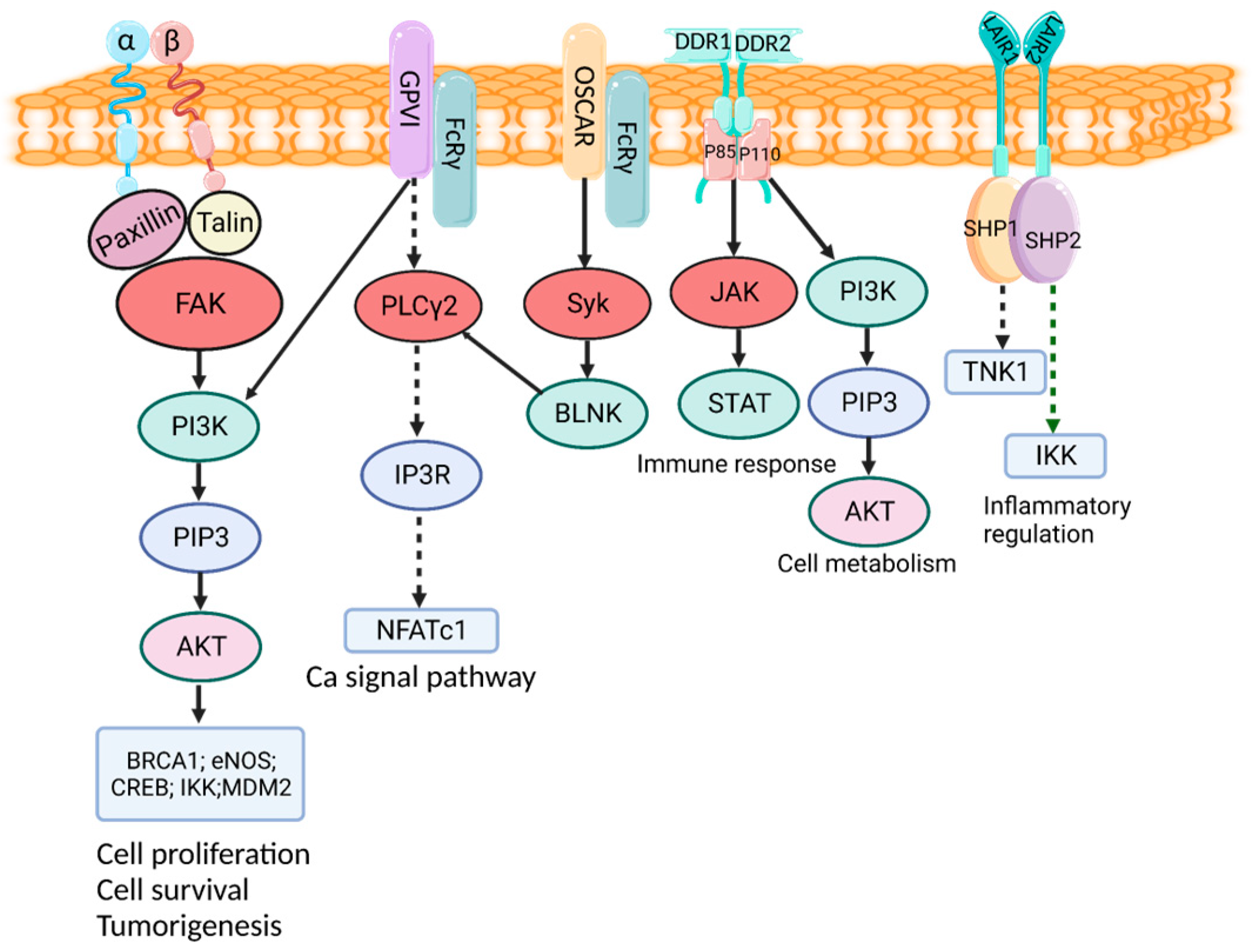
3. Cell Receptor and Function of Collagens

Collagen, as an abundant ECM protein, plays a significant biological function by interacting with cells and regulating both intrinsic and extrinsic signaling pathways. These signaling pathways mainly contain the receptors of Integrins, Discoidin domain receptor (DDR), Glycoprotein V1 (GPV1), Osteoclast-associated receptor (OSCAR), Leukocyte-associated immunoglobulin-like receptor 1 (LAIR-1), and Mannose receptor C type 2 (MRC2), all of which are involved in the regeneration mechanism and healing process of bone, cartilage, vasculature, lungs, nerve, dental tissue, and skin. Hence, the crucial receptors were necessary for the implementation of the crucial biological function of collagen. The cell receptor and function of collagen will be systematically described here (Figure 3).

3.1. Integrin Receptor

Integrins, as well-known cell surface heterodimeric receptors, are broadly distributed to almost all cells in tissues and organs such as the skin, muscles, kidneys, lungs, eyes, heart, gut, and blood vessels. [70]. Up until now, a total of 4 types of collagen-binding integrins, including α1β1, α2β1, α10β1, and α11β1, have been identified from 24 different types of integrins in human kinds, all of which play fundamental roles in regulating cell survival, migration, signal, and differentiation. These integrins transfer the signals through cells in a bidirectional way [71]. The α1β1 receptor was first discovered and is commonly expressed in many activated organs, such as lymphocytes, the liver, and the heart. Collagen binding to the integrin α1β1 receptor delivered the ability to regulate the proliferation of cells, MMP expression, and collagen synthesis [72]. The α2β1 integrin, also termed CD49b, is an extracellular matrix receptor and is mainly distributed in bone, skin, and other internal organs. Integrin α2β1 has a preferential affinity to fibrillar collagens I–III, V, and XI. The collagen motif GFOGER was responsible for the interaction between collagen and the α2β1 integrin [73]. Integrin α10β1 is a primary receptor of collagen type II, which is mostly located in the tissue of cartilage [74]. Integrin α11β1 binds with fibrillar collagen such as collagen I and XIII and is expressed in tissues including embryos, muscle, and bone [75]. Except for the four integrins mentioned above, integrin α6β4 and α5β3 have been identified to bind with collagen XVII and XIX [76]. As the receptor for collagen, the integrin signaling pathways are involved in cell proliferation, survival, and migration. In detail, the extracellular integrin/collagen conjugator could trigger the cellular signal transduction pathways, including the Akt/PI3K, protein kinase C (PKC) cascades, and MAP kinase pathways through adapter proteins such as Src, focal adhesion kinase (FAK), and integrin-linked kinase (ILK) [77].

3.2. DDR

DDR is also identified as a receptor tyrosine kinase that plays a significant role in the proliferation of cells via binding with collagens such as collagen types I–V [78]. DDR1 and DDR2 are the binding targets of collagen that provide the exact binding sites, including the juxtamembrane domain, N-terminus, and tyrosine kinase domain [79]. Furthermore, the cytoplasmic domain of DDRs interacts with the adapter proteins ShcA and Nck2 to affect the signal transduction pathway related to proliferation and survival such as the JNK/MAPK- and PI3K/Akt-signaling pathways [80]. This research has demonstrated that DDR1 and DDR2 presented overexpression in multiple cancers, such as non-small-cell lung carcinomas, ovarian tumors, breast cancer, and Hodgkin’s lymphoma [81,82]. The interaction of collagen with DDR1 triggers tyrosine phosphorylation and kinase activation of DDR1, which further initiates multiple downstream signaling pathways, including Src kinase activation for proliferation and cell migration [83].

3.3. GPVI Receptor

The GPVI receptor, belonging to the immunoglobulin-based transmembrane stimulatory receptor, is generally expressed in megakaryocytes and platelets and interacts with Gly-Pro-Hyp amino acid residues in collagen. The binding of collagen and GPVI plays an important role in platelet signaling through the interaction with PPARγ and adapter protein Syk [84]. This procedure results in the phosphorylation of the linker of T cells and consequently triggers the downstream mediators PLCλ, PI3K, and Akt. The synthesis of thrombin, serotonin, oxytocin, TXA2, and platelet-activating factors are guided by these signaling cascades, which activate platelets and enhance the entire response [85].

3.4. OSCAR

The osteoclast-associated receptor belongs to the immunoglobulin (Ig) superfamily and is expressed in various types of myeloid cells. OSCAR, a receptor that binds to the motifs of collagen types I–III, is involved in the development, maintenance, and repair of bones [86]. To be specific, OSCAR acts as a positive regulator inducing the formation of osteoclasts, which promote bone resorption through the STAT3 pathway. Collagen activation improves the interaction of OSCAR with the Fc receptor γ-chain (FcRγ), and the complex of OSCAR-FcRγ further mediates calcineurin release and CAMK IV activation to support osteoclast activation and maturation. All in all, the process of the development, maintenance, and repair of bones was regulated by the accumulation and transendothelial migration of osteoclast precursor cells, which could express RANKL and collagen I/III. Meanwhile, the conjugate of collagen and OSCAR induces the signaling cascade of DAP12/FcRγ-Syk-PLCγ, which influences calcium signaling and NFATc1 [87]. From the above information, it can be deducted that the disorder of the structure of collagen (especially type I and type III collagen) and its interaction with OSCAR may lead to osteoclast dysfunction, thereby causing skeletal system diseases such as osteoporosis.

3.5. LAIR1 Receptor

LAIR1, a member of the LRC family, is responsible for acting as an inhibitory receptor and regulating the OSCAR effects in reverse. Collagen-bound LAIR-1 stars biological signals to maintain immune tolerance by reducing phosphorylation of STAT1/4, IFN-γ, and TNF-αproduction which leads to downregulate the activity of natural killer (NK) cells [88]. Therefore, collagen has a significant impact on the decrease in cytotoxicity and the increase in immune tolerance of FNK cells. The surface phosphorylation of the collagen/LAIR complex was guided by Src family kinases and SHP-1/2 phosphatases through LAIR1 tyrosine residues and the ITIM SH2 domain [89].

3.6. MRC2 Receptor

The membrane protein receptor MRC2 (CD280) belongs to the mannose family and acts as an endocytic receptor for collagen. The interaction of CD280 and collagen leads to the enzymatic decomposition of collagen by the plasminogen activator system. The main function of lysosomal enzymes during the endocytosis process is to degrade collagen [90].
Based on the above studies, the occurrences of most diseases are directly or indirectly regulated by at least one of the collagen receptors, including Integrins, DDRs, GPV1, OSCAR, LAIR, and MRC2. Meanwhile, the collagen receptors also regulate the regeneration of tissues and the proliferation of cancer cells [91].

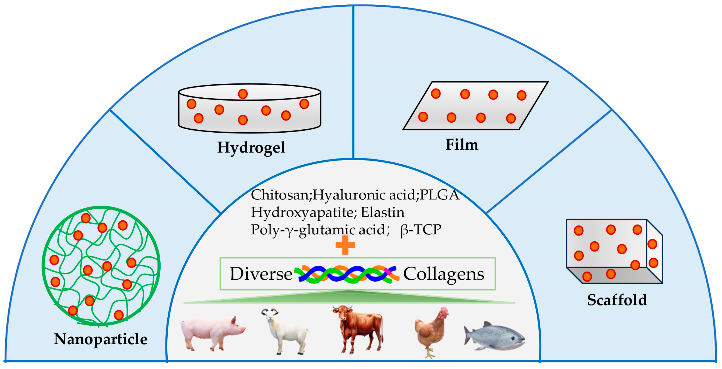
4. Collagen Application for Drug Delivery

Due to the prominent advantages of low immunogenicity, excellent biocompatibility, and biodegradability, collagen proteins have been widely used as biological materials for the delivery of small-molecule drugs, protein and peptide drugs, therapeutic cells, and genetic drugs.

4.1. Delivery of Small Molecule Drug

Collagen has a stable triple-helix structure that can form a natural network structure. These structures can realize the effective loading and slow release of small molecule drugs, which is an ideal material for drug delivery. As shown in Figure 4, the collagen hydrogel was developed to deliver tetrahydrocurcumin (THC) [92]. The nanoparticles were prepared to carry doxorubicin hydrochloride (DOX) [93]. A chitosan–collagen film was made to release local anesthetics (LA) [94]. A scaffold made of type I collagen was used to alendronate (Aln) [95].
THC is obtained by hydrogenating curcumin and has a significant impact on the pharmaceutical and cosmetic industries. However, THC possesses low solubility in water, poor absorption, and rapid elimination in the body. To improve these disadvantages of THC, a novel hydrogel was prepared with porcine skin collagen using 3D printing technique to treat wound healing [92].
In cancer chemotherapy, DOX is a well-known anticancer agent. To realize the targeted and controlled release of DOX, the nanoparticles based on chitosan and collagen peptide (CP) were prepared, which are stable under physiological conditions and have the capability of pH-controlled release. The formation of CPCN nanoparticles is attributed to hydrogen bonding and electrostatic interactions. The results of the anticancer test showed that DOX-loaded NPs have significant anti-proliferative characteristics against HeLa cells [93].
LAs are commonly utilized in clinical settings to alleviate both acute and chronic pain. Prolonging LAs remains a difficult task. A research group has initiated the development of a chitosan–collagen film for a controlled delivery of combinations of local anesthetics, including lidocaine, benzocaine, and tetracaine. The film was composed of low molecular weight chitosan and calf skin collagen and could hold and release a mixture of the local anesthetics in a proper ratio within 24 h [94].
Aln, a second-generation bisphosphonate, is well-known as an anti-osteoporosis drug based on its dual functions of restraining osteoclasts and stimulating osteoblasts. It is an engineered implantable scaffold based on type I collagen that can sustainably release Aln to repair osteoporotic bone defects and resist bone loss in osteoporosis. The release kinetics showed that Aln was released at an average rate of 2.99 μg/d in the initial 8 days and could sustainably be released for 1 month [95].
Silymarin, a bioactive compound extracted from plant milk thistle, has been widely regarded as an effective treatment for liver diseases such as alcoholic fatty liver and viral hepatitis. In addition, it possesses the properties of neuroprotection, antioxidation, and tissue regeneration. Herein, the micellar nanoparticles based on ovine collagen were prepared and applied to deliver silymarin to treat ischemic brain injury. The animal test demonstrated that nanosilymarin had a marked improvement in neuroprotection when compared to the silymarin-treated control [96].
Vancomycin, belonging to the glycopeptide antibiotic family, is used to treat a variety of infections caused by pathogenic bacteria. A system for delivering antibiotics has been created, combining elastin-like peptide and collagen-like peptide nanovesicles (ECnVs) to fight pathogenic bacteria such as Staphylococcus aureus (MRSA). Chemical synthesis was used to create collagen-like peptide in this study. This delivery system exploited the synergy effects of peptide nanocarriers and their interactions with collagen-based scaffolds [97].
To reduce the infectious risk caused by the guided bone regeneration (GBR) material, a composite restoration material made of oxidized hyaluronic acid (HA)/type I collagen integrated with tricalcium phosphate (β-TCP) was applied to cure bone regeneration. The composite had a prominent mechanical strength and biocompatibility and guided the sustainable release of tetracycline for 5 days [98].
The advantages of injectable and stimuli-responsive biomaterials for diagnostic imaging and drug delivery have made them a hot topic due to their biocompatibility and responsiveness. Herein, the Lim group developed a novel injectable and photoresponsive composite hydrogel composed of the anticancer drug Dox, imaging contrast agent MnFe2O4, type I collagen, and the multifaceted anionic polypeptide poly-γ-glutamic acid (γ-PGA). The results suggested that the novel injectable and near-infrared-responsive collagen/γ-PGA hydrogels could be used as a theranostic platform after the loading of various molecular imaging probes and therapeutic components [99].
Heterotopic ossification (HO) is a common bone syndrome in clinics and can severely damage the structure of bone tissue. The mTOR signal pathway, which was inhibited by rapamycin, may be responsible for the pathogenesis of HO. To suppress HO progression, a rapamycin (RAPA) delivery system was developed using collagen hybrid peptide (CHP) to modify the surface of poly(lactic-co-glycolic acid) (PLGA) nanoparticles. The results indicated that the CHP-PLGA-RAPA nanoparticles showed excellent pathological collagen affinity, sustained-release ability, and bioactivity, and were necessary to deliver RAPA to the injured sites and avoid disturbing the normal tendon [100].
The main component of ECM is collagen, which has a typical triple-helix structure. The thermoresponsive nanoscale vesicles were assembled from an elastin-b-collagen-like peptide (ELP-CLP) prepared by type II collagen from chicken sternal, and which were observed to dissociate at elevated temperatures to achieve the sustained release of the drug via heating. Continuous release of an encapsulated model drug was accomplished over the course of three weeks [101].

4.2. Delivery of Protein and Peptide Drug

Bone morphogenetic protein 2 (BMP2) is a crucial osteogenic factor that promotes bone formation by stimulating osteoblast differentiation. A bone repair biomaterial (HC-COL-BMP-2) was developed by the tilapia skin collagen crosslinked with heparin to specifically bind to BMP-2. The prepared HC-COL has no cytotoxicity, shows enough thermal stability, and improves the binding of BMP-2 to cure bone repair by inducing MC3T3-E1 cells with higher activity [102].
Extracellular vesicles (EVs) have received increasing attention in various areas of regenerative medicine over the past decade. Royal jelly EVs (RJ EVs) have been shown to have beneficial wound-healing properties by promoting the migration of mesenchymal stem cells and preventing the formation of pathogen biofilms [103]. Herein, RJ EVs and fibroblasts were integrated into type I collagen gel to achieve sustainable release of RJ EVs. The continuous delivery matrix made of 2 mg/mL type I collagen showed a significant potential for wound-healing therapies [104].
The treatment of bone defects like osteoporosis, arthritis, and fractures can be achieved through the use of both the bioactive molecule BMP-2 and the chemical agent Aln, which are both approved clinical drugs. Since the studies had verified that the combined application of BMP-2 and Aln for bone treatment had no additive effects on bone regeneration, the Yoon group developed a collagen–hydroxyapatite scaffold-loading BMP-2 protein and micromolecular Aln to enhance bone regeneration by releasing them in a sequential manner in the rat bone defect model. Rat tail-derived collagen type I is the source of collagen composition. The delivery system indicated an initial release of BMP-2 for a few days, followed by the sequential release of Aln after two weeks [105].
The preferred healing protein for bone defects is bone morphogenetic protein-2 (BMP-2), but BMP-7 is also a common substitute for bone grafts. In this research, the bovine type I collagen was used as the crucial composition to establish a collagen–hydroxyapatite (CHA) scaffold delivery system to compare the effect of BMP-2 homodimer and BMP-2/7 heterodimer on the osteoinductive’s potential. The CHA scaffold delivered both protein dimers for at least 20 days, resulting in decreased doses of physiological growth factors for bone fracture healing [106].
Inducing angiogenesis in ischemic tissues has a positive impact on treating myocardial infarction (MI) and stroke. In a study, porcine collagen type I was blended with alginate to form the hydrogel microsphere with angiogenic factor FGF-2. The microsphere shows complete biocompatibility and no adverse reactions when injected into mice. Furthermore, it has demonstrated the robust induction of angiogenesis in zebrafish embryos [107].
Meniscus tears are a common knee injury that further leads to arthritis. The reconstruction of the meniscus using artificial materials is an effective measure. A novel collagen nanofiber scaffold prepared by acid-soluble bovine collagen type I and harboring heparin was fabricated by electrospinning and could sustainably release three factors: PDGF-BB, TGF-β1, and CTGF. These factors integrated into collagen nanofibers resulted in their sustained release, which increased synovial cell viability, proliferation, and migration, and enhanced the regeneration of meniscal tissue [108].
The development of next-generation antibody drugs has become more attractive with the advent of nanoantibody technology. To deliver the nanoantibody to the targeted cancer, the hydrogels consisting of the cross-linking of tilapia skin collagen and chitosan were developed to deliver and release nanoantibodies 2D5 and KPU for the treatment of cancer [109].
Extracellular beta-amyloid (Aβ) plaques or intraneuronal tau tangles in the brain are the cause of Alzheimer’s disease (AD). An effective therapeutic strategy aims to prevent or clear these Aβ plaques by degrading the enzyme neprilysin. However, the blood–brain barrier is the main obstacle in delivering bioactive neprilysin into the brain. A study showed that the intranasal delivery of neprilysin loaded with collagen was very potent in clearing plaques, especially in the middle and caudal parts of the cortex. Bovine collagen type I and 4S-StarPEG were used to prepare the useful vehicles [110]

4.3. Delivery of Therapeutic Cells

In clinical practice, autologous or allogeneic cell therapy has become a promising therapy. A flexible and controllable solution for treating patients with severe cutaneous defects has been developed through the development of novel keratinocyte cell-laden microcarriers prepared with biodegradable collagen [111].
To enhance cell survival in the regeneration of articular cartilage by applying stem cell therapy, the Zhang group developed an injectable collagen-based nanocomposite hydrogel with self-healing properties for delivering mesenchymal stem cells (MSCs) to improve cartilage regeneration. Its shear-thinning capability comes from incorporating cellulose nanocrystals (CNCs), while its self-healing and stress-relaxation behaviors were achieved by the dynamic Schiff base bonds between the aldehyde groups on a-CNCs and amine groups on collagen without any additional cross-linking agents [112]. Compared to the above injectable collagen-based hydrogels, a hydrogel based on type II collagen and activated chondroitin sulfate was fabricated without the addition of any catalysts or crosslinking agents. Additionally, chondrocytes were encapsulated in these hydrogels, and the hydrogels provided a favorable growth environment for cells, resulting in increased survival, better proliferation, and more robust morphology [113].
The noninvasive injection of encapsulated cell therapy (ECT) has demonstrated obvious advantages of its treatment for retinal diseases. Despite this, the therapy was still confronted with a challenge of unsteady performance and biosafety issues. A new ECT hydrogel that uses injectable composite alginate-collagen (CAC) was created to ensure safer drug delivery. The CAC ECT system presented the advantages of mechanical stability, good encapsulation power, cell viability support, multiplexed glial-cell-derived neurotrophic factor (GDNF) dosage, and compatibility with different cell types (HEK293 and ARPE-19) without immunosuppressant. These features make the system an attractive, safe, and well-controlled platform for treating various eye diseases [114]. Similarly, the olfactory ectomesenchymal stem cells (OE-MSC) were encapsulated in the type I collagen hydrogel system to improve bone healing [115].
Neural stem cells (NSC) have been identified as an attractive potential therapy for treating defective neural tissues, such as the injured spinal cord. A study was conducted to investigate the recovery of the injured spinal cord through the injection of silk-collagen protein hydrogels. The novel hydrogels can protect NSC from the stress of injection and the severe inflammatory environment. The study highlights how silk-collagen hydrogels can help NSC differentiate and function, which aids in the recovery of injured spinal cord [116]. In another study, porous collagen–glycosaminoglycan scaffolds were created to administer embryonic neural stem cells for the treatment of spinal cord injury (SCI) [117]. Except for cell delivery, a novel injectable in situ-forming hydrogel (HG) based on alginate and rat tail type I collagen is developed and used as a cardiac vehicle to deliver the EVs from human adipose tissue-derived stem cells (hADSCs) for treating myocardial infarction. The HG increased the retention of the EVs for at least 7 days after intramyocardial administration [118].
In clinical practice of translation of tissue-engineered scaffolds, immune cell macrophages exhibit a significant immune response that influences the healing process depending on their particular subtypes. To effectively regulate the immune response of macrophages, a bovine type I collagen biomaterial was developed and crosslinked with genipin. The results indicated that the biotextiles improved the transformation of macrophages to subtype M2, which reduced fibrous capsule thickness and minimized the formation of granulation tissue [119].

4.4. Delivery of Gene Drug

Gene therapies have been a potential measure for the treatment of genetic and infectious diseases. However, clinical translation has been inhibited by low stability and limited transfection efficiency. The Sullivan team engineered a type I bovine collagen scaffold that has polyplexes bound to the collagen-mimetic peptide (CMP) to boost DNA stability by up to 400%. This finding shows that CMP modification has a significant effect on gene retention, altered release kinetics, improved serum stability, and improved gene activity in vivo [120]. In a similar fashion, a complex electrospun mat was constructed that uses different volumes of gelatin, type I collagen, and polyethylene glycol to deliver genes. These mats could effectively transport the GFP plasmid DNA and BMP-2 protein to the target cells of human myoblast (C2C12) and mouse osteoblast cells (MC3T3) [121].
Hypertrophic scarring, which is not expected, is a result of overexpression of TGF-β1 during cutaneous wound healing. To attenuate the overexpression of (TGF-β1) and fibrotic effects, a biphasic 3D collagen was developed to deliver the two plasmid DNAs encoding interleukin 10 (IL-10) and decorin (DCN) for the treatment of fibrotic diseases via suppressing the expressions of genes associated with fibrogenesis. The findings could lead to the development of a non-viral gene therapy platform that can deliver multiple genes to the diseased tissue in a wider range of tissue fibrosis-related maladies [122]. Another similar example, the pDNA encoding for IL-1 receptor antagonist (IL-1Ra) was delivered by a collagen–hydroxyapatite scaffold to mitigate the negative inflammatory effects of IL-1β and further improve the new bone formation [123].
RNA transcript therapy (RTT), as a new and safe alternative to gene therapy, has gained more attention. However, the applications of RTT therapies have been impeded by the low stability of RNA in comparison to DNA. A study reported that the vacuum-dried RNA-loaded collagen sponges, termed transcript-activated matrices (TAMs), can serve as depots for sustained delivery of RNA and sustainably produce protein for up to six days. Meanwhile, TAMs were stable at room temperature for at least 6 months. To summarize, TAMs represent a viable tool for bone regeneration and may also have other applications in regenerative medicine and tissue engineering [124].

5. Conclusions and Perspectives

Collagen that has been conserved through evolution is found in organisms ranging from unicellular prokaryotes to multicellular eukaryotes, and even in phages and animal viruses. Furthermore, collagen is also one of the most abundant proteins in ECMs of mammalian animals and plays an important role in maintaining the biological and structural integrity of tissue as the fundamental structural component. The unique triple helical structure based on GXY repeats is what allows collagen to have the above features. Although proteins with the structural characteristics of triple helix are present in less evolved prokaryotes, most of them are not able to form fibers as collagen does in eukaryotes due to their low number of GXY repeats. Therefore, the collagens found in prokaryotes are commonly known as CLPs. Moreover, CLPs present in prokaryotes are devoid of post-translational modifications, which include hydroxylation, glycosylation, and cleavage.
Type I collagen is the most abundant type among marine organisms and can be found in fish scales, skin, and bone, which are key sources of marine collagen. Compared to collagen from terrestrial animals, marine collagen has lower content of proline and hydroxyproline, which leads to low denaturing temperature and poor temperature stability.
The research focus on collagen has been mainly terrestrial mammalian collagen for the past few decades, and it has been very detailed and comprehensive. The diverse supra structures, such as fibril, fiber, FACIT, and transmembrane structure, constitute the collagen superfamily, containing 29 members. In addition to its structural function, collagens play an important bioregulatory role via interacting with variable cells mediated by cell receptors and signaling pathways. Collagen-related receptors, including integrins, DDR, MRC2, etc., are involved in the regeneration and healing of tissues and organs. Because of collagen’s important biological functions, mutations in its amino acid residues can cause dozens of diseases, including cancer.
The diverse forms and functions of collagen in various tissues have spurred a research boom to develop new biomaterials that can deliver drugs, particularly Type I collagen. These collagens are used, alone or in combination with other materials, to develop drug delivery systems such as 3D hydrogel, nanoparticle, nanovesicle, nanofiber, nanoemulsions, nanoliposomes, film, injectable hydrogel, implantable scaffold, microsphere, and sponge.
Collagen has unparalleled advantages of excellent biocompatibility, low immune response, and outstanding biodegradation in biomedical applications. It is becoming a significant material for clinical therapy and drug delivery. Targeted drug delivery, controlled release, high cellular viability, and increased bioavailability are the ways collagen-based drug delivery systems can improve clinical efficacy. Moreover, these collagen-based biomaterials are not only utilized for drug delivery but also possess biological activities that can aid in the healing of diseases. It is worth noting that collagen-based drug delivery systems present numerous challenges, including large-scale preparation, homogeneity, storage stability, controlled release mechanisms, and long-term safety. In addition, collagen has some potential limitations when it comes to its application, such as too much collagen putting pressure on the kidneys and the metal-based collagen matrix having potential cytotoxicity. To overcome these limitations, it is necessary to optimize the production process and drug delivery routes for large-scale applications. The advancement of engineering and biomaterial technology has made collagen-based products highly promising for the treatment of various diseases, drug delivery, and tissue engineering.

Author Contributions

Conceptualization, K.W. and H.D.; writing—original draft preparation, K.W. and R.C.; writing—review and editing, H.D.; visualization, R.C.; supervision, H.D.; funding acquisition, H.D. All authors have read and agreed to the published version of the manuscript.

Funding

This research was funded by the Shandong Province Science and Technology-based Small and Medium-sized Enterprises Innovation Capacity Enhancement Project (2024TSGC0896).

Institutional Review Board Statement

Not applicable.

Informed Consent Statement

Not applicable.

Data Availability Statement

The data presented in this study are available in the article.

Acknowledgments

We are grateful to the Department of Science & Technology of Shandong Province for their funding support.

Conflicts of Interest

The authors declare no conflicts of interest.

References

  1. San Antonio, J.D.; Jacenko, O.; Fertala, A.; Orgel, J.P.R.O. Collagen Structure-Function Mapping Informs Applications for Regenerative Medicine. Bioengineering 2021, 8, 3. [Google Scholar] [CrossRef] [PubMed]
  2. Exposito, J.Y.; Valcourt, U.; Cluzel, C.; Lethias, C. The Fibrillar Collagen Family. Int. J. Mol. Sci. 2010, 11, 407–426. [Google Scholar] [CrossRef] [PubMed]
  3. Wang, T.; Lew, J.; Premkumar, J.; Poh, C.L.; Win Naing, M. Production of recombinant collagen: State of the art and challenges. Eng. Biol. 2017, 1, 18–23. [Google Scholar] [CrossRef]
  4. Charalambous, B.M.; Keen, J.N.; McPherson, M.J. Collagen-like sequences stabilize homotrimers of a bacterial hydrolase. EMBO J. 1988, 7, 2903–2909. [Google Scholar] [CrossRef] [PubMed]
  5. Lukomski, S.; Nakashima, K.; Abdi, I.; Cipriano, V.J.; Ireland, R.M.; Reid, S.D.; Adams, G.G.; Musser, J.M. Identification and characterization of the scl gene encoding a group A Streptococcus extracellular protein virulence factor with similarity to human collagen. Infect. Immun. 2000, 68, 6542–6553. [Google Scholar] [CrossRef]
  6. Whatmore, A.M. Streptococcus pyogenes sclB encodes a putative hypervariable surface protein with a collagen-like repetitive structure. Microbiology 2001, 147, 419–429. [Google Scholar] [CrossRef]
  7. Wang, C.; St Leger, R.J. A collagenous protective coat enables Metarhizium anisopliae to evade insect immune responses. Proc. Natl. Acad. Sci. USA 2006, 103, 6647–6652. [Google Scholar] [CrossRef]
  8. Pizarro-Guajardo, M.; Olguín-Araneda, V.; Barra-Carrasco, J.; Brito-Silva, C.; Sarker, M.R.; Paredes-Sabja, D. Characterization of the collagen-like exosporium protein, BclA1, of Clostridium difficile spores. Anaerobe 2014, 25, 18–30. [Google Scholar] [CrossRef]
  9. Paterson, G.K.; Nieminen, L.; Jefferies, J.M.C.; Mitchell, T.J. PclA, a pneumococcal collagen-like protein with selected strain distribution, contributes to adherence and invasion of host cells. FEMS Microbiol. Lett. 2008, 285, 170–176. [Google Scholar] [CrossRef]
  10. Duncan, C.; Prashar, A.; So, J.; Tang, P.; Low Donald, E.; Terebiznik, M.; Guyard, C. Lcl of Legionella pneumophila Is an Immunogenic GAG Binding Adhesin That Promotes Interactions with Lung Epithelial Cells and Plays a Crucial Role in Biofilm Formation. Infect. Immun. 2011, 79, 2168–2181. [Google Scholar] [CrossRef]
  11. Bachert, B.A.; Choi, S.J.; Snyder, A.K.; Rio, R.V.M.; Durney, B.C.; Holland, L.A.; Amemiya, K.; Welkos, S.L.; Bozue, J.A.; Cote, C.K.; et al. A Unique Set of the Burkholderia Collagen-Like Proteins Provides Insight into Pathogenesis, Genome Evolution and Niche Adaptation, and Infection Detection. PLoS ONE 2015, 10, e0137578. [Google Scholar] [CrossRef] [PubMed]
  12. Zhao, X.; Wang, Y.; Shang, Q.; Li, Y.; Hao, H.; Zhang, Y.; Guo, Z.; Yang, G.; Xie, Z.; Wang, R. Collagen-Like Proteins (ClpA, ClpB, ClpC, and ClpD) Are Required for Biofilm Formation and Adhesion to Plant Roots by Bacillus amyloliquefaciens FZB42. PLoS ONE 2015, 10, e0117414. [Google Scholar] [CrossRef]
  13. Zhang, Q.Y.; Xiao, F.; Xie, J.; Li, Z.Q.; Gui, J.F. Complete genome sequence of lymphocystis disease virus isolated from China. J. Virol. 2004, 78, 6982–6994. [Google Scholar] [CrossRef] [PubMed]
  14. Yau, S.; Lauro, F.M.; DeMaere, M.Z.; Brown, M.V.; Thomas, T.; Raftery, M.J.; Andrews-Pfannkoch, C.; Lewis, M.; Hoffman, J.M.; Gibson, J.A.; et al. Virophage control of antarctic algal host-virus dynamics. Proc. Natl. Acad. Sci. USA 2011, 108, 6163–6168. [Google Scholar] [CrossRef] [PubMed]
  15. Stetefeld, J.; Frank, S.; Jenny, M.; Schulthess, T.; Kammerer, R.A.; Boudko, S.; Landwehr, R.; Okuyama, K.; Engel, J. Collagen Stabilization at Atomic Level: Crystal Structure of Designed (GlyProPro)10foldon. Structure 2003, 11, 339–346. [Google Scholar] [CrossRef]
  16. Berisio, R.; Vitagliano, L.; Mazzarella, L.; Zagari, A. Crystal structure of the collagen triple helix model [(Pro-Pro-Gly)(10)](3). Protein Sci. 2002, 11, 262–270. [Google Scholar] [CrossRef]
  17. Doxey, A.C.; McConkey, B.J. Prediction of molecular mimicry candidates in human pathogenic bacteria. Virulence 2013, 4, 453–466. [Google Scholar] [CrossRef]
  18. Ghosh, N.; McKillop, T.J.; Jowitt, T.A.; Howard, M.; Davies, H.; Holmes, D.F.; Roberts, I.S.; Bella, J. Collagen-Like Proteins in Pathogenic E. coli Strains. PLoS ONE 2012, 7, e37872. [Google Scholar] [CrossRef]
  19. Qiu, Y.; Zhai, C.; Chen, L.; Liu, X.; Yeo, J. Current Insights on the Diverse Structures and Functions in Bacterial Collagen-like Proteins. ACS Biomater. Sci. Eng. 2023, 9, 3778–3795. [Google Scholar] [CrossRef]
  20. Lukomski, S.; Bachert, B.A.; Squeglia, F.; Berisio, R. Collagen-like proteins of pathogenic streptococci. Mol. Microbiol. 2017, 103, 919–930. [Google Scholar] [CrossRef]
  21. Grund, M.E.; Choi, S.J.; McNitt, D.H.; Barbier, M.; Hu, G.; LaSala, P.R.; Cote, C.K.; Berisio, R.; Lukomski, S. Burkholderia collagen-like protein 8, Bucl8, is a unique outer membrane component of a putative tetrapartite efflux pump in Burkholderia pseudomallei and Burkholderia mallei. PLoS ONE 2020, 15, e0242593. [Google Scholar] [CrossRef] [PubMed]
  22. Schnicker, N.J.; Dey, M. Bacillus anthracis Prolyl 4-Hydroxylase Modifies Collagen-like Substrates in Asymmetric Patterns. J. Biol. Chem. 2016, 291, 13360–13374. [Google Scholar] [CrossRef] [PubMed]
  23. Fidler, A.L.; Darris, C.E.; Chetyrkin, S.V.; Pedchenko, V.K.; Boudko, S.P.; Brown, K.L.; Gray Jerome, W.; Hudson, J.K.; Rokas, A.; Hudson, B.G. Collagen IV and basement membrane at the evolutionary dawn of metazoan tissues. eLife 2017, 6, e24176. [Google Scholar] [CrossRef]
  24. Grau-Bové, X.; Torruella, G.; Donachie, S.; Suga, H.; Leonard, G.; Richards, T.A.; Ruiz-Trillo, I. Dynamics of genomic innovation in the unicellular ancestry of animals. eLife 2017, 6, e26036. [Google Scholar] [CrossRef] [PubMed]
  25. Cumming, M.H.; Hall, B.; Hofman, K. Isolation and Characterisation of Major and Minor Collagens from Hyaline Cartilage of Hoki (Macruronus novaezelandiae). Mar. Drugs 2019, 17, 223. [Google Scholar] [CrossRef]
  26. Subhan, F.; Ikram, M.; Shehzad, A.; Ghafoor, A. Marine Collagen: An Emerging Player in Biomedical applications. J. Food Sci. Technol. 2015, 52, 4703–4707. [Google Scholar] [CrossRef]
  27. Hashim, P.; Ridzwan, M.S.M.; Bakar, J.; Hashim, D.M. Collagen in food and beverage industries. Int. Food Res. J. 2015, 22, 1–8. [Google Scholar]
  28. Lim, Y.S.; Ok, Y.J.; Hwang, S.Y.; Kwak, J.Y.; Yoon, S. Marine Collagen as A Promising Biomaterial for Biomedical Applications. Mar. Drugs 2019, 17, 467. [Google Scholar] [CrossRef]
  29. Karim, A.A.; Bhat, R. Fish gelatin: Properties, challenges, and prospects as an alternative to mammalian gelatins. Food Hydrocoll. 2009, 23, 563–576. [Google Scholar] [CrossRef]
  30. Babu, I.R.; Ganesh, K.N. Enhanced triple helix stability of collagen peptides with 4R-aminoprolyl (Amp) residues: Relative roles of electrostatic and hydrogen bonding effects. J. Am. Chem. Soc. 2001, 123, 2079–2080. [Google Scholar] [CrossRef]
  31. Li, H.; Tian, J.; Cao, H.; Tang, Y.; Huang, F.; Yang, Z. Preparation of Enzyme-Soluble Swim Bladder Collagen from Sea Eel (Muraenesox cinereus) and Evaluation Its Wound Healing Capacity. Mar. Drugs 2023, 21, 525. [Google Scholar] [CrossRef] [PubMed]
  32. Mo, C.; Wang, Q.; Li, G.; Dong, W.; Liang, F.; Wu, C.; Wang, Z.; Wang, Y. Extraction and Characterization of Pepsin- and Acid-Soluble Collagen from the Swim Bladders of Megalonibea fusca. Mar. Drugs 2023, 21, 159. [Google Scholar] [CrossRef] [PubMed]
  33. Li, P.H.; Lu, W.C.; Chan, Y.J.; Ko, W.C.; Jung, C.C.; Huynh, D.T.L.; Ji, Y.X. Extraction and characterization of collagen from sea cucumber (Holothuria cinerascens) and its potential application in moisturizing cosmetics. Aquaculture 2020, 515, 734590. [Google Scholar] [CrossRef]
  34. Cruz-López, H.; Rodríguez-Morales, S.; Enríquez-Paredes, L.M.; Villarreal-Gómez, L.J.; True, C.; Olivera-Castillo, L.; Fernández-Velasco, D.A.; López, L.M. Swim Bladder of Farmed Totoaba macdonaldi: A Source of Value-Added Collagen. Mar. Drugs 2023, 21, 173. [Google Scholar] [CrossRef]
  35. Ampitiya, A.; Gonapinuwala, S.T.; Fernando, C.A.N.; de Croos, M.D.S.T. Extraction and characterisation of type I collagen from the skin offcuts generated at the commercial fish processing centres. J. Food Sci. Technol. 2023, 60, 484–493. [Google Scholar] [CrossRef]
  36. Li, Z.-R.; Wang, B.; Chi, C.-F.; Zhang, Q.-H.; Gong, Y.-D.; Tang, J.-J.; Luo, H.-Y.; Ding, G.-F. Isolation and characterization of acid soluble collagens and pepsin soluble collagens from the skin and bone of Spanish mackerel (Scomberomorous niphonius). Food Hydrocoll. 2013, 31, 103–113. [Google Scholar] [CrossRef]
  37. Dong, Y.; Dai, Z. Physicochemical, Structural and Antioxidant Properties of Collagens from the Swim Bladder of Four Fish Species. Mar. Drugs 2022, 20, 550. [Google Scholar] [CrossRef]
  38. Zhang, Z.; Wang, Y.-M.; Qiu, Y.-T.; Chi, C.-F.; Luo, H.-Y.; Wang, B. Gelatin From Cartilage of Siberian Sturgeon (Acipenser baerii): Preparation, Characterization, and Protective Function on Ultraviolet-A-Injured Human Skin Fibroblasts. Front. Mar. Sci. 2022, 9, 925407. [Google Scholar] [CrossRef]
  39. Salamito, M.; Nauroy, P.; Ruggiero, F. The Collagen Superfamily: Everything You Always Wanted to Know. In The Collagen Superfamily and Collagenopathies; Ruggiero, F., Ed.; Springer International Publishing: Cham, Switzerland, 2021; pp. 1–22. [Google Scholar]
  40. Alcaide-Ruggiero, L.; Molina-Hernández, V.; Granados, M.M.; Domínguez, J.M. Main and Minor Types of Collagens in the Articular Cartilage: The Role of Collagens in Repair Tissue Evaluation in Chondral Defects. Int. J. Mol. Sci. 2021, 22, 13329. [Google Scholar] [CrossRef]
  41. Kuivaniemi, H.; Tromp, G. Type III collagen (COL3A1): Gene and protein structure, tissue distribution, and associated diseases. Gene 2019, 707, 151–171. [Google Scholar] [CrossRef]
  42. Gianakas, C.A.; Keeley, D.P.; Ramos-Lewis, W.; Park, K.; Jayadev, R.; Kenny, I.W.; Chi, Q.; Sherwood, D.R. Hemicentin-mediated type IV collagen assembly strengthens juxtaposed basement membrane linkage. J. Cell Biol. 2023, 222, e202112096. [Google Scholar] [CrossRef] [PubMed]
  43. Indarwulan, N.; Savitri, M.; Ashariati, A.; Bintoro, S.U.Y.; Diansyah, M.N.; Amrita, P.N.A.; Romadhon, P.Z. Bone Mineral Density, C-Terminal Telopeptide of Type I Collagen, and Osteocalcin as Monitoring Parameters of Bone Remodeling in CML Patients Undergoing Imatinib Therapy: A Basic Science and Clinical Review. Diseases 2024, 12, 275. [Google Scholar] [CrossRef]
  44. Zhang, Z.; Hu, X.; Jin, M.; Mu, Y.; Zhou, H.; Ma, C.; Ma, L.; Liu, B.; Yao, H.; Huang, Y.; et al. Collagen Type II-Based Injectable Materials for In situ Repair and Regeneration of Articular Cartilage Defect. Biomater. Res. 2024, 28, 0072. [Google Scholar] [CrossRef]
  45. Miles, O.; Tham, S.K.; Morrison, W.; Ek, E.T.; Palmer, J.; McCombe, D. Collagen and Vascular Changes in the Scapholunate Ligament Following Injury: An Immunohistochemical Study. J. Hand Surg. Am. 2024, in press. [Google Scholar] [CrossRef]
  46. Chandrasekaran, P.; Alanazi, A.; Kwok, B.; Li, Q.; Viraraghavan, G.; Balasubramanian, S.; Frank, D.B.; Lu, X.L.; Birk, D.E.; Mauck, R.L.; et al. Type V collagen exhibits distinct regulatory activities in TMJ articular disc versus condylar cartilage during postnatal growth and remodeling. Acta Biomater. 2024, 189, 192–207. [Google Scholar] [CrossRef]
  47. Wu, Y.H.; Chou, C.Y. Collagen XI Alpha 1 Chain, a Novel Therapeutic Target for Cancer Treatment. Front. Oncol. 2022, 12, 925165. [Google Scholar] [CrossRef] [PubMed]
  48. Christiansen, H.E.; Lang, M.R.; Pace, J.M.; Parichy, D.M. Critical early roles for col27a1a and col27a1b in zebrafish notochord morphogenesis, vertebral mineralization and post-embryonic axial growth. PLoS ONE 2009, 4, e8481. [Google Scholar] [CrossRef] [PubMed]
  49. Plumb, D.A.; Ferrara, L.; Torbica, T.; Knowles, L.; Mironov, A., Jr.; Kadler, K.E.; Briggs, M.D.; Boot-Handford, R.P. Collagen XXVII organises the pericellular matrix in the growth plate. PLoS ONE 2011, 6, e29422. [Google Scholar] [CrossRef]
  50. Port, H.; He, Y.; Karsdal, M.A.; Madsen, E.A.; Bay-Jensen, A.-C.; Willumsen, N.; Nielsen, S.H. Type IX Collagen Turnover Is Altered in Patients with Solid Tumors. Cancers 2024, 16, 2035. [Google Scholar] [CrossRef]
  51. Cao, Q.; Tartaglia, G.; Alexander, M.; Park, P.H.; Poojan, S.; Farshchian, M.; Fuentes, I.; Chen, M.; McGrath, J.A.; Palisson, F.; et al. Collagen VII maintains proteostasis in dermal fibroblasts by scaffolding TANGO1 cargo. Matrix Biol. Plus 2022, 111, 226–244. [Google Scholar] [CrossRef]
  52. Dubost, A.; Micol, D.; Lethias, C.; Listrat, A. New insight of some extracellular matrix molecules in beef muscles. Relationships with sensory qualities. Animal 2016, 10, 821–828. [Google Scholar] [CrossRef] [PubMed]
  53. Ratzinger, S.; Grässel, S.; Dowejko, A.; Reichert, T.E.; Bauer, R.J. Induction of type XVI collagen expression facilitates proliferation of oral cancer cells. Matrix Biol. 2011, 30, 118–125. [Google Scholar] [CrossRef] [PubMed]
  54. Calvo, A.C.; Moreno, L.; Moreno, L.; Toivonen, J.M.; Manzano, R.; Molina, N.; de la Torre, M.; López, T.; Miana-Mena, F.J.; Muñoz, M.J.; et al. Type XIX collagen: A promising biomarker from the basement membranes. Neural Regen. Res. 2020, 15, 988–995. [Google Scholar] [CrossRef] [PubMed]
  55. Thorlacius-Ussing, J.; Jensen, C.; Madsen, E.A.; Nissen, N.I.; Manon-Jensen, T.; Chen, I.M.; Johansen, J.S.; Diab, H.M.H.; Jørgensen, L.N.; Karsdal, M.A.; et al. Type XX Collagen Is Elevated in Circulation of Patients with Solid Tumors. Int. J. Mol. Sci. 2022, 23, 4144. [Google Scholar] [CrossRef]
  56. Chou, M.Y.; Li, H.C. Genomic organization and characterization of the human type XXI collagen (COL21A1) gene. Genomics 2002, 79, 395–401. [Google Scholar] [CrossRef]
  57. Madsen, E.A.; Thorlacius-Ussing, J.; Nissen, N.I.; Jensen, C.; Chen, I.M.; Johansen, J.S.; Diab, H.M.H.; Jørgensen, L.N.; Hansen, C.P.; Karsdal, M.A.; et al. Type XXII Collagen Complements Fibrillar Collagens in the Serological Assessment of Tumor Fibrosis and the Outcome in Pancreatic Cancer. Cells 2022, 11, 3763. [Google Scholar] [CrossRef]
  58. Ceyhan, A.B.; Kaynar, A.; Altay, O.; Zhang, C.; Temel, S.G.; Turkez, H.; Mardinoglu, A. Identifying Hub Genes and Metabolic Pathways in Collagen VI-Related Dystrophies: A Roadmap to Therapeutic Intervention. Biomolecules 2024, 14, 1376. [Google Scholar] [CrossRef]
  59. Fujisaki, H.; Watanabe, T.; Yoshihara, S.; Fukuda, H.; Tomono, Y.; Tometsuka, C.; Mizuno, K.; Nishiyama, T.; Hattori, S. Laminin 511 E8 fragment promotes to form basement membrane-like structure in human skin equivalents. Regen. Ther. 2024, 26, 717–728. [Google Scholar] [CrossRef]
  60. Zhang, H.; Xu, R. A novel function of membrane-associated collagen in cancer metastasis. Oncotarget 2019, 10, 2577–2578. [Google Scholar] [CrossRef]
  61. Wang, X.; Yu, S.; Sun, R.; Xu, K.; Wang, K.; Wang, R.; Zhang, J.; Tao, W.; Yu, S.; Linghu, K.; et al. Identification of a human type XVII collagen fragment with high capacity for maintaining skin health. Synth. Syst. Biotechnol. 2024, 9, 733–741. [Google Scholar] [CrossRef]
  62. Banyard, J.; Bao, L.; Zetter, B.R. Type XXIII collagen, a new transmembrane collagen identified in metastatic tumor cells. J. Biol. Chem. 2003, 278, 20989–20994. [Google Scholar] [CrossRef] [PubMed]
  63. Keller, C.R.; Ruud, K.F.; Martinez, S.R.; Li, W. Identification of the Collagen Types Essential for Mammalian Breast Acinar Structures. Gels 2022, 8, 837. [Google Scholar] [CrossRef]
  64. Xu, T.; Pan, X.; Mi, J. Fiber anatomy and histological characteristics of the innervation of the triangular fibrocartilage complex. Surg. Radiol. Anat. 2024, 46, 2093–2101. [Google Scholar] [CrossRef]
  65. Bretaud, S.; Guillon, E.; Karppinen, S.M.; Pihlajaniemi, T.; Ruggiero, F. Collagen XV, a multifaceted multiplexin present across tissues and species. Matrix Biol. Plus 2020, 15, 100023. [Google Scholar] [CrossRef] [PubMed]
  66. Randles, M.J.; Lausecker, F.; Kong, Q.; Suleiman, H.; Reid, G.; Kolatsi-Joannou, M.; Davenport, B.; Tian, P.; Falcone, S.; Potter, P.; et al. Identification of an Altered Matrix Signature in Kidney Aging and Disease. J. Am. Soc. Nephrol. 2021, 32, 1713–1732. [Google Scholar] [CrossRef]
  67. Mohabeer, A.L.; Kroetsch, J.T.; McFadden, M.; Khosraviani, N.; Broekelmann, T.J.; Hou, G.; Zhang, H.; Zhou, Y.Q.; Wang, M.; Gramolini, A.O.; et al. Deletion of type VIII collagen reduces blood pressure, increases carotid artery functional distensibility and promotes elastin deposition. Matrix Biol. Plus 2021, 12, 100085. [Google Scholar] [CrossRef]
  68. Yi, Q.; Zhu, G.; Zhu, W.; Wang, J.; Ouyang, X.; Yang, K.; Zhong, J. Oncogenic mechanisms of COL10A1 in cancer and clinical challenges (Review). Oncol. Rep. 2024, 52, 162. [Google Scholar] [CrossRef] [PubMed]
  69. Fidler, A.L.; Boudko, S.P.; Rokas, A.; Hudson, B.G. The triple helix of collagens—An ancient protein structure that enabled animal multicellularity and tissue evolution. J. Cell Sci. 2018, 131, jcs203950. [Google Scholar] [CrossRef]
  70. Leitinger, B.; Hohenester, E. Mammalian collagen receptors. Matrix Biol. 2007, 26, 146–155. [Google Scholar] [CrossRef]
  71. Iwamoto, D.V.; Calderwood, D.A. Regulation of integrin-mediated adhesions. Curr. Opin. Cell Biol. 2015, 36, 41–47. [Google Scholar] [CrossRef]
  72. Labus, J.; Tang, K.; Henklein, P.; Krüger, U.; Hofmann, A.; Hondke, S.; Wöltje, K.; Freund, C.; Lucka, L.; Danker, K. The α(1) integrin cytoplasmic tail interacts with phosphoinositides and interferes with Akt activation. Biochim. Biophys. Acta Biomembr. 2024, 1866, 184257. [Google Scholar] [CrossRef] [PubMed]
  73. Brockhaus, K.; Hemsen, I.; Jauch-Speer, S.L.; Niland, S.; Vogl, T.; Eble, J.A. Integrin α2 is an early marker for osteoclast differentiation that contributes to key steps in osteoclastogenesis. Front. Cell Dev. Biol. 2024, 12, 1448725. [Google Scholar] [CrossRef]
  74. Ge, B.; Wei, M.; Bao, B.; Pan, Z.; Elango, J.; Wu, W. The Role of Integrin Receptor’s α and β Subunits of Mouse Mesenchymal Stem Cells on the Interaction of Marine-Derived Blacktip Reef Shark (Carcharhinus melanopterus) Skin Collagen. Int. J. Mol. Sci. 2023, 24, 9110. [Google Scholar] [CrossRef]
  75. Musiime, M.; Erusappan, P.M.; Cukierman, E.; Chang, J.; Molven, A.; Hansen, U.; Zeltz, C.; Gullberg, D. Fibroblast integrin α11β1 is a collagen assembly receptor in mechanoregulated fibrillar adhesions. Matrix Biol. 2024, 134, 144–161. [Google Scholar] [CrossRef] [PubMed]
  76. Chen, M.; Karimpour, P.A.; Elliott, A.; He, D.; Knifley, T.; Liu, J.; Wang, C.; O’Connor, K.L. Integrin α6β4 Upregulates PTPRZ1 Through UCHL1-Mediated Hif-1α Nuclear Accumulation to Promote Triple-Negative Breast Cancer Cell Invasive Properties. Cancers 2024, 16, 3683. [Google Scholar] [CrossRef] [PubMed]
  77. Zeltz, C.; Kusche-Gullberg, M.; Heljasvaara, R.; Gullberg, D. Novel roles for cooperating collagen receptor families in fibrotic niches. Curr. Opin. Cell Biol. 2023, 85, 102273. [Google Scholar] [CrossRef]
  78. Franceschi, R.T.; Hallett, S.A.; Ge, C. Discoidin domain receptors; an ancient family of collagen receptors has major roles in bone development, regeneration and metabolism. Front. Dent. Med. 2023, 4, 1181817. [Google Scholar] [CrossRef] [PubMed]
  79. Iwai, L.K.; Luczynski, M.T.; Huang, P.H. Discoidin domain receptors: A proteomic portrait. Cell. Mol. Life Sci. 2014, 71, 3269–3279. [Google Scholar] [CrossRef]
  80. Mariadoss, A.V.A.; Wang, C.Z. Exploring the Cellular and Molecular Mechanism of Discoidin Domain Receptors (DDR1 and DDR2) in Bone Formation, Regeneration, and Its Associated Disease Conditions. Int. J. Mol. Sci. 2023, 24, 14895. [Google Scholar] [CrossRef]
  81. Trono, P.; Ottavi, F.; Rosano, L. Novel insights into the role of Discoidin domain receptor 2 (DDR2) in cancer progression: A new avenue of therapeutic intervention. Matrix Biol. 2024, 125, 31–39. [Google Scholar] [CrossRef]
  82. Wu, D.; Ding, Z.; Lu, T.; Chen, Y.; Zhang, F.; Lu, S. DDR1-targeted therapies: Current limitations and future potential. Drug Discov. Today 2024, 29, 103975. [Google Scholar] [CrossRef]
  83. Chen, H.R.; Yeh, Y.C.; Liu, C.Y.; Wu, Y.T.; Lo, F.Y.; Tang, M.J.; Wang, Y.K. DDR1 promotes E-cadherin stability via inhibition of integrin-β1-Src activation-mediated E-cadherin endocytosis. Sci. Rep. 2016, 6, 36336. [Google Scholar] [CrossRef] [PubMed]
  84. Lemmens, T.P.; Luo, Q.; Wielders, S.J.H.; Scheijen, J.; Al-Nasiry, S.; Koenen, R.R.; Wenzel, P.; Cosemans, J. Platelet collagen receptors and their role in modulating platelet adhesion patterns and activation on alternatively processed collagen substrates. Thromb. Res. 2024, 244, 109201. [Google Scholar] [CrossRef]
  85. Watson, S.P.; Herbert, J.M.J.; Pollitt, A.Y. GPVI and CLEC-2 in hemostasis and vascular integrity. J. Thromb. Haemost. 2010, 8, 1457–1467. [Google Scholar] [CrossRef]
  86. Schultz, H.S.; Nitze, L.M.; Zeuthen, L.H.; Keller, P.; Gruhler, A.; Pass, J.; Chen, J.; Guo, L.; Fleetwood, A.J.; Hamilton, J.A.; et al. Collagen induces maturation of human monocyte-derived dendritic cells by signaling through osteoclast-associated receptor. J. Immunol. 2015, 194, 3169–3179. [Google Scholar] [CrossRef] [PubMed]
  87. Nedeva, I.R.; Vitale, M.; Elson, A.; Hoyland, J.A.; Bella, J. Role of OSCAR Signaling in Osteoclastogenesis and Bone Disease. Front. Cell Dev. Biol. 2021, 9, 641162. [Google Scholar] [CrossRef] [PubMed]
  88. Achieng, A.O.; Guyah, B.; Cheng, Q.; Ong’echa, J.M.; Ouma, C.; Lambert, C.G.; Perkins, D.J. Molecular basis of reduced LAIR1 expression in childhood severe malarial anaemia: Implications for leukocyte inhibitory signalling. EBioMedicine 2019, 45, 278–289. [Google Scholar] [CrossRef]
  89. Guo, N.; Zhang, K.; Gao, X.; Lv, M.; Luan, J.; Hu, Z.; Li, A.; Gou, X. Role and mechanism of LAIR-1 in the development of autoimmune diseases, tumors, and malaria: A review. Curr. Res. Transl. Med. 2020, 68, 119–124. [Google Scholar] [CrossRef]
  90. Engelholm, L.H.; Melander, M.C.; Hald, A.; Persson, M.; Madsen, D.H.; Jürgensen, H.J.; Johansson, K.; Nielsen, C.; Nørregaard, K.S.; Ingvarsen, S.Z.; et al. Targeting a novel bone degradation pathway in primary bone cancer by inactivation of the collagen receptor uPARAP/Endo180. J. Pathol. 2016, 238, 120–133. [Google Scholar] [CrossRef]
  91. Hou, Y.; Li, F.; Liu, W.; Guo, R.; Wu, H.; Huang, S.; Xu, C.; Zhu, L.; Zhang, J.; Wei, B.; et al. Unraveling the role of integrating signal peptides into natural collagen on modulating cancer cell adhesion. Int. J. Biol. Macromol. 2024, 283, 137808. [Google Scholar] [CrossRef]
  92. Andonegi, M.; Carranza, T.; Etxabide, A.; de la Caba, K.; Guerrero, P. 3D-Printed Mucoadhesive Collagen Scaffolds as a Local Tetrahydrocurcumin Delivery System. Pharmaceutics 2021, 13, 1697. [Google Scholar] [CrossRef] [PubMed]
  93. Anandhakumar, S.; Krishnamoorthy, G.; Ramkumar, K.M.; Raichur, A.M. Preparation of collagen peptide functionalized chitosan nanoparticles by ionic gelation method: An effective carrier system for encapsulation and release of doxorubicin for cancer drug delivery. Mater. Sci. Eng. C Mater. Biol. Appl. 2017, 70, 378–385. [Google Scholar] [CrossRef]
  94. Di Martino, A.; Drannikov, A.; Surgutskaia, N.S.; Ozaltin, K.; Postnikov, P.S.; Marina, T.E.; Sedlarik, V. Chitosan-collagen based film for controlled delivery of a combination of short life anesthetics. Int. J. Biol. Macromol. 2019, 140, 1183–1193. [Google Scholar] [CrossRef] [PubMed]
  95. Zeng, Y.; Zhou, M.; Mou, S.; Yang, J.; Yuan, Q.; Guo, L.; Zhong, A.; Wang, J.; Sun, J.; Wang, Z. Sustained delivery of alendronate by engineered collagen scaffold for the repair of osteoporotic bone defects and resistance to bone loss. J. Biomed. Mater. Res. A 2020, 108, 2460–2472. [Google Scholar] [CrossRef]
  96. Rathore, P.; Arora, I.; Rastogi, S.; Akhtar, M.; Singh, S.; Samim, M. Collagen Nanoparticle-Mediated Brain Silymarin Delivery: An Approach for Treating Cerebral Ischemia and Reperfusion-Induced Brain Injury. Front. Neurosci. 2020, 14, 538404. [Google Scholar] [CrossRef]
  97. Hwang, J.; Huang, H.; Sullivan, M.O.; Kiick, K.L. Controlled Delivery of Vancomycin from Collagen-tethered Peptide Vehicles for the Treatment of Wound Infections. Mol. Pharm. 2023, 20, 1696–1708. [Google Scholar] [CrossRef] [PubMed]
  98. Wei, Y.; Chang, Y.H.; Liu, C.J.; Chung, R.J. Integrated Oxidized-Hyaluronic Acid/Collagen Hydrogel with β-TCP Using Proanthocyanidins as a Crosslinker for Drug Delivery. Pharmaceutics 2018, 10, 37. [Google Scholar] [CrossRef]
  99. Cho, S.H.; Kim, A.; Shin, W.; Heo, M.B.; Noh, H.J.; Hong, K.S.; Cho, J.H.; Lim, Y.T. Photothermal-modulated drug delivery and magnetic relaxation based on collagen/poly(γ-glutamic acid) hydrogel. Int. J. Nanomed. 2017, 12, 2607–2620. [Google Scholar] [CrossRef]
  100. Chen, Y.; Shen, W.; Tang, C.; Huang, J.; Fan, C.; Yin, Z.; Hu, Y.; Chen, W.; Ouyang, H.; Zhou, Y.; et al. Targeted pathological collagen delivery of sustained-release rapamycin to prevent heterotopic ossification. Sci. Adv. 2020, 6, eaay9526. [Google Scholar] [CrossRef]
  101. Luo, T.; David, M.A.; Dunshee, L.C.; Scott, R.A.; Urello, M.A.; Price, C.; Kiick, K.L. Thermoresponsive Elastin-b-Collagen-Like Peptide Bioconjugate Nanovesicles for Targeted Drug Delivery to Collagen-Containing Matrices. Biomacromolecules 2017, 18, 2539–2551. [Google Scholar] [CrossRef]
  102. Ma, L.; Fu, L.; Gu, C.; Wang, H.; Yu, Z.; Gao, X.; Zhao, D.; Ge, B.; Zhang, N. Delivery of bone morphogenetic protein-2 by crosslinking heparin to nile tilapia skin collagen for promotion of rat calvaria bone defect repair. Prog. Biomater. 2023, 12, 61–73. [Google Scholar] [CrossRef] [PubMed]
  103. Schuh, C.M.A.P.; Aguayo, S.; Zavala, G.; Khoury, M. Exosome-like vesicles in Apis mellifera bee pollen, honey and royal jelly contribute to their antibacterial and pro-regenerative activity. J. Exp. Biol. 2019, 222, jeb208702. [Google Scholar] [CrossRef] [PubMed]
  104. Ramírez, O.J.; Alvarez, S.; Contreras-Kallens, P.; Barrera, N.P.; Aguayo, S.; Schuh, C. Type I collagen hydrogels as a delivery matrix for royal jelly derived extracellular vesicles. Drug Deliv. 2020, 27, 1308–1318. [Google Scholar] [CrossRef]
  105. Lee, D.; Wufuer, M.; Kim, I.; Choi, T.H.; Kim, B.J.; Jung, H.G.; Jeon, B.; Lee, G.; Jeon, O.H.; Chang, H.; et al. Sequential dual-drug delivery of BMP-2 and alendronate from hydroxyapatite-collagen scaffolds for enhanced bone regeneration. Sci. Rep. 2021, 11, 746. [Google Scholar] [CrossRef]
  106. Liu, Y.; Puthia, M.; Sheehy, E.J.; Ambite, I.; Petrlova, J.; Prithviraj, S.; Oxborg, M.W.; Sebastian, S.; Vater, C.; Zwingenberger, S.; et al. Sustained delivery of a heterodimer bone morphogenetic protein-2/7 via a collagen hydroxyapatite scaffold accelerates and improves critical femoral defect healing. Acta Biomater. 2023, 162, 164–181. [Google Scholar] [CrossRef] [PubMed]
  107. Ali, Z.; Islam, A.; Sherrell, P.; Le-Moine, M.; Lolas, G.; Syrigos, K.; Rafat, M.; Jensen, L.D. Adjustable delivery of pro-angiogenic FGF-2 by alginate:collagen microspheres. Biol. Open 2018, 7, bio027060. [Google Scholar] [CrossRef]
  108. Baek, J.; Lee, K.I.; Ra, H.J.; Lotz, M.K.; D’Lima, D.D. Collagen fibrous scaffolds for sustained delivery of growth factors for meniscal tissue engineering. Nanomedicine 2022, 17, 77–93. [Google Scholar] [CrossRef]
  109. Fan, X.; Liang, Y.; Cui, Y.; Li, F.; Sun, Y.; Yang, J.; Song, H.; Bao, Z.; Nian, R. Development of tilapia collagen and chitosan composite hydrogels for nanobody delivery. Colloids Surf. B Biointerfaces 2020, 195, 111261. [Google Scholar] [CrossRef]
  110. Humpel, C. Intranasal Delivery of Collagen-Loaded Neprilysin Clears Beta-Amyloid Plaques in a Transgenic Alzheimer Mouse Model. Front. Aging Neurosci. 2021, 13, 649646. [Google Scholar] [CrossRef]
  111. Martin, Y.H.; Jubin, K.; Smalley, S.; Wong, J.P.F.; Brown, R.A.; Metcalfe, A.D. A novel system for expansion and delivery of human keratinocytes for the treatment of severe cutaneous injuries using microcarriers and compressed collagen. J. Tissue Eng. Regen. Med. 2017, 11, 3124–3133. [Google Scholar] [CrossRef]
  112. Zhang, S.; Huang, D.; Lin, H.; Xiao, Y.; Zhang, X. Cellulose Nanocrystal Reinforced Collagen-Based Nanocomposite Hydrogel with Self-Healing and Stress-Relaxation Properties for Cell Delivery. Biomacromolecules 2020, 21, 2400–2408. [Google Scholar] [CrossRef] [PubMed]
  113. Gao, Y.; Li, B.; Kong, W.; Yuan, L.; Guo, L.; Li, C.; Fan, H.; Fan, Y.; Zhang, X. Injectable and self-crosslinkable hydrogels based on collagen type II and activated chondroitin sulfate for cell delivery. Int. J. Biol. Macromol. 2018, 118, 2014–2020. [Google Scholar] [CrossRef]
  114. Wong, F.S.Y.; Tsang, K.K.; Chu, A.M.W.; Chan, B.P.; Yao, K.M.; Lo, A.C.Y. Injectable cell-encapsulating composite alginate-collagen platform with inducible termination switch for safer ocular drug delivery. Biomaterials 2019, 201, 53–67. [Google Scholar] [CrossRef] [PubMed]
  115. Simorgh, S.; Milan, P.B.; Saadatmand, M.; Bagher, Z.; Gholipourmalekabadi, M.; Alizadeh, R.; Hivechi, A.; Arabpour, Z.; Hamidi, M.; Delattre, C. Human Olfactory Mucosa Stem Cells Delivery Using a Collagen Hydrogel: As a Potential Candidate for Bone Tissue Engineering. Materials 2021, 14, 3909. [Google Scholar] [CrossRef] [PubMed]
  116. Davaa, G.; Hong, J.Y.; Lee, J.H.; Kim, M.S.; Buitrago, J.O.; Li, Y.M.; Lee, H.H.; Han, D.W.; Leong, K.W.; Hyun, J.K.; et al. Delivery of Induced Neural Stem Cells Through Mechano-Tuned Silk-Collagen Hydrogels for the Recovery of Contused Spinal Cord in Rats. Adv. Healthc. Mater. 2023, 12, e2201720. [Google Scholar] [CrossRef]
  117. Kourgiantaki, A.; Tzeranis, D.S.; Karali, K.; Georgelou, K.; Bampoula, E.; Psilodimitrakopoulos, S.; Yannas, I.V.; Stratakis, E.; Sidiropoulou, K.; Charalampopoulos, I.; et al. Neural stem cell delivery via porous collagen scaffolds promotes neuronal differentiation and locomotion recovery in spinal cord injury. NPJ Regen. Med. 2020, 5, 12. [Google Scholar] [CrossRef]
  118. Gil-Cabrerizo, P.; Saludas, L.; Prósper, F.; Abizanda, G.; Echanove-González de Anleo, M.; Ruiz-Villalba, A.; Garbayo, E.; Blanco-Prieto, M.J. Development of an injectable alginate-collagen hydrogel for cardiac delivery of extracellular vesicles. Int. J. Pharm. 2022, 629, 122356. [Google Scholar] [CrossRef]
  119. Isali, I.; McClellan, P.; Wong, T.R.; Cingireddi, S.; Jain, M.; Anderson, J.M.; Hijaz, A.; Akkus, O. In Vivo Delivery of M0, M1, and M2 Macrophage Subtypes via Genipin-Cross-Linked Collagen Biotextile. Tissue Eng. 2022, 28, 672–684. [Google Scholar] [CrossRef]
  120. Urello, M.A.; Kiick, K.L.; Sullivan, M.O. ECM turnover-stimulated gene delivery through collagen-mimetic peptide-plasmid integration in collagen. Acta Biomater. 2017, 62, 167–178. [Google Scholar] [CrossRef]
  121. Tsekoura, E.K.; Dick, T.; Pankongadisak, P.; Graf, D.; Boluk, Y.; Uludağ, H. Delivery of Bioactive Gene Particles via Gelatin-Collagen-PEG-Based Electrospun Matrices. Pharmaceuticals 2021, 14, 666. [Google Scholar] [CrossRef]
  122. McArdle, C.; Abbah, S.A.; Bhowmick, S.; Collin, E.; Pandit, A. Localized temporal co-delivery of interleukin 10 and decorin genes using amediated by collagen-based biphasic scaffold modulates the expression of TGF-β1/β2 in a rabbit ear hypertrophic scarring model. Biomater. Sci. 2021, 9, 3136–3149. [Google Scholar] [CrossRef]
  123. Lackington, W.A.; Gomez-Sierra, M.A.; González-Vázquez, A.; O’Brien, F.J.; Stoddart, M.J.; Thompson, K. Non-viral Gene Delivery of Interleukin-1 Receptor Antagonist Using Collagen-Hydroxyapatite Scaffold Protects Rat BM-MSCs From IL-1β-Mediated Inhibition of Osteogenesis. Front. Bioeng. Biotechnol. 2020, 8, 582012. [Google Scholar] [CrossRef] [PubMed]
  124. Badieyan, Z.S.; Berezhanskyy, T.; Utzinger, M.; Aneja, M.K.; Emrich, D.; Erben, R.; Schüler, C.; Altpeter, P.; Ferizi, M.; Hasenpusch, G.; et al. Transcript-activated collagen matrix as sustained mRNA delivery system for bone regeneration. J. Control Release 2016, 10, 137–148. [Google Scholar] [CrossRef] [PubMed]
Figure 1. Crystal structures of the CLPs from prokaryotic organisms. (a) SclB (or Scl2) from Streptococcus pyogenes (PDB ID:4NSM); (b) the CLP from bacteriophage T4 of E. coli (PDB ID:1NAY); (c) the collagen triple helix model of Saimiriine herpesvirus 2 (PDB ID:1K6F).
Applsci 15 06472 g001
Figure 2. Comparison of domain constitution of collagens from eukaryotes and prokaryotes. NTD = N-terminal Domain; CTD = C-terminal Domain; V = Trimerization Domain; SS = Signal Sequence; OEP = Outer Membrane Efflux Protein; I = Interruption; LPXTG = LPxTG-motif protein (LMP).
Applsci 15 06472 g002
Figure 3. The schematic diagram of the main collagen receptors and related signaling pathways. These signaling pathways include FAK, PLCγ, SYK, JAK, and PI3K.
Applsci 15 06472 g003
Figure 4. Schematic illustration of the different types of micromolecular drug delivery systems derived from diverse collagens. These include nanoparticles, hydrogels, films, and scaffolds tailored for sustained and targeted drug release.
Applsci 15 06472 g004
Table 2. Collagen types and features in terrestrial animals.
Supra StructureTypesPolypeptide ChainsFunctionReferences
FibrilIα1(I) × 2 + α2(I); α1(I) × 3Providing three-dimensional scaffolds for tissues and organs[43]
IIα1(II) × 3[44]
IIIα1(III) × 3[45]
Vα1(V) × 2 + α2(V); α1(V) + α2(V) + α3(V); α1(V) × 3[46]
XIα1(XI) + α2(XI) + α3(XI)[47]
XXIVα1(XXIV) × 3[48]
XXVIIα1(XXVII) × 3[49]
Fibril associated (FACIT)IXα1(IX) + α2(IX) + α3(IX)Forming a molecular bridge to enhance the organization and stability of ECM
Promoting the adhesion and interactions between collagens
Regulating the properties of collagens
[50]
XIIα1(XII) × 3[51]
XIVα1(XIV) × 3[52]
XVIα1(XII) × 3[53]
XIXα1(XIX) × 3[54]
XXα1(XX) × 3[55]
XXIα1(XXI) × 3[56]
XXIIα1(XXII) × 3[57]
MicrofibrilVIα1(VI) + α2(VI) + α3(VI)Interacting with fibrils and cells[58]
Anchoring fibrilsVIIα1(VII) × 3Guaranteeing the integrity and stability of the basement membrane[59]
Transmembrane collagenXIIIα1(XIII) × 3Participating in cell adhesion, migration, and immune response[60]
XVIIα1(XVII) × 3[61]
XXIIIα1(XXIII) × 3[62]
XXVα1(XXV) × 3[63]
Basement membrane collagenIVα1(IV) × 2 + α2(IV); α3(IV) + α4(IV) + α5(IV) + α6(IV)Enforcing tensile strength and anchoring to the laminin network [64]
XVα1(XV) × 3[65]
XVIIIα1(XVIII) × 3[66]
Short chains collagenVIIIα1(VIII) × 2 + α2(VIII)Involving in the calcification [67]
Xα1(X) × 3[68]
Table 3. The genetic diseases caused by glycine mutations in collagen triple helix [69].
Tissues or OrgansCollagen TypesDiseases
Bone
Applsci 15 06472 i001
IOsteogenesis imperfecta
IIDysplasias, achondrogenesis, kniest dysplasia
XIStickler syndrome
XXVIISteel syndrome
Brain
Applsci 15 06472 i002
IVPorencephaly, schizencephaly
Cartilage
Applsci 15 06472 i003
IIChondrodysplasias, achondrogenesis
IXMultiple epiphyseal dysplasia
XIFibrochondrogenesis, chondrodysplasias
Eye
Applsci 15 06472 i004
IOsteogenesis imperfecta
IIDysplasias, stickler syndrome
IVAlport syndrome
XIStickler syndrome
XXVCongenital cranial dysinnervation
Inner ear
Applsci 15 06472 i005
IOsteogenesis imperfecta
IIStickler syndrome
IVAlport syndrome, X-linked deafness
XIDeafness
Joint
Applsci 15 06472 i006
IIStickler syndrome
IIIEhlers-Danlos syndrome
VEhlers-Danlos syndrome
IXMultiple epiphyseal dysplasia
XIStickler syndrome
Kidney
Applsci 15 06472 i007
IVAlport syndrome, hereditary angiopathy with nephropathy
Muscle
Applsci 15 06472 i008
IVHereditary angiopathy with nephropathy
VIBethlem myophthy, Ullrich congenital muscular dystrophy
XIIBethlem myophthy
Skin
Applsci 15 06472 i009
IIIEhlers-Danlos syndrome
VEhlers-Danlos syndrome
VIIEpidermolysis bullosa
XVIEpidermolysis bullosa
Teeth
Applsci 15 06472 i010
IOsteogenesis imperfecta, dentinogenesis imperfecta
Vasculature
Applsci 15 06472 i011
IIIEhlers-Danlos syndrome
IVCerebral small-vessel disease, Hereditary angiopathy with nephropathy
Disclaimer/Publisher’s Note: The statements, opinions and data contained in all publications are solely those of the individual author(s) and contributor(s) and not of MDPI and/or the editor(s). MDPI and/or the editor(s) disclaim responsibility for any injury to people or property resulting from any ideas, methods, instructions or products referred to in the content.

