Article

The Impact of Combined Scapular Stabilization and Breathing Training on Pain and Respiratory Function in Individuals with Upper Cross Syndrome

1
Department of Physical Therapy, The Graduate School, Daegu University, Gyeongsan-si 38453, Republic of Korea
2
Department of Physical Therapy, College of Rehabilitation Science, Daegu University, Gyeongsan-si 38453, Republic of Korea
*
Author to whom correspondence should be addressed.
Appl. Sci. 2025, 15(11), 6147; https://doi.org/10.3390/app15116147
Submission received: 6 May 2025 / Revised: 26 May 2025 / Accepted: 28 May 2025 / Published: 29 May 2025

Abstract

This study involves 32 adults with upper cross syndrome (UCS). The experimental group was asked to perform scapular stabilization accompanied by breathing training (SBG). The comparison group was asked to perform scapular stabilization accompanied by thoracic exercises (STG). After four weeks of exercise, changes in the pressure pain threshold (PPT), respiration function, and lower chest expansion (LCE) were measured again. Methods: A two-way repeated-measures analysis of variance (ANOVA) was conducted to investigate the interaction between the measurement period and measurement group, as well as the intra-group effect throughout the measurement period. The statistical significance level was set at p < 0.05. Bonferroni post hoc corrections were used to analyze the intra-group differences before and after the effect of the interventions (α = 0.025). Results: A significant difference in within-group effect validation was found when comparing the time of change between the two groups before and after the intervention. There was no significant difference in the interaction effect depending on the time and group (p > 0.025). Conclusions: Scapular stabilization combined with breathing training or thoracic exercises effectively reduces pain and improves respiratory function in upper cross syndrome.

1. Introduction

The COVID-19 pandemic has dramatically increased remote work and screen time, fostering sedentary behavior and poor postural habits. This shift, combined with the prevalence of office-based occupations, has contributed to a rise in postural dysfunctions such as upper cross syndrome (UCS) [1,2].
UCS can be defined by a pronounced pattern of muscular imbalance, characterized by tightness in the upper trapezius, levator scapulae, and pectoralis major muscles, along with weakness in the deep neck flexors, lower trapezius, and serratus anterior [3]. This musculoskeletal dysfunction may contribute to abnormal scapular kinematics, thoracic kyphosis, and compromised glenohumeral biomechanics, which can subsequently lead to shoulder impingement, pain, and restricted thoracic expansion, thereby affecting respiratory function [2,3,4,5,6].
In individuals with UCS, the shortening of the pectoralis minor and the weakening of scapular stabilizers promote scapular anterior tilt and protraction, which compromise the scapulothoracic rhythm and contribute to functional shoulder pathologies [2,3]. Pain occurs when scapular internal rotation, abnormal scapular movement, and shoulder blade posture are thought to expose the soft tissue structures in and around the shoulder joint to detrimental mechanical stress. Therefore, assessing scapular movement and posture is often included in the clinical reasoning process for clinicians who manage shoulder pain [7]. Scapular dyskinesis has been found in 32% of patients with shoulder instability and 57% of patients with shoulder impingement syndrome. Patients with shoulder instability and impingement syndrome are linked to scapular internal rotation posture, which inhibits both upward and external rotation of the scapula [8]. Normal scapular–humeral rhythm, which involves the co-ordinated movement of the scapula and humerus, is essential for shoulder motion. The scapula serves as a pivotal element in enabling the intricate functioning of the shoulder joint [9,10].
The scapular muscles may directly affect respiration. In addition, the scapular muscles connect to the inner and outer core stabilizers, indirectly affecting chest expansion and diaphragmatic mobility [11]. Abnormal spinal curves can impede the stabilization of the core musculature, significantly impairing chest mobility and reducing deep breathing. This muscular imbalance poses an increased risk for functional musculoskeletal complaints and pain, indirectly lowering cardiorespiratory exercise tolerance among young adults. Targeted exercises might influence these postural muscle changes [4]. Postural muscles have two main functions: postural control and respiration. Interestingly, the scapular muscles are a subset of the postural muscles that control upper back posture and upper chest breathing [12]. Expansion thorax exercise is one of many techniques and is very important in conventional chest physiotherapy to improve respiratory function. Passive or active chest mobilization can help to improve chest wall mobilization, flexibility, and chest capability [13].
Breathing exercises and pranayama have recently gained popularity due to their definite role in improving blood oxygenation and utilization of the greater capacity of the lungs. Pranayama is an art that consists of techniques designed to intentionally, intensely, and rhythmically move and expand the respiratory organs [14]. Breathing training usually focuses on tidal and minute volume while encouraging relaxation, home exercises, modified breathing patterns, nasal breathing, breath holding, lower rib cage engagement, and abdominal breathing. Segmental breathing is an exercise that involves inhaling deeply and exhaling gently, stimulating areas of the thorax that have decreased in function. The purpose of segmental breathing is to enhance the development of thoracic expansion and increase localized lung expansion [13].
Although numerous studies have investigated shoulder movement disorders, scapular stabilization exercises are widely recognized as effective for improving scapular position, movement, and pain [13,14,15,16]. Ghanbari et al.’s research demonstrated the impact of shoulder posture on respiratory function [17]. Furthermore, Kang et al. discovered that scapular stabilization and thoracic extension exercises affect pain levels and respiration [18]. However, no direct comparisons have been made between scapular stabilization exercises with breath training and those paired with thoracic stabilization exercises in individuals exhibiting UCS.

2. Materials and Methods

2.1. Participants

This investigation was carried out on a group of 32 individuals who demonstrated UCS and were registered at Daegu University in South Korea. Before participation, all participants were required to read and sign the human subjects consent form approved by the university. The experiment could only commence after approval from the Institutional Review Board of Daegu University (IRB number: 1040621-202401-HR-008) was obtained.
The average age of the experimental group (SBG) subjects was 23.56 ± 2.42 years. The average BMI was 22.46 ± 3.31 kg/m2. The average age of the comparison group (STG) subjects was 24.50 ± 3.06 years. The average BMI was 21.96 ± 3.36 kg/ m2. There were 7 males and 9 females in both groups.
The evaluation selection criteria were as follows: (1) a sagittal shoulder angle (SSA) of 52° or lower and (2) a craniovertebral angle (CVA) of 53° or lower. The exclusion criteria were as follows: (1) a history of trauma or surgery to the cervical vertebrae or scapular girdle in the past; (2) neurological diseases; (3) severe spinal deformity; (4) muscle diseases; and (5) currently undergoing other rehabilitation treatments related to the shoulder, neck, or posture.

2.2. Study Procedure

The subjects of the experiment were randomly selected from an opaque box containing ping-pong balls in two colors. Participants who picked the yellow ball were assigned to the SBG group, while those who picked the white ball were assigned to the STG group. A total of 32 subjects participated in the experimental data collection, which included muscle pressure pain threshold (PPT), respiratory function, and lower chest expansion (LCE). After 4 weeks of exercises, the muscle PPT, respiratory function, and LCE were measured again. The entire assessment process was conducted by professional physical therapists. In order to maintain the principle of blind selection, the assessors’ grouping of the objects remains unknown (Figure 1).

2.3. Measurements

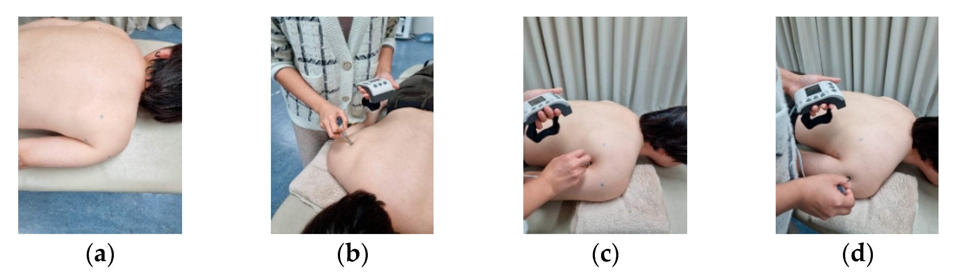
2.3.1. Measurement of the Muscle Pressure Pain Threshold

All measurements were performed using a digital pressure algometer (JTECH Medical, Midvale, UT, USA), which was periodically calibrated following the instructions of the manufacturer (Figure 2). To mark an anatomical point for each muscle, ensuring reliable pressure pain threshold (PPT) measurements at every time point, the examiner applied an increasing perpendicular pressure, at a speed of 1 kg/s, which was controlled by a metronome set to 60 beats. The participants were asked to say “stop” when the pressure applied using the algometer evoked pain; then, the assessor retracted the algometer, and the PPT was registered by another physical therapist [19]. Three measurements were taken at each point with a 30 s pause between each measurement and a 3 min pause between each muscle [20]. The PPT of the posterior deltoid (PD), teres major (TM), and infraspinatus muscles (IN) were measured with the subject in a prone position. A folded towel and cushion were utilized to provide shoulder support for the participant. The same physical therapist took measurements from start to finish. A tester marked a landmark point of each muscle on the skin using a pen to ensure reliable PPT measurements were taken at each time point. The landmarks identified the distal one-third of the infraspinatus, the midpoint of the posterior deltoid, and the belly of the infraspinatus muscle at a site located two fingers below a point halfway along the scapular spine [19,21].

2.3.2. Measurement of Respiratory Function

Respiratory functions were measured using a spirometer (Omnia software version 1.6.2, Cosmed, Rome, Italy). One of the most promising tests in a clinical setting is the forced vital capacity while the patient is seated. Before the test, the investigator taught the subjects how to accurately perform the test two or three times so they could adjust to the method and use the mouthpiece with a nasal clip. The patient was guided to inhale deeply and then exhale as quickly as possible [22]. Forced vital capacity and forced expiratory volume in 1 s were measured using pulmonary function equipment that met the American Thoracic Society’s recommendations for diagnostic spirometry [23].

2.3.3. Measurement of Lower Chest Expansion (LCE)

Lower chest expansion (LCE), the spinous process of the tenth thoracic vertebra, and the tip of the xiphoid process were used as markers [24]. The subjects received clear instructions, and the procedure was demonstrated to ensure comprehensive understanding. Measurements of chest diameter were taken after deep inspiration and expiratory maneuvers. The lower chest expansion was obtained by subtracting the inspiratory diameter from the expiratory diameter, according to the designated anatomical landmarks [24].

2.4. Intervention

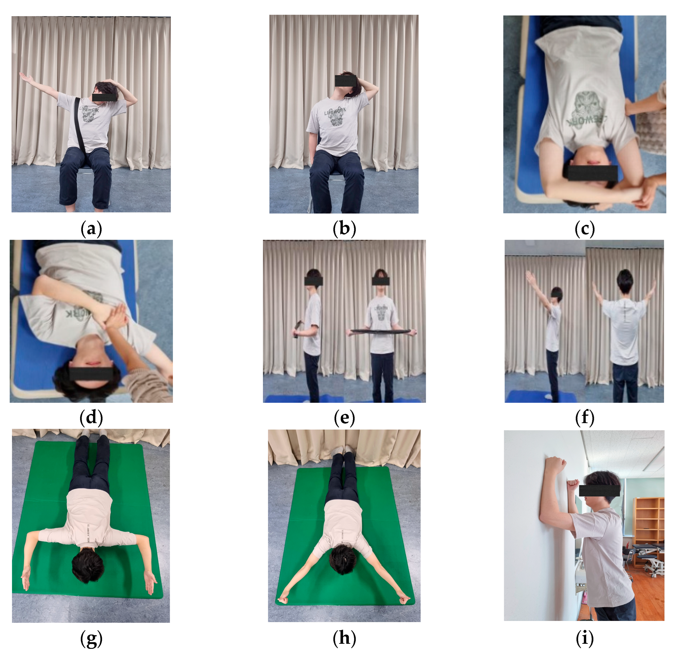
2.4.1. Scapular Stabilization Exercises

Exercises were conducted by correcting the movement of Rasoul Arshadi et al., Yasser M. Aneis et al., and Kwon et al. [25,26,27].
This study involved a stretching exercise for 45 s. Such exercises include levator scapular muscle, upper trapezius muscle, minor pectoralis (Figure 3a,b,d), and teres major stretching (Figure 3c). Participants were instructed to perform three sets of eight strengthening and movement exercises, including push-up exercises (serratus anterior strengthening) (Figure 3f), bilateral external rotation strengthening (Figure 3e), L-exercises (middle trapezius strengthening), Y-exercise (lower trapezius strengthening), and wall slide exercise (movement).

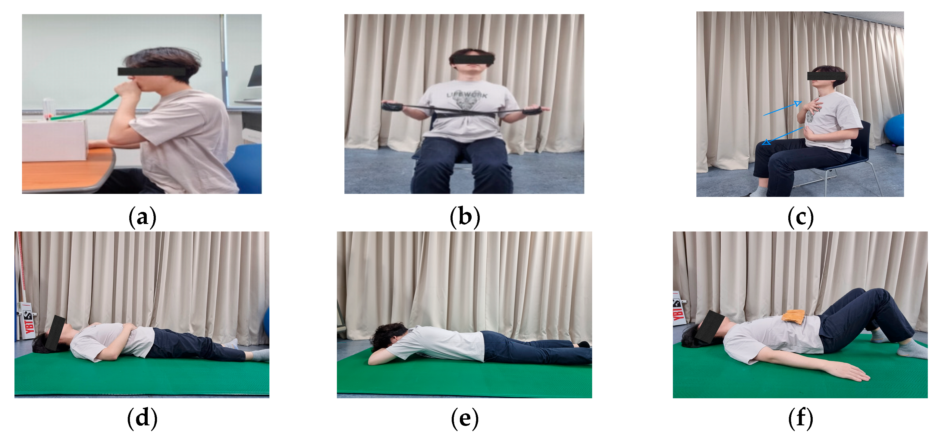
2.4.2. Breathing Training

This study involved exercise inspiratory spirometry (Figure 4a) in a seated position using a flow spirometer. Subjects were asked to hold the device upright and then inhale slowly to raise the ball inside to a set goal. The therapist demonstrated all techniques to clearly understand how to practice [28]. The exercise was divided into three sets of ten repetitions. During the rib expansion exercises (Figure 4b), the therapist first demonstrated proper breathing techniques by providing resistance with their hands and increasing it as they exhaled. The subjects remained seated and then used rubber bands for added resistance. Three sets of ten repetitions per set were completed. Three sets of ten diaphragmatic breathing exercises were carried out (including sitting, supine, prone positions, and the supine position with a 4 kg weight on the diaphragm located in the abdominal region) [25,29].

2.4.3. Thoracic Exercises

Stretching exercises (Figure 5a,b) include rectus abdominis stretching and thoracic mobility exercises for 45 s. Strengthening exercises (Figure 5c) include thoracic extensor muscle strengthening. Movement exercises (Figure 5d–g) include backward rocking, posture, and thoracic stabilization exercises. Three sets of eight were carried out, held for 15 s in each set. Movement occurs via a combination of movements by Kwon et al. and Kang [18,25].

2.5. Statistical Analysis

Statistical analyses were performed using SPSS 27.0 for Windows, and the Shapiro–Wilk test was used for normal distribution. A two-way repeated-measures analysis of variance (ANOVA) was conducted to investigate the interaction between the measurement period and measurement group, as well as the intra-group effect throughout the measurement period. The statistical significance level was set at p < 0.05. Bonferroni post hoc corrections were used to analyze the intra-group differences before and after the intervention’s effects (α = 0.025).

3. Results

A significant difference in within-group effect validation was found when comparing the time of change between the two groups before and after the intervention. Specifically, there was a significant difference in the posterior deltoid (PD) (F = 65.427, p = 0.000), teres major (TM) (F = 88.943, p = 0.000), and infraspinatus (IN) (F = 54.415, p = 0.000) based on the measurement period. There was no significant difference in the interaction effect depending on the time and group (p > 0.025). The different values of PD, TM, and IN were significant, with no differences observed between the two groups (p > 0.025) (Table 1).
A significant difference in within-group effect validation was found when comparing the time of change between the two groups before and after the intervention. Specifically, there was a significant difference in the forced vital capacity (FVC) (F = 33.322, p = 0.000), the forced expiratory volume in 1 s (FEV1) (F = 48.651, p = 0.000), FEV1/FVC% (F = 13.726, p = 0.001), and LCE (F = 96.929, p = 0.000) according to the measurement period. There was no significant difference in the interaction effect depending on the time and group (p > 0.025). The FVC, FEV1, FEV1/FVC%, and LCE were significant, with no differences between the two groups (p > 0.025) (Table 2).

4. Discussion

This study aimed to compare the effects of SBG and STG on various parameters, including the pressure pain threshold (PPT), respiratory function, and lower chest expansion (LCE), in individuals with UCS.
The PPT of the posterior deltoid, teres major, and infraspinatus muscles was significantly increased in both groups. The posterior deltoid muscle is known for its role in external rotation and horizontal abduction of the shoulder joint; the teres major muscle and infraspinatus muscle also perform the functions of external rotation and shoulder joint adduction. In subjects with UCS, when scapular tilt is excessive anteriorly or there is internal rotation, the posterior deltoid, teres major, and infraspinatus muscles are shortened [25]. Patients with posterior shoulder tightness are known to have a high sensitivity to stretching and low-pressure pain thresholds in the infraspinatus and posterior deltoid muscles [21]. Previous studies have shown an inhibitory effect of stretching on skin pain perception, while others have found that PPT increases both during and after stretching [30]. The internal rotation of the scapula in any position causes a visual increase in the tension of the spinal sheath ligament, and the opposing scapula in the retracted position increases the PPT of the infraspinatus and deltoid muscles [19]. Lee et al. also found that after 40 min of strengthening exercise, the pain threshold of the teres major muscle increased [31]. In this study, both groups underwent the same scapular stabilization exercises, including strengthening exercises and postural correction (wall slide exercises), to return the overly internally rotated scapula to its retracted position. This aimed to increase the pain threshold of the infraspinatus muscle, the posterior deltoid muscle, and the teres major muscle in both groups before and after the experiment. The results of this study indicate no differences between the two groups. Possible reasons for this include the subjects not selecting the pain-related options during their choices, which resulted in a limited amount of change. Secondly, the three muscles measured were those that cause problems due to scapular internal rotation, and breath training, along with thoracic exercises, are likely to have less of an impact on these muscles. In comparison, scapular stabilization exercises have a greater impact on these muscles.
This study showed that respiratory function and lower chest expansion were increased in both groups before and after exercise. Subjects with UCS experience excessive anterior tilt and downward rotation of the scapula. Exercises that stretch the chest muscles and strengthen the shoulder muscles are known to reduce poor shoulder posture, such as stretching of the pectoralis minor muscles. This can reduce excessive anterior tilt of the scapula and improve chest mobility, thereby increasing breathing muscle strength [32,33]. A previous study reported that the aberrant structure of the cervical and thoracic spine induces a movement disorder in the rib cage, thereby impacting respiratory function and resulting in reduced lung volume, vital capacity, and weakened respiratory muscles. The integration of scapular stabilization exercises with thoracic extension exercises enhances posture and respiratory function [18]. The serratus anterior and pectoralis minor muscles serve as accessory inspiratory muscles to assist breathing [17]. In the present study, both groups initially performed the same scapular stabilization exercises, which included strengthening exercises and stretching of the pectoralis minor muscles. These exercises contributed to improving respiratory function.
Based on current knowledge, diaphragmatic breathing exercises, as well as volume and flow-directed incentive spirometry techniques, have demonstrated efficacy in improving lung function [28]. In addition to the observed improvements in pain, the effects of the exercise intervention may also be attributed to underlying physiological mechanisms. Specifically, respiratory function may play a key role in managing UCS. Individuals with UCS often exhibit dysfunctional breathing patterns, such as upper chest breathing, which can increase tension in the accessory respiratory muscles, including the sternocleidomastoid and upper trapezius [34,35]. Targeted exercises may promote diaphragmatic breathing, reduce overactivity in these muscles, and enhance oxygen delivery, thereby contributing to improved postural control and reduced muscle strain. The SBG group underwent direct breathing training, including inspiratory exercises, rib expansion exercises, and diaphragmatic breathing exercises, which contributed to improving respiratory function and lower chest expansion. In the STG group, before the correction exercise, we first had the participants stretch their rectus abdominis muscles. During the stretching process, the subjects were asked to cooperate with abdominal breathing. Postural correction exercises, posture exercises, thoracic exercises, and backward rocking exercises were performed to reduce the structural abnormalities of the thoracic vertebrae and improve the thoracic alignment. As a result, lung function improved when the thoracic alignment was corrected, further opening the chest and enhancing the expansion of the lower chest. There was an absence of disparity between the two groups, and we concluded that for subjects with UCS, breath training and thoracic exercises have similar effects on improving respiratory function and lower chest expansion.
A limitation of this study was that no subjects who had pain were included. Since the primary aim of this study was to compare changes in the shoulders, neck muscle pain was not evaluated. Only students were selected, and no other age groups were represented. The four-week intervention period is also rather short for assessing sustained improvements in posture or respiratory function. Future studies should acknowledge these limitations and consider expanding the group of participants to include individuals experiencing pain or chronic pain.

5. Conclusions

The potential mechanisms by which the exercises impact UCS may involve improvements in respiratory function and modulation of pain thresholds. These physiological changes could contribute to postural correction and symptom relief; however, further investigation is warranted to confirm these pathways.

Author Contributions

Conceptualization, X.Y.; methodology, T.-H.K.; software, X.Y.; validation, X.Y., and T.-H.K.; formal analysis, X.Y.; investigation, X.Y.; resources, X.Y. and T.-H.K.; data curation, X.Y. and T.-H.K.; writing—original draft preparation, X.Y.; writing—review and editing, X.Y. and T.-H.K.; visualization, X.Y.; supervision, X.Y. All authors have read and agreed to the published version of the manuscript.

Funding

This research received no external funding.

Institutional Review Board Statement

The study was conducted in accordance with the Declaration of Helsinki and approved by the Institutional Review Board of Daegu University (IRB number: 1040621-202401-HR-008, approval date of the ethical protocol: 21 February 2024) for studies involving humans.

Informed Consent Statement

Informed consent was obtained from all subjects involved in the study.

Data Availability Statement

The original contributions presented in this study are included in the article. Further inquiries can be directed to the corresponding author.

Acknowledgments

We thank our colleagues and the reviewers who provided constructive criticism on drafts of this essay.

Conflicts of Interest

The authors declare no conflicts of interest.

References

  1. Sengupta, D.; Al-Khalifa, D. Pandemic imposed remote work arrangements and resultant work-life integration, future of work and role of leaders—A qualitative study of Indian millennial workers. Adm. Sci. 2022, 12, 162. [Google Scholar] [CrossRef]
  2. Kaur, S.; Chatterjee, S.; Goyal, M.; Popli, A.; Goyal, K.; Saha, M.; Garg, S.; Sushma, K.C. Physiotherapeutic approaches in upper cross syndrome: A systematic review of systematic reviews. J. Bodyw. Mov. Ther. 2025, 42, 756–762. [Google Scholar] [CrossRef]
  3. Chaudhuri, S.; Chawla, J.K.; Phadke, V. Physiotherapeutic Interventions for Upper Cross Syndrome: A Systematic Review and Meta-Analysis. Cureus 2023, 15, e45471. [Google Scholar] [CrossRef] [PubMed]
  4. Csepregi, É.; Gyurcsik, Z.; Veres-Balajti, I.; Nagy, A.C.; Szekanecz, Z.; Szántó, S. Effects of classical breathing exercises on posture, spinal and chest mobility among female university students compared to currently popular training programs. Int. J. Environ. Res. Public. Health 2022, 19, 3728. [Google Scholar] [CrossRef]
  5. Lowry, V.; Desjardins-Charbonneau, A.; Roy, J.S.; Dionne, C.E.; Fremont, P.; MacDermid, J.C.; Desmeules, F. Efficacy of workplace interventions for shoulder pain: A systematic review and meta-analysis. J. Rehabil. Med. 2017, 49, 529. [Google Scholar] [CrossRef] [PubMed]
  6. Greenberg, D.L. Evaluation and treatment of shoulder pain. Med. Clin. North. Am. 2014, 98, 487–504. [Google Scholar] [CrossRef] [PubMed]
  7. D’hondt, N.E.; Pool, J.J.; Kiers, H.; Terwee, C.B.; Veeger, H. Validity of clinical measurement instruments assessing scapular function: Insufficient evidence to recommend any instrument for assessing scapular posture, movement, and dysfunction—A systematic review. J. Orthop. Sports Phys. Ther. 2020, 50, 632–641. [Google Scholar] [CrossRef]
  8. Keshavarz, R.; Tajali, S.B.; Mir, S.M.; Ashrafi, H. The role of scapular kinematics in patients with different shoulder musculoskeletal disorders: A systematic review approach. J. Bodyw. Mov. Ther. 2017, 21, 386–400. [Google Scholar] [CrossRef]
  9. Giuseppe, L.U.; Laura, R.A.; Berton, A.; Candela, V.; Massaroni, C.; Carnevale, A.; Stelitano, G.; Schena, E.; Nazarian, A.; DeAngelis, J. Scapular dyskinesis: From basic science to ultimate treatment. Int. J. Environ. Res. Public. Health 2020, 17, 2974. [Google Scholar]
  10. Kibler, W.B.; Sciascia, A. Current concepts: Scapular dyskinesis. Br. J. Sports Med. 2010, 44, 300–305. [Google Scholar] [CrossRef]
  11. Santino, T.A.; Chaves, G.S.; Freitas, D.A.; Fregonezi, G.A.; Mendonça, K.M. Breathing exercises for adults with asthma. Cochrane Database Syst. Rev. 2020, 3, CD001277. [Google Scholar] [CrossRef] [PubMed]
  12. Srijessadarak, T.; Arayawichanon, P.; Kanpittaya, J.; Boonprakob, Y. Diaphragmatic Mobility and Chest Expansion in Patients with Scapulocostal Syndrome: A Cross-Sectional Study. Healthcare 2022, 10, 950. [Google Scholar] [CrossRef]
  13. Samosir, N.R.; Azizan, A. Effectiveness of Segmental Breathing and Expansion Thorax Exercise in Young to Middle-Aged Post-COVID Survivors. Int. J. Aging Health Mov. 2023, 5, 8–14. [Google Scholar]
  14. Dhaniwala, N.K.S.; Dasari, V.; Dhaniwala, M.N. Pranayama and Breathing Exercises-Types and Its Role in Disease Prevention & Rehabilitation. J. Evol. Med. Dent. Sci. 2020, 9, 3325–3331. [Google Scholar]
  15. Saito, H.; Harrold, M.E.; Cavalheri, V.; McKenna, L. Scapular focused interventions to improve shoulder pain and function in adults with subacromial pain: A systematic review and meta-analysis. Physiother. Theory Pract. 2018, 34, 653–670. [Google Scholar] [CrossRef]
  16. Pieters, L.; Lewis, J.; Kuppens, K.; Jochems, J.; Bruijstens, T.; Joossens, L.; Struyf, F. An update of systematic reviews examining the effectiveness of conservative physical therapy interventions for subacromial shoulder pain. J. Orthop. Sports Phys. Ther. 2020, 50, 131–141. [Google Scholar] [CrossRef]
  17. Ghanbari, A.; Ghaffarinejad, F.; Mohammadi, F.; Khorrami, M.; Sobhani, S. Effect of forward shoulder posture on pulmonary capacities of women. Br. J. Sports Med. 2008, 42, 622–623. [Google Scholar] [PubMed]
  18. Kang, N.-Y.; Im, S.-C.; Kim, K. Effects of a combination of scapular stabilization and thoracic extension exercises for office workers with forward head posture on the craniovertebral angle, respiration, pain, and disability: A randomized-controlled trial. Turk. J. Phys. Med. Rehabil. 2021, 67, 291. [Google Scholar] [CrossRef]
  19. Intelangelo, L.; Bordachar, D.J.; Mendoza, C.I.; Imaz, F.; Barone, M.; Barbosa, A.W. Effect of different postures of the scapular girdle and arm on the pressure pain threshold in the infraspinatus muscle. J. Bodyw. Mov. Ther. 2021, 28, 276–282. [Google Scholar] [CrossRef]
  20. Barone, M.; Imaz, F.; Converso, G.; Bordachar, D.; Barbero, A.; Trucco, M.; Intelangelo, L. Immediate effects of rhythmic joint mobilization of the temporomandibular joint on pain, mouth opening and electromyographic activity in patients with temporomandibular disorders. J. Bodyw. Mov. Ther. 2021, 28, 563–569. [Google Scholar] [CrossRef]
  21. Park, K.-N.; Kwon, O.-Y.; Weon, J.-H.; Choung, S.-D.; Kim, S.-H. Comparison of the effects of local cryotherapy and passive cross-body stretch on extensibility in subjects with posterior shoulder tightness. J. Sports Sci. Med. 2014, 13, 84. [Google Scholar]
  22. Pirola, A.; De Mattia, E.; Lizio, A.; Sannicolo, G.; Carraro, E.; Rao, F.; Sansone, V.; Lunetta, C. The prognostic value of spirometric tests in Amyotrophic Lateral Sclerosis patients. Clin. Neurol. Neurosurg. 2019, 184, 105456. [Google Scholar] [CrossRef]
  23. Han, J.; Park, S.; Kim, Y.; Choi, Y.; Lyu, H. Effects of forward head posture on forced vital capacity and respiratory muscles activity. J. Phys. Ther. Sci. 2016, 28, 128–131. [Google Scholar] [CrossRef] [PubMed]
  24. Derasse, M.; Lefebvre, S.; Liistro, G.; Reychler, G. Chest Expansion and Lung Function for Healthy Subjects and Individuals with Pulmonary Disease. Respir. Care 2021, 66, 661–668. [Google Scholar] [CrossRef] [PubMed]
  25. Kwon, O.-Y.; Kang, M.-H.; Kim, M.-H.; Kim, S.-J.; Kim, T.-H.; Park, K.-N.; Oh, J.-S.; Lee, W.-H.; Im, I.-Y.; Jang, J.-H.; et al. KEMA Approach for Analysis and Management of Motor Impairment 1; Hakjisa Medical: Seoul, Republic of Korea, 2022. [Google Scholar]
  26. Aneis, Y.M.; El-Badrawy, N.M.; El-Ganainy, A.A.; Atta, H.K. The effectiveness of a multimodal approach in the treatment of patients with upper crossed syndrome: A randomized controlled trial. J. Bodyw. Mov. Ther. 2022, 32, 130–136. [Google Scholar] [CrossRef]
  27. Arshadi, R.; Ghasemi, G.A.; Samadi, H. Effects of an 8-week selective corrective exercises program on electromyography activity of scapular and neck muscles in persons with upper crossed syndrome: Randomized controlled trial. Phys. Ther. Sport. 2019, 37, 113–119. [Google Scholar] [CrossRef]
  28. Shetty, N.; Samuel, S.R.; Alaparthi, G.K.; Amaravadi, S.K.; Joshua, A.M.; Pai, S. Comparison of Diaphragmatic Breathing Exercises, Volume, and—71—Flow-Oriented Incentive Spirometry on Respiratory Function in Stroke Subjects: A Non-randomized Study. Ann. Neurosci. 2020, 27, 232–241. [Google Scholar] [CrossRef]
  29. Tomas-Carus, P.; Branco, J.C.; Raimundo, A.; Parraca, J.A.; Batalha, N.; Biehl-Printes, C. Breathing exercises must be a real and effective intervention to consider in women with fibromyalgia: A pilot randomized controlled trial. J. Altern. Complement. Med. 2018, 24, 825–832. [Google Scholar] [CrossRef]
  30. Finocchietti, S.; Mørch, C.D.; Arendt-Nielsen, L.; Graven-Nielsen, T. Effects of adipose thickness and muscle hardness on pressure pain sensitivity: Correction. Clin. J. Pain. 2011, 27, 735–745. [Google Scholar] [CrossRef]
  31. Lee, H.S. The effects of aerobic exercise and strengthening exercise on pain pressure thresholds. J. Phys. Ther. Sci. 2014, 26, 1107–1111. [Google Scholar] [CrossRef]
  32. Lee, J.-H.; Cynn, H.-S.; Yoon, T.-L.; Choi, S.-A.; Choi, W.-J.; Choi, B.-S.; Ko, C.-H. Comparison of scapular posterior tilting exercise alone and scapular posterior tilting exercise after pectoralis minor stretching on scapular alignment and scapular upward rotators activity in subjects with short pectoralis minor. Phys. Ther. Sport. 2015, 16, 255–261. [Google Scholar] [CrossRef] [PubMed]
  33. Thongchote, K.; Threetepchanchai, K.; Chuwijit, A.; Lapmanee, S. Pilot study on the improvement effects of scapulothoracic exercises on the respiratory functions in sedentary young female adult with forward shoulder posture: A randomized control trial. Adv. Rehabil. 2023, 37, 23–32. [Google Scholar] [CrossRef]
  34. Arsalan, A.; Waqar, M.; Ahmad, A.; Gillani, S.A. Immediate and prolonged effects of breathing exercises on pain, quality of life and functional disability in patients of upper cross Syndrome: A randomized controlled trial. J. Riphah Coll. Rehabil. Sci. 2023, 11, 34–39. [Google Scholar]
  35. Jeong, G.H.; Lee, B.H. Effects of telerehabilitation combining diaphragmatic breathing re-education and shoulder stabilization exercises on neck pain, posture, and function in young adult men with upper crossed syndrome: A randomized controlled trial. J. Clin. Med. 2024, 13, 1612. [Google Scholar] [CrossRef]
Figure 1. Diagram of the experimental design.
Applsci 15 06147 g001
Figure 2. Measurement of muscle pressure pain threshold: (a) location of measurement; (b) infraspinatus muscle; (c) teres major muscle; and (d) posterior deltoid muscle.
Applsci 15 06147 g002
Figure 3. Scapular stabilization exercises: (a) levator scapular stretching; (b) upper trapezius stretching; (c) teres major stretching; (d) pectoralis minor stretching; (e) bilateral external rotation; (f) push-up exercises; (g) L-exercises; (h) Y-exercises; and (i) wall-slide exercises.
Applsci 15 06147 g003
Figure 4. Breath training. (a) Inspiratory training; (b) costal expansion; and (cf) diaphragmatic breathing exercises.
Applsci 15 06147 g004
Figure 5. Thoracic exercises. (a) Rectus abdominis stretching; (b) thoracic mobility exercises; (c) thoracic extensor muscle strengthening; (d) backward rocking exercises; (e,f) posture exercises; (g) thoracic stabilization exercises.
Applsci 15 06147 g005
Table 1. Comparison of the muscle pressure pain threshold of the PD, TM, and IN in the two groups.
SBGSTGTime
F(p)
Group
F(p)
Time × GroupF
(p)
PDPre9.49 ± 5.13 a9.20 ± 3.7765.427
(0.000 *)
0.107
(0.745)
0.528
(0.473)
post12.39 ± 5.3911.63 ± 3.98
t(p)−5.649 (0.000 *)−5.886 (0.000 *)
TMPre8.55 ± 3.608.24 ± 3.6588.943
(0.000 *)
0.213
(0.648)
0.947
(0.338)
Post11.52 ± 4.3810.65 ± 3.31
t(p)−6.093 (0.000 *)−8.122 (0.000 *)
INPre9.57 ± 4.018.92 ± 3.1554.415
(0.000 *)
0.193
(0.663)
0.029
(0.867)
Post11.60 ± 4.8711.05 ± 3.68
t(p)−4.845 (0.000 *)−5.645 (0.000 *)
* = p < 0.05. a Mean ± standard deviation. PD: posterior deltoid, TM: teres major, IN: infraspinatus, SBG: scapular stabilization accompanied by breathing training, STG: scapular stabilization accompanied by thoracic exercises.
Table 2. Comparison of respiratory function and lower chest expansion in the two groups.
SBGSTGTimeF
(p)
Group
F(p)
Time × Group
F(p)
FVC(L)Pre4.00 ± 1.04 a3.68 ± 0.7333.322
(0.000 *)
1.198
(0.282)
2.111
(0.157)
Post4.17 ± 1.073.79 ± 0.70
t(p)−4.933 (0.000 *)−3.172 (0.006 *)
FEV1(L)Pre3.22 ± 0.893.01 ± 0.5448.651
(0.000 *)
1.207
(0.281)
2.800
(0.105)
Post3.55 ± 0.843.21 ± 0.50
t(p)−5.920 (0.000 *)−3.881 (0.001 *)
FEV1/FVC%Pre80.52 ± 8.5882.43 ± 8.4613.726
(0.001 *)
0.107
(0.745)
0.955
(0.336)
Post85.71 ± 6.9785.45 ± 7.07
t(p)−2.741 (0.015 *)−2.623 (0.019 *)
LCEPre4.29 ± 0.624.58 ± 0.7496.929
(0.000 *)
0.150
(0.702)
0.475
(0.496)
Post6.90 ± 1.596.86 ± 1.43
t(p)−6.247 (0.000 *)−8.514 (0.000 *)
* = p < 0.05. a Mean ± standard deviation. FVC: forced vital capacity; FEV1: forced expiratory volume in 1 s; LCE: lower chest expansion.
Disclaimer/Publisher’s Note: The statements, opinions and data contained in all publications are solely those of the individual author(s) and contributor(s) and not of MDPI and/or the editor(s). MDPI and/or the editor(s) disclaim responsibility for any injury to people or property resulting from any ideas, methods, instructions or products referred to in the content.

