Article

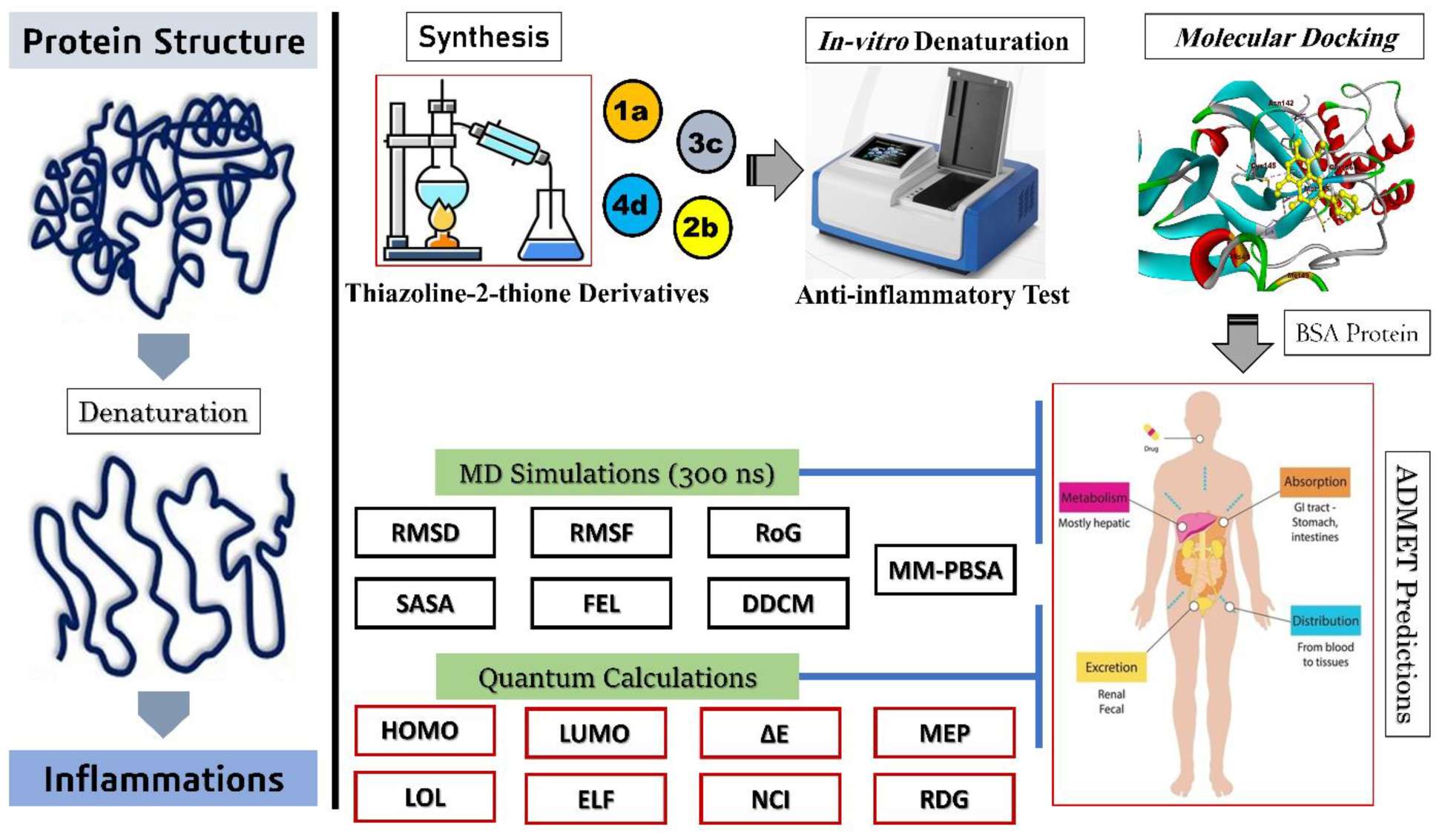
Synthesis, In Vitro Anti-Inflammatory Activity, Molecular Docking, Molecular Dynamics and DFT Calculations of Thiazoline-2-Thione Derivatives

1
Laboratory of Organic Materials and Heterochemistry, Echahid Cheikh Larbi Tebessi University, Tebessa 12000, Algeria
2
Group of Computational and Medicinal Chemistry, LMCE Laboratory, University of Biskra, Biskra 70000, Algeria
3
College of Engineering and Technology, American University of the Middle East, Egaila 54200, Kuwait
*
Authors to whom correspondence should be addressed.
Appl. Sci. 2025, 15(11), 6095; https://doi.org/10.3390/app15116095
Submission received: 12 April 2025 / Revised: 20 May 2025 / Accepted: 22 May 2025 / Published: 28 May 2025

Abstract

:
The objective of this study was to synthesize thiazoline-2-thione derivatives (1a, 2b, 3c and 4d) and examine their anti-inflammatory properties as potential alternatives to Aspirin (NSAID), which is known for its side effects, including liver damage. The study employed a multifaceted approach that integrated in vitro assays, molecular docking, ADMET predictions, molecular dynamics simulations (300 ns for each system) and detailed DFT calculations. These four molecules were initially evaluated for their effectiveness in inhibiting the denaturation of bovine serum albumin (BSA), a key indicator of their potential anti-inflammatory activity. The results show that 4d displayed notable inhibitory potential against BSA denaturation, with an IC50 value of 21.9 µg/mL, outperforming the efficacy of Aspirin (22 µg/mL). In comparison, 3c exhibited an IC50 value of 31.7 µg/mL. Molecular docking studies with the BSA active site revealed that 4d and 3c had the highest binding affinities, with binding energies (∆G) of −5.274 and −4.731 kcal.mol−1, respectively. Aspirin showed a ∆G of −4.641 kcal.mol−1. These findings suggest that 4d and 3c molecules exhibit stronger interactions with BSA, indicating superior anti-inflammatory activity compared to Aspirin. In addition, molecular dynamics simulations, cross-dynamic correlation matrix (DCCM), free energy landscape (FEL), MM-PBSA and detailed DFT calculations provided further evidence that 4d formed stable molecular interactions with the BSA receptor. These analyses highlighted the strong binding stability of 4d, indicating that it maintains consistent interactions over time. The results also suggested that 4d exhibits favorable energy profiles, good pharmacokinetic features and optimal molecular behavior within the BSA active site. Finally, the results of this study are promising for the development of new anti-inflammatory drugs, highlighting potential compounds that could offer effective and safer alternatives to existing treatments.

1. Introduction

Heterocyclic compounds are crucial in drug design, with their significance remaining evident even when the natural substrate or ligand of a biological target lacks a heterocyclic structure. Both natural and synthetic drugs often incorporate heterocyclic groups to optimize their interaction with biological targets [1]. These compounds are foundational in pharmaceutical science due to their wide range of biological activities and their role as key scaffolds in drug development. Heterocycles are extensively utilized in the creation of therapeutic agents, including antimicrobial, antiviral, anticancer, anti-inflammatory and analgesic drugs [2]. Thiazole, a prominent heterocyclic compound with a five-membered ring structure, is characterized by the presence of a sulfur atom at position one and a nitrogen atom at position three. Numerous natural and synthetic derivatives of thiazole exhibit diverse biological activities, such as anti-inflammatory properties [3].
Protein denaturation has been recognized as a primary trigger of inflammation. Evidence suggests that inflammation arises when living tissues sustain injury, manifesting as redness, swelling, heat, pain and functional impairment in the affected region. This process involves the disruption of hydrogen, electrostatic and hydrophobic bonds within the protein configuration [4,5]. Furthermore, inflammation involves a multifaceted series of processes, such as tissue degradation, release of mediators, migration of cells, activation of enzymes and subsequent repair. These mechanisms alter the protein’s molecular structure, resulting in its denaturation and vital impairment [6,7]. Thus, it can be inferred that compounds capable of preventing these structural alterations and inhibiting protein denaturation caused by thermal or heat-induced stress hold significant potential as therapeutic agents for treating inflammation.
Steroidal and non-steroidal anti-inflammatory drugs are extensively employed to treat inflammatory disorders such as rheumatoid arthritis and other infections. These medications are reported to interact with plasma albumin, effectively preventing or inhibiting its denaturation caused by heat [8,9]. However, extended use of these medications is commonly associated with harmful or secondary side effects, including liver damage, gastrointestinal disturbances and an increased risk of cardiovascular and renal complications, which can ultimately lead to failure of these organs [10,11]. Indeed, the search for novel anti-inflammatory drugs is crucial, given the widespread impact of inflammatory diseases on human health. The exploration of new compounds that can effectively modulate the inflammatory pathways, while minimizing side effects, is vital.
Computer-aided drug design (CADD) encompasses a variety of computational approaches aimed at discovering, designing and developing new pharmaceutical compounds. It plays a vital role in optimizing active ligands, identifying innovative drugs and unraveling biological processes on a biomolecular scale [12,13]. CADD techniques facilitate faster, more cost-effective and efficient drug design and development. The drug design procedure encompasses stages such as drug discovery, preclinical studies, clinical trials and regulatory approval. On average, this process is costly and spans over a decade [14,15]. For more than a decade, in silico predictions of ADMET features have been a fundamental aspect of drug design and discovery. The importance of in silico ADMET modeling has grown significantly, as these properties are now assessed earlier in the drug design process. This shift allows ADMET evaluations to guide the early stages of drug design, including the creation of combinatorial libraries where these properties are strategically considered [16]. In addition, molecular dynamics is a simulation technique designed to provide insights into the structural, dynamical and thermodynamical characteristics of molecular systems. These systems often involve biomolecules, such as proteins, enzymes or lipid assemblies forming membranes, immersed in an aqueous solvent like water or an electrolyte solution. For proteins and enzymes, MD simulations typically begin with the experimentally determined protein structures available in the Protein Data Bank (PDB) [17,18,19]. Finally, density functional theory (DFT) is a powerful computational approach that delivers precise and dependable information about various molecular characteristics. It offers detailed insights into the geometry of compounds, the energy barriers associated with rotation, vibrational frequencies and electronic properties, making it an essential tool in understanding and predicting molecular behavior [20,21].
This research focused on the synthesis of thiazoline-2-thione derivatives and a comprehensive evaluation of their anti-inflammatory properties through in vitro methods. Specifically, the study assessed its capacity to inhibit the denaturation of bovine serum albumin (BSA), a critical mechanism often associated with anti-inflammatory activity. By preventing protein denaturation, thiazoline-2-thione derivatives demonstrate their potential as candidates for further development in anti-inflammatory therapeutics. Additionally, the relationship between these anti-inflammatory activities and previously synthesized compounds was investigated in silico using molecular docking studies with the BSA protein. This approach allowed for a detailed analysis of the interactions and binding affinities, providing valuable insights into the potential efficacy of these compounds in inhibiting BSA denaturation. The drug-likeness prediction and ADMET (Absorption, Distribution, Metabolism, Excretion and Toxicity) properties of the most promising compounds, which exhibited better or comparable binding affinity to the standard drug Aspirin, were also evaluated. To gain a deeper understanding of the stability of the compounds with the target receptors (BSA protein), comprehensive molecular dynamics simulations (300 ns for each system) were conducted. These simulations included the calculation of the free energy landscape (FEL), dynamic cross-correlation matrix (DCCM) and molecular mechanics-Poisson–Boltzmann surface area (MM-PBSA) to assess the binding interactions and stability over time. Furthermore, density functional theory (DFT) calculations were carried out to provide additional insights into the electronic structure, energetics and reactivity of the compounds, complementing the molecular dynamics findings. Finally, these analyses offered a deeper understanding of the stability and energetics of the ligand–protein complexes, providing valuable insights into their interactions. This comprehensive evaluation further supports the potential of the compounds as promising candidates for anti-inflammatory therapeutics. Figure 1 provides a visual representation of the strategy applied in this study.

2. Materials and Methods

2.1. Experimental Procedures
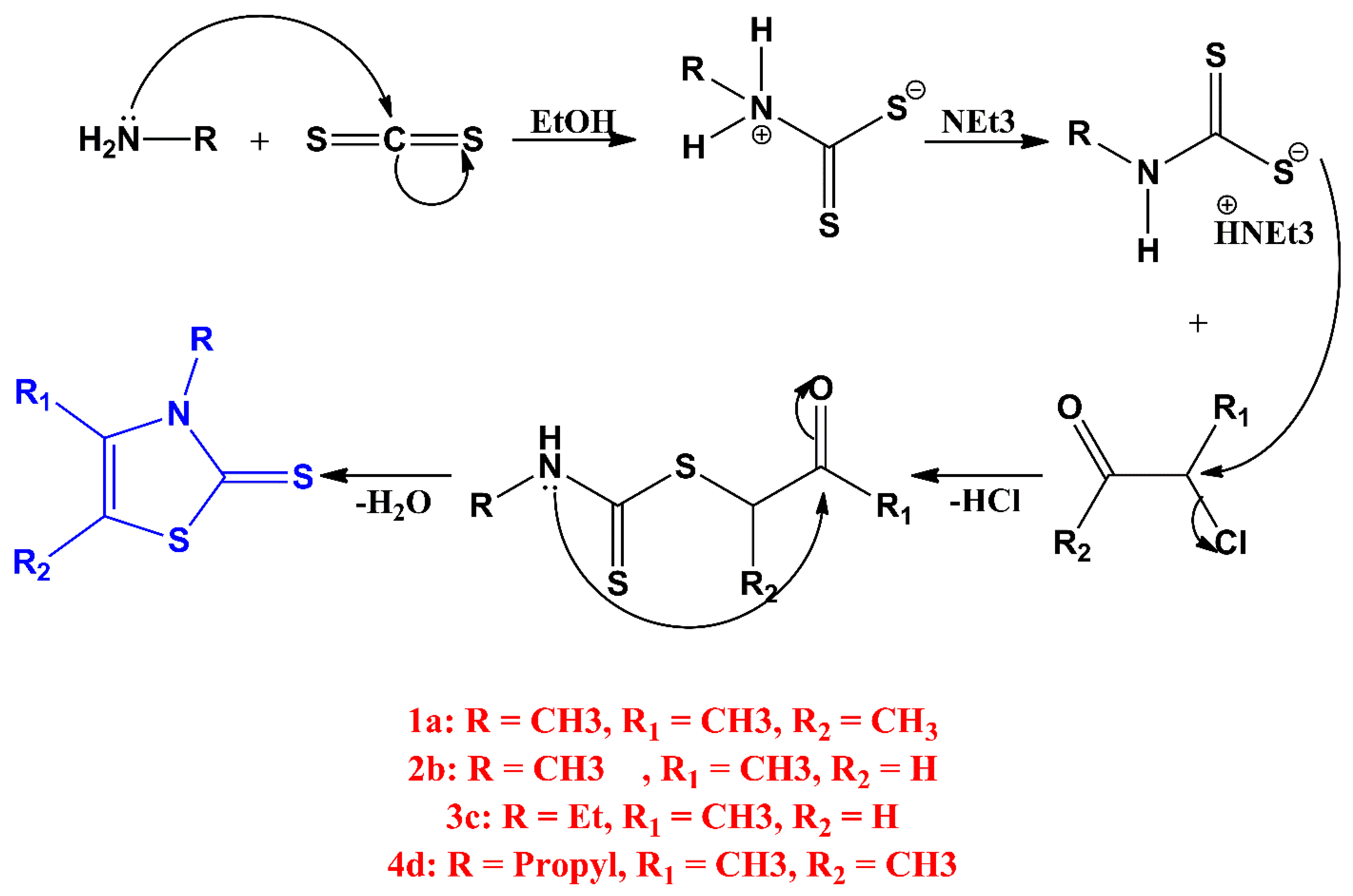
2.1.1. Synthesis

At 0 °C, a solution containing alkylamine (50 mmol) and triethylamine (7 mL, 50 mmol) in 200 mL of diethyl ether was prepared, to which carbon disulfide (CS2, 3 mL, 50 mmol) was gradually added. The resulting reaction mixture was stirred at ambient temperatures for two hours. The formed white precipitate was collected by filtration, thoroughly washed with diethyl ether and subsequently dried under reduced pressure.
The dithiocarbamate salt (28 mmol) was dissolved in 100 mL of acetonitrile, and chlorobutanone or chloroacetone was slowly introduced to the solution. The mixture, characterized by a yellow coloration, was allowed to stir overnight at room temperature. Following this, the solvent was removed through evaporation, yielding a viscous residue. This residue was then chilled to 0 °C, and 25 mL of concentrated sulfuric acid was cautiously added in a controlled dropwise manner. The reaction mixture was stirred continuously for three hours, followed by hydrolysis with ice water. The resulting solution was extracted using dichloromethane, and the organic layer was separated, washed with water and dried over anhydrous MgSO4. After solvent evaporation, the crude product was purified by column chromatography, employing a dichloromethane/hexane (1:1) mixture as the eluent, to yield thiazoline-2-thiones [22]. Figure 2 illustrates the stepwise reaction mechanism for synthesizing thiazoline-2-thione derivatives. It highlights the key intermediates and chemical transformations. This visual representation facilitates a clearer understanding of the synthetic pathway.
1a: R = CH3, R1 = CH3, R2 = CH3, white powder. Yield: 84%. mp = 99 °C. 1H NMR (CDCl3): δ 2.15 (q, 3H, CH3), 2.17 (q, 3H, CH3), 3.65 (s, 3H, CH3). 13C NMR (CDCl3): δ 11.60 (CH3), 13.01 (CH3), 34.89 (N-CH3), 117.33 (C=C), 134.58 (C=C), 185.62 (C=S) [23].
2b: R = CH3, R1 = CH3, R2 = H, white powder, yield 80%, mp = 111 °C. 1H NMR (CDCl3): δ 2.18 (s, 3H, CH3), 3.55 (s, 3H, CH3), 6.23 (s, 1H, =CH). 13C NMR (CDCl3): δ 16.15 (CH3), 34.63 (N-CH3), 106.40 (HC=C), 140.37 (HC=C), 188.29 (C=S) [23].
3c: R = Et, R1 = CH3, R2 = H, Pale brown powder, yield 90%, mp = 119° C. 1H NMR (CDCl3): δ 1.29–1.32 (t, 3H, CH3CH2), 2.29 (d, 3H, CH3), 4.21–4.26 (q, 2H, CH3CH2), 6.24–6.25 (q, 1H, C=CH). 13C NMR: δ 12.83 (CH3CH2), 15.31 (CH3), 42.30 (N-CH2), 106.31 (HC=C), 139.40 (HC=C), 187.58 (C=S).
4d: R = Propyl, R1 = CH3, R2 = CH3, brown crystal, yield 90%, mp = 85° C. 1H NMR (DMSO-d6): δ 0.89–0.92 (t, 3H, CH3CH2), 1.61–1.71 (m, 2H, CH3CH2CH2), 2.13–2.14 (m, 3H, CH3), 2.21–2.22 (m, 3H, CH3), 4.06–4.10 (m, 2H, N-CH2). 13C NMR (DMSO-d6): δ 11.44 (CH3CH2), 11.58 (CH3), 12.68 (CH3), 20.93 (CH3CH2CH2), 49.03 (N-CH2), 116.88 (C=C), 135.86 (C=C), 184.38 (C=S) (Figure S1).

2.1.2. In Vitro Inhibition of Albumin Denaturation

A 0.5% (w/v) solution of bovine serum albumin (BSA) was formulated in phosphate-buffered saline (PBS) at pH = 6.4. Next, 450 μL of this BSA stock solution was mixed with 50 μL of either the standard drug (Aspirin) or the tested molecule at various concentrations. The resulting samples were incubated at 37 °C for 20 min, followed by heating at 70 °C for an additional 20 min. Finally, the absorbance of the samples was measured at a wavelength of 660 nm to assess the reaction outcomes [24]. In addition, a control was established using the same procedure, with the sole difference being the replacement of the sample solution with 50 μL of phosphate-buffered saline (PBS). This approach allowed for a comparative assessment against the test samples. Finally, the percentage of protein denaturation inhibition was determined using the following formula:
I % = A S A C A S × 100
where I% denotes the percentage of inhibition, AS represents the absorbance value of the sample and AC indicates the absorbance value of the control.

2.1.3. Binding Free Energy and Binding Constant

The results obtained from UV–Vis spectroscopy was used to determine the binding constant (K) following the Benesi–Hildebrand equation [25]:
A S A C A S = ε S ε C ε S ε S ε C ε S 1 K C
where AC and AS represent the absorbance readings for the control and samples, respectively. εC and εS denote their corresponding extinction coefficients, [C] is the concentration of the compounds being studied and K signifies the binding constant.
The binding free energy (∆G in KJ/mol) was assessed using the equation
∆G = −RTlnK
where T denotes the ambient temperature, and R represents the universal gas constant, which is 8.32 j.mol−1K−1.

2.2. Theoretical Procedures

2.2.1. Protein and Ligand Preparation

All synthesized molecules were processed using the Schrödinger LigPrep module (v 13.4), which systematically generates optimal 3D molecular conformers, neutralizes charged moieties and precisely adjusts ionization states to correspond with a physiological pH range of 7.0 ± 2.0. Energy minimization was performed using the OPLS4e force field, ensuring optimized molecular geometries. This procedure ensures that the molecular structures are accurately prepared for downstream computational modeling and simulations [26,27]. The high-resolution three-dimensional crystal structure of bovine serum albumin (BSA), with a resolution of 2.70 Å (PDB ID: 3V03), was retrieved from the Protein Data Bank (PDB) through the RCSB PDB website (https://www.rcsb.org/) [28,29]. This structural data serves as a foundation for subsequent molecular analyses and computational studies. The BSA protein was first subjected to preprocessing using the “Protein Preparation Wizard” in Schrödinger Maestro. Heavy atoms, water molecules, cofactors and metal ions present in the protein structure were corrected as needed. Missing hydrogen atoms, side chains and protons were incorporated to restore the integrity of the protein. Further refinement and energy minimization of the structure were carried out using the OPLS4e force field, ensuring the protein’s suitability for accurate molecular modeling and computational simulations [30,31].

2.2.2. Preparation of Grid and Docking Modeling

Utilizing the receptor grid generation module, the binding sites in BSA, blind docking has been performed, with the grid size set to 110, 110 and 110 along the x-, y- and z-axes, with 0.375 Å grid spacing. The establishment of a cubic grid in the active space delineates the active site of the BSA protein. A grid box was generated at the centroid of this active site to facilitate docking calculations, ensuring accurate positioning for ligand interaction studies [32].
For molecular docking studies, Aspirin was selected as a reference ligand. The primary objective of this investigation was to evaluate the inhibitory potential of our synthesized molecules on the BSA protein relative to that of Aspirin. This comparison aims to provide insights into the efficacy of these synthesized compounds in modulating BSA protein activity. Molecular docking was conducted using the Glide program (Grid-Based Ligand Docking with Energetics) integrated within Maestro [33]. During the docking procedure, binding modes that exhibited the highest Glide scores were selected for further analysis. The Glide XP module was employed to evaluate the docking results. The XP scores of the synthesized molecules were compared to the XP score of Aspirin, a known anti-inflammatory agent and inhibitor of BSA protein, to assess the relative binding efficacy of the synthesized compounds [34]. The molecular interactions were examined using Discovery Studio software v 2024 [35]. The top-performing compounds were selected for subsequent investigations into their pharmacokinetic and toxicological profiles, with the goal of evaluating their potential for therapeutic application.

2.2.3. ADME-Tox Features

We performed a comprehensive evaluation of the ADME (Absorption, Distribution, Metabolism and Excretion) properties for each compound, with particular attention to solubility and absorptivity. These attributes are essential, as they play a pivotal role in determining the biological activity and therapeutic efficacy of the compounds, influencing their absorption and interaction within biological systems [36]. We analyzed the ADME properties using the SwissADME platform, which provides valuable insights into the pharmacokinetic profiles of the compounds under study [37]. The ProTox-II platform was employed to assess the potential toxicity of the compounds. Subsequently, the lethal dose (LD50) was quantified for both active and inactive cell types to investigate the hepatotoxicity, immunotoxicity and cytotoxicity [38].

2.2.4. MD Simulation and Stability

At this stage of the research, the goal is to analyze the stability of the complexes formed between the synthesized molecules and BSA protein, comparing these results with the reference complex of Aspirin bound to BSA. This evaluation will further allow for an assessment of the synthesized molecules’ potential to inhibit BSA protein activity and their anti-inflammatory properties. To achieve precise and reliable stability measurements, a molecular dynamics simulation was conducted over 300 ns.
The optimal synthesized molecules from the docking studies, alongside Aspirin as a reference, were further examined through molecular dynamics simulations employing Gromacs-2023 and the CHARMM36 force field to assess their interactions and stability [39]. Moreover, SwissParam was utilized to generate detailed molecular parameters for the synthesized molecule and the reference ligand, ensuring accurate input for further simulation processes [40]. The simulations were conducted for 300 ns in dodecahedral boxes using the TIP3P water model on Ubuntu (v 24.04), with the systems neutralized. Subsequently, the solvated systems underwent energy minimization via the steepest descent method until the maximum force fell below 10.0 kJ/mol. NVT equilibration was performed for 500 ps at 300 K using a V-rescale thermostat. Following this, NPT equilibration was conducted for 100 ps employing a Berenson pressure coupling method with a coupling constant of 2.0 ps [41,42]. Finally, to comprehensively assess the stability and binding affinity of each system, we carried out a series of calculations, including the root mean square deviation (RMSD), to monitor the structural variations over time, the root mean square fluctuation (RMSF) to evaluate the mobility of individual residues and the radius of gyration (Rg) to analyze the compactness of the protein structure. Additionally, we measured the solvent-accessible surface area (SASA) to determine the extent of exposure to solvent molecules. The free energy landscape (FEL) was constructed to visualize the energy profiles associated with different configurations, while the dynamic cross-correlation matrix (DCCM) was used to investigate correlated movements within the protein. Finally, we employed the MM-PBSA method to estimate the binding free energy of the complexes, providing further insights into their interactions.

2.2.5. Density Functional Theory (DFT) Calculations

The optimization of the top synthesized compound and Aspirin was conducted using Gaussian 16 W software [43], utilizing the B3LYP functional, along with a 6-311++ (d, p) basis set [44]. To gain insight into the electronic properties of the chosen molecules, several analyses were performed. These analyses included assessing molecular orbital energies, determining the highest occupied molecular orbital (HOMO) and lowest unoccupied molecular orbital (LUMO) energy levels, along with the energy gap (ΔE) between them. Additionally, the visualization of the frontier molecular orbitals (FMOs) and molecular electrostatic potential (MEP) surfaces was conducted using Gauss View 06 software [45]. Finally, to further investigate the electronic and chemical properties, such as partial electron localization function (ELF), localized orbital locator (LOL) and reduced density gradient (RDG), Multiwfn software 3.7 was utilized [46].

3. Results

3.1. Anti-Inflammatory Activity and the Experimental Validation

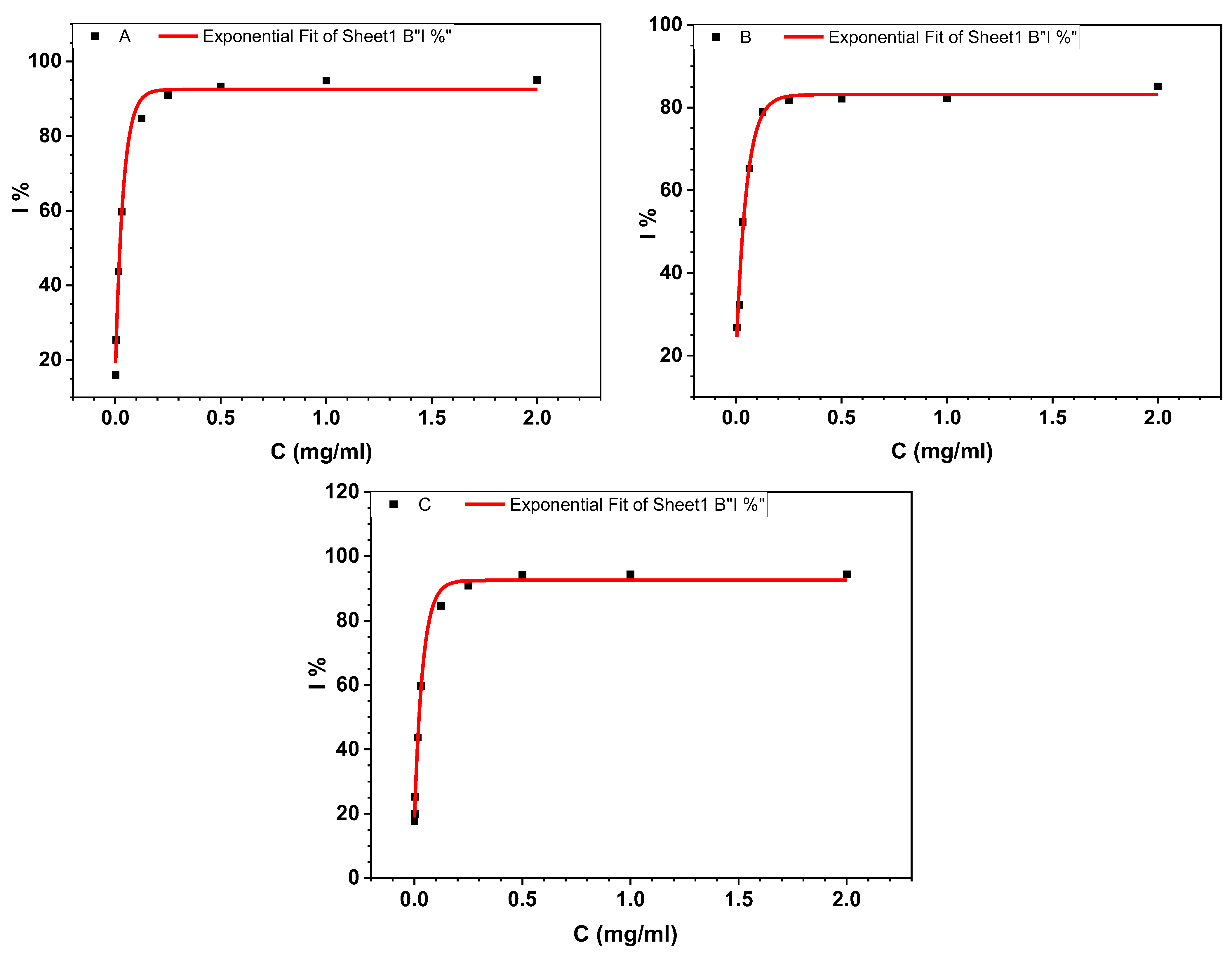
3.1.1. BSA Denaturation Assay

This section outlines an in vitro analysis conducted to assess the inhibitory potential of the synthesized molecules on BSA’s biological function. Aspirin was used as a reference compound during the study. The research aimed to compare the performance of the synthesized molecules with that of Aspirin. The findings highlight their relative effectiveness and potential applications. First, at a concentration of 2 mg/mL, Aspirin showed a maximum inhibition of BSA protein of 94.40%. In comparison, the compound 3c achieved an inhibition rate of 85.12% at the same concentration. Additionally, the molecule 4d exhibited the highest inhibition of BSA protein at 95.24% when tested at 2 mg/mL. Figure 3 represents the percentage inhibition (I %) activity of the BSA enzyme at different concentrations of the best synthesized molecules and Aspirin.
The estimated IC50 values for 3c, 4d and Aspirin were 31.7, 21.9 and 22 µg/mL, respectively (Table 1). These findings suggest that, while 4d shows a relatively strong anti-inflammatory effect, the notably higher IC50 value of 3c indicates that a significantly greater concentration is required to achieve comparable inhibition, whereas the IC50 of Aspirin is nearly equivalent to that of 4d, indicating a similar potency.

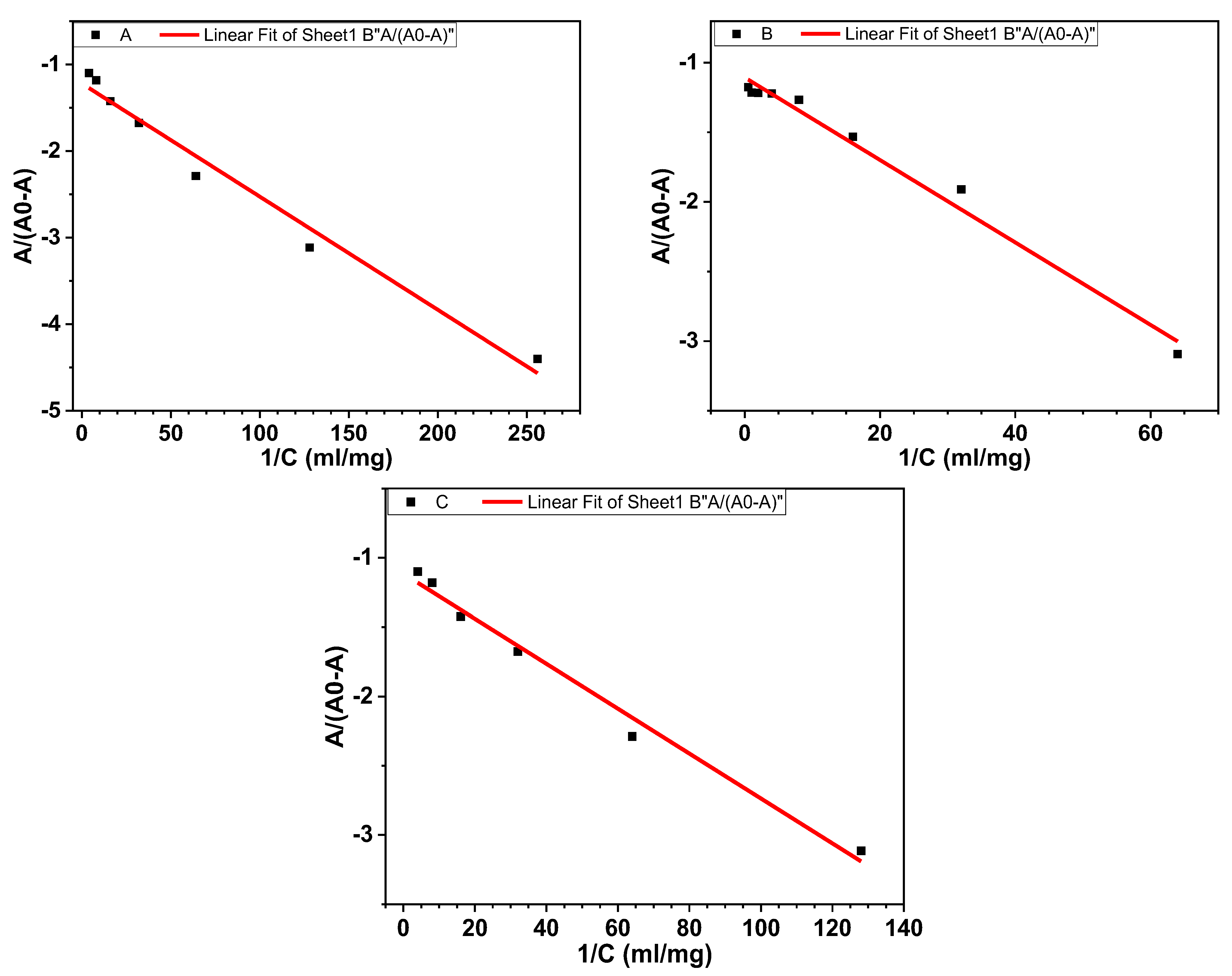
3.1.2. Binding Free Energy and Binding Constant

The K and ∆G values were determined based on changes in absorbance as the BSA concentration increased. The relationship between As/(Ac − As) and 1/[C] was plotted, as shown in Figure 4. These plots were used to calculate the values of K and ∆G. The results are summarized in Table 2. The data provide insight into the interaction between the synthesized compound and BSA. In addition, the K values were determined to be 5.95 × 10³ (M−1) for the 3c molecule, 16.30 × 10³ (M−1) for the 4d molecule and 12.40 × 10³ (M−1) for Aspirin, with corresponding binding energies of −5.16, −5.75 and −5.59 Kcal.mol−1, respectively. The results indicate that the synthesized compound 4c effectively inhibits the biological function of the BSA protein. This provides strong evidence that thiazoline thione derivatives have significant potential as novel anti-inflammatory agents, making them promising candidates for the development of future therapeutic drugs.

3.2. Molecular Docking Studies

Extensive molecular docking studies were performed on all synthesized molecules to elucidate their mechanisms in inhibiting essential functional regions of BSA, to assess their efficacy as anti-inflammatory agents and to provide a detailed analysis of their binding affinities and interaction profiles. This approach enabled a thorough investigation into the specific binding energies and the nature of interactions, shedding light on how these molecules might exert their effects on BSA’s functionality. Additionally, Aspirin was docked with the BSA protein to provide a reference for estimating the inhibitory efficacy of the synthesized compounds, allowing for a comparative analysis of their binding interactions and potential effectiveness. The docking modeling results for the top two synthesized molecules, along with the reference ligand (Aspirin), are presented in Table 3, detailing binding affinities and interaction energies. Figure 5 illustrates the specific types of interactions with the BSA active site.
The findings reveal that the two leading synthesized compounds, 4d and 3c, demonstrate binding affinities of −5.274 kcal.mol−1 and −4.731 kcal.mol−1, respectively, indicating stronger interactions with BSA compared to the reference compound (Aspirin), which has a binding affinity of −4.641 kcal.mol−1. While the docking results suggest stronger interactions of 4d and 3c with BSA, only compound 4d exhibited greater experimental inhibitory activity than Aspirin. These results underscore the importance of correlating in silico predictions with experimental data, as molecular docking does not always directly translate to biological efficacy. The molecule 4d interacted with amino acids Tyr149 and Arg256 through hydrogen bonding at distances of 2.0 Å and 2.1 Å, respectively. In addition, the molecule 3c exhibited a hydrogen bond interaction with Tyr149 and Arg256 at a distance of 2.2 Å. The reference ligand formed hydrogen bonds with amino acids Tyr149 and Arg256, with interaction distances ranging from 2.2 to 2.5 Å. In addition, a Pi–sulfur interaction was observed involving the sulfur atoms on the thiazole rings of both the 4d and 3c molecules. The molecule 4d showed Pi–sulfur interactions with amino acids Tyr149 and Arg256 at distances ranging from 2.9 to 3.1 Å. In contrast, 3c exhibited a Pi–sulfur interaction exclusively with Tyr149 at a distance of 2.1 Å. For hydrophobic interactions, both synthesized molecules and the reference compound interacted with the amino acids Tyr149, His241, Leu237, Leu259, Ile263, Ile289 and Ala290 at molecular distances ranging from 3.7 to 5.2 Å. Finally, to obtain more precise insights, the molecule 4d and Aspirin were selected for ADME-Tox analysis, molecular dynamics (MD) simulations and quantum mechanical calculations.

3.3. ADME-Tox and Bioavailability Prediction

Membrane permeability and distribution are essential pharmacokinetic parameters that influence the optimal bioactivity of compounds. SwissADME analysis revealed that the synthesized molecule 4d molecule has a consensus LogPo/w value of 2.36, indicating a higher lipophilicity compared to the reference compound, Aspirin, which has a consensus LogPo/w value of 1.28. This suggests that the 4d molecule may have improved membrane permeability and distribution characteristics relative to Aspirin, potentially enhancing its pharmacokinetic profile. Consequently, the synthesized molecule exhibits optimal aqueous solubility alongside efficient permeability through biological membranes, which suggests a strong potential for bioavailability. It is also estimated to have high gastrointestinal (GI) absorption and a robust binding affinity to plasma proteins, factors that together enhance its distribution profile and overall pharmacokinetic properties. Furthermore, the synthesized molecule does not inhibit the essential functions of liver enzymes, specifically the cytochrome P450 (CYP450) family. This characteristic suggests a reduced likelihood of metabolic interference and fewer adverse drug interactions, enhancing the molecule’s safety profile. In conclusion, the synthesized molecule is in accordance with Lipinski’s rules, indicating its potential as a viable drug candidate. The findings from the ADME study are detailed in Table 4, providing insights into the absorption, distribution, metabolism and excretion characteristics of the synthesized molecule and Aspirin.
Table 5 details the toxicity predictions for the molecule 4d as evaluated by ProTox-II. The analysis encompasses various toxicity endpoints, including hepatotoxicity, neurotoxicity, cardiotoxicity, carcinogenicity and cytotoxicity. Additionally, it provides estimates for the LD50 values and categorizes the overall class of toxicity, offering a comprehensive assessment of the molecule’s safety profile. The findings reveal that the molecule 4d does not demonstrate any of the toxicities mentioned previously. Additionally, it is categorized with a toxicity level of class 4, indicating a relatively low toxicity risk, and has an estimated lethal dose (LD50) of 690 mg/kg. This suggests a favorable safety profile for 4d.

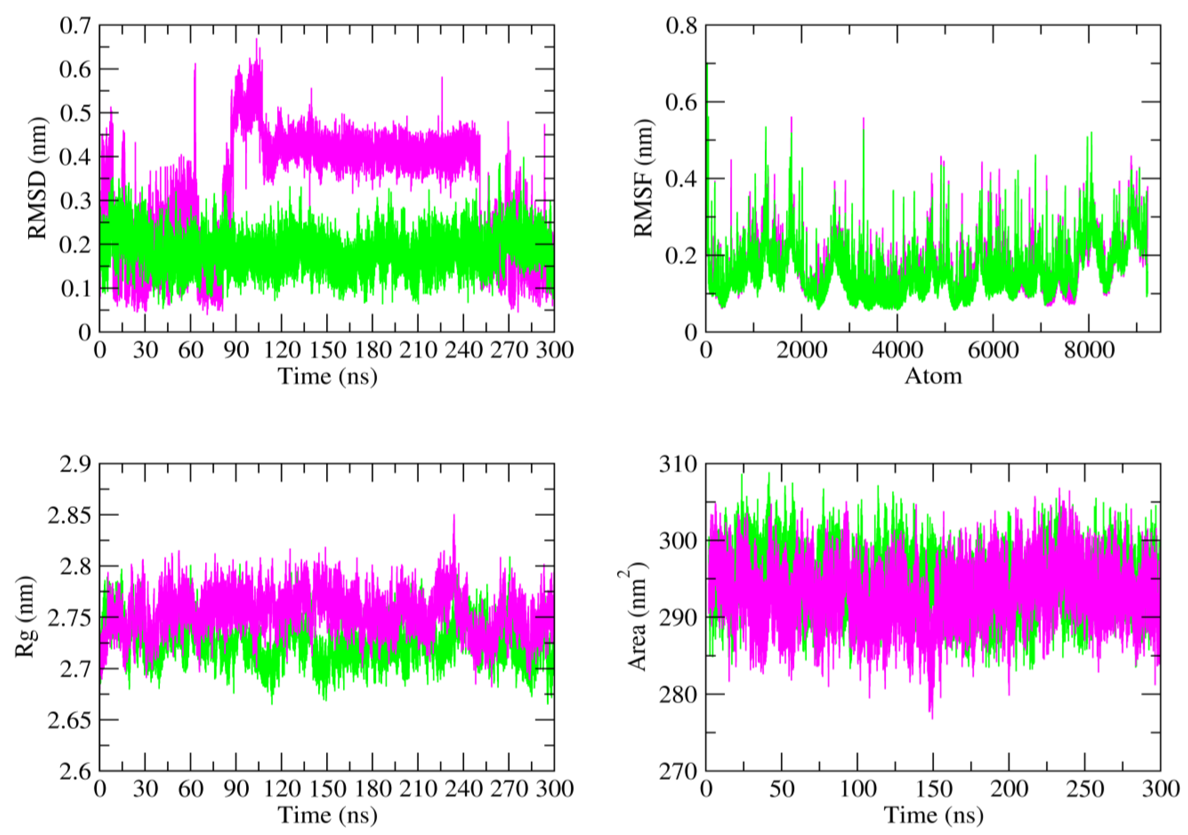
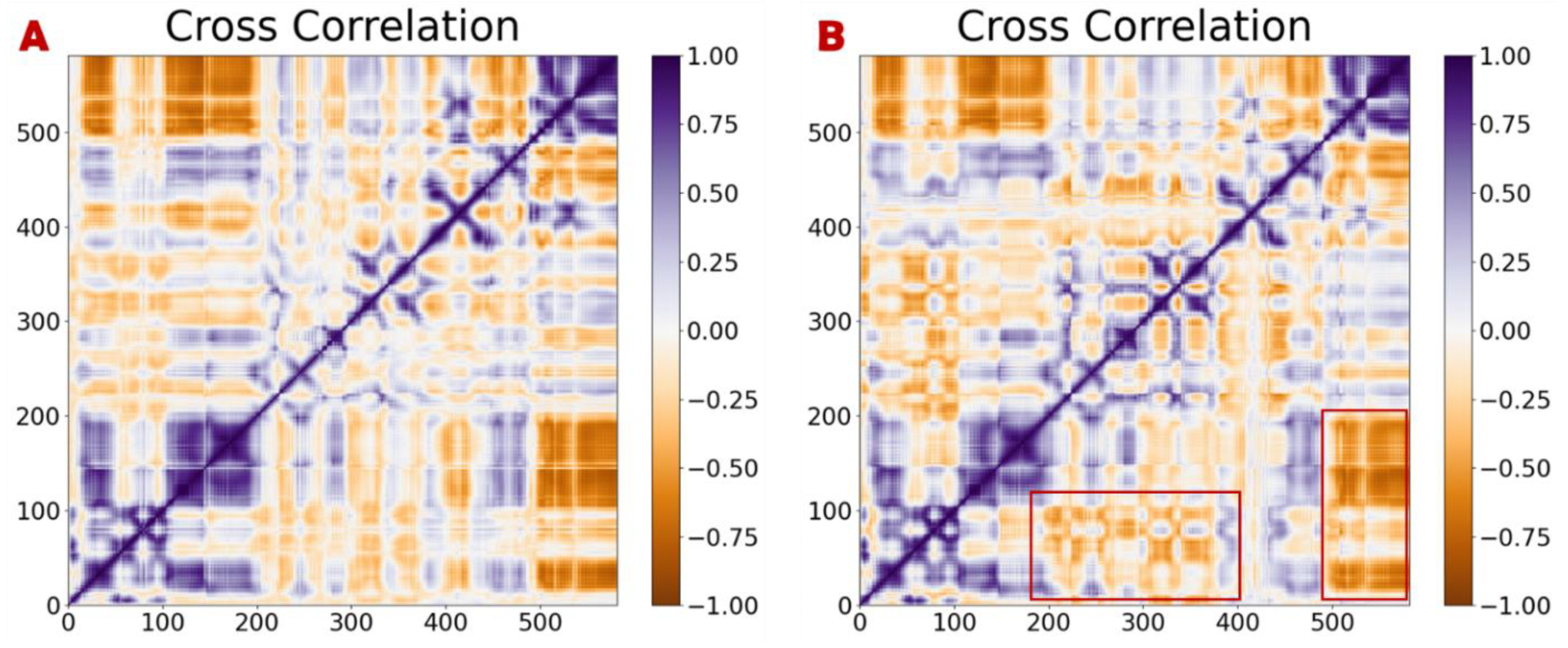
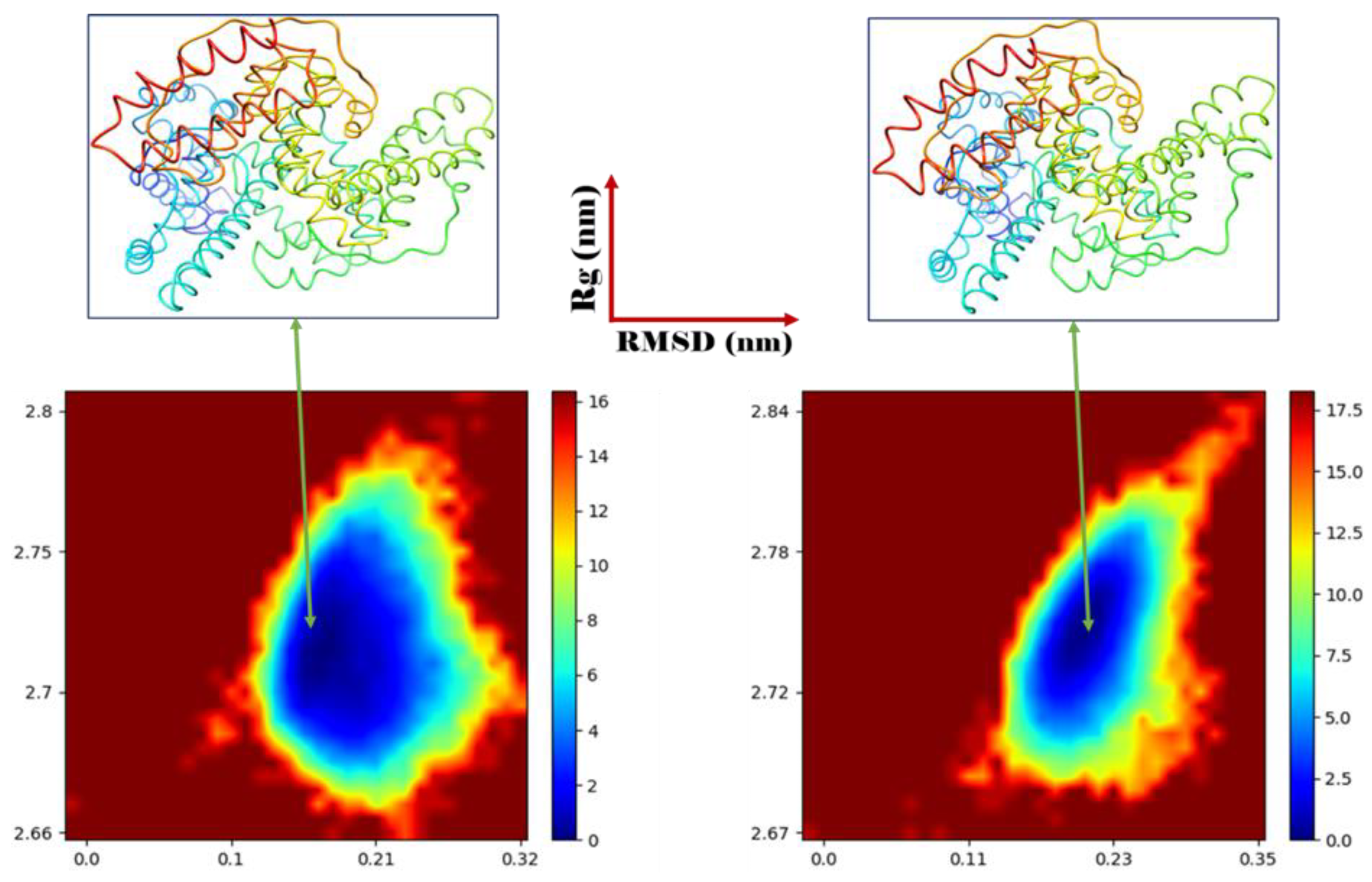
3.4. MD Simulation and Stability

Molecular dynamics simulations are carried out to comprehensively evaluate the stability and interaction dynamics of the synthesized molecules within the active site of the BSA protein. This involves detailed analyses of their binding affinities, structural stability and atomic-level mobility, which are compared to those of the reference drug (Aspirin) under similar simulation conditions. The study further aims to explore the capability of the synthesized molecules to inhibit the biological function of the BSA protein by assessing their influence on its structural and functional properties. These findings are intended to provide a deeper understanding of the molecules’ therapeutic potential and their comparative effectiveness against the reference drug [47,48,49].
Table 6 presents an in-depth analysis of the average values for four essential parameters (RMSD, RMSF, Rg and SASA), each serving a unique purpose in assessing the dynamic behavior of the generated complexes. RMSD gauges the overall stability of the structure by quantifying the deviation of atomic positions from the initial conformation, with smaller values signifying greater stability. RMSF measures the flexibility of individual residues or atoms, highlighting regions with considerable conformational variation. Rg provides insights into the compactness of the complex, where lower values indicate a more stable and tightly packed structure. SASA quantifies the degree of exposure of the molecule to the solvent, providing key information on folding and interactions with the surrounding medium. Collectively, these parameters offer a comprehensive assessment of the structural integrity and dynamic behavior of the complexes.
The RMSD results indicate that the 4d_BSA complex exhibits superior stability compared to the Aspirin_BSA complex throughout the 300 ns simulation period. The fact that the Aspirin_BSA complex deviates at 30 ns and 70 ns suggests that the complex undergoes structural adjustments or fluctuations before reaching a stable conformation. These deviations could be due to the complex undergoing conformational changes or adjustments to optimize the binding between Aspirin and the BSA protein. In contrast, the 4d_BSA complex demonstrates exceptional and constant stability throughout the simulation. The absence of significant deviations implies that the synthesized 4d molecule binds more stably within the active site of the BSA protein, maintaining its structure without undergoing major conformational changes. This may indicate that the 4d molecule has a stronger or more optimal interaction with BSA compared to Aspirin, contributing to its enhanced stability.
The average RMSD values further reinforce these observations. With an average RMSD of 0.18 nm, the 4d_BSA complex shows a high degree of structural integrity and less fluctuation compared to the Aspirin_BSA complex, which has a higher average RMSD of 0.32 nm. This suggests that 4d interacts more stably with BSA, potentially offering a more reliable and sustained binding interaction. These findings could indicate that the 4d molecule may have superior binding characteristics and could be a more stable therapeutic candidate when compared to Aspirin in terms of its interaction with the BSA protein.
The RMSF quantifies the average shifts of the BSA protein during its interaction with each ligand. The average RMSF values for the 4d_BSA and Aspirin_BSA complexes are both 0.16 nm, indicating that 4d and Aspirin binding to the BSA protein results in similar levels of flexibility or stability in the active site region. This suggests that both ligands exert comparable effects on the protein’s conformational dynamics, potentially reflecting analogous interaction modes or binding efficiencies. Furthermore, the low RMSF values emphasize the stability of these complexes, implying that the ligands maintain consistent interactions with the active site throughout the simulation.
The Rg also offers a valuable and reliable approach for evaluating the stability of the BSA protein’s folding or conformational changes when it is bound to a ligand. This metric provides insightful data on the compactness and structural integrity of the protein–ligand complex over the entire 300 ns MD simulation, making it an effective tool for assessing the dynamic stability of the interaction. Initially, the average Rg values for the 4d_BSA and Aspirin_BSA complexes were 2.75 nm and 2.72 nm, respectively. This data clearly showed a slight discrepancy between the Rg values of each protein complex, indicating that both structures remained rigid and exhibited higher compactness throughout the 300 ns MD simulation.
The SASA measures the surface area of a protein that is accessible to the solvent, providing insight into the extent to which the protein is stabilized by interactions with the solvent. The data in Table 6 reveal that the accessible surface areas for both complexes are almost identical. Specifically, the average SASA values for the 4d_BSA and Aspirin_BSA complexes were 294.60 nm2 and 292.99 nm2, respectively. This indicates that both ligands, 4d and Aspirin, interact with the BSA protein in a similar manner, with comparable solvent exposure, suggesting similar levels of stability and solvent interaction within the complexes. All the data obtained demonstrate that the synthesized molecules are capable of effectively inhibiting the biological function of the BSA protein. These findings suggest that the results of this study hold significant potential for the development of new and effective anti-inflammatory drugs. Figure 6 illustrates the biomolecular stability of each system over 300 ns of simulation.
Dynamic cross-correlation matrices were utilized to investigate the interconnected movements of structural configurations, providing insights into the stability of the BSA protein upon interaction with the reference ligand (Aspirin) and the synthesized compound (4d) derived from molecular dynamics simulations. These cross-correlation maps highlighted residual changes in the protein structure. Figure 7 depicts the movement of residues, both positive and negative, throughout the 300 ns simulation. The color scale represents the correlation levels, with blue indicating a positive correlation and red signifying a negative correlation. A positive correlation implies that residues move in the same direction, whereas a negative correlation indicates movement in opposite directions. The analysis of the dynamic cross-correlation matrix maps for both systems showed that their correlated motions were quite similar. The overall movements that displayed an indirect correlation in the 4d_BSA complex remained consistent with those in the Aspirin complex. However, the movements indicating a negative correlation significantly increased, particularly in the regions marked by red boxes. Moreover, at the atomic level, the molecule 4d formed a stable complex with the active site of BSA. The results of this study are in agreement with the molecular docking outcomes and the data from the dynamic simulations, confirming the ability of the proposed compounds to effectively inhibit the biological functions of the BSA protein.
To examine the dynamics of the BSA protein following interactions with the Aspirin and 4d molecules, we created a free energy landscape (FEL) to depict the changes in the BSA configurations. Two reaction parameters were chosen for the FEL: the RMSD of BSA, which indicates the stability of the protein over the 300 ns of the simulation, and Rg, which represents the BSA’s folding state. Figure 8 presents the free energy landscape of the interactions between the BSA protein and the 4d and Aspirin molecules. For 4d, a significant energy well is observed at RMSD values of 0.10 and 0.32 nm, with Rg values of 2.66 and 2.77 nm. Similarly, the reference molecule Aspirin shows a prominent energy well at RMSD values of 0.12 and 0.31 nm, with corresponding Rg values of 2.64 and 2.77 nm. These results indicate that both 4d and Aspirin exhibit stable binding conformations with the BSA protein, as reflected by the consistent energy wells. The slightly lower RMSD values for 4d suggest a potentially more stable interaction compared to Aspirin, which aligns with its observed compactness in the free energy landscape. Additionally, the similar Rg values for both molecules indicate that the overall structural compactness of the protein–ligand complex remains comparable in both cases. This reinforces the potential of 4d as a competitive molecule in binding stability and affinity when compared to Aspirin. Finally, the results of the free energy landscape (FEL) studies were particularly intriguing, as they provided a clear distinction between the biomolecular structures of the 4d and Aspirin complexes. These findings align closely with the outcomes of the RMSD and DCCM analyses, further validating the structural differences and dynamics observed in the interactions.
The MM-PBSA calculations were carried out to perform an extensive evaluation of the binding energies involved in the interaction between the two molecules and the BSA receptor, providing a detailed understanding of the forces that govern this interaction. The total binding energies of the 4d and Aspirin molecules, calculated as −15.91 and −14.25 kcal.mol−1 respectively, suggest that 4d binds more strongly to the BSA receptor compared to Aspirin. This difference in binding energy indicates that 4d may form a more stable complex with the protein, which could result in enhanced binding affinity and potentially greater inhibitory activity. The more negative binding energy of 4d implies stronger intermolecular interactions. In contrast, the slightly higher binding energy of Aspirin suggests that its interaction with BSA is less favorable, which could translate into a weaker or less stable binding. This analysis underscores the promising characteristics of 4d, especially in the context of its biological activity, such as its potential as a drug candidate. Finally, the van der Waals interactions are the primary contributors to the stability of the formed complexes. These interactions, although relatively weak compared to other types of bonding, play a crucial role in ensuring the proper alignment and close packing of the molecules with the BSA receptor.

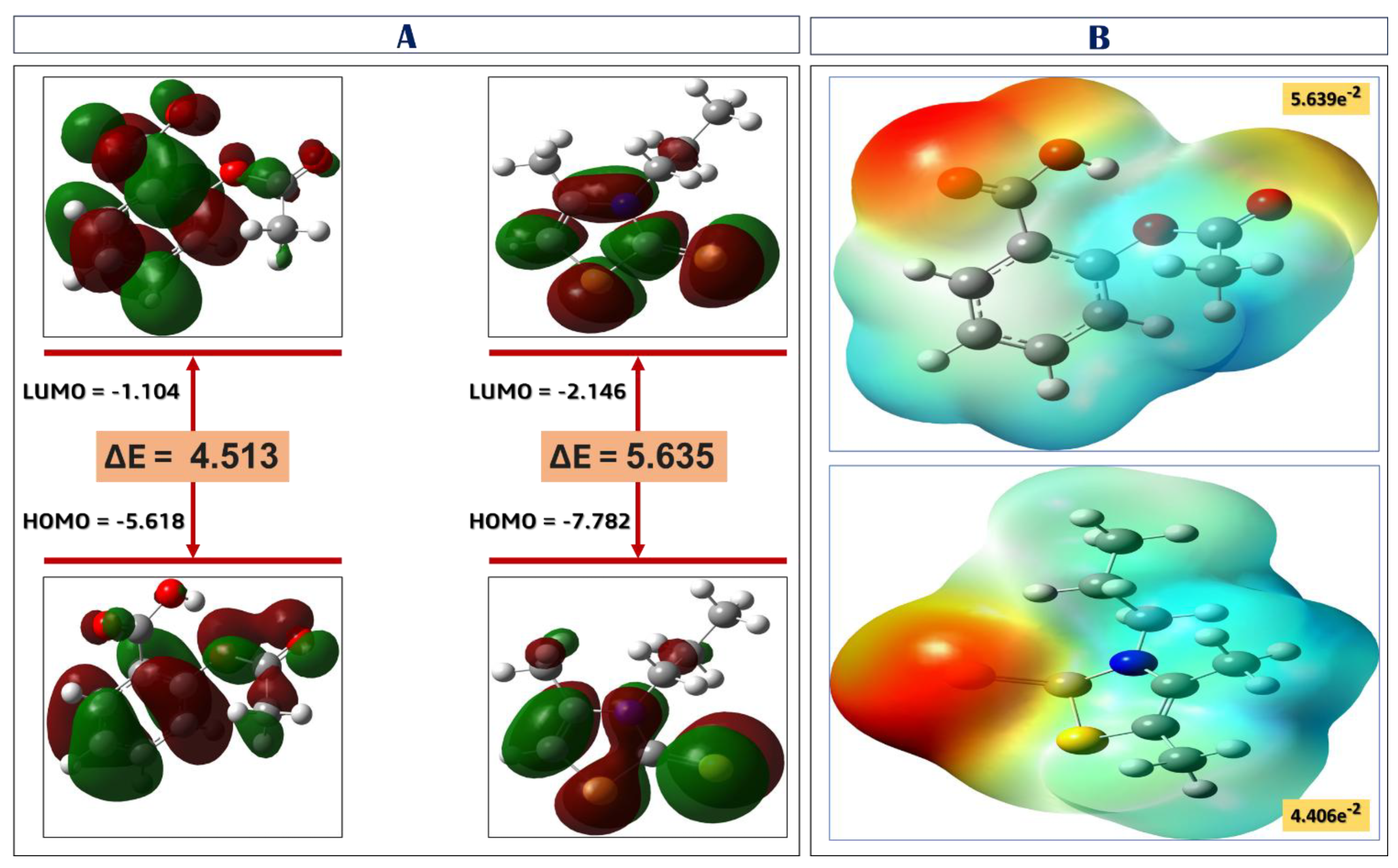
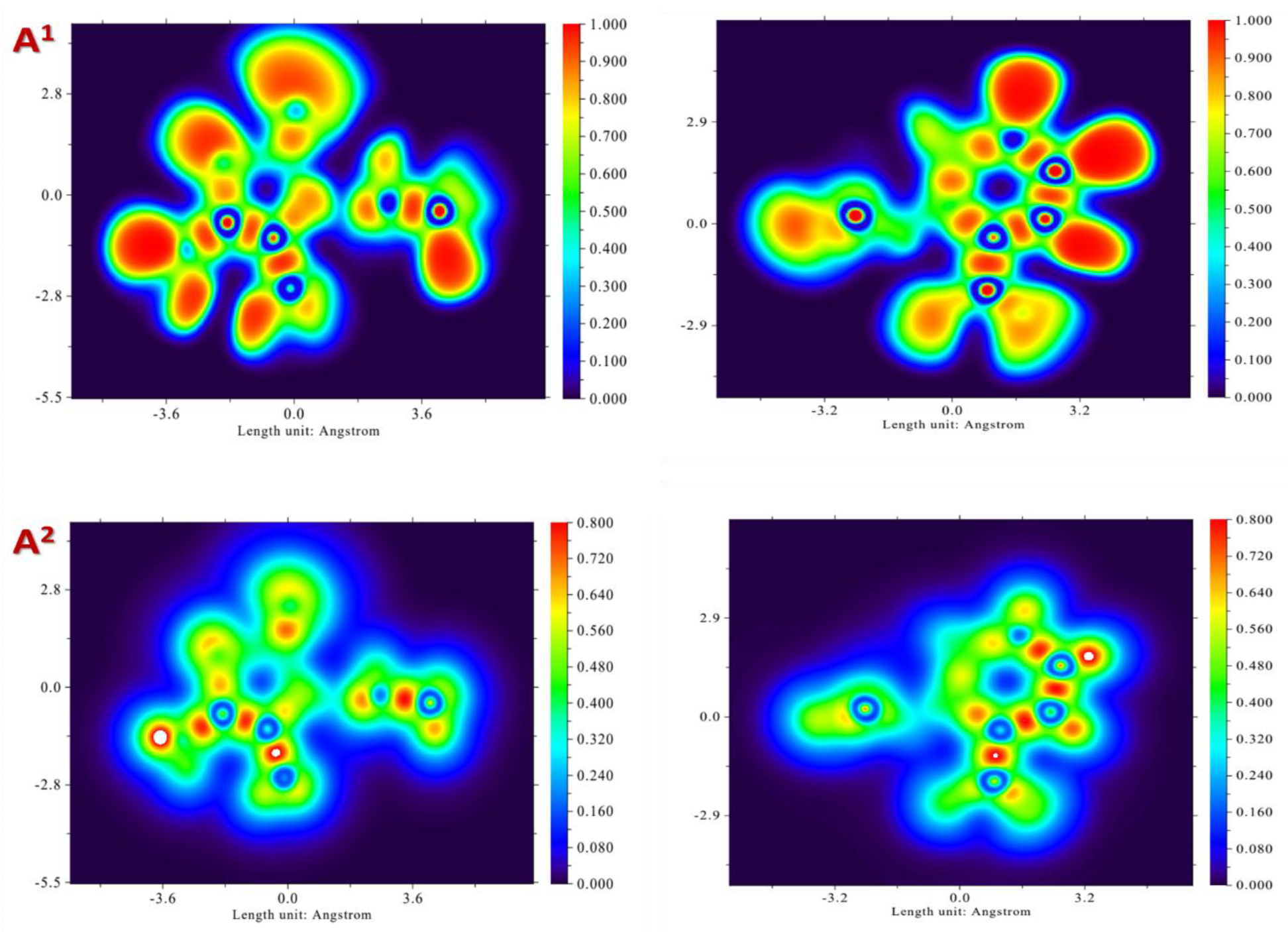
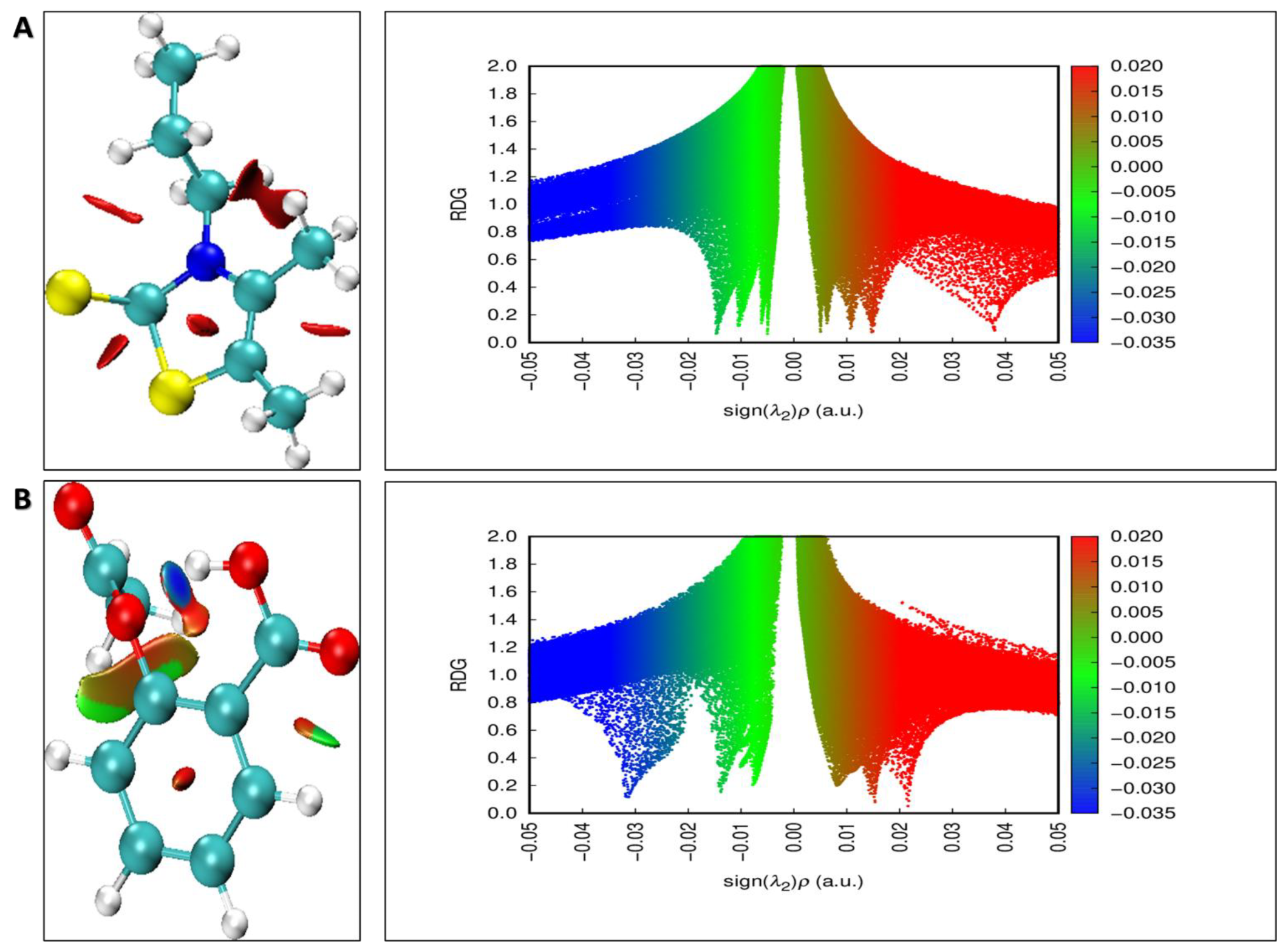
3.5. Density Functional Theory (DFT) Calculations

As per FMO theory, the HOMO and LUMO of the analyzed molecules are fundamental for determining molecular transport properties and anticipating the chemical reactivity of the conjugated system [50]. In addition, the energy gap (ΔE) signifies the fundamental energy requirement for initiating covalent bond formation. The HOMO indicates a molecule’s reactivity with electrophiles, acting as an electron donor. A higher energy HOMO typically suggests a greater electrophilic interaction. In contrast, a lower energy level corresponds to a reduced tendency to react with nucleophiles, making the molecules less reactive in that regard. The calculated energy values for 4d show a HOMO of −5.618 eV and a LUMO of −1.104 eV. In comparison, the Aspirin molecule has a HOMO of −7.785 eV and a LUMO of −2.146 eV (Figure 9A). These values indicate the energy levels associated with electron donation and acceptance for each molecule. The difference between the HOMO and LUMO helps to assess the molecule’s reactivity. Molecules with a smaller energy gap are typically more polarizable and exhibit higher chemical reactivity. The ΔE values indicate that the 4d molecule, with a ΔE of 4.513 eV, is more reactive than Aspirin, which has a ΔE of 5.635 eV. This enhanced reactivity of 4d allows it to form hydrogen bonds with the BSA protein more efficiently and rapidly compared to Aspirin, suggesting stronger and faster interactions between 4d and the protein. The HOMO orbitals of the 4d molecule are predominantly situated on the thiazole group thione and (–CH2CH3) groups, while the LUMO is spread across the majority of the molecule. These results are in agreement with docking studies, which revealed interactions between these specific groups and the BSA-binding site, suggesting that these regions play a key role in the binding process.
The MEP surface is crucial for studying how a substrate interacts with its protein. It reveals the distribution of electric charge across a molecule, which is essential for understanding hydrogen bonding, reactivity and the overall polarity. Additionally, MEP helps in identifying molecular interactions, including polarizability and structure–activity relationships, offering valuable insights into how molecules behave and interact with one another. The areas with greater nucleophilic potential, shown in red in Figure 9B, are found around the thione (C=S) and carbonyl (C=O) groups in both molecules, indicating these regions as possible sites for electrophilic reactions, highlighting their reactivity in chemical interactions. On the other hand, the regions with higher electrophilic potential are located in the aromatic ring and thiazole group of both molecules, marking these sites as likely targets for nucleophilic reactions. This is validated by molecular docking, which reveals that these groups form hydrogen bonds with the amino acids of the BSA protein.
The ELF (electron localization function) is a useful method for evaluating the likelihood of electron pair localization and gaining insights into electron behavior in multielectronic systems. Similarly, the LOL is widely used to characterize the molecular bonding, reactivity and chemical structure. Both ELF (localized orbital locator) and LOL rely on kinetic energy density to examine the electron distribution [51,52,53]. Nevertheless, the LOL map presents a more straightforward and clearer visualization than ELF. While ELF emphasizes the density of electron pairs, LOL highlights the gradient of localized orbitals, making it especially beneficial when orbitals overlap. Figure 10 presents a comprehensive depiction of the ELF and LOL maps for both the 4d molecule and Aspirin, shedding light on their electronic localization and bonding features. The ELF map ranges from 0 to 1, with regions exhibiting values below 0.5 signifying delocalized electronic zones, reflecting reduced localization of electrons in those areas. In contrast, when the electron density is more tightly concentrated, the LOL phase rises above 0.5, highlighting areas where electron localization is more pronounced. The increased LOL value signifies regions with robust covalent bonding or nuclear layers, marked by a notable concentration of electron density, which indicates a higher degree of electron localization in those areas. In the study of the 4d and Aspirin molecules, the electron cloud was extensively delocalized across certain carbon, oxygen, nitrogen and sulfur atoms, with blue regions marking the areas of electron dispersion. Alternatively, the red and orange regions around the hydrogen atoms exhibit higher ELF values, pointing to a strong covalent bond character with a significant density of electrons concentrated in these areas. In addition, the central areas of some hydrogen atoms in the molecules appeared white, signifying that their electron density exceeded the upper limit of the color scale (0.80), as illustrated in Figure 10.
The stability of the compounds in this study was mainly ascribed to weak inter/intramolecular interactions. These interactions were investigated via the RDG analysis, which focuses on non-covalent interactions. The λ2 sign was utilized to distinguish between bonded, λ2 < 0 and non-bonded, λ2 > 0 interactions. In addition, the λ2 sign the ρ function varied between −0.05 and 0.05 a.u., as shown on the RDG plot (Figure 11).
Furthermore, red peaks (λ2 > 0; ρ > 0) signify steric repulsion effects within the ring structure. Green peaks (λ2 = 0; ρ = 0) are associated with Van der Waals interactions, whereas blue peaks (ρ > 0; λ2 < 0) indicate electrostatic interactions, such as hydrogen or halogen bonds [54,55]. The RDG plots (Figure 11) reveal that the blue surface in the 4d molecule emphasizes strong intermolecular H-bonding for N–H…N. The RDG scatter plot further supports this, showing blue contours for −0.05 a.u. < λ2ρ < −0.01 a.u., indicating robust H-bonding. Van der Waals (VDW) interactions are identified by red–green mixed spikes, ranging from −0.02 a.u. < λ2ρ < 0.02 a.u. These spikes are observed between the hydrogen atoms. The range of λ2ρ values confirms the existence of these interactions. These interactions further demonstrate the proximity and interaction of hydrogen atoms in the molecule. Steric effects, highlighted by red coloration, were detected in the range of 0.005 a.u. < λ2ρ < 0.045 a.u. These effects were especially prominent at the core of the thiazole group. The red regions correspond to areas of significant steric repulsion.

4. Conclusions

This study aimed to synthesize thiazoline-2-thione derivatives and evaluate their anti-inflammatory properties as potential alternatives to the non-steroidal anti-inflammatory drug Aspirin, which is associated with adverse effects such as liver toxicity. This study highlights that the 4d molecule, one of the synthesized thiazoline-2-thione derivatives, demonstrates remarkable anti-inflammatory properties. Its performance suggests it as a promising candidate for further development as an alternative to conventional anti-inflammatory drugs. Furthermore, molecular docking studies reveal that the 4d molecule has a strong binding affinity to the BSA receptor, with a binding free energy (∆G) of −5.274 kcal.mol−1, surpassing that of the reference drug Aspirin (∆G of −4.641 kcal.mol−1). These results are consistent with the in vitro findings, supporting the potential of 4d as an effective inhibitor of critical inflammatory pathways. Quantum chemical calculations, encompassing HOMO, LUMO and MPE analyses, combined with molecular dynamics simulations, provide additional evidence of the 4d molecule’s stability and strong interactions within the active sites of BSA. These findings not only highlight its superior binding characteristics compared to the reference drug but also reinforce its potential as a stable and effective anti-inflammatory agent. Moreover, the results from predictive modeling and ADMET studies validate the drug-like properties of these compounds, ensuring they meet the criteria outlined by Lipinski’s Rule of Five. These combined insights not only highlight the potential of 4d as an effective anti-inflammatory agent but also provide a strong foundation for its further optimization and clinical development.

Supplementary Materials

The following supporting information can be downloaded at https://www.mdpi.com/article/10.3390/app15116095/s1: Figure S1: 1H and 13C NMR spectra of the four synthesized compounds.

Author Contributions

Conceptualization, B.F. and B.L.; methodology, G.A.; software, B.F.; validation, G.A., H.S. and F.M.; formal analysis, B.T.; investigation, B.F.; resources, G.A.; data curation, B.F.; writing—original draft preparation, C.Z. and M.A.O.A.; writing—review and editing, B.F.; visualization, G.A.; supervision, G.A.; funding acquisition, J.M. All authors have read and agreed to the published version of the manuscript.

Funding

Authors Jestin Mandumpal is employed by the American University of the Middle-East, which provided funding for the publication fees associated with this research.

Data Availability Statement

The original contributions presented in this study are included in the article. Further inquiries can be directed to the corresponding authors.

Conflicts of Interest

The authors declare no conflicts of interest.

References

  1. Broughton, H.B.; Watson, I.A. Selection of Heterocycles for Drug Design. J. Mol. Graph. Model. 2004, 23, 51–58. [Google Scholar] [CrossRef] [PubMed]
  2. Kumar, S.; Arora, A.; Sapra, S.; Kumar, R.; Singh, B.K.; Singh, S.K. Recent Advances in the Synthesis and Utility of Thiazoline and Its Derivatives. RSC Adv. 2024, 14, 902–953. [Google Scholar] [CrossRef]
  3. Chugh, V.; Pandey, G.; Rautela, R.; Mohan, C. Heterocyclic Compounds Containing Thiazole Ring as Important Material in Medicinal Chemistry. Mater. Today Proc. 2022, 69, 478–481. [Google Scholar] [CrossRef]
  4. Amin, M.Z.; Rity, T.I.; Uddin, M.R.; Rahman, M.d.M.; Uddin, M.J. A Comparative Assessment of Anti-Inflammatory, Anti-Oxidant and Anti-Bacterial Activities of Hybrid and Indigenous Varieties of Pumpkin (Cucurbita Maxima Linn.) Seed Oil. Biocatal. Agric. Biotechnol. 2020, 28, 101767. [Google Scholar] [CrossRef]
  5. Lekouaghet, A.; Boutefnouchet, A.; Bensuici, C.; Gali, L.; Ghenaiet, K.; Tichati, L. In Vitro Evaluation of Antioxidant and Anti-Inflammatory Activities of the Hydroalcoholic Extract and Its Fractions from Leuzea Conifera L. Roots. S. Afr. J. Bot. 2020, 132, 103–107. [Google Scholar] [CrossRef]
  6. Singh, H.; Kumar, S.; Arya, A. Evaluation of Antibacterial, Antioxidant, and Anti-Inflammatory Properties of GC/MS Analysis of Extracts of Ajuga. Integrifolia Buch.-Ham. Leaves. Sci. Rep. 2024, 14, 16754. [Google Scholar] [CrossRef]
  7. Bakhouche, I.; Aliat, T.; Boubellouta, T.; Gali, L.; Şen, A.; Bellik, Y. Phenolic Contents and in Vitro Antioxidant, Anti-Tyrosinase, and Anti-Inflammatory Effects of Leaves and Roots Extracts of the Halophyte Limonium Delicatulum. S. Afr. J. Bot. 2021, 139, 42–49. [Google Scholar] [CrossRef]
  8. Kaur, R.; Desai, D.; Amin, S.; Raza, K.; Bhalla, A.; Yadav, P.; Kaushal, N. Selenocoxib-3, a Novel Anti-Inflammatory Therapeutic Effectively Resolves Colitis. Mol. Cell Biochem. 2023, 478, 621–636. [Google Scholar] [CrossRef]
  9. Aladejana, E.B. Biological Properties of Polyherbal Formulations: A Review of Their Antimicrobial, Anti-Inflammatory, Antioxidant, and Toxicological Activities. Pharmacogn. J. 2023, 15, 933–963. [Google Scholar] [CrossRef]
  10. Bindu, S.; Mazumder, S.; Bandyopadhyay, U. Non-Steroidal Anti-Inflammatory Drugs (NSAIDs) and Organ Damage: A Current Perspective. Biochem. Pharmacol. 2020, 180, 114147. [Google Scholar] [CrossRef]
  11. Panchal, N.K.; Prince Sabina, E. Non-Steroidal Anti-Inflammatory Drugs (NSAIDs): A Current Insight into Its Molecular Mechanism Eliciting Organ Toxicities. Food Chem. Toxicol. 2023, 172, 113598. [Google Scholar] [CrossRef] [PubMed]
  12. Muhammed, M.T.; Aki-Yalcin, E. Molecular Docking: Principles, Advances, and Its Applications in Drug Discovery. Lett. Drug Des. Discov. 2024, 21, 480–495. [Google Scholar] [CrossRef]
  13. Prieto-Martínez, F.D.; López-López, E.; Eurídice Juárez-Mercado, K.; Medina-Franco, J.L. Computational Drug Design Methods—Current and Future Perspectives. In Silico Drug Design; Elsevier: Amsterdam, The Netherlands, 2019; pp. 19–44. [Google Scholar]
  14. Deore, A.B.; Dhumane, J.R.; Wagh, R.; Sonawane, R. The Stages of Drug Discovery and Development Process. Asian J. Pharm. Res. Dev. 2019, 7, 62–67. [Google Scholar] [CrossRef]
  15. Ferreira, L.; Dos Santos, R.; Oliva, G.; Andricopulo, A. Molecular Docking and Structure-Based Drug Design Strategies. Molecules 2015, 20, 13384–13421. [Google Scholar] [CrossRef]
  16. Alqahtani, S. In Silico ADME-Tox Modeling: Progress and Prospects. Expert. Opin. Drug Metab. Toxicol. 2017, 13, 1147–1158. [Google Scholar] [CrossRef] [PubMed]
  17. Van Gunsteren, W.F.; Berendsen, H.J.C. Computer Simulation of Molecular Dynamics: Methodology, Applications, and Perspectives in Chemistry. Angew. Chem. Int. Ed. Engl. 1990, 29, 992–1023. [Google Scholar] [CrossRef]
  18. Kuhlman, B.; Bradley, P. Advances in Protein Structure Prediction and Design. Nat. Rev. Mol. Cell Biol. 2019, 20, 681–697. [Google Scholar] [CrossRef]
  19. Berman, H.M. The Protein Data Bank. Nucleic Acids Res. 2000, 28, 235–242. [Google Scholar] [CrossRef] [PubMed]
  20. Tegegn, D.F.; Belachew, H.Z.; Salau, A.O. DFT/TDDFT Calculations of Geometry Optimization, Electronic Structure and Spectral Properties of Clevudine and Telbivudine for Treatment of Chronic Hepatitis B. Sci. Rep. 2024, 14, 8146. [Google Scholar] [CrossRef]
  21. Siddique, F.; Anwaar, A.; Bashir, M.; Nadeem, S.; Rawat, R.; Eyupoglu, V.; Afzal, S.; Bibi, M.; Bin Jardan, Y.A.; Bourhia, M. Revisiting Methotrexate and Phototrexate Zinc15 Library-Based Derivatives Using Deep Learning in-Silico Drug Design Approach. Front. Chem. 2024, 12, 1380266. [Google Scholar] [CrossRef]
  22. Chou, L.-K.; Quijada, M.A.; Clevenger, M.B.; de Oliveira, G.F.; Abboud, K.A.; Tanner, D.B.; Talham, D.R. Dication Salts of the Organic Donor Bis(Ethylenedithio)Tetrathiafulvalene. Chem. Mater. 1995, 7, 530–534. [Google Scholar] [CrossRef]
  23. Bellec, N.; Lorcy, D.; Boubekeur, K.; Carlier, R.; Tallec, A.; Los, S.z.; Pukacki, W.; Trybula, M.; Piekara-Sady, L.; Robert, A. Dicationic State of Dithiadiazafulvalene within a TCNQ Charge-Transfer Complex: Generation and Characterization. Chem. Mater. 1999, 11, 3147–3153. [Google Scholar] [CrossRef]
  24. Anokwah, D.; Kwatia, E.A.; Amponsah, I.K.; Jibira, Y.; Harley, B.K.; Ameyaw, E.O.; Obese, E.; Biney, R.P.; Mensah, A.Y. Evaluation of the Anti-Inflammatory and Antioxidant Potential of the Stem Bark Extract and Some Constituents of Aidia Genipiflora (DC.) Dandy (Rubiaceae). Heliyon 2022, 8, e10082. [Google Scholar] [CrossRef]
  25. Benesi, H.A.; Hildebrand, J.H. A Spectrophotometric Investigation of the Interaction of Iodine with Aromatic Hydrocarbons. J. Am. Chem. Soc. 1949, 71, 2703–2707. [Google Scholar] [CrossRef]
  26. Ahmad, S.; Raza, K. Identification of 5-Nitroindazole as a Multitargeted Inhibitor for CDK and Transferase Kinase in Lung Cancer: A Multisampling Algorithm-Based Structural Study. Mol. Divers. 2024, 28, 1189–1202. [Google Scholar] [CrossRef]
  27. Chitre, T.S.; Hirode, P.V.; Lokwani, D.K.; Bhatambrekar, A.L.; Hajare, S.G.; Thorat, S.B.; Priya, D.; Pradhan, K.B.; Asgaonkar, K.D.; Jain, S.P. In-Silico Studies of 2-Aminothiazole Derivatives as Anticancer Agents by QSAR, Molecular Docking, MD Simulation and MM-GBSA Approaches. J. Biomol. Struct. Dyn. 2024, 42, 11396–11414. [Google Scholar] [CrossRef] [PubMed]
  28. Musthafa, M.; Aneesrahman, K.N.; Perumalsamy, B.; Ramasamy, T.; Ganguly, R.; Sreekanth, A. Synthesis, Crystal Structure, DFT Study, in Vitro and in Silico Molecular Docking of Novel Bis (Aroyl Selenourea) Derivatives. J. Mol. Struct. 2019, 1180, 585–594. [Google Scholar] [CrossRef]
  29. Hossain, M.d.J.; Sultan, M.d.Z.; Rashid, M.A.; Kuddus, M.d.R. Interactions of Linagliptin, Rabeprazole Sodium, and Their Formed Complex with Bovine Serum Albumin: Computational Docking and Fluorescence Spectroscopic Methods. Anal. Sci. Adv. 2021, 2, 480–494. [Google Scholar] [CrossRef] [PubMed]
  30. Madhavi Sastry, G.; Adzhigirey, M.; Day, T.; Annabhimoju, R.; Sherman, W. Protein and Ligand Preparation: Parameters, Protocols, and Influence on Virtual Screening Enrichments. J. Comput. Aided Mol. Des. 2013, 27, 221–234. [Google Scholar] [CrossRef]
  31. Kumar, S.; Manoharan, A.; J, J.; Abdelgawad, M.A.; Mahdi, W.A.; Alshehri, S.; Ghoneim, M.M.; Pappachen, L.K.; Zachariah, S.M.; Aneesh, T.P.; et al. Exploiting Butyrylcholinesterase Inhibitors through a Combined 3-D Pharmacophore Modeling, QSAR, Molecular Docking, and Molecular Dynamics Investigation. RSC Adv. 2023, 13, 9513–9529. [Google Scholar] [CrossRef]
  32. Amani, A.M.; Gholamzadeh, S.; Zarenezhad, M.; Malekpour, A.; Javidnezhad, E. DFT and Molecular Docking Studies on Biological Active Acridines: The Interaction with Bovine Serum Albumin. J. Adv. Med. Sci. Appl. Technol. 2015, 1, 78. [Google Scholar] [CrossRef]
  33. B, M.; Manandhar, S.; Hari, G.; Priya, K.; Kumar B, H.; Pai, K.S.R. Virtual Structure-Based Docking, WaterMap, and Molecular Dynamics Guided Identification of the Potential Natural Compounds as Inhibitors of Protein-Tyrosine Phosphatase 1B. J. Mol. Struct. 2021, 1226, 129396. [Google Scholar] [CrossRef]
  34. Ni, Y.; Zhu, R.; Kokot, S. Competitive Binding of Small Molecules with Biopolymers: A Fluorescence Spectroscopy and Chemometrics Study of the Interaction of Aspirin and Ibuprofen with BSA. Analyst 2011, 136, 4794. [Google Scholar] [CrossRef]
  35. Ayodele, P.F.; Bamigbade, A.; Bamigbade, O.O.; Adeniyi, I.A.; Tachin, E.S.; Seweje, A.J.; Farohunbi, S.T. Illustrated Procedure to Perform Molecular Docking Using PyRx and Biovia Discovery Studio Visualizer: A Case Study of 10kt With Atropine. Progress. Drug Discov. Biomed. Sci. 2023, 6. [Google Scholar] [CrossRef]
  36. Xu, L.; Zhao, L.; Che, J.; Zhang, Q.; Cao, R.; Li, X. Identification of Novel Influenza Polymerase PB2 Inhibitors Using Virtual Screening Approach and Molecular Dynamics Simulation Analysis of Active Compounds. Bioorg Med. Chem. 2021, 52, 116515. [Google Scholar] [CrossRef]
  37. Daina, A.; Michielin, O.; Zoete, V. SwissADME: A Free Web Tool to Evaluate Pharmacokinetics, Drug-Likeness and Medicinal Chemistry Friendliness of Small Molecules. Sci. Rep. 2017, 7, 42717. [Google Scholar] [CrossRef] [PubMed]
  38. Banerjee, P.; Eckert, A.O.; Schrey, A.K.; Preissner, R. ProTox-II: A Webserver for the Prediction of Toxicity of Chemicals. Nucleic Acids Res. 2018, 46, W257–W263. [Google Scholar] [CrossRef]
  39. Huang, J.; Rauscher, S.; Nawrocki, G.; Ran, T.; Feig, M.; de Groot, B.L.; Grubmüller, H.; MacKerell, A.D. CHARMM36: An Improved Force Field for Folded and Intrinsically Disordered Proteins. Biophys. J. 2017, 112, 175a–176a. [Google Scholar] [CrossRef]
  40. Bugnon, M.; Goullieux, M.; Röhrig, U.F.; Perez, M.A.S.; Daina, A.; Michielin, O.; Zoete, V. SwissParam 2023: A Modern Web-Based Tool for Efficient Small Molecule Parametrization. J. Chem. Inf. Model. 2023, 63, 6469–6475. [Google Scholar] [CrossRef]
  41. Bussi, G.; Donadio, D.; Parrinello, M. Canonical Sampling through Velocity Rescaling. J. Chem. Phys. 2007, 126, 014101. [Google Scholar] [CrossRef]
  42. Bourougaa, L.; Ouassaf, M.; Alhatlani, B.Y. Clinical Informatics and Molecular Hybridization of Established Clinical DPP-4 Inhibitors to Generate next-Level Diabetes Type 2 Drugs. Chem. Pap. 2024, 78, 8485–8503. [Google Scholar] [CrossRef]
  43. Elangovan, N.; Sowrirajan, S.; Arumugam, N.; Rajeswari, B.; Mathew, S.; Priya, C.G.; Venkatraman, B.R.; Mahalingam, S.M. Theoretical Investigation on Solvents Effect in Molecular Structure (TD-DFT, MEP, HOMO-LUMO), Topological Analysis and Molecular Docking Studies of N-(5-((4-Ethylpiperazin-1-Yl)Methyl)Pyridin-2-Yl)-5-Fluoro-4-(4-Fluoro-1-Isopropyl-2-Methyl-1H-Benzo[d] Imidazol-6-Yl) Pyrimidin-2-Amine. Polycycl. Aromat. Compd. 2024, 44, 4467–4490. [Google Scholar] [CrossRef]
  44. Becke, A.D. Density-Functional Thermochemistry. III. The Role of Exact Exchange. J. Chem. Phys. 1993, 98, 5648–5652. [Google Scholar] [CrossRef]
  45. Castaneda, S.M.B.; Alvarenga, E.S.; Demuner, A.J.; Guimaraes, L.M. Vibrational Spectra and Theoretical Calculations of a Natural Pentacyclic Triterpene Alcool Isolated from Mucuna Pruriens. Struct. Chem. 2020, 31, 599–607. [Google Scholar] [CrossRef]
  46. Lu, T.; Chen, F. Multiwfn: A Multifunctional Wavefunction Analyzer. J. Comput. Chem. 2012, 33, 580–592. [Google Scholar] [CrossRef]
  47. Ouassaf, M.; Bourougaa, L.; Bahaz, F.; Alhatlani, B.Y. Exploring the Antiviral Potential of Artemisia Annua Through JAK-STAT Pathway Targeting: A Network Pharmacology Approach. Pharmaceuticals 2024, 17, 1539. [Google Scholar] [CrossRef]
  48. Lotfi, B.; Mebarka, O.; Khan, S.U.; Htar, T.T. Pharmacophore-Based Virtual Screening, Molecular Docking and Molecular Dynamics Studies for the Discovery of Novel Neuraminidase Inhibitors. J. Biomol. Struct. Dyn. 2024, 42, 5308–5320. [Google Scholar] [CrossRef] [PubMed]
  49. Lotfi, B.; Mebarka, O.; Alhatlani, B.Y.; Abdallah, E.M.; Kawsar, S.M.A. Pharmacoinformatics and Breed-Based De Novo Hybridization Studies to Develop New Neuraminidase Inhibitors as Potential Anti-Influenza Agents. Molecules 2023, 28, 6678. [Google Scholar] [CrossRef]
  50. Choudhary, V.; Bhatt, A.; Dash, D.; Sharma, N. DFT Calculations on Molecular Structures, HOMO–LUMO Study, Reactivity Descriptors and Spectral Analyses of Newly Synthesized Diorganotin(IV) 2-chloridophenylacetohydroxamate Complexes. J. Comput. Chem. 2019, 40, 2354–2363. [Google Scholar] [CrossRef]
  51. Feixas, F.; Matito, E.; Duran, M.; Solà, M.; Silvi, B. Electron Localization Function at the Correlated Level: A Natural Orbital Formulation. J. Chem. Theory Comput. 2010, 6, 2736–2742. [Google Scholar] [CrossRef]
  52. Poater, J.; Duran, M.; Solà, M.; Silvi, B. Theoretical Evaluation of Electron Delocalization in Aromatic Molecules by Means of Atoms in Molecules (AIM) and Electron Localization Function (ELF) Topological Approaches. Chem. Rev. 2005, 105, 3911–3947. [Google Scholar] [CrossRef] [PubMed]
  53. Janani, S.; Rajagopal, H.; Muthu, S.; Aayisha, S.; Raja, M. Molecular Structure, Spectroscopic (FT-IR, FT-Raman, NMR), HOMO-LUMO, Chemical Reactivity, AIM, ELF, LOL and Molecular Docking Studies on 1-Benzyl-4-(N-Boc-Amino)Piperidine. J. Mol. Struct. 2021, 1230, 129657. [Google Scholar] [CrossRef]
  54. Reeda, V.J.; Sakthivel, S.; Divya, P.; Javed, S.; Jothy, V.B. Conformational Stability, Quantum Computational (DFT), Vibrational, Electronic and Non-Covalent Interactions (QTAIM, RDG and IGM) of Antibacterial Compound N-(1-Naphthyl)Ethylenediamine Dihydrochloride. J. Mol. Struct. 2024, 1298, 137043. [Google Scholar] [CrossRef]
  55. Suja, R.; Rathika, A.; Reeda, V.S.J.; Kumar, A.A.; Divya, P. Synthesis, Spectroscopic Analysis (FT-IR, FT-Raman, UV, NMR), Non-Covalent Interactions (RDG, IGM) and Dynamic Simulation on Bis(8-hydroxy Quinoline) Salicylate Salicylic Acid. J. Mol. Struct. 2024, 1310, 138231. [Google Scholar] [CrossRef]
Figure 1. Visual representation of the strategy used in this study.
Applsci 15 06095 g001
Figure 2. Illustration of the stepwise reaction mechanism for synthesizing thiazoline-2-thione derivatives.
Applsci 15 06095 g002
Figure 3. Percentage inhibition (I %) activity of the BSA enzyme at different concentrations. (A) 4d, (B) 3c and (C) Aspirin.
Applsci 15 06095 g003
Figure 4. The relationship between As/(Ac − As) and 1/[C]. (A) 4d, (B) 3c and (C) Aspirin.
Applsci 15 06095 g004
Figure 5. Molecular interactions of the two best synthetized molecules and Aspirin within the active site of the BSA protein. (A) Aspirin, (B) 4d and (C) 3c.
Applsci 15 06095 g005
Figure 6. Evaluation of biomolecular stability during 300 ns of simulation, including RMSD, RMSF, Rg and SASA. The 4d_BSA complex is represented in green, while the Aspirin_BSA complex is shown in magenta.
Applsci 15 06095 g006
Figure 7. Cross-correlation maps based on Cα atom positions of the BSA protein: (A) 4d and (B) Aspirin.
Applsci 15 06095 g007
Figure 8. Free energy profile derived from RMSD and Rg (in nm), with the 4d_BSA complex shown on the left and the Aspirin_BSA complex on the right. The structures corresponding to the minimum energy regions were obtained.
Applsci 15 06095 g008
Figure 9. FMO analysis. (A) HOMO and LUMO energies obtained for 4d (left) and Aspirin (right). (B) MEP surface of both molecules.
Applsci 15 06095 g009
Figure 10. (A1) ELF (4d in the left and Aspirin in the right) and (A2) LOL (4d in the right and Aspirin in the right).
Applsci 15 06095 g010
Figure 11. Non-covalent interactions (NCIs) plot (left) and reduced density gradient (RDG) plot (right) for (A) 4d and (B) Aspirin molecules.
Applsci 15 06095 g011
Table 1. IC50 values of the best synthesized molecules and Aspirin against the BSA protein.
CompoundsEquationsR2IC50 (µg/mL)
4d y = 92.479 77.254 e x p ( 27.241 x ) 0.99321.9
3c y = 83.129 63.395 e x p ( 20.472 x ) 0.99031.7
Aspirin y = 92.541 75.542 e x p ( 26.0095 x ) 0.99622
Table 2. Binding free energies and binding constants for the exanimated molecules with the BSA protein via UV–Visible spectroscopy.
CompoundsEquationsR2K (M−1)ΔG (Kcal.mol−1)
4d y = 1.220 0.013 X 0.97916.30 × 103−5.75
3c y = 1.107 0.030 X 0.9845.95 × 103−5.16
Aspirin y = 1.115 0.016 X 0.98712.40 × 103−5.59
Table 3. The molecular interactions between the best synthetized molecules and Aspirin with the BSA active site.
Molecules∆G (XP)H-BondDistance (Å)Pi–sulfurDistance (Å)HydrophobicDistance (Å)
4d−5.274Tyr149, Arg256[2.0–2.1]Tyr149, [2.9–3.1]Tyr149, Leu237, His241, Ile263, Ile289, Ala290, [3.7–4.8]
3c−4.731Tyr149
Arg256
2.2Tyr1492.1Tyr149, His241, Leu237, Leu259, Ile263, Ile289, Ala290[4.1–5.0]
Aspirinref−4.641Tyr149, Arg256[2.2–2.5]--Leu237 and Ala290[4.3–5.2]
Table 4. Pharmacokinetic and physicochemical parameters determined through SwissADME.
4dAspirin
MW (g/mol)187.33180.16
Consensus Log Po/w2.361.28
Log S−2.77−1.85
Bioavailability0.550.85
GI absorptionHighHigh
Cytochrome P450 inhibitionNoNo
LipinskiYesYes
Synthetic accessibility2.471.52
Table 5. Toxicity prediction of the top synthesized molecule via the ProTox-II webserver.
HepatotoxicityNeurotoxicityCardiotoxicityCarcinogenicityCytotoxicityLD50 (mg/kg)Class
4dInactiveInactiveInactiveInactiveInactive9604
Table 6. The average values of RMSD, RMSF, Rg and SASA for BSA–ligand complexes over the 300 ns simulation period.
4d_BSA ComplexAspirin_BSA Complex
RMSD (nm)0.180 ± 0.010.324 ± 0.02
RMSF (nm)0.161 ± 0.010.169 ± 0.01
Rg (nm)2.757 ± 0.032.725 ± 0.02
SASA (nm2)294.606 ± 1.25292.994 ± 1.10
Disclaimer/Publisher’s Note: The statements, opinions and data contained in all publications are solely those of the individual author(s) and contributor(s) and not of MDPI and/or the editor(s). MDPI and/or the editor(s) disclaim responsibility for any injury to people or property resulting from any ideas, methods, instructions or products referred to in the content.

