Article

Preventive Capacity of Citrus paradisi Juice for Male Reproductive Damage Induced by Cadmium Chloride in Mice

by
Isela Álvarez-González
1,*,
José David García-García
1,
Beatriz A. Espinosa-Ahedo
1,
Luis S. Muñoz-Carrillo
1,
José A. Morales-González
2,
Eduardo O. Madrigal-Santillán
2,
Felipe de Jesús Carrillo-Romo
3,
Antonieta García-Murillo
3,
Rogelio Paniagua-Pérez
4 and
Eduardo Madrigal-Bujaidar
1,*
1
Laboratorio de Genética, Escuela Nacional de Ciencias Biológicas, Instituto Politécnico Nacional, Av. Wilfrido Massieu s/n, Zacatenco, Ciudad de Mexico 07738, Mexico
2
Laboratorio de Medicina de la Conservación, Escuela Superior de Medicina, Instituto Politécnico Nacional, Plan de San Luis y Díaz Mirón s/n, Casco de Santo Tomás, Ciudad de Mexico 11340, Mexico
3
CIITEC IPN—Instituto Politécnico Nacional, Cerrada de Cecati s/n, Col. Santa Catarina, Azcapotzalco, Ciudad de Mexico 02250, Mexico
4
Servicio de Bioquímica, Instituto Nacional de Rehabilitación “Luis Guillermo Ibarra Ibarra”, Av. Mexico-Xochimilco 289, Col. Arenal de Guadalupe, Tlalpan, Ciudad de Mexico 14389, Mexico
*
Authors to whom correspondence should be addressed.
Appl. Sci. 2025, 15(11), 6071; https://doi.org/10.3390/app15116071
Submission received: 28 March 2025 / Revised: 26 May 2025 / Accepted: 27 May 2025 / Published: 28 May 2025

Abstract

:
Previous studies have shown mouse antigenotoxic and chemopreventive potential with the administration of Citrus paradisi juice (GJ). To evaluate another activity, the aim of the present report was to determine the beneficial effect of GJ on male mouse reproductive damage induced by cadmium chloride (CC). Seven groups of mice were intragastrically (IG) administered for 11 days. A control group was administered purified water daily, three groups were administered GJ daily (4.1, 16.6, and 33.2 µL/g), plus a single administration of CC (3 mg/kg) on the fifth day of the assay, another group was treated daily with 33.2 µL/g GJ, and a positive control group was treated with 3 mg/kg of CC on day 5 of the experiment. The results with the high GJ dose on the CC-treated mice showed a mean reduction of 88% in sperm quality endpoints (viability, motility, malformations) and a 94% sperm concentration increase. With the same dose, we also determined an 81% reduction in the DNA breaking potential and in the number of micronuclei in the spermatids. We also found an 87% decrease in lipoperoxidation and a 68% decrease in protein oxidation with respect to the CC damage, and a strong DPPH scavenging ability. Our results suggest the potential involvement of the GJ antioxidant in the observed effect.

1. Introduction

Infertility is a significant problem worldwide, affecting approximately 15% of couples, which corresponds to approximately 48.5 million couples with fertility failure [1]. It is known that the condition has a multifactorial origin, including the presence of diverse congenital physiological, anatomical, and hormonal disturbances, the presence of sexually transmitted infections, advanced age, and the participation of genetic and epigenetic factors, as well as the presence of cancer and other diseases. Furthermore, it is also known that exposure to a variety of environmental agents may be of importance in reproductive damage. In this panorama, a problem to resolve is that more than 40% of cases are of unknown etiology and are usually classified as idiopathic infertility [2,3]. In infertility, the male factor participates in approximately 40–50%, and it has been suggested that its participation is increasing, as observed by the persistent decrease in sperm count and quality [4,5].
Exposure to a variety of chemicals has been found to deteriorate reproductive capacity. With respect to metals, a review that included 33 chemicals found reproductive alterations related to exposure to lead, mercury, molybdenum, cobalt, cadmium, chromium, nickel, strontium and barium [6,7].
Cadmium is a transition metal found in tobacco smoke, industrial products, and as a byproduct of mining, smelting, and refining various metals [8]. Concern about its participation in health status has increased because the metal can reach the organism through food, drinking water, or contaminated soil and dust, and because its elimination half-life can be 10–30 years [9]. Moreover, with respect to dietary exposure, cadmium has been observed in excess in crustaceans and other seafood, and in various types of meat, cereals, vegetables, grains, coffee, tea, or cocoa [10]. Recently, health damage by cadmium has been observed in a number of organs and functions, including the kidney, liver, and bones, as well as alterations in the gastrointestinal and immune systems [11,12]. In the human reproductive system, cadmium has shown controversial results; however, it has been reported as an endocrine-disrupting agent with the capacity to increase the amount of reactive oxygen species (ROS), a property that has shown negative effects on sperm motility, concentration, and morphology, and it also causes alterations in the Sertoli and Leydig cells, damages that can be related to serious disturbances in the reproductive process [13]. Moreover, in previous research, we demonstrated significant damage induced by cadmium chloride (CC) in mouse sperm quantity and quality, as well as in mouse DNA and chromosome integrity. In the latter case, we observed a micronuclei increase, a genotoxic marker that represents chromosome fragmentation or the presence of an abnormally displaced whole chromosome during cell division [14].
The diagnosis of male reproductive anomalies is usually initiated with a clinical examination and semen analysis, although other genetic or cellular studies can be performed, including hormone tests or genetic and chromosome studies. Clinical anomalies, such as cryptorchidism, varicocele, or obstructions in ejaculatory conducts, can be corrected with surgical procedures, and in other cases, infertility may be treated with a number of in vitro insemination methods.
On the other hand, preventive measures against reproductive damages have been experimented on by means of plant extracts, plant phytochemicals, or other mixtures. Our present report belongs to this field. Grapefruit (Citrus paradisi Var. Ruby Red) is a low-calorie, high-nutrient plant that belongs to the Rutaceae family and mainly grows in tropical and subtropical regions of the world. Its peel, seed, and juice have been found to possess different biomedical activities, such as antimicrobial, antiparasitic, antioxidant, antiulcerogenic, or antiglycemic properties, as well as attributes that may be of help in human cardiac and pancreas disturbances [15]. Moreover, with respect to its preventive capacity against genotoxic damage, a number of grapefruit juice (GJ) reports showed positive results in in vitro and in vivo assays when the micronucleus test, sister chromatid exchange analysis, and the comet assay were used [16,17,18,19,20]. In these reports, the blocking of reactive oxygen and nitrogen species has been suggested as a protective mechanism of action, as well as GJ’s capacity to modulate xenobiotic activities by means of various CYP450s. In addition, GJ has also been reported to have chemopreventive properties that include cytotoxic effects in various carcinogenic cell lines, such as neuroblastoma and leukemia, as well as to decrease the number of rodent colon aberrant crypts [15,21].
Based on the previously mentioned antioxidant effect of GJ, as well as its beneficial health properties, including DNA damage prevention in somatic cells, and the colon cancer chemoprevention effect, we undertake the present research to evaluate GJ’s effect on reproductive anomalies induced by CC in mice. With this aim in mind, we determined the sperm quality and quantity, the DNA integrity in spermatozoids, the number of micronuclei in the spermatids, the testicular oxidation of lipids and proteins, and the in vivo 2,2-diphenyl-1-picrylhydrazyl (DPPH) radical scavenging capacity.

2. Material and Methods

2.1. Chemicals, Grapefruit Juice, and Mice

Phosphate-buffered saline (PBS), eosin Y, DL-dithiothreitol, Triton X-100, Trisma base, low melting point agarose (LMPA), normal melting point agarose (NMPA), sodium hydroxide, guanidine hydrochloride, 4,4-dinitrophenyl hydrazine (DNFH), trichloroacetic acid (TCA), nigrosine, ethanol, xylene, albumin, DPPH, and L-ascorbic acid were obtained from Sigma-Aldrich (St Louis, MO, USA). Sodium metabisulfite was obtained from Mallinckrodt Inc. (St. Louis, MO, USA). Cadmium chloride (CC) was purchased from Fermont (Monterrey, Mexico). Sodium chloride, EDTA, physiological saline solution (SS), dibasic potassium phosphate, monobasic potassium phosphate, HCl, and ethyl acetate were purchased from J.T. Baker (Mexico City, Mexico). Basic fuchsin and hematoxylin stains were obtained from Hycel (Mexico City, Mexico). Thiobarbituric acid (TBA) was obtained from MP Biomedicals (Mexico City, Mexico). Entellan and periodic acid were purchased from Merck (Mexico City, Mexico).
Grapefruit var. Ruby red was purchased once from San Bartolo Atepehuacan Market (Mexico City, Mexico), from a cultivar with sandy loam soil, well drained and without limestone. Subsequently, the peel was carefully removed, and the GJ was obtained daily before administration to mice, by hand, squeezing various segments of the fruit through a gauze type VII.
Regarding the biological model, we used CD1 male mice with a mean weight of 25 g obtained from the Autonomous University of Hidalgo State (Mexico). Mice were first acclimated for a week in the vivarium of the Genetics Laboratory of the National School of Biological Sciences. For this purpose, the organisms were placed in polypropylene cages at 23 °C, with 50% relative humidity, and in a 12 h dark–light cycle; in addition, animals were permitted to freely drink purified water and ingest standard food (Rodent diet, 5001. Purina). The experiment was approved by the Ethics Committee of the Autonomous University of Hidalgo State (CICUAL-V-1/013/2023. Pachuca, Hidalgo State, Mexico) and it followed international recommendations shown in Albus (2011) [22], as well as the ARRIVE Guidelines [23].

2.2. Experimental Design

The study was performed under the conditions indicated before for the acclimatization period and based on a previous report [14], in which we demonstrated the pertinence of the present strategy for inducing cadmium testicular damage and its protection. The total treatment time was 11 days, and the test substances were intragastrically (IG) administered. Seven groups of mice with 10 individuals each were organized as follows: a control group (1) was administered 10 µL/g of purified water daily, a positive control group (2) was treated with 3 mg/kg of CC on day 5 of the assay, group 3 was treated daily with 33.2 µL/g of GJ for 11 days, and three more groups, 4, 5, and 6, were administered 4.1, 16.6, or 33.2 µL/g of GJ daily, respectively, and on the fifth day of administration, mice in the three groups (4, 5, and 6) were treated once with 3 mg/kg of CC made in purified water. Five animals from each group were used to determine the sperm parameters, DNA breaking, and micronuclei number, while the other 5 mice (group 7) were chosen to carry out the oxidative assays and the DPPH test. The tested doses of the agents were selected according to preliminary assays, as well as previous reports that used these substances [14,24]. At the end of the treatment (11 days), mice were euthanized by cervical dislocation, and the testes and epididymis of each mouse were dissected. Testicles were placed in PBS, homogenized, and kept on ice to carry out the test, while the epididymis was also placed in PBS. However, in mice to be used for the DPPH assay, we first obtained cardiac blood, as will be outlined in the description of the corresponding assay. Figure 1 shows a schematic representation of the used methodology, as well as the obtained results, which will be described in the corresponding section.

2.3. Spermatic Concentration and Quality

The sperm obtained from the epididymis of each mouse were dispersed in 500 µL of PBS to carry out concentration and quality observations according to the criteria established by the World Health Organization (2021) [25]. Microscopic observations were made with a Carl Zeiss microscope (Axioscope, Jena, Germany) as follows: (1) Sperm concentration was determined at 400× in a 10 µL suspension per mouse placed in a Neubauer chamber, and the results were expressed as millions of spermatozoa/mL/per mouse. (2) Viability was determined at 400× in 100 sperm per mouse. For this examination, 10 µL of the sperm suspension was stained with 1% nigrosine made in PBS 0.01 M for 2 min to prepare a smear. In this test, the absence of color indicates live sperm, while dark blue staining shows dead sperm. (3) Progressive motility (spermatozoids that move in straight lines or large circles) was determined at 400× in 100 sperm per mouse. (4) Normal or abnormal sperm morphology (head, midpiece, and flagellum) was observed at 1000× in 200 spermatozoids per mouse. For this purpose, we made a smear from 10 µL of the cellular suspension, which was stained for 2 min with 10 µL of 1% eosin Y made in PBS 0.01 M.

2.4. Sperm DNA and Micronuclei Evaluation

These methods were applied to demonstrate the level of DNA and chromosome fragmentation induced by CC and the protection exerted by the administration of GJ.

2.4.1. Sperm Comet Assay

Sperm from the epididymis of each mouse previously kept in PBS were used for the assay, which followed the general criteria described by Urióstegui-Acosta et al. (2014) [26]. The method was carried out in the dark. In fully frosted slides, we placed 110 µL of NMPA coated with coverslips, which were left to solidify for 10 min at 4 °C. Next, we placed on top a second layer formed by 85 µL LMPA plus 15 µL of the sperm suspension, which was left to solidify for 5 min at 4 °C. As a final step in this part of the process, we put a third layer on top of the previous one, conformed by 100 µL of NMPA and left to solidify for 10 min at 4 °C. Later, the slides were placed for 24 h at 4 °C in a lysis solution composed of NaCl 2.5 M, EDTA 50 mM, and Tris-HCl 10 mM at pH 10, together with 1% Triton X-100 and 1% DL-dithiothreitol 20 mM. In the next step, the DNA molecule was denatured by placing the slides in a solution of 300 mM NaOH plus EDTA at pH ≥ 13 for 20 min at 4 °C. In the same solution, sperm were then subjected to electrophoresis at 24 V and 300 mA for 15 min at 4 °C to express the broken DNA chains. This process was neutralized with three washes of 5 min each, using a solution of Tris-HCl 0.4 M pH 7.5. Finally, the cells were dehydrated with 50% methanol and stained with 50 µL of ethidium bromide per slide.
Observations were made in 100 nucleoids per mouse at 400× by means of a fluorescence Axioscope microscope with excitation–emission filters, 450–500 nm (Carl Zeiss, Jena, Germany), adjusted to the software Image Pro-Plus, version 5.0 (Media Cybernetics, Rockville, MD, USA). The DNA damage was determined by identifying the length-to-width index. This index was obtained in each nucleoid by dividing the comet length measure by the nucleoid diameter [14,27].

2.4.2. Spermatid Micronucleus Test

The indicated test was carried out according to the description by Russo (2000) [28], with slight modifications. Testicles in PBS were opened to liberate the spermatids, which were centrifuged in 12 mL of PBS three times at 1500 rpm for 10 min each time. Later, we prepared three slides, each with 500 µL of the suspension, and left them at room temperature for 24 h. Slides were then washed with purified and distilled water (5 min in each type of water) before they were placed in 1% periodic acid for 10 min, washed again in purified and distilled water for 5 min each, placed in Schiff’s reagent for 15 min at room temperature, followed by 2 min in 5% sodium metabisulfite, washed in purified and distilled water, dried, and finally stained with 0.5% hematoxylin for 2 min, and mounted in entellan.
With this procedure, the acrosome was stained pink, and the nuclei and micronuclei were stained blue. To determine the induction of micronuclei, we counted their number in 1000 spermatids per mouse in the Golgi or cap stages.

2.5. Oxidative Assays

Molecular oxidation may result in an imbalance between the systemic manifestation of reactive oxygen species and the ability of biological systems to readily detoxify, provoking a number of testicular and sperm damages that may give rise to infertility. Therefore, in the present report, we first determined the amount of proteins, and later we determined the protein and the lipid oxidation levels of the experimental groups. Moreover, we also performed the DPPH assay to determine the antioxidant potential of GJ through the scavenging of free radicals.

2.5.1. Testicular Protein Content

For this purpose, we used the method of Bradford (1976) [29], with slight modifications. Five hundred microliters of the sperm suspension was centrifuged at 2000 rpm for 20 min, and the supernatant (100 µL) per triplicate was treated with 2.5 mL of Bradford’s reagent for 5 min, to be read at 595 nm. In this technique, a blank with 100 µL of PBS plus 2.5 mL of Bradford’s reagent was included. Moreover, a calibration curve with bovine serum (0.1 to 1.0 mg/mL) was developed for the interpolation of the obtained results. Calculations were obtained in triplicate, and the results were expressed in mg of protein/g of tissue.

2.5.2. Testicular Protein Oxidation

For this test, we followed the indications described by Levine et al. (1990) [30]. The procedure was performed in triplicate with slight modifications. The testicular homogenate (100 µL) was mixed with 500 µL of 10 mM DNFN and left for 1 h in the dark at room temperature with gentle vortexing of the mix every 15 min. Next, 500 µL of 20% TCA was added to each sample to generate hydrazones, and the mixture was centrifuged at 9000 rpm for 10 min. In the next step, the pellet was washed three times with ethyl acetate/ethanol 1:1 and centrifuged at 9000 rpm for 10 min each. We then eliminated the supernatant and added 1 mL of 6 M guanidine for 15 min at 37 °C and centrifuged the mix for 10 min at 9000 rpm. The absorbance was read at 361 nm in parallel with a blank in which DNFH was substituted for 2 M HCl. The concentration of the carbonyl groups was calculated using a molar extinction coefficient equal to 22,000 M−1 cm−1, and the results were expressed in nmol carbonyls/mg of protein.

2.5.3. Testicular Malondialdehyde Content

This assay was performed in triplicate in the testicular homogenate according to the indications of Buege and Aust (1978) [31], with minor changes. We combined 100 µL of homogenate plus 2 mL of the TBARS reactive solution (15% TCA and 0.375% TBA made in HCl). The mix was subjected to boiling for 15 min and, later, to ice temperature for another 15 min. The samples were centrifuged at 4000 rpm for 10 min and spectrophotometrically read at 532 nm. The blank was the TBARS reactive solution in PBS, and the obtained results were expressed as nmol of malondialdehyde (MDA)/mg of protein. The MDA concentration was calculated using a molar extinction coefficient equal to 1.56 × 10⁵ M−1 cm−1, and the results were expressed in nmol of MDA/mg of protein.

2.5.4. 2,2-Diphenyl-1-Picrylhydrazyl (DPPH) Test

The assay is based on spectrophotometric measurements related to the capacity of the tested antioxidant to scavenge the stable, purple-colored DPPH radical. When captured by an antioxidant, the electron of the radical is removed, and the original purple color changes to yellow.
Two hours before the end of the experimental treatment, a group of 5 mice with the same characteristics as the other groups (group 7) were orally administered 2.57 mg/kg vitamin C, in order to be used as the reference group. After this time, these animals, together with the remaining mice for this assay, were anesthetized in a chamber with vaporized ethyl ether and placed in a supine position to obtain 1 mL of blood by cardiac puncture. Mice were finally euthanized with ethyl ether.
The test was performed according to previous descriptions [32,33], with minor changes. The coagulated blood was centrifuged at 9500 rpm for 10 min at 4 °C to recuperate the serum; next, 200 µL of this was combined with 200 µL of 9.5 M acetonitrile and centrifuged as previously indicated. In the next step, and by triplicate, 25 µL of the supernatant plus 5 µL of 0.01 M DPPH and 970 µL of methanol were left at room temperature for 30 min to be spectrophotometrically read at 517 nm. In this method, the blank used was methanol, and as a control, we used DPPH plus methanol. The results were expressed as percentage antioxidant capacity [32,33].

2.6. Statistical Analysis

In the cases of sperm concentration, DNA damage, oxidized proteins and lipids, and the DPPH assay, data were expressed as mean ± standard deviation of the mean (SDM). Significant differences between the means of each independent group were determined by one-way analysis of variance, followed by Tukey’s test for comparison between groups. For the ANOVA, we considered that each group was normally distributed, with a similar variance, and that observations within each group were independent. In the Tukey test, we explored differences between multiple groups while controlling the experiment-wise error rate (false positives). The method adjusted the level of confidence of each interval, in order to reach a simultaneous confidence level equal to the specified value. Statistically significant differences were identified when p ≤ 0.05. With respect to sperm motility and viability, data were also expressed as mean ± standard deviation of the mean (SDM). However, in these assays, we applied chi-squared and Fisher tests. Statistically significant differences were identified when p ≤ 0.05. For the analysis, we used the GraphPad Prism 8.0.1 software (Dotmatics, San Diego, CA, USA).
Results in reproductive protective studies may be presented in various forms, including numerical data or percentages related to the control values, as in, for example, Arican et al. (2020) [34]. The percentages show the level of protection given by GJ against the damage induced by CC (3 mg/kg). For the calculation, we considered that the value of the control group (group 1) corresponded to 100% of protection.

3. Results

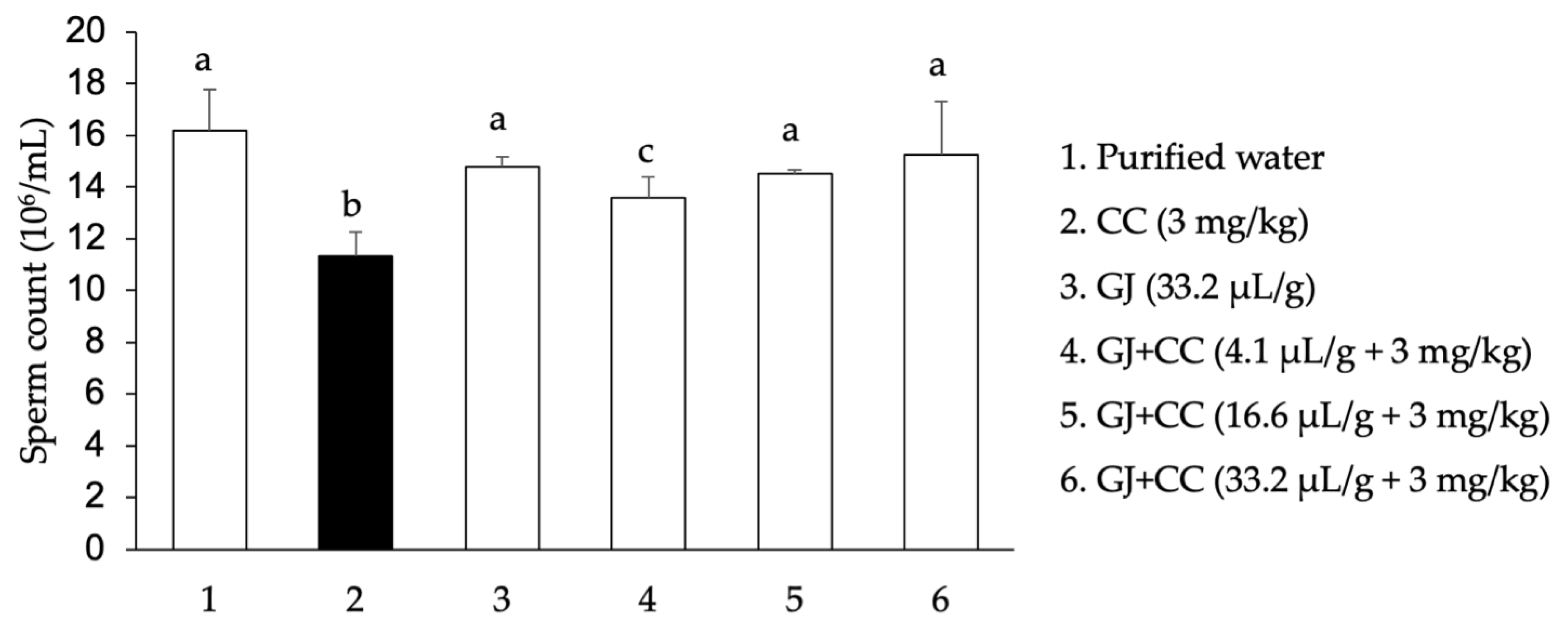
3.1. Sperm Concentration and Quality

With respect to the amount of spermatozoids, we determined a mean of 16 million/mL in the control mice, while the administration of CC reduced this level by 30%. The mice administered only GJ (33.2 µL/g) were found in the range determined for the control mice, while the three doses of GJ plus CC showed a significant concentration recovery. With the high dose, the sperm concentration recovery was 94% with respect to the CC value (Figure 2).
The results for viability, progressive motility, and malformations are presented in Table 1. Strong cell mortality due to CC was produced, while a normal amount of viable sperm was observed in the mice treated with GJ only. In the mice administered the three doses of GJ plus CC, we found a significant, dose-dependent recuperation of viable sperm. The recovery rates with respect to the CC damage level with the low to high GJ doses were 70, 82, and 89%, respectively. A highly significant decrease in sperm progressive motility, approximately three times, was induced by CC in comparison with the control level, and no damage was shown with the administration of GJ alone. In mice administered GJ plus CC, no protection was observed with the lowest GJ dose; however, the two high doses strongly improved motility, and with the highest dose, the restoration reached 90% with respect to the value observed with the CC treatment. Finally, we identified the amount of morphological disturbances produced by the action of CC that was prevented by the administration of GJ. The anomalies were determined in the sperm flagellum, mid-piece, and head (Table 1). While the effect of CC gave rise to an 83% decrease in normal sperm, the treatment with GJ alone produced a similar amount of normal sperm to that registered in the control mice, and with respect to the combined treatment groups (GJ plus CC), we observed a dose-dependent improvement in the malformed sperm effects produced by CC. A statistically significant improvement was found in the two high doses, with an 87% recovery determined with the high dose (33.2 µL/g) in comparison with the damage induced by CC.

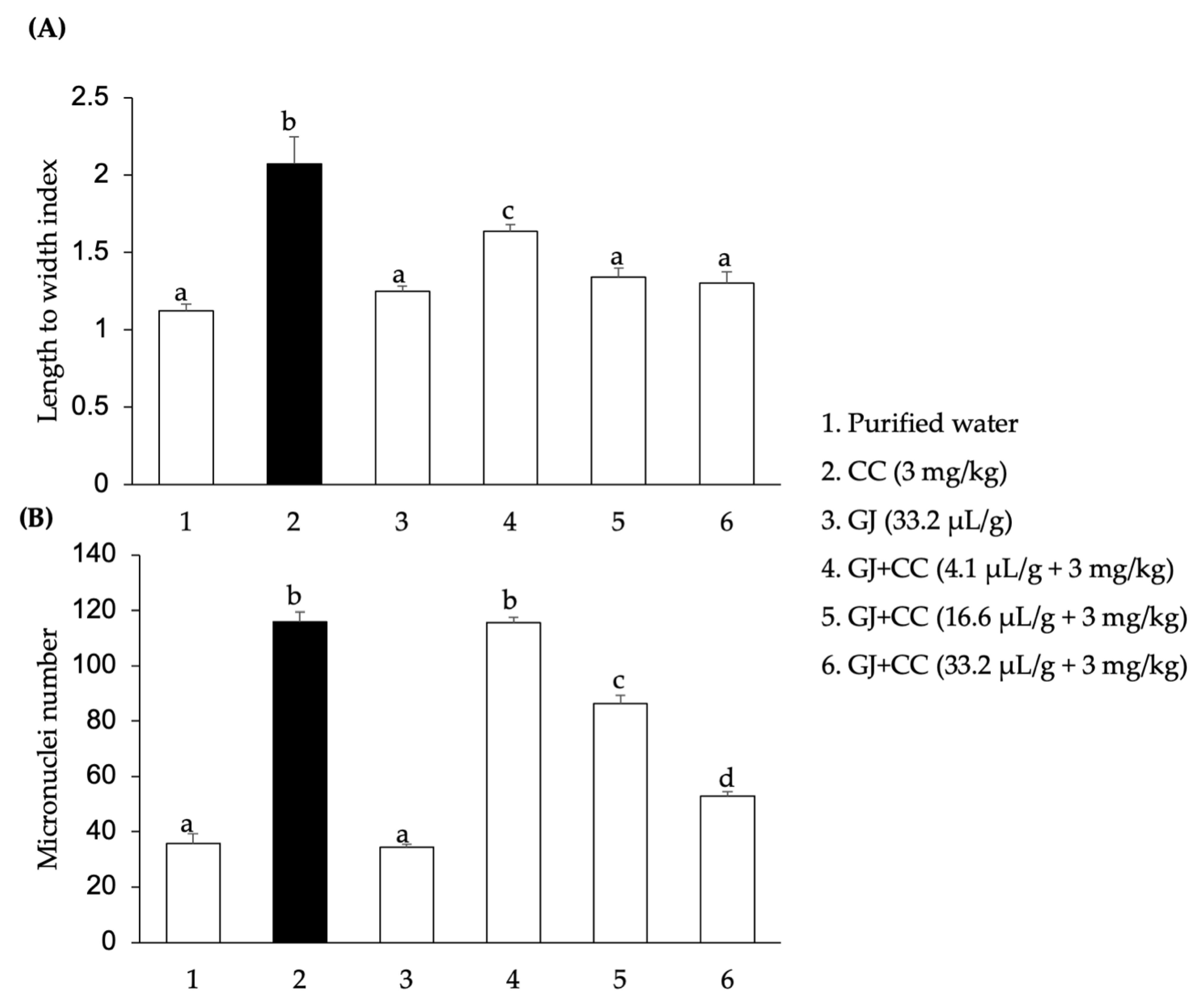
3.2. DNA Damage and Micronuclei in Spermatids

The indicated DNA parameter was examined by means of the alkaline comet assay. Figure 3A shows an approximation of the unit length-to-width index in the control mice, indicating an almost complete absence of DNA damage, a result that was similar to that found in the animals administered only GJ. These data contrast with the statistically significant duplication of the comet damage shown in the case of the CC-treated animals. Regarding the effect induced in the GJ-plus-CC-treated mice, we determined a significant level of protection exerted with the three tested GJ doses, with a mean preventive effect of 68% with respect to the level found in the CC-treated mice.
In Figure 3B, we present the results obtained in the cytogenetic assay concerning the number of micronuclei in the mouse spermatids. The control and GJ-alone-administered animals had similarly low amounts of micronuclei, confirming that the juice exerted no damage in this parameter. In contrast, the CC-treated animals showed a threefold elevation in the amount of micronuclei with respect to the control value. Regarding the combined groups (GJ plus CC), although we observed no protection with the lower dose of GJ, a statistically significant decrease in the number of micronuclei was found with the two high doses with respect to the CC-administered mice. At this endpoint, the high dose of GJ (33.2 µL/g) showed a better protective effect, with a 79% micronuclei reduction in comparison with the value observed in the CC-treated mice.

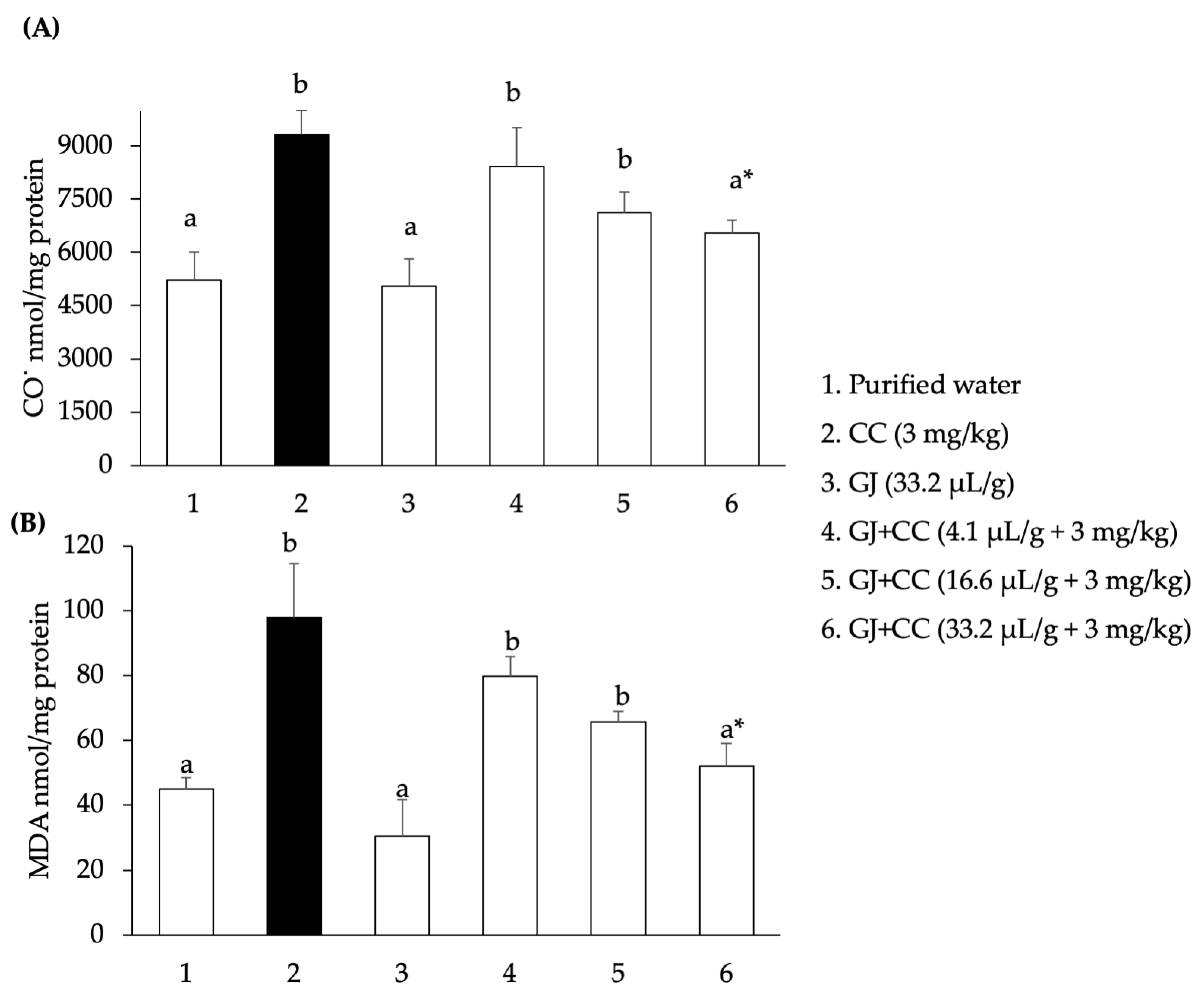
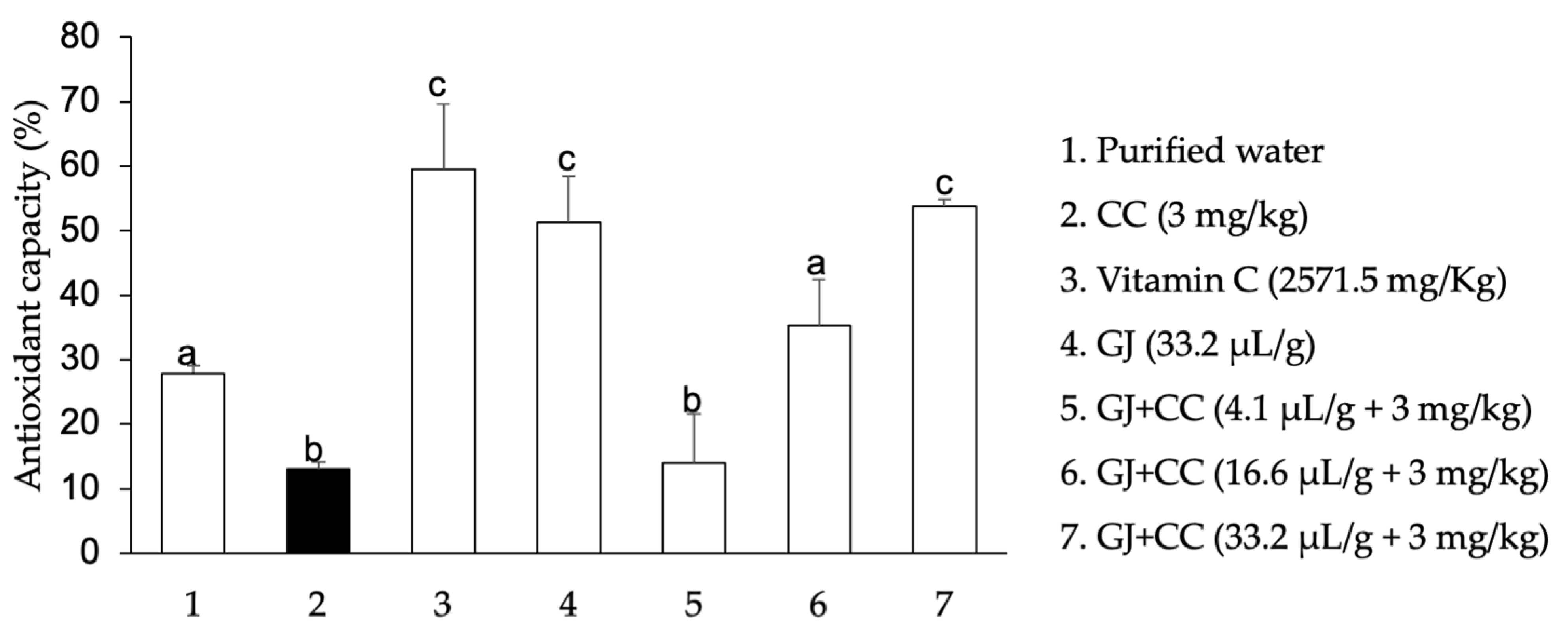
3.3. Oxidation of Biomolecules in Mouse Testes

Initially, we determined a homogeneous protein content in the experimental groups with the Bradford’s method. In a subsequent step, we identified the content of oxidized proteins, as shown by the concentration of reactive carbonyls. Figure 4A shows similar data in the control and GJ-treated groups, while the administration of CC almost doubled this value. Concerning the effect of GJ on the CC-treated mice, we found a dose-dependent decrease in the value reached with the metal, with the high dose being the most effective because its reductive power reached 68% in comparison with the CC level.
We also examined the effect of GJ on the lipoperoxidation of the mice administered CC, as shown by the MDA content. Figure 4B shows that the application of CC to the mice gave rise to more than a doubling of the level shown in the control animals. It was also observed that the GJ administration produced even lower lipid oxidation than that detected in the control animals, and finally, we detected dose-dependent protection when GJ plus CC was administered; in this case, the levels of protection against CC were 34%, 61%, and 87% with 4.1, 16.6, and 33.2 µL/g GJ, respectively.
Finally, we evaluated the antioxidant potential of the tested agents by means of the in vivo DPPH assay. Figure 5 shows that the administration of CC to the mice decreased this capacity by more than 50%, which is contrary to the significant increase found with the administration of the reference chemical (vitamin C), which corresponded to more than a duplication of the control value, as well as the antioxidant potential of GJ, which was slightly lower than that induced by the reference chemical. Regarding the treatment with GJ plus CC, our results showed a recuperation of the antioxidant potential with respect to that determined with the CC administration, showing statistical significance with the two high GJ doses. Regarding this effect, it seems interesting to note that the high GJ dose (33.2 µL/g) was found in the range of the value obtained for GJ and for vitamin C.

4. Discussion

Exposure to metals has been described among the numerous factors involved in fertility failures, and cadmium has been mentioned as one of the chemicals involved in the problem. This is understandable in light of the deleterious effects of the metal in a number of different organs and functions, its long biological half-life in humans, its potential formation of a variety of organic complexes, such as those structured with amines, sulfur, chloro, or chelates, and because of its high level of use in a number of human devices and its inefficient recycling [9].
In the present report, we confirmed the poisonous properties of cadmium against the various examined sperm and testicle endpoints. In terms of the sperm quality and concentration parameters, CC strongly damaged normal sperm in regard to motility, viability, and morphology. We found damage greater than 75%, with the exception of the spermatozoid amount, which was 30%. Additionally, with respect to DNA and chromosome structure, the administration of CC gave rise to more than an 85% damage increase regarding that observed in control mice. Moreover, our study also suggests that such an effect may be connected with CC’s oxidative potential, as indicated by its elevated values in lipids and proteins, which were close to a duplication in comparison with the control data, as well as with the cell antioxidant decrease induced by CC. In fact, the high sensitivity of the male reproductive apparatus to the studied metal has been previously shown by various authors, as well as its participation, together with other environmental toxicants, in the declining fertility of animals, including humans [35].
In contrast, our results with GJ administration showed a strong potential to reduce the reproductive damage induced by CC. All the examined endpoints that were severely altered by exposure to the metal were highly improved with exposure to the juice. According to our calculations, the low, intermediate, and high GJ doses correspond to 1, 4, and 8 glasses of 250 mL of juice ingested by a human weighing 60 kg.
Regarding the sperm quality observations, the three tested doses were probably related to the antigenotoxic and DNA repair capacities of various GJ constituents, and with respect to motility and morphologic anomalies, a significant improvement was detected with the two high doses. In addition, the sperm concentration decrease produced by the CC was significantly ameliorated with the three tested doses of GJ. Moreover, our data also demonstrated a significant antigenotoxic effect of GJ on the damaging capacity of CC. This was shown by the significant inhibition of DNA fragmentation with the three doses of GJ, as well as the significant reduction in the CC-induced micronuclei number with the two high doses of GJ. With respect to DNA fragmentation, GJ had a mean amelioration of 68%, and with respect to micronuclei, the mean reduction corresponded to approximately 58%.
In our study, a dose-dependent preventive decrease was also demonstrated by GJ with respect to the testicle lipid and protein oxidation produced by the CC administered to the mice, as well as the antioxidant capacity (shown with the DPPH assay), which significantly improved with the two high doses of GJ. These last three results suggest that the oxidative properties of CC were most likely involved in the observed reproductive damage and that such damage was efficiently counteracted by GJ-induced antioxidant molecular and cellular activities.
It is known that the normal functioning of organisms requires a well-controlled and delicate balance between oxidation and antioxidation. In cases in which the former activity increases in excess, oxidative stress may develop, giving rise to metabolic disturbances that can be reflected in significant reproductive pathologies, including infertility [1,36]. Here, we confirmed the oxidative potential of CC, which may have been involved in the observed sperm and spermatid anomalies. However, on the contrary, we showed the protection against this damage exerted by GJ, including significant antioxidant potential, probably related to the effect of the constituents of GJ. This property agrees with similar observations in somatic cells [15], and may support the observed beneficial capacity in cadmium-induced sperm damage.
The relevance of fertility failures has raised the importance of constantly developing preventive and curative measures to cope with the problem. In this effort, in addition to clinical advances, a number of reports with plant extracts have shown specific improvements in cadmium-affected spermatogenesis or hormonal production failures. Examples of these extracts include species from the families Moraceae, Polygalaceae, Moringaceae, Euphorbiaceae, Amaryllidaceae, Morchellaceae, and Spirulinaceae [37,38,39,40,41,42,43].
In regard to plant juices, publications on the matter are scarce; however, studies on goat semen cryopreservation for insemination purposes have shown improvements in sperm motility and acrosome and membrane integrity, as well as reduction in sperm abnormalities with the addition of pineapple and orange juices to semen extenders [44]. Additionally, Annona muricata juice used for the same purpose in rooster semen has shown antioxidant capacity and improvement in spermatozoid motility [45]. In addition, a report by Al-Olayan et al. (2014) [46] showed that pomegranate juice protected against the carbon tetrachloride depletion of rat testicular hormones, improved the level of antioxidant enzymes, and reduced sperm deformities, and two reports by Lamas et al., in 2015 and 2017 [47,48], have shown that commercial grape concentrate given to rats for at least 50 days alleviated the histological testicular and epididymal disruption caused by CC and improved the sperm count and structure. Our present report with GJ confirms these useful attributes, in a significantly lower time, determining the importance of its antioxidant capacity and showing for the first time a correlation between sperm damage inhibition and antigenotoxic effects. The chemical composition of GJ and its health-beneficial attributes suggest that its effects could be extended to other disruptive reproductive agents, and this is confirmed by the report by Sakr et al. (2013) [49], showing that rat-induced sperm head anomalies and testicular histological alterations by the administration of the antiarrhythmic drug amiodarone were ameliorated by a 5-week administration of the juice.
Regarding GJ’s properties, it is relevant to mention that a number of its specific components have been reported to have antioxidant, antigenotoxic, or curative human health disease properties. This has been demonstrated with respect to vitamins A and C, to the flavonoids naringin, naringenin, quercetin, and hesperetin, to various furanocoumarins, mainly bergamottin, sterols such as beta-sitosterol, and to the carotenoids lycopene, beta-carotene, zeaxanthin, and lutein, among other chemicals [50,51,52,53,54,55,56,57,58]. Vitamin C in particular is known to protect spermatogenesis from various toxins by preventing sperm agglutination, restoring damaged sperm and testes, and increasing testosterone synthesis (U.S. Department of Agriculture 2020b) [58].
The present report and others suggest that the beneficial effect on reproductive health of GJ relies on its strong antioxidant effect and that GJ’s chemical composition and its specific effects could give rise to a synergistic interaction subjacent to the origin of the observed protective effect. Therefore, it may be assumed that its supplementation could be of help in fertility problems related to oxidative etiology, an interesting possibility in light of fruit accessibility. However, it is always pertinent to note the need for caution with respect to juice consumption when it is combined with a number of medications because of GJ’s reported inhibition of CYP3A4 activity, which may give rise to reduced intestinal metabolism of the involved drug and, consequently, to bioavailability and absorption increases, which may produce overdoses [59]. In some cases, however, such interactions may be useful. According to a report on the administration of the antipsychotic drug aripiprazole [37], the interaction with GJ may potentiate its therapeutic activity by increasing its bioavailability and, thus, it may reduce the side effects of the drug because of the need for a lower dosage.
During their formation and transit, sperm are surrounded by endogenously and exogenously induced ROS. While low levels of these chemicals are required for normal physiological processes, high amounts may generate oxidative stress, which is a highly damaging factor that affects sperm function and reduces male fertility potential. Regrettably, a number of agents and human conducts have been found to possess oxidative potential, which may negatively affect the fertilization process. These include environmental toxins from the cosmetics-, plastic-, and metal-related industries, as well as exposure to alcohol, tobacco, or drugs and excessive exercise [60,61,62]. This brief recount of oxidative agents that produce deleterious effects on the male reproductive process support the importance of finding protective agents against this problem, including natural products.

5. Conclusions

In the present report, the strong preventive effect of GJ on various mouse male reproductive endpoints damaged by CC was demonstrated. The decrease in sperm number, motility, and viability was significantly improved by juice administration, and the sperm abnormality increase was also highly reduced. In addition, we found a correlation of these alterations with sperm DNA fragmentation and micronuclei induction in spermatids, damages that were also reduced by the action of the juice. Moreover, this report provides information on GJ’s testicular antioxidant capacity and suggests that this activity may explain the observed protection. Therefore, our results may stimulate research on GJ, including antioxidant molecular insights by examining the modulation of oxidative stress via the Nrf2/ARE, the inhibition of peroxidation enzymes, or the reduction of mitochondrial ROS. It may also be relevant to extend studies on GJ’s protection against other oxidant agents.

Author Contributions

Conceptualization and writing, E.M.-B. and I.Á.-G.; methodology, visualization, J.D.G.-G., R.P.-P. and B.A.E.-A.; validation and formal analysis, L.S.M.-C., E.O.M.-S. and J.A.M.-G.; supervision, software, and formal analysis, R.P.-P., F.d.J.C.-R. and A.G.-M. All authors have read and agreed to the published version of the manuscript.

Funding

This research received no external funding.

Institutional Review Board Statement

The study was conducted in accordance with the Declaration of Helsinki, and approved by the Ethics Committee of the Autonomous University of Hidalgo State (CICUAL/F0119/28 February 2022; Pachuca, Hidalgo State, Mexico).

Informed Consent Statement

Not applicable.

Data Availability Statement

The original contributions presented in this study are included in the article. Further inquiries can be directed to the corresponding author.

Conflicts of Interest

The authors declare no conflicts of interest.

References

  1. Agarwal, A.; Mulgund, A.; Hamada, A.; Chyatte, M.R. A unique view on male infertility around the globe. Reprod. Biol. Endocrinol. 2015, 13, 37. [Google Scholar] [CrossRef] [PubMed]
  2. Badar, A.; Khilwani, B.; Lohiya, N.K.; Ansari, A.S. Etiology of male infertility; A review. Int. J. Reprod. Contracept. Obstet. Gynecol. 2022, 11, 2320–2328. [Google Scholar] [CrossRef]
  3. Bellver, J.; Donnez, J. Introduction: Infertility etiology and offspring health. Fertil. Steril. 2019, 111, 1033–1035. [Google Scholar] [CrossRef]
  4. Babakhanzadeh, E.; Nazari, M.; Ghasemifar, S.; Khodadadian, A. Some of the factors involved in male infertility: A prospective review. Int. J. Gen. Med. 2020, 13, 29–41. [Google Scholar] [CrossRef] [PubMed]
  5. Geoffroy-Siraudin, C.; Loundou, A.D.; Romain, F.; Achard, V.; Courbière, B.; Perrard, M.H.; Durand, P.; Guichaoua, M.R. Decline of semen quality among 10 932 males consulting for couple infertility over a 20-year period in Marseille, France. Asian J. Androl. 2012, 14, 584–590. [Google Scholar] [CrossRef]
  6. Marić, T.; Fučić, A.; Aghayanian, A. Environmental and occupational exposures associated with male infertility. Arh. Hig. Rada Toksikol. 2021, 72, 101–113. [Google Scholar] [CrossRef]
  7. López-Botella, A.; Velasco, I.; Acién, M.; Sáez-Espinosa, P.; Todolí-Torró, J.L.; Sánchez-Romero, R.; Gómez-Torres, M.J. Impact of heavy metals on human male fertility-an overview. Antioxidants 2021, 10, 1473. [Google Scholar] [CrossRef]
  8. Genchi, G.; Sinicropi, M.S.; Lauria, G.; Carocci, A.; Catalano, A. The effects of cadmium toxicity. Int. J. Environ. Res. Public Health 2020, 17, 3782. [Google Scholar] [CrossRef]
  9. Rafati Rahimzadeh, M.; Rafati Rahimzadeh, M.; Kazemi, S.; Moghadamnia, A.A. Cadmium toxicity and treatment: An update. Casp. J. Intern. Med. 2017, 8, 135–145. [Google Scholar] [CrossRef]
  10. Schaefer, H.R.; Dennis, S.; Fitzpatrick, S. Cadmium: Mitigation strategies to reduce dietary exposure. J. Food Sci. 2020, 85, 260–267. [Google Scholar] [CrossRef]
  11. Wang, Z.; Sun, Y.; Yao, W.; Ba, Q.; Wang, H. Effects of cadmium exposure on the immune system and immunoregulation. Front. Immunol. 2021, 12, 695484. [Google Scholar] [CrossRef] [PubMed]
  12. Schaefer, H.R.; Flannery, B.M.; Crosby, L.; Jones-Dominic, O.E.; Punzalan, C.; Middleton, K. A systematic review of adverse health effects associated with oral cadmium exposure. Regul. Toxicol. Pharmacol. 2022, 134, 105243. [Google Scholar] [CrossRef] [PubMed]
  13. Zhu, Q.; Li, X.; Ge, R.S. Toxicological effects of cadmium on mammalian testis. Front. Genet. 2020, 11, 527. [Google Scholar] [CrossRef] [PubMed]
  14. Espinosa-Ahedo, B.A.; Madrigal-Bujaidar, E.; Sánchez-Gutiérrez, M.; Izquierdo-Vega, J.A.; Morales-González, J.A.; Madrigal-Santillán, E.O.; Álvarez-González, I. Potential protective effect of beta-caryophyllene against cadmium chloride-induced damage to the male reproductive system in mouse. Reprod. Toxicol. 2022, 110, 19–30. [Google Scholar] [CrossRef]
  15. Cristóbal-Luna, J.M.; Álvarez-González, I.; Madrigal-Bujaidar, E.; Chamorro-Cevallos, G. Grapefruit and its biomedical, antigenotoxic and chemopreventive properties. Food Chem. Toxicol. 2018, 112, 224–234. [Google Scholar] [CrossRef]
  16. Álvarez-González, I.; Madrigal-Bujaidar, E.; Martino-Roaro, L.; Espinosa-Aguirre, J.J. Antigenotoxic and antioxidant effect of grapefruit juice in mice treated with daunorubicin. Toxicol. Lett. 2004, 152, 203–211. [Google Scholar] [CrossRef]
  17. Miyata, M.; Takano, H.; Guo, L.Q.; Nagata, K.; Yamazoe, Y. Grapefruit juice intake does not enhance but rather protects against aflatoxin B1-induced liver DNA damage through a reduction in hepatic CYP3A activity. Carcinogenesis 2004, 25, 203–209. [Google Scholar] [CrossRef]
  18. Álvarez-González, I.; Madrigal-Bujaidar, E.; Sánchez-García, V.Y. Inhibitory effect of grapefruit juice on the genotoxic damage induced by ifosfamide in mouse. Plant Foods Hum. Nutr. 2010, 65, 369–373. [Google Scholar] [CrossRef]
  19. Argüelles, N.; Álvarez-González, I.; Chamorro, G.; Madrigal-Bujaidar, E. Protective effect of grapefruit juice on the teratogenic and genotoxic damage induced by cadmium in mice. J. Med. Food 2012, 15, 887–893. [Google Scholar] [CrossRef]
  20. Diab, K.A. In vitro studies on phytochemical content, antioxidant, anticancer, immunomodulatory, and antigenotoxic activities of lemon, grapefruit, and mandarin citrus peels. Asian Pac. J. Cancer Prev. 2016, 17, 3559–3567. [Google Scholar]
  21. Stanly, C.; Alfieri, M.; Ambrosone, A.; Leone, A.; Fiume, I.; Pocsfalvi, G. Grapefruit-derived micro and nanovesicles show distinct metabolome profiles and anticancer activities in the A375 human melanoma cell line. Cells 2020, 9, 2722. [Google Scholar] [CrossRef] [PubMed]
  22. Albus, U. Guide for the care and use of laboratory animals (8th ed.). Lab. Anim. 2011, 46, 267–268. [Google Scholar] [CrossRef]
  23. ARRIVE Guidelines, Animal Research: Reporting of In Vivo Experiments. 2021. Available online: https://arriveguidelines.org/ (accessed on 3 January 2025).
  24. Álvarez-Gonzalez, I.; Mojica, R.; Madrigal-Bujaidar, E.; Camacho-Carranza, R.; Escobar-García, D.; Espinosa-Aguirre, J.J. The antigenotoxic effects of grapefruit juice on the damage induced by benzo(a)pyrene and evaluation of its interaction with hepatic and intestinal Cytochrome P450 (Cyp) 1a1. Food Chem. Toxicol. 2011, 49, 807–811. [Google Scholar] [CrossRef]
  25. World Health Organization. Laboratory Manual for the Examination and Processing of Human Semen, 6th ed.; WHO: Geneva, Switzerland, 2021. [Google Scholar]
  26. Urióstegui-Acosta, M.; Hernández-Ochoa, I.; Solís-Heredia, M.d.J.; Martínez-Aguilar, G.; Quintanilla-Vega, B. Comparative effect of technical and commercial formulations of methamidophos on sperm quality and DNA integrity in mice. Environ. Toxicol. 2014, 29, 942–949. [Google Scholar] [CrossRef]
  27. Álvarez-González, I.; Camacho-Cantera, S.; Gómez-González, P.; Rendon-Barrón, M.J.; Morales-González, J.A.; Madrigal-Santillán, E.O.; Paniagua-Pérez, R.; Madrigal-Bujaidar, E. Genotoxic and oxidative effect of duloxetine on mouse brain and liver tissues. Sci. Rep. 2021, 11, 6897. [Google Scholar] [CrossRef] [PubMed]
  28. Russo, A. In vivo cytogenetics: Mammalian germ cells. Mutat. Res. 2000, 455, 167–189. [Google Scholar] [CrossRef]
  29. Bradford, M.M. A rapid and sensitive method for the quantitation of microgram quantities of protein utilizing the principle of protein-dye binding. Anal. Biochem. 1976, 72, 248–254. [Google Scholar] [CrossRef]
  30. Levine, R.L.; Garland, D.; Oliver, C.N.; Amici, A.; Climent, I.; Lenz, A.G.; Ahn, B.W.; Shaltiel, S.; Stadtman, E.R. Determination of carbonyl content in oxidatively modified proteins. Methods Enzymol. 1990, 186, 464–478. [Google Scholar] [CrossRef]
  31. Buege, J.A.; Aust, S.D. Microsomal lipid peroxidation. Methods Enzymol. 1978, 52, 302–310. [Google Scholar] [CrossRef]
  32. Chen, F.; Huang, G.; Yang, Z.; Hou, Y. Antioxidant activity of Momordica charantia polysaccharide and its derivatives. Int. J. Biol. Macromol. 2019, 138, 673–680. [Google Scholar] [CrossRef]
  33. Chrzczanowicz, J.; Gawron, A.; Zwolinska, A.; de Graft-Johnson, J.; Krajewski, W.; Krol, M.; Markowski, J.; Kostka, T.; Nowak, D. Simple method for determining human serum 2,2-diphenyl-1-picryl-hydrazyl (DPPH) radical scavenging activity—Possible application in clinical studies on dietary antioxidants. Clin. Chem. Lab. Med. 2008, 46, 342–349. [Google Scholar] [CrossRef] [PubMed]
  34. Arıcan, E.Y.; Gökçeoğlu Kayalı, D.; Ulus Karaca, B.; Boran, T.; Öztürk, N.; Okyar, A.; Ercan, F.; Özhan, G. Reproductive effects of subchronic exposure to acetamiprid in male rats. Sci. Rep. 2020, 10, 8985. [Google Scholar] [CrossRef]
  35. Bhardwaj, J.K.; Paliwal, A.; Saraf, P. Effects of heavy metals on reproduction owing to infertility. J. Biochem. Mol. Toxicol. 2021, 35, e22823. [Google Scholar] [CrossRef] [PubMed]
  36. Gulcin, İ.; Alwasel, S.H. DPPH radical scavenging assay. Processes 2023, 11, 2248. [Google Scholar] [CrossRef]
  37. Ola-Mudathir, K.F.; Suru, S.M.; Fafunso, M.A.; Obioha, U.E.; Faremi, T.Y. Protective roles of onion and garlic extracts on cadmium-induced changes in sperm characteristics and testicular oxidative damage in rats. Food Chem. Toxicol. 2008, 46, 3604–3611. [Google Scholar] [CrossRef]
  38. El-Neweshy, M.S.; El-Maddawy, Z.K.; El-Sayed, Y.S. Therapeutic effects of date palm (Phoenix dactylifera L.) pollen extract on cadmium-induced testicular toxicity. Andrologia 2013, 45, 369–378. [Google Scholar] [CrossRef]
  39. Adaramoye, O.A.; Akanni, O.O. Protective effects of Artocarpus altilis (Moraceae) on cadmium-induced changes in sperm characteristics and testicular oxidative damage in rats. Andrologia 2016, 48, 152–163. [Google Scholar] [CrossRef] [PubMed]
  40. Olaniyan, O.T.; Kunle-Alabi, O.T.; Raji, Y. Protective effects of methanol extract of Plukenetia conophora seeds and 4H-Pyran-4-One 2,3-Dihydro-3,5-Dihydroxy-6-Methyl on the reproductive function of male Wistar rats treated with cadmium chloride. JBRA Assist. Reprod. 2018, 22, 289–300. [Google Scholar] [CrossRef]
  41. Elblehi, S.S.; El Euony, O.I.; El-Nahas, A.F. Partial ameliorative effect of Moringa leaf ethanolic extract on the reproductive toxicity and the expression of steroidogenic genes induced by subchronic cadmium in male rats. Environ. Sci. Pollut. Res. Int. 2019, 26, 23306–23318. [Google Scholar] [CrossRef]
  42. Akinola, A.O.; Oyeyemi, A.W.; Daramola, O.O.; Raji, Y. Effects of the methanol root extract of Carpolobia lutea on sperm indices, acrosome reaction, and sperm DNA integrity in cadmium-induced reproductive toxicity in male Wistar rats. JBRA Assist. Reprod. 2020, 24, 454–465. [Google Scholar] [CrossRef]
  43. Iqbal, T.; Jahan, S.; Ain, Q.U.; Ullah, H.; Li, C.; Chen, L.; Zhou, X. Ameliorative effects of morel mushroom (Morchella esculenta) against cadmium-induced reproductive toxicity in adult male rats. Braz. J. Biol. 2021, 82, e250865. [Google Scholar] [CrossRef] [PubMed]
  44. Daramola, J.O.; Adekunde, E.O.; Onagbesan, O.M.; Oke, O.E.; Ladokun, A.O.; Abiona, J.A.; Abioja, M.O.; Oyewusi, I.K.; Oyewusi, J.A.; Isah, O.A.; et al. Protective effects of fruit juices on sperm viability of West African Dwarf goat bucks during cryopreservation. Anim. Reprod. 2016, 13, 7–13. [Google Scholar] [CrossRef]
  45. Jimoh, O.A.; Nwachukwu, C.U. Efficacy of soursop juice extender on rooster semen quality, oxidative activity and spermatozoa kinematics. Transl. Anim. Sci. 2022, 6, txac046. [Google Scholar] [CrossRef] [PubMed]
  46. Al-Olayan, E.M.; El-Khadragy, M.F.; Metwally, D.M.; Abdel Moneim, A.E. Protective effects of pomegranate (Punica granatum) juice on testes against carbon tetrachloride intoxication in rats. BMC Complement. Altern. Med. 2014, 14, 164. [Google Scholar] [CrossRef] [PubMed]
  47. Lamas, C.A.; Gollücke, A.P.; Dolder, H. Grape juice concentrate (G8000(®)) intake mitigates testicular morphological and ultrastructural damage following cadmium intoxication. Int. J. Exp. Pathol. 2015, 96, 301–310. [Google Scholar] [CrossRef]
  48. Lamas, C.A.; Cuquetto-Leite, L.; do Nascimento da Silva, E.; Thomazini, B.F.; Cordeiro, G.D.S.; Predes, F.S.; Gollücke, A.P.B.; Dolder, H. Grape juice concentrate alleviates epididymis and sperm damage in cadmium-intoxicated rats. Int. J. Exp. Pathol. 2017, 98, 86–99. [Google Scholar] [CrossRef]
  49. Sakr, S.A.; Zoil, M.E.S.; El-Shafey, S.S. Ameliorative effect of grapefruit juice on amiodarone-induced cytogenetic and testicular damage in albino rats. Asian Pac. J. Trop. Biomed. 2013, 3, 573–579. [Google Scholar] [CrossRef]
  50. Duester, K.C. Avocado fruit is a rich source of beta-sitosterol. J. Am. Diet. Assoc. 2001, 101, 404–405. [Google Scholar] [CrossRef]
  51. Bacanlı, M.; Başaran, A.A.; Başaran, N. The antioxidant and antigenotoxic properties of citrus phenolics limonene and naringin. Food Chem. Toxicol. 2015, 81, 160–170. [Google Scholar] [CrossRef]
  52. Sarker, S.K.; Tun, K.D.; Eva, E.O.; Paul, R. Grapefruit juice: Nutritional values and drug interactions. Int. J. Integr. Med. Sci. 2015, 2, 186–189. [Google Scholar] [CrossRef]
  53. Singh, Z.; Sharma, S.; Kaur, A. Antitoxic effects of naringin: A flavonoid with diverse biological activities. World J. Pharm. Res. 2018, 7, 484–489. [Google Scholar] [CrossRef]
  54. Ko, J.H.; Arfuso, F.; Sethi, G.; Ahn, K.S. Pharmacological utilization of bergamottin, derived from grapefruits, in cancer prevention and therapy. Int. J. Mol. Sci. 2018, 19, 4048. [Google Scholar] [CrossRef] [PubMed]
  55. Khan, U.M.; Sevindik, M.; Zarrabi, A.; Nami, M.; Ozdemir, B.; Kaplan, D.N.; Selamoglu, Z.; Hasan, M.; Kumar, M.; Alshehri, M.M.; et al. Lycopene: Food sources, biological activities, and human health benefits. Oxidative Med. Cell. Longev. 2021, 2021, 2713511. [Google Scholar] [CrossRef]
  56. Saini, R.K.; Ranjit, A.; Sharma, K.; Prasad, P.; Shang, X.; Gowda, K.G.M.; Keum, Y.S. Bioactive compounds of citrus fruits: A review of composition and health benefits of carotenoids, flavonoids, limonoids, and terpenes. Antioxidants 2022, 11, 239. [Google Scholar] [CrossRef]
  57. Khalil, M.N.A.; Farghal, H.H.; Farag, M.A. Outgoing and potential trends of composition, health benefits, juice production and waste management of the multi-faceted Grapefruit Citrus x paradisi: A comprehensive review for maximizing its value. Crit. Rev. Food Sci. Nutr. 2022, 62, 935–956. [Google Scholar] [CrossRef]
  58. Martins, R.V.L.; Silva, A.M.S.; Duarte, A.P.; Socorro, S.; Correia, S.; Maia, C.J. Natural products as protective agents for male fertility. BioChem 2021, 1, 122–147. [Google Scholar] [CrossRef]
  59. Zargar, S.; Al-Majed, A.A.; Wani, T.A. Potentiating and synergistic effect of grapefruit juice on the antioxidant and anti-inflammatory activity of aripiprazole against hydrogen peroxide induced oxidative stress in mice. BMC Complement. Altern. Med. 2018, 18, 106. [Google Scholar] [CrossRef]
  60. Darbandi, M.; Darbandi, S.; Agarwal, A.; Sengupta, P.; Durairajanayagam, D.; Henkel, R.; Sadeghi, M.R. Reactive oxygen species and male reproductive hormones. Reprod. Biol. Endocrinol. 2018, 16, 87. [Google Scholar] [CrossRef]
  61. Aitken, R.J.; Drevet, J.R.; Moazamian, A.; Gharagozloo, P. Male infertility and oxidative stress: A focus on the underlying mechanisms. Antioxidants 2022, 11, 306. [Google Scholar] [CrossRef]
  62. Ribas-Maynou, J.; Yeste, M. Oxidative stress in male infertility: Causes, effects in assisted reproductive techniques, and protective support of antioxidants. Biology 2020, 9, 77. [Google Scholar] [CrossRef]
Figure 1. Schematic representation of the used methodology. The administration of grapefruit juice was performed for 11 days, and cadmium was administered once, on day 5 of the study. Seven groups with 5 mice each were used in the assay, as described in Section 2.2.
Applsci 15 06071 g001
Figure 2. Preventive effect of grapefruit juice (GJ) on mouse sperm concentration decrease produced by cadmium chloride (CC). Each bar represents the mean ± SDM obtained in five mice per group, 100 spermatozoids per mouse. Different letters show statistical differences, one-way ANOVA and post hoc Tukey test, p ≤ 0.05.
Applsci 15 06071 g002
Figure 3. Preventive effect of grapefruit juice (GJ) on the DNA damage induced by cadmium chloride (CC) (A), and preventive effect on the number of micronucleated spermatids induced by CC (B). In (A), each bar represents the mean ± SDM obtained in five mice per group, 100 spermatozoids per mouse, and in (B), each bar represents the mean ± SDM obtained in five mice per group, 1000 spermatids per mouse. Different letters show statistical differences, one-way ANOVA and post hoc Tukey test, p ≤ 0.05.
Applsci 15 06071 g003
Figure 4. Preventive effect of grapefruit juice (GJ) on the mouse testicular protein carbonyl content (CO˙) induced by cadmium chloride (CC) (A), and protective effect of GJ on the malondialdehyde (MDA) content induced by CC (B). Each bar represents the mean ± SDM in five mice per group. a Statistical difference when comparing with the positive control, b statistical difference when comparing with the negative control, and * statistical difference when comparing with group 4. One-way ANOVA and post hoc Tukey test, p ≤ 0.05.
Applsci 15 06071 g004
Figure 5. DPPH scavenging capacity of grapefruit juice (GJ) in the sera of mice administered cadmium chloride (CC). Each bar represents the mean ± SDM obtained in five mice per group. Different letters show statistical differences, one-way ANOVA and post hoc Tukey test, p ≤ 0.05.
Applsci 15 06071 g005
Table 1. Mouse sperm quality determined with exposure to grapefruit juice (GJ) and cadmium chloride (CC).
GroupTreatmentsNormal MorphologyProgressive MotilityViability
1Purified water/10 µL/g16 ± 1.41 a51.4 ± 2.7 a94 ± 1.58 a
2CC/3 mg/kg2.8 ± 0.84 b13 ± 2.83 b14.8 ± 1.64 b
3GJ/33.28 µL/g15.6 ± 1.52 a50.4 ± 6.42 a89 ± 1.58 a
4 GJ   +   CC
4.16 µL/g    3 mg/kg
6.8 ± 0.83 c12.8 ± 2.05 b66.2 ± 2.59 c
5 GJ   +   CC
16.64 µL/g    3 mg/kg
12.6 ± 1.67 d39 ± 4.30 c77.8 ± 4.92 d
6 GJ   +   CC
33.28 µL/g    3 mg/kg
14 ± 1 e46.6 ± 2.88 a83.4 ± 3.21 e
For morphology, each value represents the mean ± SDM obtained in 200 spermatozoids per mouse (five mice per group). For motility and viability, each value represents the mean ± SDM obtained in 100 spermatozoids per mouse (five mice per group). Values are expressed in percentage form. Different letters show statistical differences. For morphology, one-way ANOVA, and post hoc Tukey tests, p ≤ 0.05 were used. For motility and viability, chi-squared and Fisher tests, p ≤ 0.05 were used.
Disclaimer/Publisher’s Note: The statements, opinions and data contained in all publications are solely those of the individual author(s) and contributor(s) and not of MDPI and/or the editor(s). MDPI and/or the editor(s) disclaim responsibility for any injury to people or property resulting from any ideas, methods, instructions or products referred to in the content.

