Article

Lymph Node Involvement Prediction Using Machine Learning: Analysis of Prostatic Nodule, Prostatic Gland, and Periprostatic Adipose Tissue (PPAT)

by
Eliodoro Faiella
1,2,
Giulia D’amone
1,2,
Raffaele Ragone
1,2,*,
Matteo Pileri
1,2,
Elva Vergantino
1,2,
Bruno Beomonte Zobel
1,2,
Rosario Francesco Grasso
1,2 and
Domiziana Santucci
1,2,*
1
Unit of Radiology and Interventional Radiology, Fondazione Policlinico Universitario Campus Bio-Medico, Via Alvaro del Portillo, 00128 Rome, Italy
2
Research Unit of Radiology, Department of Medicine and Surgery, Università Campus Bio-Medico di Roma, Via Alvaro del Portillo, 00128 Rome, Italy
*
Authors to whom correspondence should be addressed.
Appl. Sci. 2025, 15(10), 5426; https://doi.org/10.3390/app15105426
Submission received: 18 March 2025 / Revised: 25 April 2025 / Accepted: 6 May 2025 / Published: 13 May 2025
(This article belongs to the Special Issue Advances in Diagnostic Radiology)

Abstract

:
Background: Prostate cancer is a major cause of cancer-related mortality among men, with approximately 15% of newly diagnosed patients having pelvic lymph node metastasis (PLNM). For this reason, PLNM identification before localized PCa treatment would significantly impact treatment planning, clinical judgment, and patient outcome prediction. Radiomics has gained popularity for its ability to predict tumor behavior and prognosis without invasive procedures. Magnetic resonance imaging (MRI) is widely used in radiomic workups, particularly for prostate cancer. This study aims to predict lymph node invasion in prostate cancer patients using clinical information and mp-MRI radiomics features extracted from the suspicious nodule, prostate gland, and periprostatic adipose tissue (PPAT). Methods: A retrospective review of 85 patients who underwent mp-MRI at our radiology department between 2016 and 2022 was conducted. This study included patients who underwent prostatectomy and lymphadenectomy with complete histological examination and previous staging mp-MRI and were divided into two groups based on lymph node status (positive/negative). Data were collected from each patient, including clinical information, radiomics, and semantic data (such as tumor MRI characteristics, histological tumor details, and lymph node status (LNS)). MRI exams were conducted using a 1.5-T system and were used to study the prostate gland. A three-year resident manually segmented the prostate nodule, prostatic gland, and periprostatic tissue using an open-source segmentation program. A random forest (RF) machine learning model was developed and tested using Chat-GPT version 4.0 software. The model’s performance in predicting LNS was assessed using accuracy, precision, recall, F1 score, and area under the curve (AUC) receiver operating characteristic (ROC), with sensitivity and specificity evaluated using DeLong’s test. Results: Random forest demonstrated the best performance in prediction considering features extracted from DWI nodules (67% of accuracy, 0.83 AUC), from T2 fat (78% of accuracy, 0.86 AUC), and from T2 glands (78% of accuracy, 0.97 AUC). The combination of the three sequences in the nodule evaluation was more accurate compared with the single sequences (88%). Combining all the nodule features with gland and PPAT features, an accuracy of 89% with AUC near 1 was obtained. Compared with the analysis of the nodule and the PPAT, the whole-gland evaluation had the best performance (p ≤ 0.05) in predicting LNS when compared with the nodule. Conclusions: Precise nodal staging is essential for PCa patients’ prognosis and therapeutic strategy. When compared with a radiologist’s assessment, radiomics models enhance the diagnostic accuracy of lymph node staging for prostate cancer. Although data are still lacking, deep learning models may be able to further improve on this.

1. Introduction

Prostate cancer (PCa) [1] is the second leading cause of cancer-related mortality among men and remains one of the most frequently diagnosed malignancies worldwide. In the United States alone, 2023 saw 609,820 cancer-related deaths [2] and 1,958,310 new cancer cases [3]. Approximately 15% of patients with newly diagnosed PCa present with pelvic lymph node metastases (PLNM), a major prognostic factor linked to distant metastases and biochemical recurrence after curative treatment [4]. Therefore, accurate pre-treatment identification of PLNM is crucial, as it significantly influences clinical decision-making and treatment strategies [5,6].
Radiomics is a rapidly developing field that uses advanced computational techniques and artificial intelligence to extract quantitative imaging features, providing non-invasive biomarkers of tumor phenotype [7,8]. In prostate cancer, radiomics has shown potential in improving diagnosis, risk stratification, and treatment planning [9]. Among imaging modalities, magnetic resonance imaging (MRI) stands out for its high soft-tissue contrast and multiparametric capabilities, making it ideal for radiomic analysis [10]. Sequences such as T2-weighted imaging (T2W), diffusion-weighted imaging (DWI), and apparent diffusion coefficient (ADC) maps allow for the extraction of relevant features related to tumor texture, shape, and intensity [11].
In prostate MRI, two anatomical regions—the peripheral zone (PZ) and transition zone (TZ)—are typically evaluated, with suspicious lesions often appearing hyperintense on DWI and ADC sequences [12,13].
Beyond tumor tissue, recent evidence suggests that peritumoral environments, such as periprostatic adipose tissue (PPAT), may play a critical role in cancer progression [14]. PPAT and adipose stromal cells (ASCs) have been shown to promote tumor growth and invasiveness through neovascularization, extracellular matrix remodeling, immune modulation, and paracrine signaling. In particular, PPAT-derived soluble factors and lipid transfer mechanisms have been implicated in enhancing PCa aggressiveness and local spread [15,16].
This study aims to investigate whether radiomic features extracted from the tumor nodule, the entire prostate gland, and the surrounding PPAT can predict lymph node invasion in patients with prostate cancer.

2. Materials and Methods

This study adhered to the guidelines of the Declaration of Helsinki. Ethical review and approval were not required due to its retrospective design.
A retrospective analysis was performed on patients who underwent multiparametric MRI (mp-MRI) for prostate cancer staging at our Radiological Department between 2016 and 2022. The inclusion criteria encompassed patients who subsequently underwent prostatectomy and lymphadenectomy, yielding a final sample size of 85 individuals. Patients were excluded if they had received hormonal therapy, radiation therapy, or random biopsy prior to mp-MRI. Additional exclusion criteria included prior radio- or chemotherapy, transurethral resection of the prostate (TURP), partial gland surgical removal, or cases where lymphadenectomy was not performed.
The clinical and demographic characteristics of the patient cohort are summarized in Table 1. Data collected for each patient included clinical information (age and PSA levels prior to mp-MRI), tumor MRI features [17] (signal intensity on T2-weighted images, signal intensity on DWI/ADC maps, and PIRADS score [18]), and histopathological tumor characteristics (Gleason score, TNM staging, including extracapsular extension, seminal vesicle invasion, neurovascular bundle involvement, and histological subtype). These characteristics were categorized as “semantic features” [19]. The lymphadenectomy was performed at a mean distance of 102 days (min 91 and max 115) by mp-MRI.
Based on lymph node status at the time of lymphadenectomy, patients were divided into two groups:
-
Lymph node positive status (n = 35): at least one lymph node showed metastatic involvement.
-
Lymph node negative status (n = 50): all examined lymph nodes were free of metastases.

2.1. Magnetic Resonance Imaging

MRI examinations were performed using a 1.5-T system (Magnetom Aera, Siemens, Gurgaon, Haryana, version syngo MR E11) with a pelvic phased-array coil. Patients were positioned supine and underwent appropriate preparatory procedures before imaging.
The prostate gland was imaged in axial, coronal, and sagittal planes using T2-weighted turbo spin-echo sequences with the following parameters: repetition time (TR) of 3520 ms, echo time (TE) of 114 ms, field of view (FOV) of 200 mm, slice count of 30, slice thickness of 3 mm, and a gap factor of 10%. Subsequently, T1-weighted fast spin-echo transverse images were acquired with TR of 426 ms, TE of 11 ms, FOV of 330 mm, slice count of 30, slice thickness of 3 mm, and a gap factor of 10%. The imaging protocol also included diffusion-weighted imaging (DWI) with b-values of 500, 1000, 1500, and 2000 s/mm2, as well as apparent diffusion coefficient (ADC) maps. Additionally, 3D volumetric interpolated breath-hold examination (VIBE) T1-weighted fat-suppressed images in the axial plane were obtained (TR 4.3 ms, TE 1.62 ms, FOV 260 mm) following an intravenous bolus injection of 0.15 mL/kg gadoteric acid (0.5 mmol/mL) at a rate of 3 mL/s.
For the purposes of this analysis, only axial T2-weighted images, DWI, and ADC maps were utilized. Written informed consent was obtained from all patients prior to mp-MRI. Preoperative mp-MRI was conducted 4–5 weeks after ultrasound-guided transrectal biopsy.

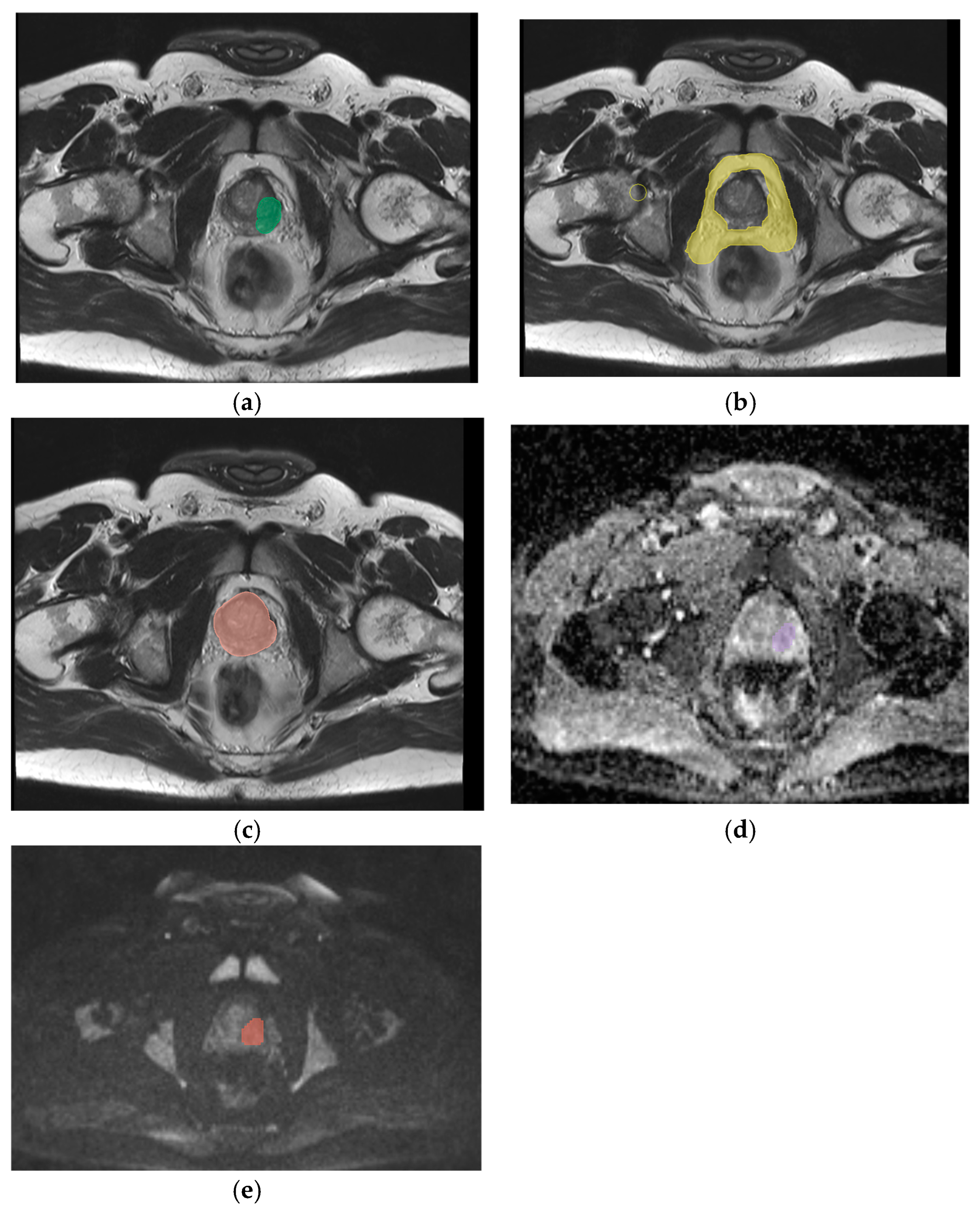
2.2. Segmentation, Feature Extraction and Selection

All mp-MRI examinations were transferred to a dedicated workstation. A three-year radiology resident (R.R.) manually segmented the prostate nodule previously (identified as a key image on T2-weighted sequences, DWI, and ADC maps), the entire prostate gland (on T2-weighted sequences), and the periprostatic adipose tissue (PPAT) (on T2-weighted sequences) using the open-source software 3D Slicer (v. 5.0.1). A volume of interest (VOI) was generated for each segmentation (Figure 1 and Figure 2).
Two radiologists, with 12 years (E.F.) and 5 years (D.S.) of experience, independently reviewed all prostate MRI scans and validated segmentation. Target lesions were identified in all sequences, measured, and recorded as key images.
A total of 131 radiomic features were extracted from each VOI using the Pyradiomics application (v. 3) integrated into the software. Features were automatically selected based on relevance. Both semantic features and radiomics features were included in the analysis, by capturing intratumoral histopathological characteristics, such as tumor shape and intensity.
Radiomics Features:
Shape Features: Describing geometric properties of the region of interest (ROI), including surface area, total volume, maximum diameter, elongation, sphericity, and surface-to-volume ratio;
First-Order Statistics: Histogram-based features representing the distribution of voxel intensities in the ROI, including metrics such as energy, entropy, mean, interquartile range, skewness, kurtosis, and uniformity;
Second-Order Textural Features: Capturing the statistical interrelationships between neighboring voxels. These include:
-
Gray-Level Co-Occurrence Matrix (GLCM): Analyzes spatial gray-level intensity distributions within a 3D image;
-
Gray-Level Run-Length Matrix (GLRLM): Quantifies contiguous voxels with the same gray-level value in multiple directions;
-
Gray-Level Size Zone Matrix (GLSZM): Measures zones of connected voxels with identical gray-level intensity in a 3D space;
-
Gray-Tone Difference Matrix (NGTDM): Evaluates differences between voxel intensity and the average intensity of neighboring voxels within a set distance;
-
Gray-Level Dependence Matrix (GLDM): Assesses the degree of dependence between neighboring voxels at varying distances.

2.3. Radiomics Analysis and Model Development

Following feature extraction and selection, a random forest (RF) machine learning model was developed and training was performed using the artificial intelligence platform ChatGPT version 4.0. The dataset was randomly divided into training and testing sets with an 80:20 ratio. To enhance robustness and reduce variability, a 5-fold cross-validation was performed on the training set to validate model performance and optimize hyperparameters. The dataset presented an unbalanced distribution between patients with and without lymph node involvement, and this class imbalance was preserved in both the training and test sets to reflect real-world clinical conditions. During model development, data augmentation techniques were employed to mitigate this issue. Model performance was evaluated using metrics such as accuracy, precision, recall, F1 score, and area under the receiver operating characteristic curve (AUC). Sensitivity and specificity were also assessed across individual imaging sequences and their combinations (Table 2).
DeLong’s test was employed to compare the highest AUCs obtained from analyses of the nodule, whole gland, and PPAT. Statistical significance was set at p < 0.05.
Data augmentation was simulated using boosting techniques, including XGBoost and CatBoost. These techniques informed the development of the RF model, enhancing predictions and identifying the most significant features for lymph node invasion prediction.
Feature importance analysis, based on boosting algorithms and RF modeling, identified key predictive variables. This analysis improved the model’s interpretability and overall prediction accuracy.

3. Results

All performance metrics reported below refer to model evaluation on the independent test set, following an 80:20 train–test split with internal five-fold cross-validation on the training data.
The random forest (RF) model was evaluated by accuracy, AUC, precision, recall, F1-score, specificity, and sensitivity.
As summarized in Table 2, the RF model demonstrated optimal performance in predicting lymph node involvement using features extracted from specific sequences. Notable results included:
-
DWI nodule features: Accuracy of 67% and AUC of 0.83;
-
T2-weighted PPAT features: Accuracy of 78% and AUC of 0.86;
-
T2-weighted whole gland features: Accuracy of 78% and AUC of 0.97.
When combining features from all three sequences for nodule evaluation, the accuracy increased to 88%, although the AUC was lower than that of individual sequences. However, integrating nodule features from all sequences with features derived from the T2-weighted whole gland and T2-weighted PPAT resulted in an overall accuracy of 89% and an AUC approaching 1.
To rigorously compare model performance, DeLong’s test was employed to assess the sensitivity and specificity of the models by comparing the AUCs of their respective ROC curves. This non-parametric statistical test allowed for a robust evaluation of whether observed differences between models were statistically significant.
The comparison of the best performances from nodule, whole gland, and PPAT analyses revealed that whole-gland evaluation had the greatest predictive impact, with a statistically significant difference (p ≤ 0.05) when compared with nodule analysis alone. These findings highlight the importance of comprehensive gland assessment in predicting lymph node involvement in prostate cancer patients.

4. Discussion

Lymph node involvement in prostate cancer [20] is currently assessed through regional lymphadenectomy, as recommended by the latest clinical guidelines. Specifically, the European Association of Urology (EAU), European Society for Radiotherapy and Oncology (ESTRO), EAU Section of Urological Research (ESUR), and International Society of Geriatric Oncology (SIOG) advocate for an extended pelvic lymph node dissection (e-PLND) in patients with a risk of nodal involvement exceeding 5% [21]. The National Comprehensive Cancer Network (NCCN), however, recommends e-PLND for patients with a risk greater than 2% [22]. Lymphadenectomy typically targets specific nodal stations, such as the inguinal and internal obturator lymph nodes. Despite its diagnostic and therapeutic significance, the procedure is associated with substantial complications, prompting the exploration of alternative, non-invasive methods for predicting lymph node involvement.
Algorithms combining clinical, anatomo-pathological, and radiological data (e.g., Briganti or MSKCC nomograms [23]) have been developed to estimate the likelihood of lymph node involvement, sparing unnecessary surgical interventions. ChatGPT has been used as a tool for analysis, although in terms of radiological and imaging performance, it does not have sufficient performance to replace a radiologist [24]. Among imaging modalities, MRI has shown utility in evaluating lymph node status with varying success, often dependent on the radiologist’s expertise. Diffusion-weighted imaging (DWI) [23,25], a functional imaging technique that quantifies water molecule movement through apparent diffusion coefficient (ADC) maps, has demonstrated the ability to differentiate between benign and malignant lymph nodes. However, its role remains limited due to difficulties in detecting micrometastases, necessitating reliance on surgical staging for definitive evaluation.
Advancements in artificial intelligence (AI) [26] and radiomics have significantly improved the capacity to extract and analyze quantitative data from imaging [7,27]. Radiomics allows for the evaluation of diverse tumor characteristics, including shape, intensity, and texture features [28], offering a detailed representation of tumor microenvironments. In this study, we employed a machine learning model of random forest type [29,30] to predict lymph node involvement by combining MRI-derived radiomics [31] features with clinical, anatomopathological, and radiological information.
The use of a train–test split combined with internal cross-validation strengthened the reliability of the reported model performance, reducing the risk of overfitting and supporting the generalizability of our findings. Our approach extended beyond the analysis of prostate nodules to include the entire gland and periprostatic adipose tissue (PPAT), recognizing the paracrine influence of PPAT [32] in prostate cancer progression.
We identified significant radiomics descriptors from prostate nodules, the gland, and PPAT using machine learning to integrate these features for improved lymph node involvement prediction. Notably, the inclusion of PPAT features alongside gland and nodule data yielded the highest AUC values, supporting the notion that lymph node and adjacent organ involvement is influenced not only by primary tumor characteristics, but also by the surrounding microenvironment [33,34].
Consistent with us, recent studies have highlighted that radiomic features extracted from periprostatic adipose tissue (PPAT) can provide additional information on prostate cancer aggressiveness and local disease spread [35,36]. Shahait, M. et al. [37], in their radiomic analysis on T1-weighted images, demonstrated that different patterns of periprostatic fat significantly correlated with prostate cancer (Gleason ≥ 7), distinguishing indolent tumors with high predictive accuracy (AUC ~0.82). Similarly, Arslan, A. et al. [38] supported that integrating radiomic features from the PPAT with tumor nodule features improved prognostic stratification. They introduced a radiomics nomogram combining intratumoral and periprostatic radiomic features from mpMRI for the prediction of biochemical recurrence-free survival, obtaining very high AUC values (0.85–0.92 at 1–5 years) [38].
These findings suggest that PPAT radiomics may serve as an additional biomarker of tumor aggressiveness and risk stratification, reflecting the interactions between the tumor and its local adipose environment.
Emerging evidence highlights the role of PPAT in facilitating tumor progression [39] through mechanisms such as neovascularization [33], extracellular matrix remodeling, and epithelial–mesenchymal transition (EMT). PPAT secretes fatty acids and other soluble factors, enhancing tumor aggressiveness and regional spread [39]. For instance, Ribeiro et al. revealed diminished immune surveillance in PPAT associated with complement factor H (CFH) downregulation. These findings underscore the biological relevance of PPAT in prostate cancer pathophysiology [36].
Other studies support the evidence that radiomics analysis should be extended to the entire prostate gland and surrounding tissues to capture signals of locoregional disease spread.
Some studies have shown that peritumoral radiomics features (i.e., features extracted from a defined ring of tissue surrounding the tumor) provide crucial information on extracapsular extension (ECE). In the study by Zhao W et al. involving prostatectomy patients, a radiomic model incorporating peritumoral features from mpMRI more accurately predicted ECE compared with intratumoral radiomics alone [40].
We demonstrated an accuracy of about 89% and an AUC of 100% when all masks were combined, while the performances of both were lower if a single nodule, gland, or PPAt were analyzed. However, especially for single analysis, a disparity between accuracy and AUC in our results was noted. This underscores the complementary nature of these metrics: while accuracy reflects the overall proportion of correct predictions, it is sensitive to class imbalance. Conversely, AUC provides a threshold-independent measure of the model’s discriminative power, particularly valuable in imbalanced datasets. The apparent discrepancy between accuracy and AUC values observed for DWI and ADC sequences can be attributed to the intrinsic limitations of using these modalities in isolation, as well as the class imbalance in our dataset. While accuracy is threshold-dependent and sensitive to the dominant class, AUC provides a threshold-independent assessment of the model’s discriminative ability. This divergence highlights the importance of evaluating multiple performance metrics and supports our choice to integrate radiomic features from multiple anatomical regions to enhance predictive power.
Currently, there is a growing interest in AI method development for predicting and detecting occult lymph node metastases preoperatively in many oncological fields [40].
However, only few studies about PCa are present in the literature, although with very encouraging results. Cysouw et al. [6] achieved an AUC of 0.86 for predicting nodal or distant metastases, and Liu et al. [23] reported an AUC of 0.83 in subgroups with small lymph nodes using radiomics features. Faiella et al. developed a machine learning model integrating radiomic features from a prostate tumor on mpMRI with clinical data that predicted lymph node invasion (LNI) with high accuracy (AUC ~0.91 in the test set) [41].
Our study [41] demonstrates higher AUC values, reinforcing the potential of radiomics-based approaches in improving diagnostic accuracy.
Our results demonstrated elevated specificity compared with sensitivity, indicating the model’s robustness in identifying true negatives, i.e., patients without lymph node involvement. This is clinically significant as it enables the sparing of unnecessary lymphadenectomies and their associated complications. The radiomic data derived from MRI could be incorporated into clinical workflows to predict lymph node involvement even before biopsy, offering a non-invasive alternative to traditional methods [20].
The aim is to stratify patients who may avoid extensive pelvic lymph node dissection (ePLND) without compromising staging accuracy and potentially reduce unnecessary ePLND procedures by more accurately identifying high-risk cases. This radiomics-based model may assist clinicians in the preoperative decision-making process by identifying patients at higher risk of lymph node involvement, potentially reducing the number of unnecessary extended pelvic lymph node dissections (ePLNDs) and their related complications.
These results underscore the added value of extending radiomics analysis beyond the primary tumor to include the entire gland and PPAT, providing imaging biomarkers for locoregional disease dissemination. The integration of tumor-derived and surrounding tissue radiomics improves the ability to predict both extracapsular extension and lymph node involvement, supporting more tailored clinical decision-making in prostate cancer management.
Nevertheless, this study has limitations, including the small sample size and class imbalance within the dataset. To mitigate these issues, we employed a wrapper feature selection method to prevent overfitting and implemented data augmentation techniques to address class imbalance. Despite these limitations, our findings align with emerging evidence on the role of PPAT in prostate cancer aggressiveness and highlight the potential of radiomics in refining risk stratification and guiding personalized management strategies.
Despite the promising results, our study has several limitations that should be acknowledged. First, the model was developed and tested using data from a single institution, and no external validation was performed. This limits the generalizability of our findings and highlights the need for multicentric prospective studies. Second, although segmentations were reviewed by two experienced radiologists, the initial segmentation was performed by a single resident, and no formal inter-observer variability analysis was conducted. Third, we did not perform a comparative analysis with other machine learning algorithms beyond the random forest model. Future work should include benchmarking against alternative models to confirm the robustness and comparative advantage of our approach.
Future research should focus on validating these findings in larger cohorts and exploring the integration of radiomics with genomic data to further enhance predictive accuracy and inform novel therapeutic approaches. The characterization of PPAT’s role in prostate cancer progression holds promise for advancing both diagnostic precision and therapeutic interventions.

5. Limitations

This study has several limitations.
First, it lacks external validation, which limits the generalizability of the results.
Second, the segmentation process was performed by a single individual, potentially introducing subjective bias.

6. Conclusions

Accurate lymph node staging is essential for effective prostate cancer management, yet conventional imaging techniques often show limited sensitivity and specificity. In this study, we developed a radiomics-based model integrating features from the prostate nodule, whole gland, and periprostatic adipose tissue (PPAT), which demonstrated high predictive performance. While the results showed promising accuracy, AUC, sensitivity, and specificity, these findings must be interpreted with caution due to the relatively small sample size, class imbalance, and lack of external validation. Nevertheless, the model illustrates the potential of radiomics to improve preoperative risk stratification and assist in tailoring surgical decisions, including the indication for extended pelvic lymph node dissection (ePLND). Further validation on larger, multicentric cohorts is warranted to confirm its clinical applicability.
Radiomics-based prediction of lymph node involvement may serve as a valuable tool to guide surgical planning and support more individualized treatment strategies.

Author Contributions

E.F. and R.F.G.: Conceptualization, methodology, formal analysis. R.R.: data curation. G.D.: writing—original draft preparation. D.S.: writing—review and editing. M.P. and E.V.: English editing. E.F. and B.B.Z.: visualization, supervision and project administration. All authors have read and agreed to the published version of the manuscript.

Funding

This research received no external funding.

Institutional Review Board Statement

The study was conducted according to the guidelines of the Declaration of Helsinki. Ethical review and approval were waived for this study, due to the retrospective nature of the study.

Informed Consent Statement

This is a retrospective study and there are not any personal information of patients. According to our Institution (Campus Biomedico di Roma), for retrospective studies, not other approval except for informed consent for MRI execution is needed.

Data Availability Statement

The authors confirm that the data supporting the findings of this study are available within the article and its references.

Acknowledgments

The authors have no further information to disclose.

Conflicts of Interest

The authors declare no conflicts of interest.

References

  1. Mohler, J.L.; Armstrong, A.J.; Bahnson, R.R.; D’Amico, A.V.; Davis, B.J.; Eastham, J.A.; Enke, C.A.; Farrington, T.A.; Higano, C.S.; Horwitz, E.M.; et al. Prostate Cancer, Version 1.2016. J. Natl. Compr. Cancer Netw. 2016, 14, 19–30. [Google Scholar] [CrossRef] [PubMed]
  2. Kraujalis, V.; Ruzgas, T.; Milonas, D. Mortality Rate Estimation Models for Patients with Prostate Cancer Diagnosis. Balt. J. Mod. Comput. 2022, 10, 170–184. [Google Scholar] [CrossRef]
  3. Siegel, R.L.; Miller, K.D.; Wagle, N.S.; Jemal, A. Cancer statistics, 2023. CA Cancer J. Clin. 2023, 73, 17–48. [Google Scholar] [CrossRef]
  4. Calabrese, A.; Santucci, D.; Landi, R.; Zobel, B.B.; Faiella, E.; de Felice, C. Radiomics MRI for lymph node status prediction in breast cancer patients: The state of art. J. Cancer Res. Clin. Oncol. 2021, 147, 1587–1597. [Google Scholar] [CrossRef] [PubMed]
  5. Bianchi, L.; Gandaglia, G.; Fossati, N.; Suardi, N.; Moschini, M.; Cucchiara, V.; Bianchi, M.; Damiano, R.; Schiavina, R.; Shariat, S.F.; et al. Pelvic Lymph Node Dissection in Prostate Cancer: Indications, Extent and Tailored Approaches. Urol. J. 2017, 84, 9–19. [Google Scholar] [CrossRef]
  6. Preisser, F.; van den Bergh, R.C.N.; Gandaglia, G.; Ost, P.; Surcel, C.I.; Sooriakumaran, P.; Montorsi, F.; Graefen, M.; van der Poel, H.; de la Taille, A.; et al. Effect of Extended Pelvic Lymph Node Dissection on Oncologic Outcomes in Patients with D’Amico Intermediate and High Risk Prostate Cancer Treated with Radical Prostatectomy: A Multi-Institutional Study. J. Urol. 2020, 203, 338–343. [Google Scholar] [CrossRef] [PubMed]
  7. Subrahmanya, S.V.G.; Shetty, D.K.; Patil, V.; Hameed, B.M.Z.; Paul, R.; Smriti, K.; Naik, N.; Somani, B.K. The role of data science in healthcare advancements: Applications, benefits, and future prospects. Ir. J. Med Sci. 2021, 191, 1473–1483. [Google Scholar] [CrossRef]
  8. Santucci, D.; Faiella, E.; Cordelli, E.; Sicilia, R.; de Felice, C.; Zobel, B.B.; Iannello, G.; Soda, P. 3T MRI-Radiomic Approach to Predict for Lymph Node Status in Breast Cancer Patients. Cancers 2021, 13, 2228. [Google Scholar] [CrossRef] [PubMed] [PubMed Central]
  9. Liberini, V.; Laudicella, R.; Balma, M.; Nicolotti, D.G.; Buschiazzo, A.; Grimaldi, S.; Lorenzon, L.; Bianchi, A.; Peano, S.; Bartolotta, T.V.; et al. Radiomics and artificial intelligence in prostate cancer: New tools for molecular hybrid imaging and theragnostics. Eur. Radiol. Exp. 2022, 6, 27. [Google Scholar] [CrossRef] [PubMed] [PubMed Central]
  10. Faiella, E.; Santucci, D.; Calabrese, A.; Russo, F.; Vadalà, G.; Zobel, B.B.; Soda, P.; Iannello, G.; de Felice, C.; Denaro, V. Artificial Intelligence in Bone Metastases: An MRI and CT Imaging Review. Int. J. Environ. Res. Public Health 2022, 19, 1880. [Google Scholar] [CrossRef] [PubMed] [PubMed Central]
  11. Santucci, D.; Faiella, E.; Calabrese, A.; Zobel, B.B.; Ascione, A.; Cerbelli, B.; Iannello, G.; Soda, P.; de Felice, C. On the Additional Information Provided by 3T-MRI ADC in Predicting Tumor Cellularity and Microscopic Behavior. Cancers 2021, 13, 5167. [Google Scholar] [CrossRef] [PubMed] [PubMed Central]
  12. Ren, J.; Huan, Y.; Wang, H.; Zhao, H.; Ge, Y.; Chang, Y.; Liu, Y. Diffusion-weighted imaging in normal prostate and differential diagnosis of prostate diseases. Abdom. Imaging 2008, 33, 724–728. [Google Scholar] [CrossRef]
  13. Iglesias, Á.S.; Macías, V.M.; Peris, A.P.; Fuster-Matanzo, A.; Infante, A.N.; Soria, R.M.; Bataller, F.B.; Pomar, M.D.; Meléndez, C.C.; Huertas, R.Y.; et al. Prostate Region-Wise Imaging Biomarker Profiles for Risk Stratification and Biochemical Recurrence Prediction. Cancers 2023, 15, 4163. [Google Scholar] [CrossRef] [PubMed]
  14. Vertulli, D.; Santucci, D.; Esperto, F.; Beomonte Zobel, B.; Grasso, R.F.; Faiella, E. Impact of adipose tissue distribution on prostate cancer recurrence after radical prostatectomy. Actas Urol. Esp. 2023, 47, 104–110. [Google Scholar] [CrossRef] [PubMed]
  15. Faiella, E.; Vaccarino, F.; Ragone, R.; D’amone, G.; Cirimele, V.; Piccolo, C.L.; Vertulli, D.; Grasso, R.F.; Zobel, B.B.; Santucci, D. Can Machine Learning Models Detect and Predict Lymph Node Involvement in Prostate Cancer? A Comprehensive Systematic Review. J. Clin. Med. 2023, 12, 7032. [Google Scholar] [CrossRef] [PubMed] [PubMed Central]
  16. Faiella, E.; Vergantino, E.; Vaccarino, F.; Bruno, A.; Perillo, G.; Grasso, R.F.; Zobel, B.B.; Santucci, D. A Review of the Paradigmatic Role of Adipose Tissue in Renal Cancer: Fat Measurement and Tumor Behavior Features. Cancers 2024, 16, 1697. [Google Scholar] [CrossRef] [PubMed] [PubMed Central]
  17. Verma, S.; Choyke, P.L.; Eberhardt, S.C.; Oto, A.; Tempany, C.M.; Turkbey, B.; Rosenkrantz, A.B. The Current State of MR Imaging–targeted Biopsy Techniques for Detection of Prostate Cancer. Radiology 2017, 285, 343–356. [Google Scholar] [CrossRef]
  18. Chen, M.; Dang, H.-D.; Wang, J.-Y.; Zhou, C.; Li, S.-Y.; Wang, W.-C.; Zhao, W.-F.; Yang, Z.-H.; Zhong, C.-Y.; Li, G.-Z. Prostate cancer detection: Comparison of t2-weighted imaging, diffusion-weighted imaging, proton magnetic resonance spectroscopic imaging, and the three techniques combined. Acta Radiol. 2008, 49, 602–610. [Google Scholar] [CrossRef]
  19. Li, Q.; Lu, H.; Choi, J.; Gage, K.; Feuerlein, S.; Pow-Sang, J.M.; Gillies, R.; Balagurunathan, Y. Radiological semantics discriminate clinically significant grade prostate cancer. Cancer Imaging 2019, 19, 81. [Google Scholar] [CrossRef]
  20. Chiacchio, G.; Castellani, D.; Nedbal, C.; De Stefano, V.; Brocca, C.; Tramanzoli, P.; Galosi, A.B.; da Silva, R.D.; Teoh, J.Y.-C.; Tiong, H.Y.; et al. Radiomics vs radiologist in prostate cancer. Results from a systematic review. World J. Urol. 2023, 41, 709–724. [Google Scholar] [CrossRef]
  21. Sanda, M.G.; Cadeddu, J.A.; Kirkby, E.; Chen, R.C.; Crispino, T.; Fontanarosa, J.; Freedland, S.J.; Greene, K.; Klotz, L.H.; Makarov, D.V.; et al. Clinically Localized Prostate Cancer: AUA/ASTRO/SUO Guideline. Part I: Risk Stratification, Shared Decision Making, and Care Options. J. Urol. 2018, 199, 683–690. [Google Scholar] [CrossRef]
  22. Cheung, D.C.; Fleshner, N.; Sengupta, S.; Woon, D. A narrative review of pelvic lymph node dissection in prostate cancer. Transl. Androl. Urol. 2020, 9, 3049–3055. [Google Scholar] [CrossRef]
  23. Liu, X.; Tian, J.; Wu, J.; Zhang, Y.; Wang, X.; Zhang, X.; Wang, X. Utility of diffusion weighted imaging-based radiomics nomogram to predict pelvic lymph nodes metastasis in prostate cancer. BMC Med. Imaging 2022, 22, 190. [Google Scholar] [CrossRef]
  24. Kufel, J.; Paszkiewicz, I.; Bielówka, M.; Bartnikowska, W.; Janik, M.; Stencel, M.; Czogalik, Ł.; Gruszczyńska, K.; Mielcarska, S. Will ChatGPT pass the Polish specialty exam in radiology and diagnostic imaging? Insights into strengths and limitations. Pol. J. Radiol. 2023, 88, e430–e434. [Google Scholar] [CrossRef] [PubMed] [PubMed Central]
  25. Caglic, I.; Barrett, T. Diffusion-weighted imaging (DWI) in lymph node staging for prostate cancer. Transl. Androl. Urol. 2018, 7, 814–823. [Google Scholar] [CrossRef] [PubMed]
  26. Bedrikovetski, S.; Dudi-Venkata, N.N.; Maicas, G.; Kroon, H.M.; Seow, W.; Carneiro, G.; Moore, J.W.; Sammour, T. Artificial intelligence for the diagnosis of lymph node metastases in patients with abdominopelvic malignancy: A systematic review and meta-analysis. Artif. Intell. Med. 2021, 113, 102022. [Google Scholar] [CrossRef] [PubMed]
  27. Ferro, M.; de Cobelli, O.; Musi, G.; del Giudice, F.; Carrieri, G.; Busetto, G.M.; Falagario, U.G.; Sciarra, A.; Maggi, M.; Crocetto, F.; et al. Radiomics in prostate cancer: An up-to-date review. Ther. Adv. Urol. 2022, 14, 175628722211090. [Google Scholar] [CrossRef] [PubMed]
  28. Smith, C.P.; Czarniecki, M.; Mehralivand, S.; Stoyanova, R.; Choyke, P.L.; Harmon, S.; Turkbey, B. Radiomics and radiogenomics of prostate cancer. Abdom. Imaging 2019, 44, 2021–2029. [Google Scholar] [CrossRef]
  29. Rigatti, S.J. Random Forest. J. Insur. Med. 2017, 47, 31–39. [Google Scholar] [CrossRef] [PubMed]
  30. Chen, S.; Jian, T.; Chi, C.; Liang, Y.; Liang, X.; Yu, Y.; Jiang, F.; Lu, J. Machine Learning-Based Models Enhance the Prediction of Prostate Cancer. Front. Oncol. 2022, 12, 941349. [Google Scholar] [CrossRef] [PubMed] [PubMed Central]
  31. Lay, N.S.; Tsehay, Y.; Greer, M.D.; Turkbey, B.; Kwak, J.T.; Choyke, P.L.; Pinto, P.; Wood, B.J.; Summers, R.M. Detection of prostate cancer in multiparametric MRI using random forest with instance weighting. J. Med. Imaging 2017, 4, 024506. [Google Scholar] [CrossRef] [PubMed] [PubMed Central]
  32. Cao, H.; Wang, Y.; Zhang, D.; Liu, B.; Zhou, H.; Wang, S. Periprostatic Adipose Tissue: A New Perspective for Diagnosing and Treating Prostate Cancer. J. Cancer 2024, 15, 204–217. [Google Scholar] [CrossRef]
  33. Passos, G.R.; Ghezzi, A.C.; Antunes, E.; de Oliveira, M.G.; Mónica, F.Z. The Role of Periprostatic Adipose Tissue on Prostate Function in Vascular-Related Disorders. Front. Pharmacol. 2021, 12, 626155. [Google Scholar] [CrossRef]
  34. Toren, P.; Venkateswaran, V. Periprostatic Adipose Tissue and Prostate Cancer Progression: New Insights into the Tumor Microenvironment. Clin. Genitourin. Cancer 2014, 12, 21–26. [Google Scholar] [CrossRef] [PubMed]
  35. Sacca, P.A.; Calvo, J.C. Periprostatic Adipose Tissue Microenvironment: Metabolic and Hormonal Pathways During Prostate Cancer Progression. Front. Endocrinol. 2022, 13, 863027. [Google Scholar] [CrossRef] [PubMed] [PubMed Central]
  36. Ribeiro, R.; Monteiro, C.; Catalán, V.; Hu, P.; Cunha, V.; Rodríguez, A.; Gómez-Ambrosi, J.; Fraga, A.; Príncipe, P.; Lobato, C.; et al. Obesity and prostate cancer: Gene expression signature of human periprostatic adipose tissue. BMC Med. 2012, 10, 108. [Google Scholar] [CrossRef] [PubMed]
  37. Shahait, M.; Usamentiaga, R.; Tong, Y.; Sandberg, A.; Lee, D.I.; Udupa, J.K.; Torigian, D.A. Periprostatic Adipose Tissue MRI Radiomics-Derived Features Associated with Clinically Significant Prostate Cancer. J. Endourol. 2023, 37, 1156–1161. [Google Scholar] [CrossRef] [PubMed]
  38. Arslan, A.; Karaarslan, E.; Güner, A.L.; Sağlıcan, Y.; Tuna, M.B.; Kural, A.R. Comparing the Diagnostic Performance of Multiparametric Prostate MRI Versus 68Ga-PSMA PET-CT in the Evaluation Lymph Node Involvement and Extraprostatic Extension. Acad. Radiol. 2022, 29, 698–704. [Google Scholar] [CrossRef]
  39. Saha, A.; Kolonin, M.G.; DiGiovanni, J. Obesity and prostate cancer—Microenvironmental roles of adipose tissue. Nat. Rev. Urol. 2023, 20, 579–596. [Google Scholar] [CrossRef] [PubMed]
  40. Zhao, W.; Hou, M.; Wang, J.; Song, D.; Niu, Y. Interpretable machine learning model for predicting clinically significant prostate cancer: Integrating intratumoral and peritumoral radiomics with clinical and metabolic features. BMC Med. Imaging 2024, 24, 353. [Google Scholar] [CrossRef] [PubMed] [PubMed Central]
  41. Santucci, D.; Ragone, R.; Vergantino, E.; Vaccarino, F.; Esperto, F.; Prata, F.; Scarpa, R.M.; Papalia, R.; Zobel, B.B.; Grasso, F.R.; et al. Comparison between Three Radiomics Models and Clinical Nomograms for Prediction of Lymph Node Involvement in PCa Patients Combining Clinical and Radiomic Features. Cancers 2024, 16, 2731. [Google Scholar] [CrossRef] [PubMed]
Figure 1. Segmentation of the index prostatic nodule (green zone) in TSW (a), in ADC (violet zone in (d)) and in DWI (red zone in (e)), the PPTA (yellow zone) in T2W (b), prostate gland (orange zone) in T2W (c), in a 63-year-old patient with a PSA value of 10.3 ng/mL. The histological analysis showed an adenocarcinoma with a Gleason score of 7 (4 + 3), with involvement of neurovascular bundle and negative lymph node involvement.
Applsci 15 05426 g001
Figure 2. Segmentation of the index prostatic nodule (green zone) in TSW (a), in ADC (violet zone in (d)), and in DWI (red zone in (e)), the PPTA (yellow zone) in T2W (b), prostate gland (orange zone) in T2W (c), in a 55-year-old patient with a PSA value of 7 ng/mL. The histological analysis showed an adenocarcinoma with a Gleason score of 7 (4 + 3), with a locoregional involvement of neurovascular bundle and positive lymph node involvement.
Applsci 15 05426 g002
Table 1. Patients’ characteristics. LNS: lymph node status.
CategoryNumber
Patients with positive LNS35
Patients with negative LNS50
PSA (ng/mL) (Median, range)4.5
Gleason Grade (Median)7
Tumor target Zone Peripheral33
Tumor target Zone Transition12
Disease Grade (TNM)T3 (16)
T2 (10)
T3.5 (19)
Table 2. The performances for the different MRI sequences of prostatic nodules, for the whole gland, and for the PPAT reported in terms of accuracy, AUC, Recall, and F1.
SeqAccuracyAUCPrecisionRecallF1-ScoreSpecificitySensitivity
ADC78%0.7221.0000.3330.50010033
DWI67%0.830.500.330.4010033
T2 nod78%0.780.500.330.4010033
all nodule seq88508719310050
T2 PPAT78%0.861.0000.330.5010033
T2 gland78%0.9721.0000.3330.50010033
ALL mask88.89%11.0000.670.80100100
Disclaimer/Publisher’s Note: The statements, opinions and data contained in all publications are solely those of the individual author(s) and contributor(s) and not of MDPI and/or the editor(s). MDPI and/or the editor(s) disclaim responsibility for any injury to people or property resulting from any ideas, methods, instructions or products referred to in the content.

