Article

Plasma-Assisted Decoration of Gold Nanoparticles on Bioinspired Polydopamine Nanospheres as Effective Catalyst for Organic Pollutant Removal

1
Institute of Materials Science, Vietnam Academy of Science and Technology, 18 Hoang Quoc Viet, Cau Giay, Hanoi 100000, Vietnam
2
Department of Biotechnology, College of Engineering, The University of Suwon, Hwaseong 18323, Republic of Korea
3
Plasma Bioscience Research Center, Department of Electrical and Biological Physics, Kwangwoon University, Seoul 01897, Republic of Korea
*
Authors to whom correspondence should be addressed.
Appl. Sci. 2025, 15(10), 5280; https://doi.org/10.3390/app15105280
Submission received: 18 April 2025 / Revised: 2 May 2025 / Accepted: 7 May 2025 / Published: 9 May 2025
(This article belongs to the Section Applied Biosciences and Bioengineering)

Abstract

:
Polydopamine (PDA) is an emerging biomimetic material that stimulates the distinctive physicochemical properties of the blue mussel byssus. In this study, we report a rapid and facile method for the decoration of gold nanoparticles (AuNPs) onto the mussel-inspired polydopamine nanospheres (PDA NSs) via cold atmospheric plasma treatment. After 10 min of plasma treatment, AuNPs with a size of 10.3 ± 2.0 nm were formed on the surface of PDA NSs. This reaction was performed without the need for any additional reducing agents, thereby eliminating the use of harsh chemicals during the process. The synthesized AuNP-decorated PDA nanohybrids (PDA-Au) exhibit effective catalytic activity for the decoloration of Rhodamine B, with a pseudo-first-order rate constant of 1.405 min−1. The green synthesis approach in this work highlights the potential of plasma-assisted methods for decorating biomimetic materials with metallic nanoparticles for catalytic and environmental applications.

1. Introduction

Organic dyes are colored substances that are extensively used in industries such as pharmaceuticals, textiles, cosmetics, and food coloring [1,2,3,4]. However, their accumulation in wastewater poses significant concerns for both human health and the environment. The catalytic reduction process of these dyes into non-toxic byproducts using biomimetic nanomaterials has emerged as a promising approach [5,6,7,8]. Biomimetic materials are synthetic substances inspired by nature to simulate desirable properties. Among them, a polymer so-called PDA, which is inspired by the adhesive proteins in blue mussel byssus, has gained attention as an excellent support material for nanocatalyst stabilization and decoration [9]. Owing to its high biocompatibility and robust adhesive properties, PDA serves as an environmentally friendly platform for various applications such as catalysis, biomedicine, or sensing [10]. It is commonly synthesized as a coating layer, enabling the facile functionalization of surfaces and the attachment of diverse molecules and nanoparticles [11]. Due to the presence of catechol and amine groups, PDA can act both as a reducing agent and a stabilizing matrix for metal nanoparticles. Additionally, PDA can be synthesized in the form of nanospheres with tunable sizes, which serve as effective templating materials and are well dispersed in aqueous solutions [12,13,14]. PDA NSs can be used as suitable substrates for the in situ decoration of nanoparticles such as Au, Pt, Pd, Rh, Ru, and CdS to obtain high-efficient hybrid nanocatalysts [15,16,17,18,19]. In this context, AuNPs are of particular interest in catalysis because of their high specific surface area that enables higher catalytic activity [20]. Moreover, the applications of PDA-Au have been extended not only for catalysis but also for a variety range of applications. For instance, Yang et al. decorated AuNPs on mussel-inspired PDA as a sensitive SERS substrate for the selective detection of phthalate plasticizers [21]. Gao et al. develop Au nanostars based on PDA-Au for highly sensitive cancer biomarker detection [22]. Liu et al. used PDA-Au as a substrate for the construction of Co2+-based copeptin immunosensing [23].
In dye degradation reactions, in the presence of NaBH4 as a reducing agent, AuNPs act as catalysts to accelerate the electron transfer from NaBH4 to dye molecules, leading to their decomposition and decolorization of the polluted solution [24,25,26]. It is well known that smaller AuNPs exhibit higher catalytic efficiency due to their increased surface-to-volume ratio and greater number of surface-active sites [27,28,29]. However, small-size nanoparticles are prone to aggregation, thus limiting their catalytic activity and reusability. Immobilizing AuNPs onto a substrate such as PDA is a suitable approach for preventing nanoparticle aggregation and facilitating catalyst recollection. Additionally, substrate interactions can enhance the electronic properties of AuNPs, thereby improving their catalytic performance [30]. For example, Mei et al. reported that gold nanoparticles supported by hollow PDA nanoreactors is a highly efficient catalyst for dye degradation [31]. Several studies also have investigated the in situ formation of gold ions into metallic AuNPs on PDA surfaces [32,33]. However, due to the mild reducing capability of PDA, the in situ synthesis of AuNPs often requires a long reaction time ranging from a few hours to a day. For instance, Han et al. demonstrated that AuNP decoration on PDA NSs required up to 24 h to complete [16]. Therefore, improving the decoration process of AuNPs on PDA NSs is necessary to fully realize the potential of PDA-Au hybrids.
To overcome the limitation of the slow reduction process, we propose a new strategy using cold atmospheric pressure plasma as an alternative approach to induce Au ion reduction. Plasma generates reactive species and high-energy electrons, which can dissolve in a liquid medium in contact with the plasma. These plasma-induced species can efficiently reduce metal ions in solution to form zero-valent nanoparticles, including AuNPs [34]. In this context, plasma treatment can be used as a replacement for reducing agents, which are generally harsh and hazardous chemicals. In our previous study, we demonstrated that adding a dopamine monomer during plasma treatment could enhance AuNP synthesis through the simultaneous polymerization of the monomer into PDA [35]. In this study, we present a plasma-assisted method to decorate small and narrow distributed AuNPs on PDA NSs to form PDA-Au nanohybrids (Scheme 1). This approach offers an environmentally friendly route for the decoration of metallic nanoparticles on biomimetic materials without the need of using harsh chemicals.

2. Materials and Methods

2.1. Materials

Tetrachloroauric (III) acid (HAuCl4), dopamine hydrochloride, ammonia solution (NH4OH, 28–30%), and ethanol were purchased from Sigma-Aldrich, Burlington, MA, USA. Isopropanone, Rhodamine B (RhB), methylene blue (MB), and sodium sulfate (Na2SO4) were obtained from Shanghai Macklin Biochemical, Shanghai, China. All chemicals were used directly without further purification.

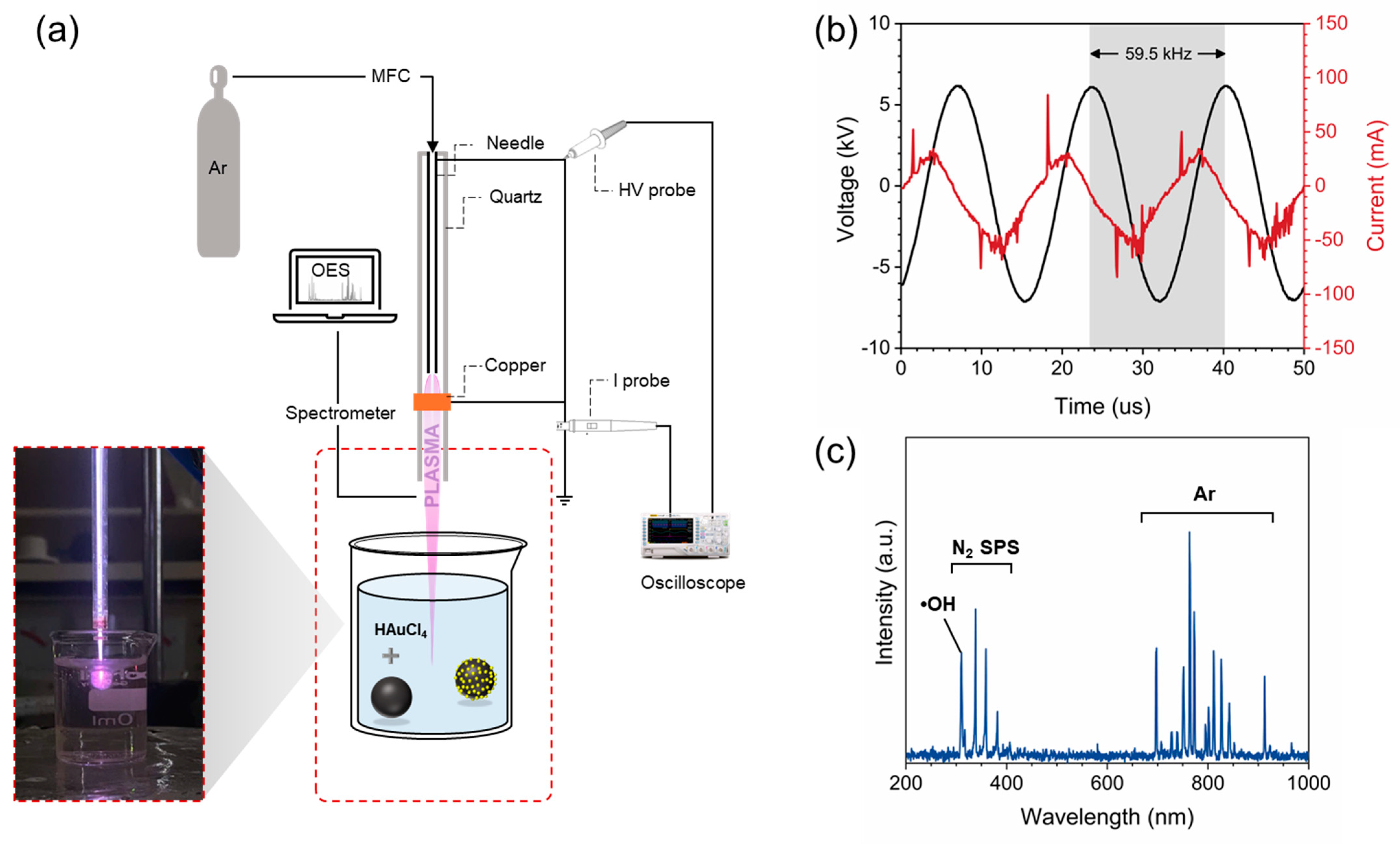
2.2. Plasma Experimental Setup

A custom-built plasma jet system was employed for the plasma-assisted synthesis of PDA-Au. The plasma jet consisted of a high-voltage electrode made from a stainless-steel syringe needle, placed inside a quartz capillary that served as the dielectric barrier. The diameters and the wall thickness of the quartz capillary were 4 mm and 1 mm, respectively. A copper ring was positioned externally around the quartz tube, acting as the grounded electrode. Argon was used as the working gas and was regulated at a flow rate of 1400 sccm using a mass flow controller. A homemade high-voltage AC power supply was applied between the needle and the copper electrodes to trigger the plasma discharge. The plasma effluent extended beyond the quartz nozzle with the length of about 10 mm, making it suitable for liquid-phase treatment.

2.3. Plasma Characterizations

The electrical characteristics of the plasma jet setup were measured using an SDS 8012 oscilloscope (Owon, Zhangzhou, China) in conjunction with a P6015 voltage probe (Tektronix, Beaverton, OR, USA) and a P6021A current probe (Tektronix, Beaverton, OR, USA) to measure the applied voltage and discharge current, respectively. An Aurora 4000 spectrometer (CNI Laser, Jilin, China) was used to record the optical emission spectroscopy (OES) spectrum of the plasma effluent. The spectrometer was connected to an optical fiber positioned perpendicular to the plasma effluent.

2.4. Synthesis of Polydopamine PDA Nanospheres

PDA-NSs were prepared using a previously reported chemical synthesis method with slight modifications [36]. A mixture of 40 mL ethanol (99.9%), 90 mL deionized water, and 2 mL NH4OH (28%) was prepared and stirred for 30 min. Subsequently, 500 mg of dopamine hydrochloride was dissolved in deionized water (10 mL) and mixed with the previously prepared mixture. Upon mixing, the solution gradually turned brown, indicating the ongoing polymerization of dopamine. The reaction was allowed to proceed for 24 h, resulting in a black-colored solution containing PDA-NSs. The PDA-NSs were then separated by using centrifuge, washed and dried in a glass desiccator. The final PDA-NSs powder could be easily stored and redispersed in water for further use.

2.5. Decoration of PDA with AuNPs by Plasma–Liquid Interaction

PDA-Au was synthesized using a facile plasma-assisted synthesis process. PDA NSs were dissolved in DI water at a concentration of 20 mg·mL−1. A 10 mL mixture containing 0.5 mL of PDA NSs (20 mg·mL−1), 0.5 mL of HAuCl4 (10 mM), and 9 mL of deionized water was prepared. The final concentrations of PDA NSs and HAuCl4 in the solution were 1 mg·mL−1 and 0.5 mM, respectively. The PDA–HAuCl4 solution was treated by the plasma system for a duration of 10 min, resulting in the formation of Au–PDA. The plasma treatment was carried out at room temperature. The Au–PDA product was then collected by centrifugation, washed and then dried in a desiccator.

2.6. Nanoparticle Characterizations

Scanning electron microscopy (SEM) analysis was performed using a JSM-7100F FE-SEM microscope (Jeol, Tokyo, Japan) operated at an acceleration voltage of 15 kV. The sample was prepared by drop-casting an aliquot of nanoparticles onto a Si substrate and drying it in a desiccator. X-ray diffraction (XRD) patterns were recorded using a D8 Advance X-ray diffraction platform (Bruker, Karlsruhe, Germany) over a 2θ range of 20° to 80°. For XRD analysis, the sample was similarly prepared by drop-casting nanoparticles onto a Si substrate and drying it in a desiccator. Fourier transform infrared spectroscopy (FTIR) analysis was conducted using an InfraLUM FT-08 spectrometer (Lumex, Mission, BC, Canada). In this case, the nanoparticle powder was mixed with KBr and pressed into a transparent pellet for measurement. UV-Vis absorption spectroscopy was performed using a USB4000 spectrometer (Ocean Optics, Orlando, FL, USA) combined with a DH-2000 deuterium–tungsten halogen light source (Ocean Optics, Orlando, FL, USA). The sample was injected into a quartz cuvette with a 10 mm light path length for measurement.

2.7. Catalytic Reduction

The catalytic activity of PDA-Au was evaluated for the reduction of Rhodamine B (RhB) using NaBH4 as the reducing agent. Briefly, 3 mL of an aqueous RhB solution (10 ppm) was mixed with 0.5 mL of freshly prepared NaBH4 solution (0.1 M) in a quartz cuvette. The reaction was initiated by adding 1 mg of PDA-Au catalyst, and the reaction kinetics were monitored using UV-Vis spectroscopy. The pseudo-first-order kinetic rate constant (k) was calculated using the following equation:
l n C C 0 = k t
where C and C0 are the dye concentrations at a given time and the initial time, respectively. The recyclability of PDA-Au was tested by recovering the catalyst via centrifugation, washing with deionized water, and reusing it for multiple cycles.

3. Results and Discussion

3.1. Plasma System Characterizations

Figure 1a illustrates the plasma jet system used for preparing PDA-Au. The current-voltage waveforms recorded from the plasma jet are shown in Figure 1b. The applied voltage was sinusoidal with a period of 16.8 μs, corresponding to a frequency of 59.5 kHz and a maximum applied voltage of approximately 6 kV (equivalent to a root-mean-square voltage of about 4.2 kV). The maximum discharge current was measured at around 85 mA. Figure 1c demonstrates the OES spectrum of the plasma effluent that extended beyond the quartz nozzle. The spectrum shows intensive emission lines in the 700–1000 nm range, which are attributed to the transition of Ar 2p–1s. The emission band observed at 309.2 nm is associated with hydroxyl radicals (•OH), which are formed through the plasma-driven dissociation of water vapor present in the surrounding air. These •OH radicals can subsequently recombine to produce hydrogen peroxide (H2O2), which may diffuse into the liquid phase. Additionally, the bands at 300–400 nm are attributed to the emissions of the nitrogen second positive system. These bands arise from electronic transitions between the excited C3Πu state and the B3Πg state of N2 molecules in ambient air, which are excited through interactions with high-energy plasma constituents.

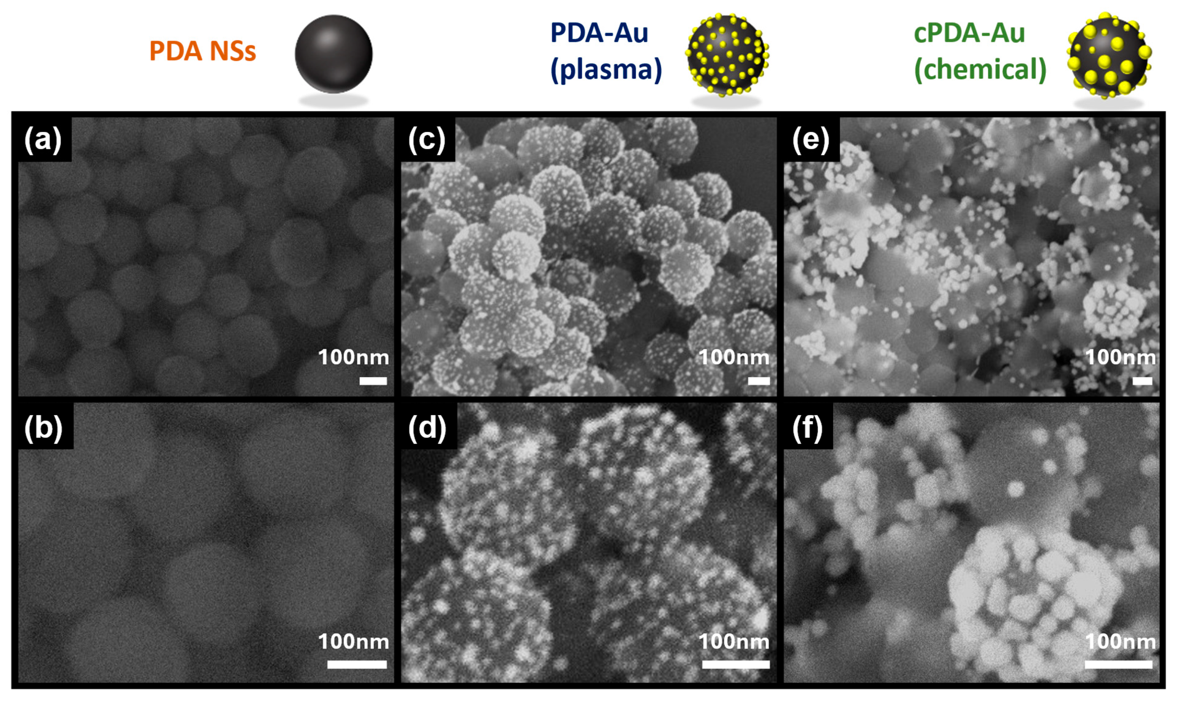
3.2. Morphological and Structural Characterizations

Figure 2a,b display SEM images of PDA nanospheres prepared through the chemical polymerization procedure using dopamine hydrochloride as a monomer. SEM images were analyzed using ImageJ 1.54g software to estimate the size distribution of the PDA nanospheres, which was found to be 205.0 ± 20.9 nm. The PDA nanospheres exhibited a spherical shape with a smooth surface (Figure S1). In contrast, the SEM images of PDA-Au (Figure 2c,d) reveal small AuNPs anchored on the surface of the PDA nanospheres, confirming the success of the plasma-assisted decoration process. The average size of AuNPs in the plasma-assisted PDA-Au (PDA-Au) was estimated to be 10.3 ± 2.0 nm. For comparison, AuNPs decorated on PDA using an in situ chemical synthesis approach (cPDA-Au) were also prepared. For cPDA-Au, the same PDA–HAuCl4 precursor solution was stirred for 24 h without plasma treatment. SEM images of cPDA-Au (Figure 2e,f) show that the average diameter of the AuNPs was approximately 35.3 ± 11.5 nm, significantly larger than those on plasma-synthesized PDA-Au. The histograms of size distribution for both conditions are presented in Figure S2. These results provide evidence that the plasma-assisted method yields smaller and more homogeneously distributed AuNPs compared to the conventional in situ chemical reduction approach, suggesting that plasma treatment promotes a more uniform nucleation of AuNPs on the PDA NS surface.
Figure 3a demonstrates the XRD patterns of the prepared PDA NSs and PDA-Au for comparison. The XRD pattern of PDA exhibits no distinct diffraction peaks, indicating the amorphous nature of PDA. In contrast, the XRD pattern of PDA-Au displays characteristic diffraction peaks at 38.3°, 44.4°, 64.6°, and 77.5°, corresponding to the (111), (200), (220), and (311) planes of standard gold structure, as referenced by JCPDS card number 00-004-0784. We noted that the XRD of cPDA-Au prepared via chemical synthesis by Han et al. also exhibited similar characteristic peaks [16]. Thus, the obtained pattern further verifies the formation of crystalline metallic Au on the PDA structure by plasma treatment. Moreover, the presence of sharp and intense peaks indicates the good crystallinity of the Au nanoparticles formed on the PDA surface. The strong peak at 2θ value of 38.3° suggests that the (111) plane is the dominant orientation of the gold nanocrystals, a characteristic commonly associated with plasma-induced AuNPs [35,37]. The crystallite size of small AuNPs on PDA structures was calculated using Scherrer’s equation:
D = K λ β c o s θ
where D is the crystallite size, K is the shape factor (~0.9 for spherical-like nanoparticles), λ is the X-ray wavelength (0.154 nm), θ is the Bragg angle and β is the full width at half maximum at 2θ [38]. From the obtained XRD pattern, the average crystallite size was calculated to be about 7.4 nm, which is smaller than the mean nanoparticle size of 10.3 nm.
FTIR analysis was carried out to identify the functional groups present in the prepared samples. Figure 3b shows the FTIR spectra of PDA (orange line) and Au–PDA (blue line). In the PDA spectrum, the broad absorption peak in the 3200–3500 cm−1 range can be attributed to the O–H stretching vibration of the catechol group and the N–H stretching vibration of the amine group in the PDA molecular structure [39]. The peaks at 1630 cm−1 and 1380 cm−1 correspond to the vibrational modes of the unsaturated C=C stretching and saturated C–C stretching of the aromatic ring, respectively. Similar features are observed in the PDA-Au spectrum, confirming the successful preparation of AuNPs on PDA NSs via plasma treatment.

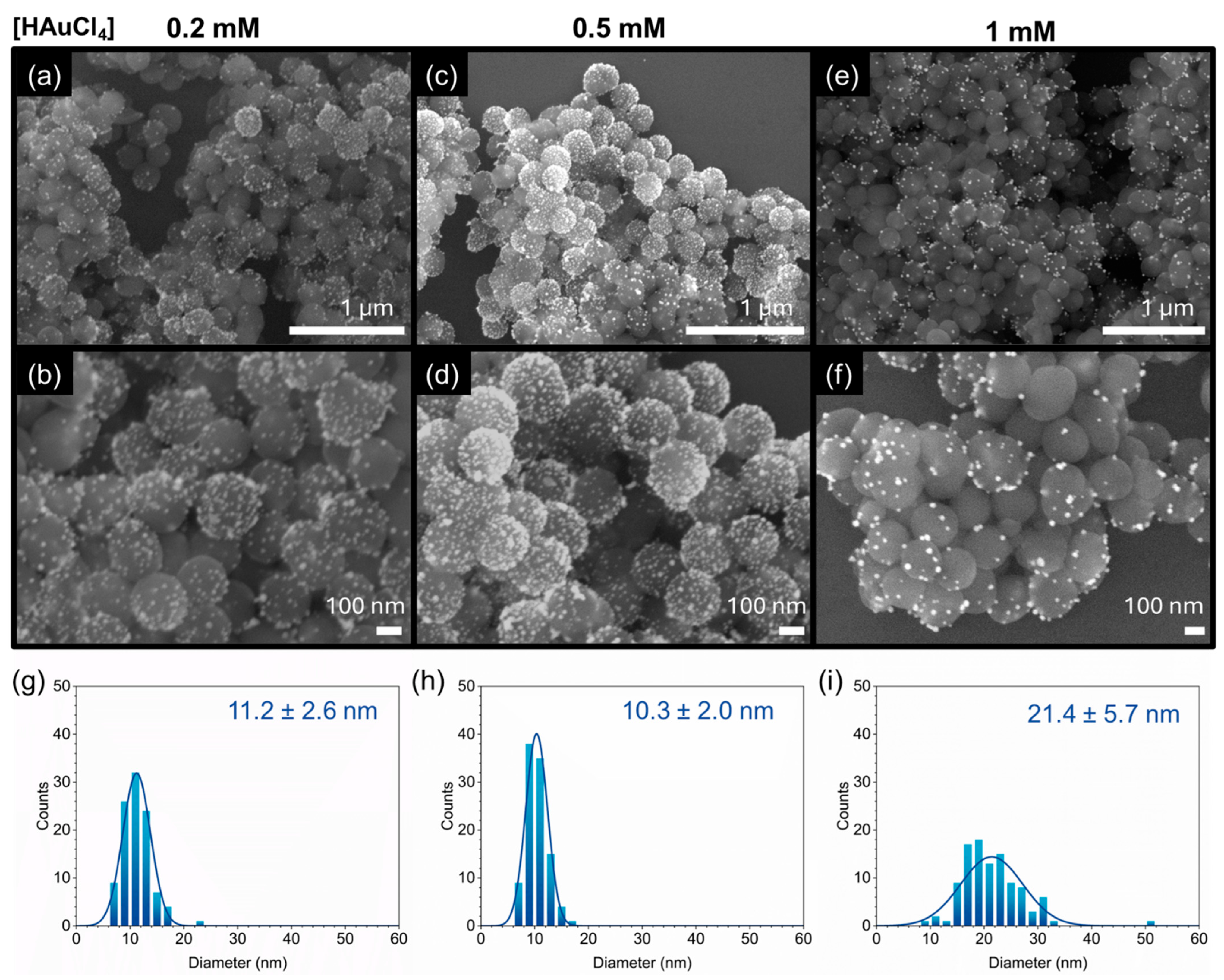
3.3. Impact of HAuCl4 Concentrations on the Morphology of PDA-Au

Previous studies on the plasma-assisted synthesis of AuNPs emphasized that this method is affected by various factors such as the type of plasma reactors, operating parameters, working gas, and the concentration of precursors. Among these, the concentration of HAuCl4 is considered the most crucial factor influencing the formation and morphology of AuNPs. Thus, we further investigate the impact of the Au ion precursor on the morphology of PDA-Au by altering the HAuCl4 concentration (0.2, 0.5, and 1 mM).
Figure 4a–f present the SEM images of PDA-Au synthesized at different HAuCl4 concentrations (0.2 mM, 0.5 mM, and 1 mM). The average particles size of the decorated AuNPs for HAuCl4 0.2 mM, 0.5 mM and 1 mM are 10.3 ± 2.0 nm, 11.2 ± 2.6 nm, 21.4 ± 5.7 nm, respectively (Figure 4g–i). As the precursor concentration increases, significant changes in both the morphology and distribution of AuNPs on the PDA surface are observed. At the lowest HAuCl4 concentration of 0.2 mM (Figure 4a,b), a sparse and relatively homogeneous distribution of small AuNPs can be observed on the PDA surface. The nanoparticles are well dispersed with low aggregation. When the concentration increases to 0.5 mM, the density of AuNPs on PDA NSs increases significantly (Figure 4c,d). In this condition, the AuNPs appear more closely packed with a larger number of particles, thus covering a larger portion of the PDA surface compared to 0.2 mM. At the highest HAuCl4 concentration of 1 mM, a different morphology is observed as larger particles are formed on the PDA surface (Figure 4e,f). However, in this case, the number of AuNPs on the PDA NS surface is much lower compared to the two lower concentrations. These findings suggest that the HAuCl4 concentration significantly influences the size, distribution, and uniformity of AuNPs formed on PDA. This can be attributed to the fact that the reduction of HAuCl4 during plasma treatment is highly dependent on the concentration of Au3+ ions. An excessive concentration of HAuCl4 generally leads to the formation of a fewer number of nanoparticles with larger particle sizes [40,41]. We evidenced that the HAuCl4 concentration of 0.5 mM yielded the most homogeneous decoration of AuNPs, whereas an excessive concentration of 1 mM resulted in the formation of larger particles. These larger particles may affect the performance of the final product in applications such as catalysis or sensing.

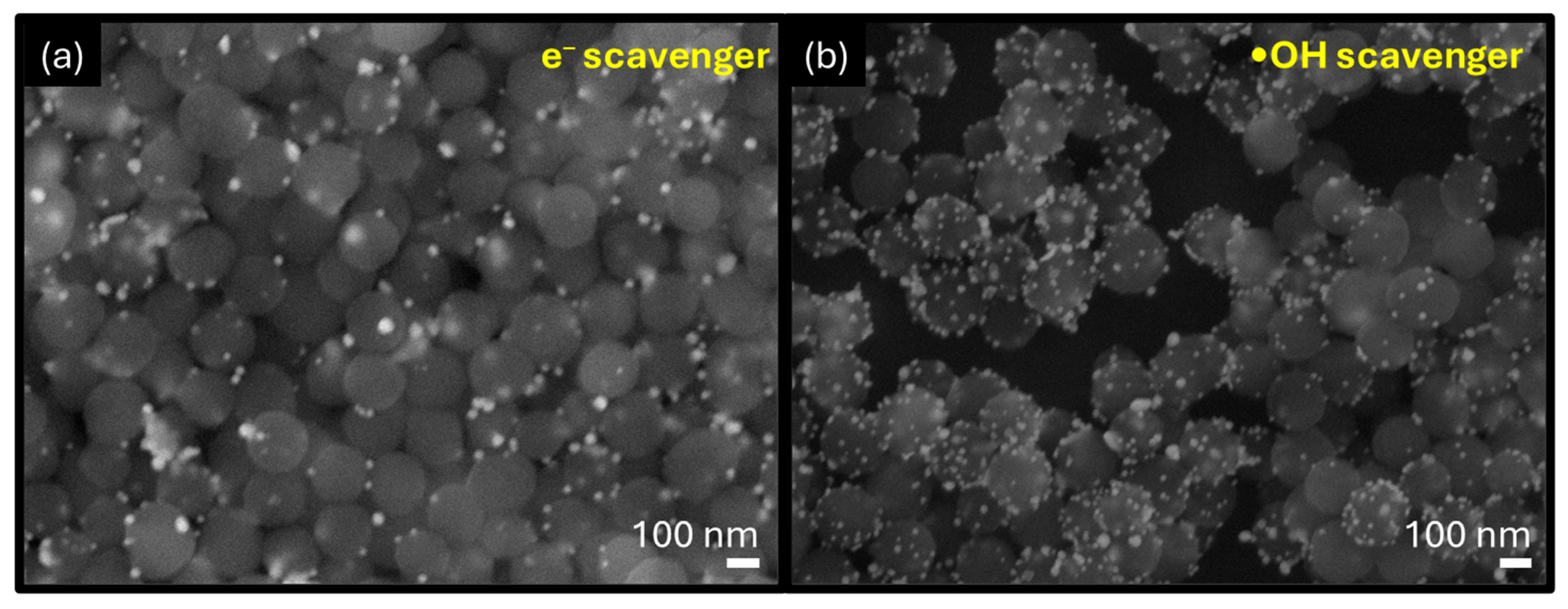
3.4. Insight into Formation of PDA-Au by Plasma-Assisted Method

We then investigated the reaction mechanism for the preparation of PDA-Au via a plasma-assisted approach. The growth pathway of colloidal AuNPs in this method is generally attributed to the reduction of Au3+ ions by plasma-induced reactive species, such as electrons and H2O2 [35,40,42]. In order to elucidate the key components involved in the formation of AuNPs, Na2SO4 and isopropanol were added to the precursor solution as scavengers for electrons (e) and •OH, respectively [43]. Under the presence of an e scavenger, the number of AuNPs on PDA reduced significantly (Figure 5a). In contrast, with the presence of an •OH scavenger, the amount of AuNPs only decreased slightly (Figure 5b). Since H2O2 is produced through the recombination of •OH radicals, the presence of isopropanol could hinder the formation of H2O2 in plasma-treated aqueous solutions [44]. This observation indicates that plasma-induced electrons are the main contributors for the decoration of AuNPs on PDA NSs.
We then performed the plasma treatment of HAuCl4 without the presence of PDA NSs. After 10 min of treatment, the yellowish precursor turned into a dark red wine color, indicating the formation of AuNPs. This result indicated that the plasma–liquid interaction is an effective approach for the synthesis of AuNPs. The UV-Vis spectrum of the plasma-synthesized AuNPs shows a surface plasmon resonance peak at around 546 nm, suggesting the presence of large-sized particles (Figure S3). SEM imaging further confirms that the AuNPs range in size from approximately 70 to 110 nm (Figure S3). We evidenced that the size of free AuNPs in the solution is significantly larger than the size of AuNPs formed on the surface of PDA NSs. A previous study by Sun et al. demonstrated that anchored Au3+ ions yield smaller particle sizes after plasma treatment compared to unanchored ones [41]. Accordingly, the difference in AuNP size observed in the presence and absence of PDA NSs can be explained by the interaction of PDA and the Au3+ precursor. Upon mixing, Au3+ ions can form complexes with the catechol moieties of PDA. These Au3+–catechol complexes on the PDA NS surface act as nucleation sites for AuNP formation. The Au3+–catechol complex can be reduced to Au0 by PDA. However, due to the mild reducing potential of catechol (E0 = −0.699 V), the in situ reduction may take longer time to finish, possibly leading to the formation of irregular and larger particles.
By applying plasma treatment, the reduction of the Au3+–catechol complex can be significantly accelerated. This is because solvated electrons, generated during plasma exposure, are among the strongest reducing species, with a high reduction potential of E0 = −2.9 V [45]. Additionally, H2O2 generated during plasma treatment may contribute to the reduction of Au3+, as confirmed by previous studies [42]. Our previous studies have shown that the Ar plasma jet can produce H2O2 with the concentration of 9.4 mM within 10 min [43]. The reduction of the Au3+ complex can be described by the following reactions (1–2):
Au3+ + 3e → Au0
Au3+ + H2O2 → Au0
Based on this, we proposed a plausible reaction mechanism for the rapid decoration of AuNPs on PDA NSs by plasma–liquid interactions (Scheme 2). Firstly, HAuCl4 reacts with the catechol moieties on PDA NS’s surface to form a Au3+–catechol complex. After this, under the plasma treatment, solvated electrons and H2O2 can directly reduce the Au3+–catechol complex into AuNPs anchored on PDA NS’s surface.

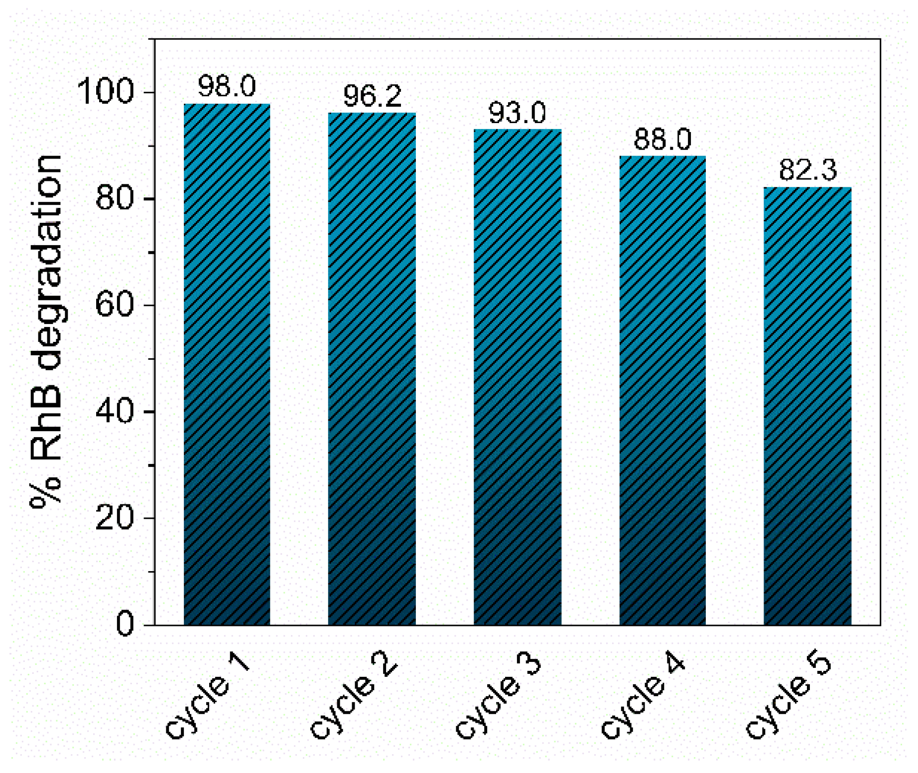
3.5. Catalytic Activity for Organic Dye Removal

The catalytic performance of PDA-Au was evaluated using the reduction of Rhodamine B (RhB) with NaBH4 as the reducing agent. UV-Vis absorption spectroscopy was used to examine the changes in the main absorption peak of RhB at 554 nm. For comparison, we also used PDA NSs as catalysts with the same reaction conditions. Figure 6a shows the temporal evolution of the RhB absorption spectrum using PDA-Au as the catalyst. After 3 min, approximately 98% of RhB was removed, indicating the high efficiency of PDA-Au. On the other hand, the degradation of RhB by PDA nanospheres took more than 35 min to complete. Interestingly, in the case of PDA-Au, a new absorption peak appeared at 420 nm, which was not observed with PDA NSs. This new peak can be attributed to intermediates formed during the decomposition process, which are subsequently degraded [24]. The degradation rates are demonstrated in Figure 6c. The kinetic rate constant for the PDA-Au catalyst in the reduction of RhB is 1.405 min−1, which is significantly higher than that of PDA NSs (0.051 min−1) (Figure 6d). Compared to several previously reported catalysts for RhB degradation, the plasma-synthesized PDA-Au catalyst demonstrates relatively high catalytic activity (Table 1). Additionally, we also investigated the catalytic degradation of MB by PDA-Au in the presence of NaBH4 (Figure S4). In this case, the complete decoloration of MB was achieved within 7 min using the PDA-Au catalyst. In contrast, the decomposition process using PDA NSs required 60 min to be completed. The rate constant for MB decomposition with PDA-Au is approximately 0.560 min−1, while PDA NSs exhibit a significantly lower rate of 0.066 min−1 (Figure S5). These results further highlight the effective catalytic activity of the plasma-synthesized PDA-Au catalyst in the degradation of organic dyes.
Another important factor for evaluating a catalyst is its reusability over time. The recyclability of the PDA-Au catalyst was assessed through five successive RhB degradation cycles. After each catalytic cycle, PDA-Au was recollected by centrifugation, washed, and resuspended under identical conditions. As shown in Figure 7, PDA-Au retained over 82% of its catalytic performance after five cycles, highlighting the reusability of this plasma-synthesized catalyst. The loss of catalytic performance is attributed to the loss of catalysts after the recollection and redispersion processes.

4. Conclusions

In this work, we developed a rapid, environmentally friendly strategy for the decoration of bio-inspired PDA NSs with AuNPs using a plasma-assisted method. Specifically, 10.3 ± 2.0 nm AuNPs can be decorated on PDA NSs to form hybrid PDA-Au within 10 min of direct plasma treatment. The characterization results from SEM, XRD, and FTIR confirmed successful AuNP formation on the PDA NS surface. The impact of Auric ion precursor concentration on the morphology of PDA-Au was also investigated. The formation of AuNPs on PDA NSs is attributed to the plasma-induced reducing agents such as solvated electrons and H2O2. The synthesized PDA-Au nanocatalysts demonstrated excellent catalytic activity for the degradation of Rhodamine B with the pseudo-first-order kinetic rate of 1.405 min−1, confirming their potential for the removal of organic pollutants in aqueous environment.

Supplementary Materials

The following supporting information can be downloaded at https://www.mdpi.com/article/10.3390/app15105280/s1, Figure S1: Particle size distribution histogram of PDA-NSs. Figure S2: Particle size histogram of PDA-Au (prepared by plasma-assisted method) and cPDA-Au (prepared by chemical synthesis method). Figure S3: (a) UV-Vis spectrum and (b) SEM image of AuNPs synthesized by plasma without PDA NSs. Figure S4: Catalytic degradation of MB by (a) PDA-Au and (b) PDA NSs with presence of NaBH4. Figure S5: (a) Degradation rate of RhB under catalysis of PDA-Au and PDA NSs. (b) The pseudo-first-order kinetic for the removal of RhB of PDA-Au and PDA NSs.

Author Contributions

Conceptualization, L.N.N.; methodology, T.M.N., L.T.N. and G.T.N.; software, T.M.N.; validation, N.K.; formal analysis, T.H.N.; investigation, N.K. and H.S.P.; resources, E.H.C.; writing—original draft preparation, T.M.N. and L.N.N.; writing—review and editing, N.K.K. and L.N.N.; visualization, N.K.; supervision, N.K.K. and L.N.N.; project administration, L.N.N.; funding acquisition, L.N.N. All authors have read and agreed to the published version of the manuscript.

Funding

This work is supported by the Vietnam Academy of Science and Technology under project number THTEXS.06/22-25. The present research was conducted by the Research Grant of Kwangwoon University in 2025 and National Research Foundation (NRF) of Korea (2021R1A6A1A03038785).

Institutional Review Board Statement

Not applicable.

Informed Consent Statement

Not applicable.

Data Availability Statement

The raw data supporting the conclusions of this article will be made available by the authors on request.

Conflicts of Interest

The authors declare no conflicts of interest.

Abbreviations

The following abbreviations are used in this manuscript:
AuNPsGold nanoparticles
PDAPolydopamine
PDA NSsPolydopamine nanospheres
PDA-AuAuNP decorated on PDA nanospheres (synthesized by plasma-assisted method)
cPDA-AuAuNP decorated on PDA nanospheres (synthesized by chemical synthesis method)
SEMScanning electron microscopy
XRDX-ray diffraction
FTIRFourier transform infrared spectroscopy

References

  1. Rezania, S.; Darajeh, N.; Rupani, P.F.; Mojiri, A.; Kamyab, H.; Taghavijeloudar, M. Recent Advances in the Adsorption of Different Pollutants from Wastewater Using Carbon-Based and Metal-Oxide Nanoparticles. Appl. Sci. 2024, 14, 11492. [Google Scholar] [CrossRef]
  2. Katheresan, V.; Kansedo, J.; Lau, S.Y. Efficiency of Various Recent Wastewater Dye Removal Methods: A Review. J. Environ. Chem. Eng. 2018, 6, 4676–4697. [Google Scholar] [CrossRef]
  3. Oller, I.; Malato, S.; Sánchez-Pérez, J.A. Combination of Advanced Oxidation Processes and Biological Treatments for Wastewater Decontamination—A Review. Sci. Total Environ. 2011, 409, 4141–4166. [Google Scholar] [CrossRef]
  4. Martínez-Huitle, C.A.; Brillas, E. Decontamination of Wastewaters Containing Synthetic Organic Dyes by Electrochemical Methods: A General Review. Appl. Catal. B Environ. 2009, 87, 105–145. [Google Scholar] [CrossRef]
  5. Chen, W.; Mo, J.; Du, X.; Zhang, Z.; Zhang, W. Biomimetic Dynamic Membrane for Aquatic Dye Removal. Water Res. 2019, 151, 243–251. [Google Scholar] [CrossRef]
  6. Ma, W.; Li, Y.; Zhang, M.; Gao, S.; Cui, J.; Huang, C.; Fu, G. Biomimetic Durable Multifunctional Self-Cleaning Nanofibrous Membrane with Outstanding Oil/Water Separation, Photodegradation of Organic Contaminants, and Antibacterial Performances. ACS Appl. Mater. Interfaces 2020, 12, 34999–35010. [Google Scholar] [CrossRef]
  7. Li, N.; Chen, G.; Zhao, J.; Yan, B.; Cheng, Z.; Meng, L.; Chen, V. Self-Cleaning PDA/ZIF-67@PP Membrane for Dye Wastewater Remediation with Peroxymonosulfate and Visible Light Activation. J. Membr. Sci. 2019, 591, 117341. [Google Scholar] [CrossRef]
  8. Lv, Y.; Zhang, C.; He, A.; Yang, S.-J.; Wu, G.-P.; Darling, S.B.; Xu, Z.-K. Photocatalytic Nanofiltration Membranes with Self-Cleaning Property for Wastewater Treatment. Adv. Funct. Mater. 2017, 27, 1700251. [Google Scholar] [CrossRef]
  9. Lee, H.; Dellatore, S.M.; Miller, W.M.; Messersmith, P.B. Mussel-Inspired Surface Chemistry for Multifunctional Coatings. Science 2007, 318, 426–430. [Google Scholar] [CrossRef]
  10. Chin, H.-K.; Lin, P.-Y.; Chen, J.; Kirankumar, R.; Wen, Z.-H.; Hsieh, S. Polydopamine-Mediated Ag and ZnO as an Active and Recyclable SERS Substrate for Rhodamine B with Significantly Improved Enhancement Factor and Efficient Photocatalytic Degradation. Appl. Sci. 2021, 11, 4914. [Google Scholar] [CrossRef]
  11. Chae, W.R.; Song, Y.-J.; Lee, N.Y. Polydopamine-Mediated Gold Nanoparticle Coating Strategy and Its Application in Photothermal Polymerase Chain Reaction. Lab Chip 2025, 25, 1429–1438. [Google Scholar] [CrossRef]
  12. Wu, M.; Wang, T.; Müller, L.; Müller, F.A. Adjustable Synthesis of Polydopamine Nanospheres and Their Nucleation and Growth. Colloids Surf. A Physicochem. Eng. Asp. 2020, 603, 125196. [Google Scholar] [CrossRef]
  13. Jiang, X.; Wang, Y.; Li, M. Selecting Water-Alcohol Mixed Solvent for Synthesis of Polydopamine Nano-Spheres Using Solubility Parameter. Sci. Rep. 2014, 4, 6070. [Google Scholar] [CrossRef]
  14. Ghorbani, F.; Zamanian, A.; Behnamghader, A.; Joupari, M.D. A Facile Method to Synthesize Mussel-Inspired Polydopamine Nanospheres as an Active Template for in Situ Formation of Biomimetic Hydroxyapatite. Mater. Sci. Eng. C 2019, 94, 729–739. [Google Scholar] [CrossRef]
  15. Wang, Z.; Wang, H. Pt, Pd, and Rh Nanoparticles Supported on Polydopamine Nanospheres as Catalysts for Transfer Hydrogenolysis. ACS Appl. Nano Mater. 2022, 5, 11797–11808. [Google Scholar] [CrossRef]
  16. Han, X.; Chen, X.; Yan, M.; Liu, H. Synergetic Effect of Polydopamine Particles and In-Situ Fabricated Gold Nanoparticles on Charge-Dependent Catalytic Behaviors. Particuology 2019, 44, 63–70. [Google Scholar] [CrossRef]
  17. Wang, W.; Dong, X.; Huang, X.; Wu, H.; Zhu, D. Polydopamine-Mediated CdS Crystal Growth Behavior and the Synergistic Photocatalytic–Adsorption Mechanisms in Removing Rhodamine B. Energy Fuels 2023, 37, 18184–18193. [Google Scholar] [CrossRef]
  18. Wang, Z.; Wang, W.; Wamsley, M.; Zhang, D.; Wang, H. Colloidal Polydopamine Beads: A Photothermally Active Support for Noble Metal Nanocatalysts. ACS Appl. Mater. Interfaces 2022, 14, 17560–17569. [Google Scholar] [CrossRef]
  19. Wang, Z.; Wang, H. Phase-Controlled Ruthenium Nanocrystals on Colloidal Polydopamine Supports and Their Catalytic Behaviors in Aerobic Oxidation Reactions. ACS Appl. Mater. Interfaces 2023, 15, 36676–36687. [Google Scholar] [CrossRef]
  20. Abood, E.A.; Essa, W.K.; Alsuraifi, A.; Yasin, S.A. A Novel Nanogold Composite Fabrication, Its Characterization, and Its Application in the Removal of Methylene Blue Dye from an Aqueous Solution. Appl. Sci. 2024, 14, 5229. [Google Scholar] [CrossRef]
  21. Yang, Y.-Y.; Li, Y.-T.; Li, X.-J.; Zhang, L.; Kouadio Fodjo, E.; Han, S. Controllable in Situ Fabrication of Portable AuNP/Mussel-Inspired Polydopamine Molecularly Imprinted SERS Substrate for Selective Enrichment and Recognition of Phthalate Plasticizers. Chem. Eng. J. 2020, 402, 125179. [Google Scholar] [CrossRef]
  22. Gao, Y.; Wu, Y.; Huang, P.; Wu, F.-Y. Colorimetric and Photothermal Immunosensor for Sensitive Detection of Cancer Biomarkers Based on Enzyme-Mediated Growth of Gold Nanostars on Polydopamine. Anal. Chim. Acta 2023, 1279, 341775. [Google Scholar] [CrossRef]
  23. Liu, Y.; Haghighatbin, M.A.; Shen, W.; Cui, H. Functionalized Polydopamine Nanospheres with Chemiluminescence and Immunoactivity for Label-Free Copeptin Immunosensing. ACS Appl. Nano Mater. 2020, 3, 4681–4689. [Google Scholar] [CrossRef]
  24. Zhang, K.; Suh, J.M.; Choi, J.-W.; Jang, H.W.; Shokouhimehr, M.; Varma, R.S. Recent Advances in the Nanocatalyst-Assisted NaBH4 Reduction of Nitroaromatics in Water. ACS Omega 2019, 4, 483–495. [Google Scholar] [CrossRef]
  25. Anbu Anjugam Vandarkuzhali, S.; Karthikeyan, G.; Pachamuthu, M.P. Efficient Catalytic Reduction of Xanthene Based Dyes, Methylene Blue, and 4-Nitrophenol Using Gold Nanoparticles Supported on Mesoporous Amine Functionalized SBA-15. Mater. Sci. Eng. B 2024, 302, 117208. [Google Scholar] [CrossRef]
  26. Stolle, H.L.K.S.; Kluitmann, J.J.; Csáki, A.; Köhler, J.M.; Fritzsche, W. Shape-Dependent Catalytic Activity of Gold and Bimetallic Nanoparticles in the Reduction of Methylene Blue by Sodium Borohydride. Catalysts 2021, 11, 1442. [Google Scholar] [CrossRef]
  27. Wang, L.; Liu, H.; Zhuang, J.; Wang, D. Small-Scale Big Science: From Nano- to Atomically Dispersed Catalytic Materials. Small Sci. 2022, 2, 2200036. [Google Scholar] [CrossRef]
  28. Suchomel, P.; Kvitek, L.; Prucek, R.; Panacek, A.; Halder, A.; Vajda, S.; Zboril, R. Simple Size-Controlled Synthesis of Au Nanoparticles and Their Size-Dependent Catalytic Activity. Sci. Rep. 2018, 8, 4589. [Google Scholar] [CrossRef]
  29. Liang, C.; Cheong, J.Y.; Sitaru, G.; Rosenfeldt, S.; Schenk, A.S.; Gekle, S.; Kim, I.-D.; Greiner, A. Size-Dependent Catalytic Behavior of Gold Nanoparticles. Adv. Mater. Interfaces 2022, 9, 2100867. [Google Scholar] [CrossRef]
  30. Sankar, M.; He, Q.; Engel, R.V.; Sainna, M.A.; Logsdail, A.J.; Roldan, A.; Willock, D.J.; Agarwal, N.; Kiely, C.J.; Hutchings, G.J. Role of the Support in Gold-Containing Nanoparticles as Heterogeneous Catalysts. Chem. Rev. 2020, 120, 3890–3938. [Google Scholar] [CrossRef]
  31. Mei, S.; Kochovski, Z.; Roa, R.; Gu, S.; Xu, X.; Yu, H.; Dzubiella, J.; Ballauff, M.; Lu, Y. Enhanced Catalytic Activity of Gold@Polydopamine Nanoreactors with Multi-Compartment Structure Under NIR Irradiation. Nano-Micro Lett. 2019, 11, 83. [Google Scholar] [CrossRef]
  32. Wang, J.-G.; Hua, X.; Li, M.; Long, Y.-T. Mussel-Inspired Polydopamine Functionalized Plasmonic Nanocomposites for Single-Particle Catalysis. ACS Appl. Mater. Interfaces 2017, 9, 3016–3023. [Google Scholar] [CrossRef]
  33. Hemmati, S.; Heravi, M.M.; Karmakar, B.; Veisi, H. In Situ Decoration of Au NPs over Polydopamine Encapsulated GO/Fe3O4 Nanoparticles as a Recyclable Nanocatalyst for the Reduction of Nitroarenes. Sci. Rep. 2021, 11, 12362. [Google Scholar] [CrossRef]
  34. Zubair, M.; Rafique, M.S.; Khalid, A.; Yaqub, T.; Alomar, S.Y.; Gohar, H. Synthesis of Gold-PVP Nanostructured Composites by Microplasma: A Test to Study Their Inhibiting Tendency of Avian Influenza Virus Activity. Appl. Sci. 2022, 12, 5352. [Google Scholar] [CrossRef]
  35. Nguyen, L.N.; Kaushik, N.; Lamichhane, P.; Mumtaz, S.; Paneru, R.; Bhartiya, P.; Kwon, J.S.; Mishra, Y.K.; Nguyen, L.Q.; Kaushik, N.K.; et al. In Situ Plasma-Assisted Synthesis of Polydopamine-Functionalized Gold Nanoparticles for Biomedical Applications. Green Chem. 2020, 22, 6588–6599. [Google Scholar] [CrossRef]
  36. Liu, Y.; Ai, K.; Liu, J.; Deng, M.; He, Y.; Lu, L. Dopamine-Melanin Colloidal Nanospheres: An Efficient Near-Infrared Photothermal Therapeutic Agent for In Vivo Cancer Therapy. Adv. Mater. 2013, 25, 1353–1359. [Google Scholar] [CrossRef]
  37. Lee, S.Y.; Do, H.T.; Kim, J.H. Microplasma-Assisted Synthesis of TiO2–Au Hybrid Nanoparticles and Their Photocatalytic Mechanism for Degradation of Methylene Blue Dye under Ultraviolet and Visible Light Irradiation. Appl. Surf. Sci. 2022, 573, 151383. [Google Scholar] [CrossRef]
  38. Oliveira, A.E.F.; Pereira, A.C.; Resende, M.A.C.; Ferreira, L.F. Gold Nanoparticles: A Didactic Step-by-Step of the Synthesis Using the Turkevich Method, Mechanisms, and Characterizations. Analytica 2023, 4, 250–263. [Google Scholar] [CrossRef]
  39. Zhou, L.-J.; Wang, Y.-Y.; Li, S.-L.; Cao, L.; Jiang, F.-L.; Maskow, T.; Liu, Y. Core–Shell Polydopamine/Cu Nanometer Rods Efficiently Deactivate Microbes by Mimicking Chloride-Activated Peroxidases. ACS Omega 2022, 7, 29984–29994. [Google Scholar] [CrossRef]
  40. Patel, J.; Němcová, L.; Maguire, P.; Graham, W.G.; Mariotti, D. Synthesis of Surfactant-Free Electrostatically Stabilized Gold Nanoparticles by Plasma-Induced Liquid Chemistry. Nanotechnology 2013, 24, 245604. [Google Scholar] [CrossRef]
  41. Sun, D.; McLaughlan, J.; Zhang, L.; Falzon, B.G.; Mariotti, D.; Maguire, P.; Sun, D. Atmospheric Pressure Plasma-Synthesized Gold Nanoparticle/Carbon Nanotube Hybrids for Photothermal Conversion. Langmuir 2019, 35, 4577–4588. [Google Scholar] [CrossRef]
  42. De Vos, C.; Baneton, J.; Witzke, M.; Dille, J.; Godet, S.; Gordon, M.J.; Sankaran, R.M.; Reniers, F. A Comparative Study of the Reduction of Silver and Gold Salts in Water by a Cathodic Microplasma Electrode. J. Phys. D Appl. Phys. 2017, 50, 105206. [Google Scholar] [CrossRef]
  43. Nguyen, M.T.; Nguyen, M.D.; Nguyen, T.H.; Gao, M.; Kaushik, N.; Vasilyevich, O.V.; Petrovna, A.A.; Andreevich, O.N.; Kaushik, N.K.; Nguyen, T.T.; et al. Microwave-Assisted Synthesis of Copper Ferrite Nanocatalysts for Synergistic Plasma-Fenton Degradation of Rhodamine B. Colloids Surf. A Physicochem. Eng. Asp. 2025, 705, 135681. [Google Scholar] [CrossRef]
  44. Qi, Z.; Zhang, Q.; Zhu, D.; Ding, Z.; Niu, J.; Liu, D.; Zhao, Y.; Xia, Y.; Zhao, Z.; Wang, X. The Reaction Pathways of H2O2(Aq) in the He Plasma Jet with a Liquid System. Plasma Chem. Plasma Process. 2020, 40, 1001–1018. [Google Scholar] [CrossRef]
  45. Skiba, M.; Zaporozhets, J.; Vorobyova, V. Gold Nanoparticles with Natural Polymer Synthesized by Plasma–Liquid Interactions: Size-Control, Characterization and Colorimetric Detection of Melamine Based on the Size Effect of Gold Nanoparticles. Nano-Struct. Nano-Objects 2024, 37, 101113. [Google Scholar] [CrossRef]
  46. Bhargava, A.; Jain, N.; Khan, M.A.; Pareek, V.; Dilip, R.V.; Panwar, J. Utilizing Metal Tolerance Potential of Soil Fungus for Efficient Synthesis of Gold Nanoparticles with Superior Catalytic Activity for Degradation of Rhodamine B. J. Environ. Manag. 2016, 183, 22–32. [Google Scholar] [CrossRef]
  47. Nguyen, T.T.-N.; Vo, T.-T.; Nguyen, B.N.-H.; Nguyen, D.-T.; Dang, V.-S.; Dang, C.-H.; Nguyen, T.-D. Silver and Gold Nanoparticles Biosynthesized by Aqueous Extract of Burdock Root, Arctium Lappa as Antimicrobial Agent and Catalyst for Degradation of Pollutants. Environ. Sci Pollut. Res. 2018, 25, 34247–34261. [Google Scholar] [CrossRef]
  48. Liu, C.-C.; Wang, W.-Y.; Hu, C.-C.; Chiu, T.-C. Tannic Acid-Decorated Bimetallic Copper–Gold Nanoparticles with High Catalytic Activity for the Degradation of 4-Nitrophenol and Rhodamine B. ACS Omega 2024, 9, 24970–24977. [Google Scholar] [CrossRef]
  49. Yan, Q.; Wang, X.-Y.; Feng, J.-J.; Mei, L.-P.; Wang, A.-J. Simple Fabrication of Bimetallic Platinum-Rhodium Alloyed Nano-Multipods: A Highly Effective and Recyclable Catalyst for Reduction of 4-Nitrophenol and Rhodamine B. J. Colloid Interface Sci. 2021, 582, 701–710. [Google Scholar] [CrossRef]
  50. Li, Z.; Yuan, C.-G.; Guo, Q.; Wei, X. Preparation of Stable AgNPs@PAN/GO-SH Nanocompsite by Electrospinning for Effective Degradation of 4-Nitrophenol, Methylene Blue and Rhodamine B. Mater. Lett. 2020, 265, 127409. [Google Scholar] [CrossRef]
  51. Velidandi, A.; Pabbathi, N.P.P.; Baadhe, R.R. Study of Parameters Affecting the Degradation of Rhodamine-B and Methyl Orange Dyes by Annona Muricata Leaf Extract Synthesized Nanoparticles as Well as Their Recyclability. J. Mol. Struct. 2021, 1236, 130287. [Google Scholar] [CrossRef]
  52. Vijayan, R.; Joseph, S.; Mathew, B. Green Synthesis of Silver Nanoparticles Using Nervalia Zeylanica Leaf Extract and Evaluation of Their Antioxidant, Catalytic, and Antimicrobial Potentials. Part. Sci. Technol. 2019, 37, 809–819. [Google Scholar] [CrossRef]
Scheme 1. Schematic illustration of preparation of PDA-Au using plasma-assisted synthesis process.
Applsci 15 05280 sch001
Figure 1. The plasma jet system for the preparation of PDA-Au. (a) An illustration of the plasma jet setup. (b) The current–voltage waveform of the plasma jet recorded by an oscilloscope. (c) The OES spectrum of the plasma effluent obtained from the spectrometer.
Applsci 15 05280 g001
Figure 2. Scanning electron microscope images of (a,b) PDA NSs, (c,d) PDA-Au prepared by plasma-assisted method, and (e,f) cPDA-Au prepared by conventional chemical synthesis for comparison.
Applsci 15 05280 g002
Figure 3. Characterizations of plasma synthesized PDA-Au and PDA NSs: (a) X-ray diffraction patterns and (b) Fourier-transform infrared spectra.
Applsci 15 05280 g003
Figure 4. Scanning electron microscope images of plasma-synthesized PDA-Au with different HAuCl4 concentrations of (a,b) 0.2 mM, (c,d) 0.5 mM, and (e,f) 1 mM. Size distribution histogram of PDA-Au with HAuCl4 of (g) 0.2 mM, (h) 0.5 mM, and (i) 1 mM.
Applsci 15 05280 g004
Figure 5. Scanning electron microscope images of plasma-synthesized PDA-Au in presence of (a) e scavenger and (b) •OH scavenger.
Applsci 15 05280 g005
Scheme 2. Proposed reaction mechanisms for the plasma-assisted preparation of PDA-Au.
Applsci 15 05280 sch002
Figure 6. Catalytic activity of (a) PDA-Au and (b) PDA NSs for removal of 10 ppm RhB. (c) Degradation rate of RhB under catalysis of PDA-Au and PDA NSs. (d) Pseudo-first-order kinetic for removal of RhB of PDA-Au and PDA NSs.
Applsci 15 05280 g006
Figure 7. The reusability of PDA-Au for RhB removal after 5 repeated catalytic cycles.
Applsci 15 05280 g007
Table 1. Comparison of reported catalysts for degradation of RhB with NaBH4.
CatalystRhB ConcentrationTreatment VolumeDegradation TimeDegradation EfficiencyPseudo-First-Order Rate ConstantRef.
AuNPs2.5 × 10−5 M3 mL7 min-0.762 min−1 *[46]
Br-AuNPs0.1 mM3 mL12 min-0.364 min−1 *[47]
BR-AgNPs0.1 mM3 mL10 min-0.424 min−1 *[47]
TA-CuAu NPs0.1 mM4 mL10 min-0.2628 min−1[48]
PtRh ANMPs0.3 mM2.36 mL12 min97.22%0.354 min−1[49]
Pt black0.3 mM2.36 mL40 min47.29%0.056 min−1[49]
AgNPs@PAN/Go-SH10 mg L−150 mL14 min-0.266 min−1[50]
Ag-AgCl NPs50 mg/100 mL5 mL5 min96%0.748 min−1[51]
AgNPs0.05 mM3.5 mL10 min-0.600 min−1[52]
PDA-Au10 ppm3.5 mL7 min>98%1.405 min−1This work
* Converted from reported rate constant value in s−1.
Disclaimer/Publisher’s Note: The statements, opinions and data contained in all publications are solely those of the individual author(s) and contributor(s) and not of MDPI and/or the editor(s). MDPI and/or the editor(s) disclaim responsibility for any injury to people or property resulting from any ideas, methods, instructions or products referred to in the content.

