Article

Analysis of Glucocorticoids as Potential Adulterants in Cosmetic Products: A Dual Approach for Qualitative and Quantitative Evaluation Based on ELISA and HPLC-MS Methods

by
Seyedeh Rojin Shariati Pour
1,
Afsaneh Emamiamin
1,
Martina Zangheri
1,2,
Donato Calabria
3,
Massimo Guardigli
3,4,
Emanuele Porru
5,
Jessica Fiori
1,6,* and
Mara Mirasoli
1,4
1
Department of Chemistry “Giacomo Ciamician”, Alma Mater Studiorum, University of Bologna, Tecnopolo di Rimini, Via Dario Campana 71, 47922 Rimini, Italy
2
Interdepartmental Centre for Industrial Agrofood Research (CIRI AGRO), Alma Mater Studiorum, University of Bologna, Via Quinto Bucci 336, 47521 Cesena, Italy
3
Department of Chemistry “Giacomo Ciamician”, Alma Mater Studiorum, University of Bologna, Via Francesco Selmi 2, 40126 Bologna, Italy
4
Interdepartmental Centre for Industrial Research in Renewable Resources, Environment, Sea, and Energy (CIRI FRAME), Alma Mater Studiorum, University of Bologna, Via Sant’Alberto 163, 48123 Ravenna, Italy
5
Occupational Medicine Unit, Department of Medical and Surgical Science, Alma Mater Studiorum, University of Bologna, 40138 Bologna, Italy
6
Laboratory of Proteomics Metabolomics and Bioanalytical Chemistry, IRCCS Institute of Neurological Sciences of Bologna, 40126 Bologna, Italy
*
Author to whom correspondence should be addressed.
Appl. Sci. 2025, 15(1), 414; https://doi.org/10.3390/app15010414
Submission received: 12 November 2024 / Revised: 19 December 2024 / Accepted: 24 December 2024 / Published: 4 January 2025
(This article belongs to the Special Issue Analytical Chemistry: Techniques and Applications)

Abstract

:
The analysis of cosmetic products represents an important field of analytical chemistry, since the demand for new formulations is continuously increasing. Regulations about prohibited/regulated compounds are applied in each country. Among the substances that are banned in cosmetics, corticosteroids represent a potential harm for consumers since the prolonged exposure to these compounds can affect health status. However, corticosteroids can be found in cosmetics as an illegal addition since they are able to alleviate the symptoms of inflammatory skin problems. In this work, two different approaches for detecting corticosteroids as potential adulterants in cosmetic products were compared. First, a reversed-phase HPLC-MS method was optimized and fully validated in order to identify and quantify eight corticosteroids (methylprednisolone, beclomethasone, flunisolide, budesonide, betamethasone 17-valerate, beclomethasone dipropionate, flumethasone, and dexamethasone). This reference method was then compared with an enzyme-linked immunosorbent assay (ELISA). Indeed, immunological techniques allow for rapid, low-cost, and sensitive detection of target analytes even in complex matrices, and they can be performed with simple instrumentation and by non-skilled personnel. The application of these methods on spiked cosmetic products was compared in terms of performance and advantages in order to evaluate the possibility of exploiting a complementary approach for optimizing the time for and costs of the analysis.

1. Introduction

Glucocorticoids, also known as corticosteroids, exhibit several therapeutic uses in the treatment of chronic inflammatory and immune diseases due to their immunosuppressive properties. These compounds inhibit the production and activity of inflammatory cells, decreasing the number of immune cells [1,2]. Glucocorticoids are widely found in allergy, dermatology, endocrinology, hematology, and immunology medicines, administered orally, topically, or in inhalation solutions [3,4,5,6,7]. However, studies show that corticosteroids can be toxic and cause severe side effects, namely, osteoporosis, Cushing’s syndrome, hypertension, hyperglycemia, increased risk of infection, skin atrophy, erythema, delayed wound healing, etc. Chronic use of these compounds can be of substantial risk since the main key factors that influence the adverse effect of corticosteroids are dose and duration of therapy [8,9,10,11,12,13,14]. As for definition, cosmetics cannot contain active substances with pharmacological properties; European legislation banned these substances in cosmetic formulations [15]. Also, other countries have regulated the presence of such substances in cosmetic products [16]. Despite this, because of their anti-inflammatory, antiaging, and depigmenting properties, they might be present in counterfeit products as adulterants [17,18,19,20,21]. Although the concentration of glucocorticoids in counterfeit products is usually low, prolonged use of them by consumers unaware of the adulteration constitutes a real health risk. Corticosteroids are added in counterfeit products as a mixture of low contents of corticosteroids to enhance the effect. Therefore, in order to verify the presence of such substances suspected to be illegally added to cosmetic products, the analytical method must be sensitive and selective enough to detect nanograms of the illegal drug and to distinguish similar structures. Several qualitative and quantitative analytical methods have been introduced to determine corticosteroids in different matrices using well-established instrumental techniques such as gas chromatography (GC) [22] and high-performance liquid chromatography (HPLC) coupled with diode array detector (DAD) [23] or mass spectrometry (MS) [24,25,26] as well as NMR spectroscopy [27]. Among these, some have been developed to detect trace amounts in different formulations, such as creams, gels, and liquid formulations. Giaccone et al. proposed a method based on LC-ESI-MS/MS for the determination of several corticosteroids in cosmetic products [28]. Kang et al. [29] developed a method using homogeneous ionic liquid microextraction high-performance liquid chromatography equipped with a photo-diode array detector to determine corticosteroids in liquid and gel-like cosmetic products. In another study, Ivković et al. optimized and validated an RP-HPLC method with UV detection for the determination of corticosteroids in ambiphilic creams using ultrasonic bath as the sample preparation technique [30]. Kim et al. reported an ultra-performance liquid chromatography–tandem mass spectrometry (UPLC-MS/MS) to analyze trace amounts of 43 corticosteroids in different cosmetic products such as creams, gels, and powders [20]. As an alternative, bioanalytical techniques have been reported to detect corticosteroids in different matrices [31,32]. Indeed, immunoassays have been widely employed for detecting prohibited or restricted ingredients in the cosmetic field [33,34,35,36,37,38,39,40,41,42,43,44,45]. Despite this, to the best of our knowledge, only few of them were applied to cosmetic formulations. Zhang et al. developed a competitive indirect enzyme-linked immunosorbent assay (ELISA) for screening of hydrocortisone in body lotion, moisture cream, and smoothing toner samples [46]. In another study, Wang et al. reported a lateral flow immunoassay to rapidly test dexamethasone in commercial facial masks. The results obtained by this method were reported to be consistent with those acquired by LC-MS [37]. Immunological techniques offer some advantages with respect to the traditional chromatography-based methods, such as easier and faster protocol, as well as suitability to analyze a large number of samples. The specificity of the antigen–antibody reaction makes this approach ideal for the detection of small traces of the target analyte in very complex matrices [47]. Furthermore, immunological methods can be performed easily and rapidly (the tests are generally completed in a few hours) and the protocol is suitable for complete automation and high-throughput screening. Finally, execution of immunoassays requires an aqueous environment, avoiding the use of organic solvents and toxic chemicals and thus allowing for working with biocompatible and green procedures. There are different formats in which immunological methods can be implemented, but the most used for routinary applications is the ELISA. An enzyme (such as horseradish peroxidase, HRP) is used as a label in order to catalyze a reaction in the presence of a specific enzyme substrate in order to generate a signal that is measured for quantitative detection of the target analyte. The assay is performed in a microwell plate platform, and a multistep procedure comprising the addition of the sample and of the immunoreagents followed by several washing steps is required. Finally, the addition of the enzymatic substrate allows for the development of the analytical signal, which can be detected by exploiting a multi-well plate reader. Depending on the nature of the enzymatic substrate, it is possible to exploit different detection principles (e.g., colorimetric, chemiluminescent, or electrochemiluminescent detection). The traditional reference techniques, such as HPLC, GC coupled with MS, and immunoassays, show a limitation in sensitivity. Indeed, the limit of detection (LOD) and quantification (LOQ) are generally higher than the ones offered by instrumental-based techniques. Another problem is represented by the cross-reactivity phenomenon, which implies that the antibody recognizes the target antigen and similar species, leading to a lack of specificity. However, immunoassays can be very useful as preliminary screening methods to be applied in series on a great number of samples in order to identify batches suspected of adulteration, which can then be subjected to further analysis [48]. In this study, an HPLC-MS method was developed for the simultaneous determination of eight corticosteroids (Figure 1): betamethasone valerate (BM 17-V), beclomethasone (BC), beclomethasone dipropionate (BCDP), methylprednisolone (MP), budesonide (BD), flunisolide (FN), flumethasone (FM), and dexamethasone (DM) in cosmetic formulations without any sample pre-treatment. The method was validated in terms of linearity, sensitivity, accuracy, precision, and matrix effect on three cosmetic products (two creams and one serum) available on the market. Moreover, an alternative method based on ELISA was employed to detect the aforementioned corticosteroids. This technique was adopted to simplify the analysis of cosmetic formulations to determine the presence of glucocorticoids without the need for a chromatography instrument, which might not be available in every cosmetic lab. The ELISA was developed starting from a commercial kit for the measurement of flumethasone in plasma, extending its applicability to cosmetic matrices and to a multi-target screening (5 corticosteroids: BM 17-V, BC, MP, FM, and DM). The ELISA protocol in use was optimized and validated on two steroid-free commercial cosmetic products (cream and serum). For the first time, an ELISA was validated for the preliminary detection of a group of corticosteroids at the same time. This method can be of great support for the rapid control of samples suspected of adulteration with glucocorticoids in order to increase the action of the product claim.

2. Materials and Methods

2.1. Materials

HPLC-grade acetonitrile, formic acid, tetrahydrofuran, isopropanol, and sodium phosphate were purchased from Sigma-Aldrich (Milan, Italy). Betamethasone 17-valerate (BM 17-V), beclomethasone (BC), beclomethasone dipropionate (BCDP), methylprednisolone (MP), budesonide (BD), flunisolide (FN), flumethasone (FM), and dexamethasone (DM) were purchased from Sigma-Aldrich (Milan, Italy). Purified water from the Milli-RX system (Millipore, Milford, MA, USA) was used to prepare mobile phase and standard solutions. Nitrocellulose syringe filters (0.20 mm) were purchased from Phenomenex (Torrance, CA, USA). Commercial cosmetic products, namely, cream A, cream B, and serum, were available on the market; the ingredient lists are reported in Table S1 of the Supplementary Material. An ELISA kit was purchased from Neogen (Lansing, MI, USA). It contained the EIA buffer (phosphate-buffered saline solution with bovine serum and a preservative), the washing solution (phosphate-buffered saline solution with a surfactant), the substrate (stabilized 3,3′,5,5′-tetramethylbenzidine (TMB) plus hydrogen peroxide), the drug–enzyme conjugate (drug horseradish peroxidase concentrate), the antibody-coated plate, and the stop solution (non-acidic reagent).

2.2. Standard Solutions and Sample Preparation

HPLC-MS. Stock standard solutions were prepared by dissolving 1 mg of each standard corticosteroid in 1 mL of a mixture of 0.1% formic acid in acetonitrile/0.1% formic acid in water 60:40. These stock solutions were diluted at different concentrations in a mixture of 0.1% formic acid in acetonitrile/0.1% formic acid in water 20:80 to a final concentration in the range 0.1–20 ng/mL for calibration. To prepare the sample solutions, 300 mg of the cosmetic formulation (cream A, B, and serum) were diluted in 10 mL of tetrahydrofuran. The solution was sonicated for 20 min and then filtered using a 0.2 mm nitrocellulose syringe filter. The filtered solution was diluted 1:5 in a mixture of 0.1% formic acid in acetonitrile/0.1% formic acid in water 60:40. Spiked sample solutions were obtained by adding corticosteroid solution at five different concentration levels to two steroid-free cosmetic products (two creams and one serum) available on the market (final concentrations as in the standard solutions). After corticosteroid addition, samples were treated as described before. Spiked samples were used for method validation.
ELISA. Stock standard solutions were prepared by dissolving each standard corticosteroid in a mixture composed of 80% PBS 0.1 M and 20% THF. Samples were treated by dissolving 300 mg of sample (cream A, B, and serum) in 10 mL of tetrahydrofuran (THF) and diluting this solution 1:5 in PBS 1 M.

2.3. Apparatus and Analytical Procedures

HPLC-MS. Liquid chromatography analyses were performed on an Agilent 1260 Infinity II Chromatograph (Agilent, CA, USA) coupled with an Agilent InfinityLab MSD XT mass spectrometer (Agilent, CA, USA). This setup featured an electrospray ionization (ESI) source operated with an analyzer. The ESI system employed a 4.5 kV (positive polarity) spray voltage and a heated capillary temperature of 200 °C. The sheath gas and the auxiliary gas (nitrogen) flow rate were set to 0.75 and 1.2 L/min, respectively. The analytical parameters were based on the article from the same authors [26] with some modifications. Mass chromatograms were obtained in both scan mode (50–600 m/z) and single-ion monitoring mode using the most abundant ion that corresponded to either pseudo molecular ion: [M + H]+ or [M-H2O + H]+. [M + H]: 435 m/z for FN and 411 m/z for FM. [M-H2O + H]: 357 m/z for MP, 391 m/z for BC, 413 m/z for BD, 457 m/z for BM 17-V, 503 m/z for BCDP, and 373 m/z for DM. An XSELECT CSH C18 column (150 mm × 2.1 mm) was used for the chromatographic analyses. The mobile phases included A (0.1% formic acid in acetonitrile), B (0.1% formic acid in water) with a linear gradient from A-B 25:75 (V/V) to A-B 95:5 (V/V) over 30 min, and a flow rate of 0.3 mL/min. A 3 min re-equilibrium time was considered between runs, and the injection volume was 10 µL.
ELISA. A commercial corticosteroid group kit was used to detect steroids. The 96-well plate was pre-coated with anti-flumethasone antibody. The kit was kept in the fridge below 5 °C until use and the entire procedure was conducted at room temperature. A total of 20 µL of sample solutions were added to the microwells. The conjugate enzyme was diluted 1:180 in a PBS-based buffer. A total of 180 µL of the diluted conjugate was added to the sample solutions in each well. The plate was incubated for 45 min at room temperature. The liquid was removed from the wells and each well was washed three times with 300 µL of washing solution diluted 1:10 in distilled water. A total of 150 µL of substrate were added to each well, and the plate was incubated for 30 min. A total of 50 µL of stop solution were added and the absorbance was measured at 650 nm using a spectrophotometer. For each analysis, a blank sample (corresponding to the maximum signal obtainable in the absence of the target analyte) and the signal related to the absorbance of the colorimetric substrate were measured.

2.4. Method Validation

HPLC-MS. Linearity was verified by generating calibration curves for each reference standard across five successive dilutions of the stock solution. Seven concentrations of the standard steroids (ranging from 0.1 to 20 ng/mL, see Section 2.2) were injected, and calibration graphs were plotted using the peak area of each analyte obtained from mass chromatogram (SIM mode) versus the concentrations. Linearity was evaluated on real spiked samples at the same concentrations. Sensitivity as limit of detection (LOD, signal/noise = 3) and limit of quantitation (LOQ, signal/noise = 10) were obtained by triplicate injections of serial dilution of stock standard solutions and real samples. To investigate the accuracy of the method, recovery studies were performed by spiking real samples with low (0.3 and 0.5 ng/mL), medium (10 ng/mL), and high (20 ng/mL) concentrations of standard steroid solutions. After treating the sample as described in Section 2.2, the obtained results of the analysis were compared to those of the standard solutions at the same concentration. Matrix effect (ME) was also determined through adding different concentrations of standards (low, medium, and high) to the solutions obtained after sample extraction. ME% was calculated as the percent ratio between the response in spiked samples subtracted by the standard response. Precision was evaluated through analyzing three sets of standard solutions at four different concentrations three times a day (intra-day) for three consecutive days (inter-day), and the corresponding relative standard deviations (CV%) were calculated.
ELISA. Calibration curves were generated by spiking known amounts of the target analytes in two different matrices obtained after the pretreatment described in Section 2.2. Calibration graphs were plotted using the B/B0 ratio, in which B represents the signal obtained for a specific target analyte concentration and B0 is the maximum signal measured in the absence of the target analyte against the logarithm of the target concentration and fitting the experimental data with a four-parameter logistic equation. The LODs of the assay were calculated as the concentration, yielding a B/B0 corresponding to that of the blank minus three times its standard deviation. These calibration curves were then compared with a calibration curve obtained by spiking each corticosteroid in a mixture composed of 80% PBS 0.1 M and 20% THF in order to evaluate the matrix effect. To investigate the recovery obtained with the proposed procedure of sample pretreatment, recovery studies were performed by directly spiking the cosmetic products with low, medium, and high concentrations of each corticosteroid.

3. Results and Discussion

3.1. LC-MS Method Development

Eight corticosteroids BM 17-V, BC, BCDP, MP, BD, FN, FM, and DM were separated using reversed phase chromatography. The conditions are reported in Section 2.3. The analysis was performed on a C18 column, and a gradient elution was selected to resolve the peaks of the analytes with similar structures and formulation ingredients. As shown in Figure S1, all the analytes were separated, including the two isomers of BCDP [26]. As mentioned before, a mass spectrometer was used as the detector in this method. Total ion current mode was used to identify the analytes, and single-ion monitoring (SIM) was selected to increase the specificity and sensitivity for quantitative purposes. SIM analyses were performed based on the most abundant ion, which was either the pseudo-molecular ion [M + H]+, or [M-H2O + H]+, given by positive ESI. The monitored [M + H]+ ions were 435 m/z for FN, 411 m/z for FM, [M-H2O + H]+ 357 m/z for MP, 391 m/z for BC, 413 m/z for BD, 457 m/z for BM 17-V, 503 m/z for BCDP, and 373 m/z for DM. The TIC chromatograms of a standard mixture and a representative spiked commercial cosmetic product (cream A) are shown in Figure S1.

3.2. HPLC-MS Method Validation

The full method validation involved the assessment of linearity, limit of detection (LOD), limit of quantification (LOQ), intra-day variability (repeatability), inter-day variability (intermediate precision), and recovery (trueness). Although the lack of use of the internal standard may seem to be a limitation of the method, the validation results, presented below, demonstrate that the method is still suitable and reliable for the use proposed in this work.
Linearity. Linearity was verified on both standard and spiked sample solutions. Five dilutions of a mixture of stock standard corticosteroid solutions (BM 17-V, BC, BCDP, MP, BD, FN, FM, and DM) in a mixture of 0.1% formic acid in acetonitrile/0.1% formic acid in water 20:80 at the following concentrations: 0.1, 0.3, 1.0, 5.0, 10.0, 20.0 ng/mL and spiked samples (cream A, cream B, and serum) at the same final concentration were prepared. The solutions were injected in HPLC, and calibration curves were obtained for each steroid by using the least-squares method. The linear correlation coefficients (R2) were between 0.99 and 0.98, and the results are reported in Table S2. The limit of detection (LOD) and limit of quantitation (LOQ) were determined by lowering the concentration of steroid solutions or spiked sample solutions to reach signal/noise = 3 and signal/noise = 10 for LOD and LOQ, respectively. The results for sensitivity (LOQ) can be found in Table 1.
Recovery precision and matrix effect. Recovery values and the matrix effect were evaluated in order to assess the applicability of the methods on real samples. Recovery measures the ability of a method to extract an analyte from a matrix and it is expressed as the percentage of the known amount of the analyte carried through the sample extraction and processing steps of the method. Four concentrations (low, medium, and high levels) of a mixture of standard steroids were added to commercial samples (cream A, cream B, and serum). The samples were treated as described in Section 2.2. The final solution of the samples containing the spiked steroids were analyzed twice along with the standard solutions to compare the results and determine the recovery percentage. The results are reported in Table 1. The mean values for cream A, cream B, and serum were 87.3%, 88.7%, and 93.1%, respectively. In order to evaluate the presence of significative differences between the obtained results and the real concentration values, paired t-tests were performed, with no critical issues found for any of the analyzed samples (n = 2 and p = 0.01). It is important to point out that there is not a minimum established value for recovery since an analytical method with a low recovery could be suitable for a certain analyte if the sensitivity of the detection technique is high enough. Nevertheless, recovery of the analyte should be precise, reproducible, and consistent over the calibration range. Indeed, as happens with ME, the guidelines do not focus on the recovery value but rather on demonstrating that the obtained values are consistent [49,50]. The precision of the method was assessed through multiple intra-day (n = 3) and inter-day (n = 3) analyses of the spiked commercial samples. The values were compared with those of standard solutions. The results expressed in CV% can be found in Table S3. The matrix effect was evaluated for all three samples as well. The sample solutions were spiked with standard steroids at low, medium, and high concentration levels after sample extraction. The ME% values ranged from −1 to 5% (Table 2), which are considered acceptable since they are below 10%.

3.3. ELISA Method Validation

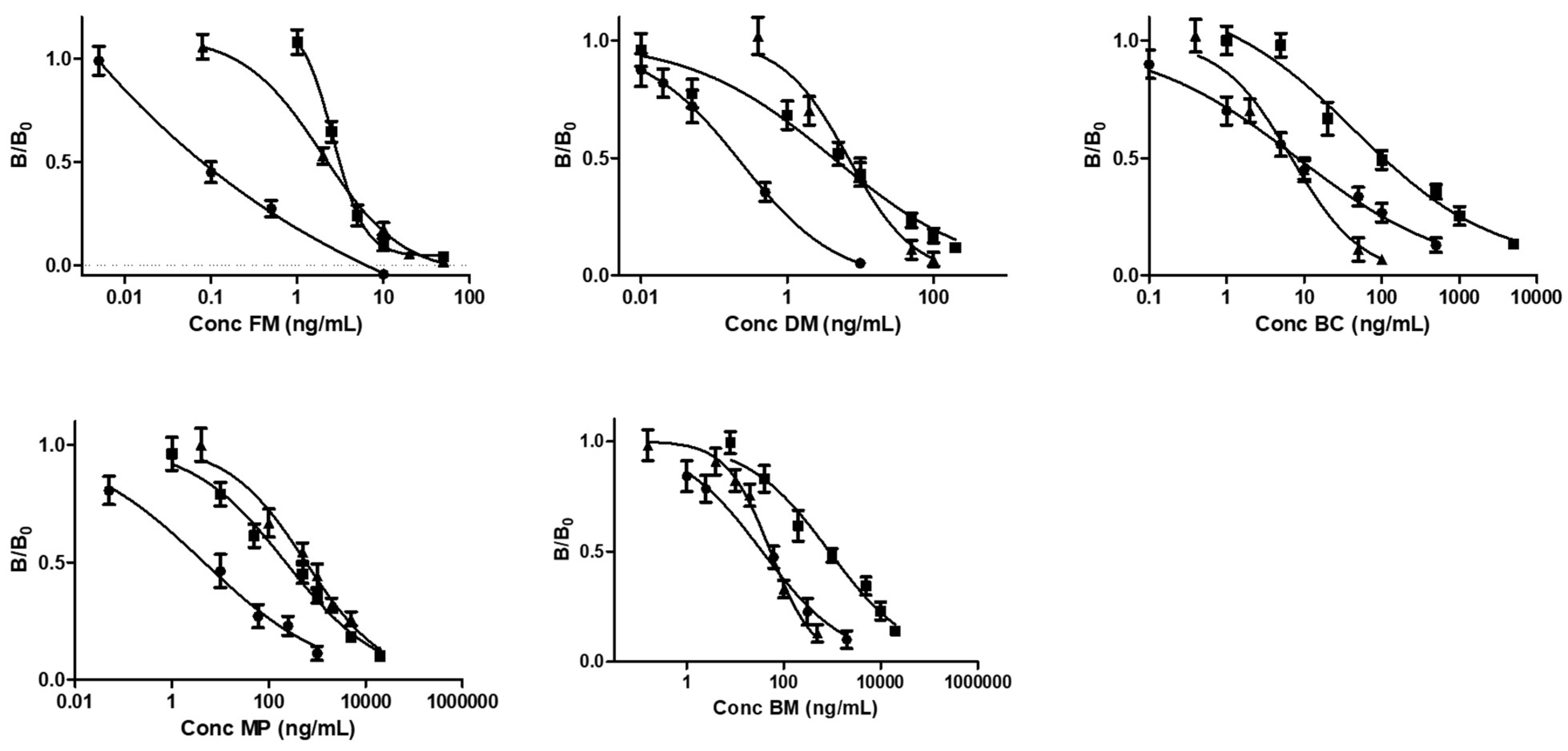
A protocol for using the ELISA kit has been successfully developed and optimized for its use in the analysis of corticosteroids in cosmetic formulations. Calibration curves were generated by spiking a known amount of target analytes in the cosmetic matrices (facial cream corresponding to cream A used for the HPLC-MS method and serum) suitably dissolved in THF and diluted 1:5 (v/v) in PBS. The matrices were selected based on the fact that generally the illegal addition of corticosteroids involves preparations for facial application [35]. These calibration curves were compared with those obtained by spiking target analytes in a mixture of PBS/THF. In the case of FN, BD, and DP, it was not possible to generate calibration curves since there were no significant changes in the signal even in the presence of high concentrations of these targets with respect to the blank. The primary antibody immobilized on the microwell plate was probably not able to recognize these corticosteroids. Indeed, the specifications of the used commercial kit did not report these target analytes among those that can be quantified.
The calibration curves (Figure 2) showed a high matrix effect; indeed, the LODs and dynamic ranges proved that the matrix significantly influences the performance of the method. Table 3 shows the LOD values and upper limits obtained for all the target analytes in the different matrices. As expected, the best performances were obtained by spiking corticosteroids in the solvent PBS/THF without the matrices. The dynamic ranges for some analytes were wide (up to 3 orders of magnitude), thus showing that to obtain a significant change in the signal a large difference in concentration of the target analyte is required. For FM and DM, the lowest LODs were obtained; and for the other target analytes the minimum detectable concentrations were also compatible with the concentrations of corticosteroids that are commonly added to cosmetic preparations.
In the case of immunoassay, the pretreatment of the sample represents a critical step since most of the extractive methods reported in literature applied to cosmetic samples are based on the use of organic solvents. Since immunoassays are based on the use of biological probes, organic solvents are not ideal for developing such bioassays. For this reason, the proposed pre-treatment procedure was based on the extraction of target analytes using THF followed by a 1:5 dilution in PBS in order to avoid an undesired effect of THF on the immunoassay performances. Therefore, in order to evaluate the recovery of the target analytes obtained through this procedure, cream and serum samples spiked with low, medium, and high concentration of different corticosteroids were analyzed. The added concentrations were selected considering the dynamic ranges reported for the different corticosteroids. The results are shown in Table 4. The recovery values were between 70 and 92%, and the performance was better in the case of medium and high concentrations. Paired t-tests were performed to evaluate the difference between the real concentration and the obtained values, and it was found that there were no significative differences for all the analyzed samples (n = 3 and p = 0.01). The assay also showed a satisfactory precision, providing a mean CV% of below 25%.

3.4. Comparison Between HPLC-MS Method and Immunoassay

Several observations can be made based on the results obtained using the two different methods for detecting corticosteroids within cosmetic matrices. As regards performance in terms of detection limits, these appear to be comparable for the two approaches. Indeed, depending on the analyte, the immunological assay guarantees the detection of the target even at low concentrations, showing the possibility of competing with the reference HPLC-MS technique. However, from the point of view of the matrix effect, the bioassay is greatly affected by the different type of cosmetic, thus making calibration necessary for each different type of matrix. Given the possibility of testing several samples in a single analysis and given the reproducibility of the data, the need to perform a specific calibration for each matrix is not a problem. Furthermore, despite having a significant matrix effect, the LODs and LOQs are suitable for the detection of these target analytes in the different matrices; therefore, the ELISA method is sufficiently sensitive for its application in this field. Recovery and precision are also higher in the case of the HPLC-MS method. The selectivity of the reference HPLC-MS method is high, but the ELISA test is not able to identify and discriminate different corticosteroids, instead only providing their generic presence. As regards quantification, the immunoassay for some analytes (BC, BM 17-V, MP) presents very wide dynamic ranges that correspond to low sensitivities and to a more imprecise quantification compared to that obtainable by HPLC-MS. Despite this, however, considerations must be made regarding ease of use and rapidity of analysis, which are very different for the two approaches. In fact, the immunoassay requires less complex instrumentation, high-throughput screening, and an easy-to-use procedure, which therefore considerably simplifies the analysis process. In particular, these analyses can also be conducted by non-specialized personnel and outside of centralized laboratories, unlike HPLC-MS techniques, which instead require specific training for qualified personnel [51]. These two techniques therefore offer different advantages, which makes them complementary and suitable for different contexts. Furthermore, the expansion of the use of the commercial ELISA kit that allows for the broadening of the spectrum of targets and matrices to be detected and quantified represents a novelty of this study.
Regarding the applicability of the two methods to different types of formulations, we analyzed spiked samples at medium concentration. The results were in line with the cosmetic products involved in the method validation. In particular, we tested four more creams—two oil/water and two water/oil emulsions—and three more serums.

4. Conclusions

The great variety of cosmetic products and the complexity of their composition leads to the need for sensitive and versatile analytical methods suitable for the detection of different target analytes. The continuous evolution of regulated and prohibited substances for cosmetic purposes also necessitates frequent checks and large-scale screening, which can highlight the presence of any substances added illegally to the formulations. For this reason, a rapid and easy-to-use method such as immunoassay can represent a valid alternative to reference benchtop methods such as HPLC-MS. In this work, we therefore developed and compared two analytical methods based on different principles. The first was an ad-hoc optimized HPLC-MS method to identify and quantify eight corticosteroids. The second was a bioanalytical immunoassay based on ELISA technology for the detection of corticosteroids in cosmetic matrices.
Compared to previous works [20,28,29,30] (Table 5), the LC-MS sensitivity is similar if not greater, in the order of 0.02–0.2 ng/mL (LOD) for creams and 0.1–0.5 ng/mL (LOD) for serum. Together with the high sensitivity, another advantage of the presented method over the previous ones is the simple sample preparation without any pretreatment like solid-phase extraction [52,53], surfactant-based ultrasound-assisted dispersive liquid–liquid microextraction [54], or microwave-assisted extraction [55].
The detectability offered by the biospecific assay is comparable to those in the literature [35,44] and that obtained by HPLC-MS. In this context, the detected concentration ranges for both methods are compatible with the concentrations of corticosteroids that are commonly added to cosmetics illegally. Indeed, to obtain a significant effect on skin aesthetics, the concentration of corticosteroids should comprise between 0.2 and 2% (w/v) [56].
However, as regards the performance of the method in terms of specificity, precision, recovery, and matrix effect in the quantification of the target analytes, the HPLC-MS technique performs much better. Despite this, the possibility of exploiting a rapid, easy-to-use, low-cost, and high-throughput technique such as immunoassay for large-scale preliminary screening would be useful for the purposes of frequent checks on cosmetic products. With ELISA, the instrumental configuration allowed approximately one hundred samples to be analyzed in 1 min using the 96-well plates. At this stage, the identification of the corticosteroid present would not be important since all corticosteroids are prohibited in cosmetic formulations.
After this preliminary multi-analyte screening, the samples suspected to contain one or more corticosteroids among FM, DM, BC, BM 17-V, and MP could then be subjected to more expensive and time-consuming analyses via HPLC-MS in order to accurately identify and quantify these analytes. This complementary approach based on two analytical techniques could in the future also be extended to other target analytes of cosmetic interest, as regulated substances are continually increasing and frequent controls are urgently needed to avoid counterfeit products on the market.

Supplementary Materials

The following supporting information can be downloaded at: https://www.mdpi.com/article/10.3390/app15010414/s1, Figure S1. TIC chromatograms of (a) MP, (b) BC, (c) FN, (d) BD, (e) BM 17-V, (f) BCDP, (g) FM, (h) DM, (i) blank cream A, (j) blank cream B, (k) serum; Table S1. List of ingredients in the cosmetic products (A, B, and serum). Table S2. Slope and R2 obtained for calibration curves generated with standard and spiked solutions in the concentration range of 0.1–20 ng/mL; Table S3. Precision of the method assessed through multiple intra-day (n = 3) and inter-day (n = 3) analyses of the spiked samples.

Author Contributions

Conceptualization, J.F. and M.M.; methodology, M.Z. and M.G.; validation, S.R.S.P., A.E., E.P. and D.C.; formal analysis, S.R.S.P., E.P. and D.C.; investigation, M.Z. and M.G.; data curation, S.R.S.P., A.E. and D.C.; writing—original draft preparation, S.R.S.P. and A.E.; writing—review and editing, M.Z. and M.G.; supervision, J.F. and M.M.; project administration, J.F. and M.M.; funding acquisition, J.F. All authors have read and agreed to the published version of the manuscript.

Funding

This work was supported by the 2023 Ricerca Fondamentale Orientata—University of Bologna, Italy.

Institutional Review Board Statement

Not applicable.

Informed Consent Statement

Not applicable.

Data Availability Statement

The original contributions presented in this study are included in the article. Further inquiries can be directed to the corresponding author.

Conflicts of Interest

The authors declare no conflicts of interest.

References

  1. Barnes, P.J. How corticosteroids control inflammation: Quintiles Prize Lecture 2005. Br. J. Pharmacol. 2006, 148, 245–254. [Google Scholar] [CrossRef]
  2. Kapugi, M.; Cunningham, K. Corticosteroids. Orthop. Nurs. 2019, 38, 336–339. [Google Scholar] [CrossRef] [PubMed]
  3. del Rosso, J.; Friedlander, S.F. Corticosteroids: Options in the era of steroid-sparing therapy. J. Am. Acad. Dermatol. 2005, 53, S50–S58. [Google Scholar] [CrossRef] [PubMed]
  4. Fokkens, W.J.; Godthelp, T.; Holm, A.F.; Blom, H.; Klein-Jan, A. Allergic rhinitis and inflammation: The effect of nasal corticosteroid therapy. Allergy 1997, 52, 29–32. [Google Scholar] [CrossRef] [PubMed]
  5. Rowe, B.H.; Edmonds, M.L.; Spooner, C.H.; Diner, B.; Camargo, C.A. Corticosteroid therapy for acute asthma. Respir. Med. 2004, 98, 275–284. [Google Scholar] [CrossRef] [PubMed]
  6. Möricke, A.; Zimmermann, M.; Valsecchi, M.G.; Stanulla, M.; Biondi, A.; Mann, G.; Locatelli, F.; Cazzaniga, G.; Niggli, F.; Aricò, M.; et al. Dexamethasone vs prednisone in induction treatment of pediatric ALL: Results of the randomized trial AIEOP-BFM ALL 2000. Blood 2016, 127, 2101–2112. [Google Scholar] [CrossRef]
  7. Benedek, T.G. History of the development of corticosteroi therapy. Clin. Exp. Rheumatol. 2011, 29 (Suppl. S68), 5–12. [Google Scholar]
  8. Williams, D.M. Clinical Pharmacology of Corticosteroids. Respir. Care 2018, 63, 655–670. [Google Scholar] [CrossRef] [PubMed]
  9. Van Staa, T.P.; Leufkens, H.G.M.; Cooper, C. The Epidemiology of Corticosteroid-Induced Osteoporosis: A Meta-analysis. Osteoporos. Int. 2002, 13, 777–787. [Google Scholar] [CrossRef]
  10. Peppa, M.; Krania, M.; Raptis, S.A. Hypertension and other morbidities with Cushing’s syndrome associated with corticosteroids: A review. Integr. Blood Press. Control 2011, 4, 7–16. [Google Scholar] [CrossRef]
  11. Clore, J.N.; Thurby-Hay, L. Glucocorticoid-Induced Hyperglycemia. Endocr. Pract. 2009, 15, 469–474. [Google Scholar] [CrossRef] [PubMed]
  12. Schoepe, S.; Schäcke, H.; May, E.; Asadullah, K. Glucocorticoid therapy-induced skin atrophy. Exp. Dermatol. 2006, 15, 406–420. [Google Scholar] [CrossRef] [PubMed]
  13. Hengge, U.R.; Ruzicka, T.; Schwartz, R.A.; Cork, M.J. Adverse effects of topical glucocorticosteroids. J. Am. Acad. Dermatol. 2006, 54, 1–15. [Google Scholar] [CrossRef]
  14. Youm, J.; Park, K.; Uchida, Y.; Chan, A.; Mauro, T.M.; Holleran, W.M.; Elias, P.M. Local blockade of glucocorticoid activation reverses stress- and glucocorticoid-induced delays in cutaneous wound healing. Wound Repair Regen. 2013, 21, 715–722. [Google Scholar] [CrossRef] [PubMed]
  15. Curtis, J.R.; Westfall, A.O.; Allison, J.; Bijlsma, J.W.; Freeman, A.; George, V.; Kovac, S.H.; Spettell, C.M.; Saag, K.G. Population-based assessment of adverse events associated with long-term glucocorticoid use. Arthrit. Care Res. 2006, 55, 420–426. [Google Scholar] [CrossRef] [PubMed]
  16. Available online: https://www.mfds.go.kr/eng/index.do (accessed on 1 December 2024).
  17. Gaudiano, M.C.; Lucente, D.; Antoniella, E.; Bertocchi, P.; Muleri, N.; Manna, L.; Bartolomei, M.; Alimonti, S.; Valvo, L.; Rodomonte, A.L. “For export only” medicines come back to Europe: A RP-LC method for the screening of six glucocorticoids in illegal and counterfeit anti-inflammatory and lightening creams. J. Pharm. Biomed. Anal. 2010, 53, 158–164. [Google Scholar] [CrossRef]
  18. Wei, C.; Ding, S.; You, H.; Zhang, Y.; Wang, Y.; Yang, X.; Yuan, J. An Immunoassay for Dibutyl Phthalate Based on Direct Hapten Linkage to the Polystyrene Surface of Microtiter Plates. PLoS ONE 2011, 6, e29196. [Google Scholar] [CrossRef] [PubMed]
  19. Nam, Y.S.; Kwon, I.K.; Lee, K.B. Monitoring of clobetasol propionate and betamethasone dipropionate as undeclared steroids in cosmetic products manufactured in Korea. Forensic Sci. Int. 2011, 210, 144–148. [Google Scholar] [CrossRef] [PubMed]
  20. Kim, N.S.; Yoo, G.J.; Lee, J.H.; Park, H.-J.; Cho, S.; Shin, D.W.; Kim, Y.; Baek, S.Y. Determination of 43 prohibited glucocorticoids in cosmetic products using a simultaneous LC-MS/MS method. Anal. Methods 2017, 9, 2104–2115. [Google Scholar] [CrossRef]
  21. Pellegrini, M.; Marchei, E.; Pacifici, R.; Rotolo, M.C.; Pichini, S. Advances in the analysis of non-allowed pharmacologically active substances in cosmetic products. J. Pharm. Biomed. Anal. 2011, 55, 842–847. [Google Scholar] [CrossRef]
  22. Johansson, M.; Fransson, D.; Rundlöf, T.; Huynh, N.-H.; Arvidsson, T. A general analytical platform and strategy in search for illegal drugs. J. Pharm. Biomed. Anal. 2014, 100, 215–229. [Google Scholar] [CrossRef]
  23. Jalili, R.; Miraghaei, S.; Mohamadi, B.; Babaei, A.; Bahrami, G. Detection of corticosteroid compounds and phosphodiesterase inhibitors (PDH-5) as counterfeit in herbal products available in Iranian market by HPLC method. J. Rep. Pharm. Sci. 2015, 4, 75–81. [Google Scholar] [CrossRef]
  24. Lee, J.H.; Jung, E.J.; Ham, H.J.; Yang, Y.J.; Kim, N.S.; Kim, H.I.; Baek, S.Y. Application of LC–MS/MS and UHPLC-Q-Orbitrap methods for determining 54 steroids in illegal dietary supplements and other sample types. Rapid Commun. Mass Spectrom. 2022, 36, e9334. [Google Scholar] [CrossRef]
  25. Chan, C.Y.; Ng, S.W.; Ching, C.K.; Mak, T.W.L. Detection of 28 Corticosteroids in Pharmaceutical and Proprietary Chinese Medicinal Products Using Liquid Chromatography–Tandem Mass Spectrometry. J. Chromatogr. Sci. 2021, 59, 548–554. [Google Scholar] [CrossRef]
  26. Fiori, J.; Andrisano, V. LC–MS method for the simultaneous determination of six glucocorticoids in pharmaceutical formulations and counterfeit cosmetic products. J. Pharm. Biomed. Anal. 2014, 91, 185–192. [Google Scholar] [CrossRef]
  27. McEwen, I.; Elmsjö, A.; Lehnström, A.; Hakkarainen, B.; Johansson, M. Screening of counterfeit corticosteroid in creams and ointments by NMR spectroscopy. J. Pharm. Biomed. Anal. 2012, 70, 245–250. [Google Scholar] [CrossRef]
  28. Giaccone, V.; Polizzotto, G.; Macaluso, A.; Cammilleri, G.; Ferrantelli, V. Determination of ten corticosteroids in illegal cosmetic products by a simple, rapid, and high-performance LC-MS/MS method. Int. J. Anal. Chem. 2017, 2017, 3531649. [Google Scholar] [CrossRef] [PubMed]
  29. Kang, M.; Sun, S.; Li, N.; Zhang, D.; Chen, M.; Zhang, H. Extraction and determination of hormones in cosmetics by homogeneous ionic liquid microextraction high-performance liquid chromatography. J. Sep. Sci. 2012, 35, 2032–2039. [Google Scholar] [CrossRef]
  30. Ivković, B.; Crevar, M.; Cvetanović, A.; Ubavkić, K.; Marković, B. Development and validation of RP-HPLC method for quantification of trace levels of topical corticosteroids in ambiphilic cream. Acta Chromatogr. 2023, 35, 46–51. [Google Scholar] [CrossRef]
  31. Li, M.; Wang, L.; Wang, M.; Zhao, H.; Zhao, F. Advances on Hormones in Cosmetics: Illegal Addition Status, Sample Preparation, and Detection Technology. Molecules 2023, 28, 1980. [Google Scholar] [CrossRef] [PubMed]
  32. Ashraf, S.S.; El-Gahany, W.; Alraeesi, A.; Al-Hajj, L.; Al-Maidalli, A.; Shah, I. Analysis of illicit glucocorticoid levels in camel hair using competitive ELISA—Comparison with LC–MS/MS. Drug Test. Anal. 2020, 12, 458–464. [Google Scholar] [CrossRef] [PubMed]
  33. Duffort, O.A.; Polo, F.; Lombardero, M.; Díaz-Perales, A.; Sánchez-Monge, R.; García-Casado, G.; Salcedo, G.; Barber, D. Immunoassay to quantify the major peach allergen Pru p 3 in foodstuffs. Differential allergen release and stability under physiological conditions. J. Agric. Food Chem. 2002, 50, 7738–7741. [Google Scholar] [CrossRef] [PubMed]
  34. Ashley, J.; D’Aurelio, R.; Piekarska, M.; Temblay, J.; Pleasants, M.; Trinh, L.; Rodgers, T.; Tothill, I. Development of a β-Lactoglobulin Sensor Based on SPR for Milk Allergens Detection. Biosensors 2018, 8, 32. [Google Scholar] [CrossRef]
  35. Sharma, G.M.; Rallabhandi, P.; Williams, K.M.; Herrmann, M.; Sadrieh, N. Gluten Quantitation in Cosmetic Products by Enzyme-Linked Immunosorbent Assay. J. AOAC Int. 2016, 99, 586–590. [Google Scholar] [CrossRef]
  36. Tranquet, O.; Lupi, R.; Echasserieau-Laporte, V.; Pietri, M.; Larré, C.; Denery-Papini, S. Characterization of Antibodies and Development of an Indirect Competitive Immunoassay for Detection of Deamidated Gluten. J. Agric. Food Chem. 2015, 63, 5403–5409. [Google Scholar] [CrossRef] [PubMed]
  37. Wang, M.; Guo, L.; Yu, M.; Zhao, H. The application of a lateral flow immunographic assay to rapidly test for dexamethasone in commercial facial masks. Anal. Bioanal. Chem. 2019, 411, 5703–5710. [Google Scholar] [CrossRef] [PubMed]
  38. Zhang, S.; Yao, T.; Wang, S.; Feng, R.; Chen, L.; Zhu, V.; Hu, G.; Zhang, H.; Yang, G. Upconversion luminescence nanoparticles-based immunochromatographic assay for quantitative detection of triamcinolone acetonide in cosmetics. Spectrochim. Acta A Mol. Biomol. Spectrosc. 2019, 214, 302–308. [Google Scholar] [CrossRef] [PubMed]
  39. Wang, Y.; Yang, H.; Pschenitza, M.; Niessner, R.; Li, Y.; Knopp, D.; Deng, A. Highly sensitive and specific determination of mercury(II) ion in water, food and cosmetic samples with an ELISA based on a novel monoclonal antibody. Anal. Bioanal. Chem. 2012, 403, 2519–2528. [Google Scholar] [CrossRef] [PubMed]
  40. Zhang, J.; Wang, M.; Yao, X.; Deng, A.; Li, J. Highly sensitive electroluminescence immunoassay for Hg(II) ions based on the use of CdSe quantum dots, the methylmercury-6-mercaptonicotinic acid-ovalbumin conjugate, and a specific monoclonal antibody. Mikrochim. Acta 2015, 182, 469–477. [Google Scholar] [CrossRef]
  41. English, D.; Scalici, C.; Hamilton, J.; Destro, C.; Jimenez, L. Evaluation of the TECRA™ visual immunoassay for detecting Staphylococcus aureus in cosmetic/pharmaceutical raw materials and finished products. J. Rapid Methods Autom. Microbiol. 1999, 7, 193–203. [Google Scholar] [CrossRef]
  42. Li, Q.; Zhu, R.; Li, J.; Wang, X.; Xu, L.; Li, Y.; Li, P. Highly Specific Chemiluminescence Immunoassay for the Determination of Chloramphenicol in Cosmetics. Int. J. Anal. Chem. 2019, 2019, 1–6. [Google Scholar] [CrossRef] [PubMed]
  43. Xun, Y.P.; Yan, H.; Liu, J.; Shi, L.L.; Chen, P.; Du, H.W. Development of a High-Throughput Immunoassay for Rapid Detection of Multiple Antibiotic Residues in Cosmetics. Adv. Mater. Res. 2014, 1073–1076, 357–361. [Google Scholar] [CrossRef]
  44. Le Loir, Y.; Baron, F.; Gautier, M. Staphylococcus aureus and food poisoning. Genet. Mol. Res. 2003, 2, 63–76. [Google Scholar]
  45. Dayan-Kenigsberg, J.; Bertocchi, A.; Garber, E.A.E. Rapid detection of ricin in cosmetics and elimination of artifacts associated with wheat lectin. J. Immunol. Methods 2008, 336, 251–254. [Google Scholar] [CrossRef]
  46. Zhang, S.; Wu, Z.Y.; Zhou, K.; Luo, L.; Xu, Z.L. Development of a competitive indirect ELISA for high-throughput screening of hydrocortisone in cosmetic sample. Food Agric. Immunol. 2019, 30, 594–605. [Google Scholar] [CrossRef]
  47. Calabretta, M.M.; Zangheri, M.; Calabria, D.; Lopreside, A.; Montali, L.; Marchegiani, E.; Trozzi, I.; Guardigli, M.; Mirasoli, M.; Michelini, E. Paper-Based Immunosensors with Bio-Chemiluminescence Detection. Sensors 2021, 21, 4309. [Google Scholar] [CrossRef] [PubMed]
  48. Zangheri, M.; Calabretta, M.M.; Calabria, D.; Fiori, J.; Guardigli, M.; Michelini, E.; Melandri, S.; Maris, A.; Mirasoli, M.; Evangelisti, L. Immunological Analytical Techniques for Cosmetics Quality Control and Process Monitoring. Processes 2021, 9, 1982. [Google Scholar] [CrossRef]
  49. González, O.; Blanco, M.E.; Iriarte, G.; Bartolomé, L.; Maguregui, M.I.; Alonso, R.M. Bioanalytical chromatographic method validation according to current regulations, with a special focus on the non-well defined parameters limit of quantification, robustness and matrix effect. J. Chromatogr. A 2014, 1353, 10–27. [Google Scholar] [CrossRef]
  50. Raposo, F.; Ibelli-Bianco, C. Performance parameters for analytical method validation: Controversies and discrepancies among numerous guidelines. TrAC Trends Anal. Chem. 2020, 129, 115913. [Google Scholar] [CrossRef]
  51. Pour, S.R.S.; Calabria, D.; Nascetti, A.; Caputo, D.; de Cesare, G.; Guardigli, M.; Zangheri, M.; Mirasoli, M. Easy-to-Use Chemiluminescent-Based Assay for a Rapid and Low-Cost Evaluation of the Antioxidant Activity of Cosmetic Products. Chemosensors 2024, 12, 25. [Google Scholar] [CrossRef]
  52. Shao, B.; Cui, X.; Yang, Y.; Zhang, J.; Wu, Y. Validation of a solid-phase extraction and ultra-performance liquid chromatographic tandem mass spectrometric method for the detection of 16 glucocorticoids in pig tissues. J. AOAC Int. 2009, 92, 604–611. [Google Scholar] [CrossRef] [PubMed]
  53. Andersen, J.H.; Hansen, L.G.; Pedersen, M. Optimization of solid phase extraction clean up and validation of quantitative determination of corticosteroids in urine by liquid chromatography–tandem mass spectrometry. Anal. Chim. Acta 2008, 617, 216–224. [Google Scholar] [CrossRef]
  54. Qin, H.; Yu, S.; Hu, X.; Yang, Y. Surfactant-based ultrasound-assisted dispersive liquid-liquid microextraction determination of corticosteroids followed by HPLC-DAD. Anal. Lett. 2013, 46, 589–604. [Google Scholar] [CrossRef]
  55. Guedes-Alonso, R.; Sosa-Ferrera, Z.; Santana-Rodríguez, J.J. Determination of steroid hormones in fish tissues by microwave-assisted extraction coupled to ultra-high performance liquid chromatography tandem mass spectrometry. Food Chem. 2017, 237, 1012–1020. [Google Scholar] [CrossRef]
  56. El-Alfy, M.; Deloche, C.; Azzi, L.; Bernard, B.A.; Bernerd, F.; Coutet, J.; Chaussade, V.; Martel, C.; Leclaire, J.; Labrie, F. Skin responses to topical dehydroepiandrosterone: Implications in antiageing treatment? BJD 2010, 163, 968–976. [Google Scholar] [CrossRef]
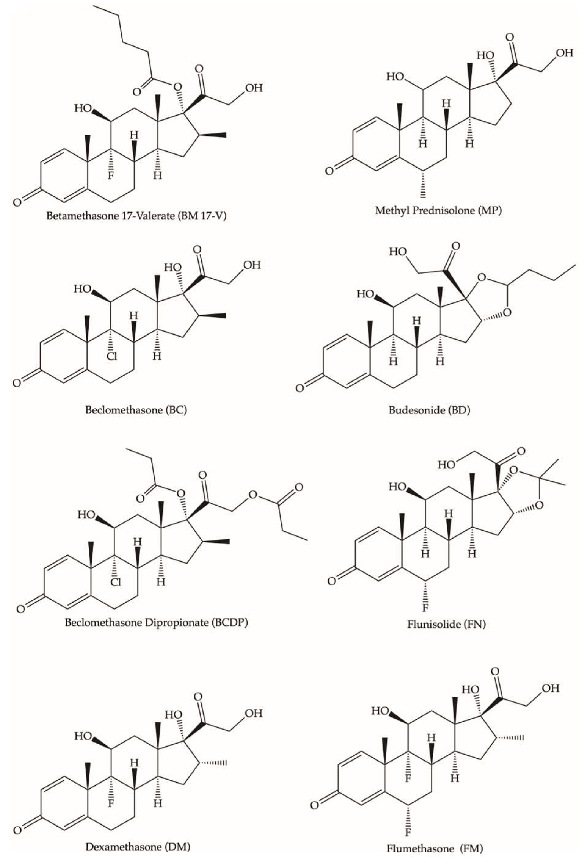
Figure 1. Corticosteroid chemical structures.
Applsci 15 00414 g001
Figure 2. Calibration curves obtained in PBS/THF (circle symbols), cream A (triangular symbols), and serum (square symbols) for the different corticosteroids.
Applsci 15 00414 g002
Table 1. Recovery for spiked BM 17-V, BC, BCDP, MP, BD, FN, FM, and DM concentrations; LOD; and LOQ obtained in different matrices.
SteroidSpiked Concentration
(ng/mL)
Recovery (±CV%)LOD (ng/mL)LOQ (ng/mL)
Cream ACream BSerumCream ACream BSerumCream ACream BSerum
BM 17-V0.388.1 (±4.3)65.3 (±5.5)70.2 (±9.2)0.020.0670.190.070.130.36
0.595.1 (±7.6)71.2(±7.8)78.2 (±3.4)
1083.5 (±5.2)62.5 (±7.7)76.7 (±5.7)
2084.9 (±5.3)68.6 (±4.9)73.5 (±3.7)
BC0.391.5 (±8.2)120.8 (±9.6)108.7 (±9.1)0.070.150.280.20.490.51
0.589.1 (±4.3)109.9 (±7.7)103.2 (±6.6)
1095.3 (±5.6)115.7 (±6.7)105.1 (±3.6)
2099.8 (±8.9)113.5 (±5.4)112.9 (±3.9)
BCDP0.3112.1 (±7.1)101.4 (±7.5)77.8 (±8.2)0.020.040.480.080.131.16
0.5101.1 (±8.4)99.6 (±3.9)80.6 (±7.5)
10105.1 (±8.6)93.8 (±4.1)75.0 (±8.3)
20111.8 (±4.4)98.6 (±8.4)85.3 (±9.1)
MP0.376.0 (±3.4)77.9 (±2.9)79.5 (±7.3)0.050.040.100.150.130.32
0.570.3 (±8.2)85.8 (±6.4)73.5 (±8.3)
1079.2 (±10.1)81.9 (±4.6)71.3 (±8.4)
2072.4 (±3.7)83.4 (±8.8)75.3 (±5.6)
BD0.377.4 (±9.2)78.0 (±7.2)106.0 (±4.6)0.120.160.130.420.530.45
0.572.4 (±6.2)75.3 (±7.7)115.2 (±7.1)
1075.0 (±5.5)82.8 (±9.2)110.3 (±9.1)
2079.0 (±9.8)79.2 (±7.6)111.2 (±8.8)
FN0.372.4 (±9.8)102.0 (±7.8)96.0 (±4.8)0.040.080.130.130.250.43
0.575.4 (±8.9)100.6 (±8.8)106.5 (±4.4)
1082.5 (±9.6)106.7 (±9.1)103.3 (±8.1)
2079.0 (±5.8)105.4 (±8.3)99.2 (±8.4)
FM0.393.5 (±3.9)74.2 (±8.8)99.9 (±5.5)0.050.070.210.120.220.72
0.589.4 (±4.3)74.8 (±9.9)102.0 (±7.5)
10101.1 (±4.9)76.7 (±2.8)109.9 (±8.4)
2096.6 (±5.9)72.1 (±3.7)103.54(±8.2)
DM0.385.7 (±6.6)94.1 (±8.8)96.1 (±8.8)0.020.050.120.180.360.41
0.591.4 (±6.9)82.9 (±8.4)91.7 (±7.2)
1079.1 (±7.1)93.8 (±9.4)93.7 (±6.2)
2089.7 (±8.3)89.0 (±4.8)87.6 (±4.4)
Table 2. Matrix effect on spiked samples in different matrices.
SteroidSpiked Concentration
(ng/mL)
Matrix Effect %
Cream ACream BSerum
BM 17-V0.30.47−0.48−0.47
0.50.26−0.250.21
10.00.04−0.320.26
20.00.01−0.01−0.29
BC0.30.460.810.71
0.50.210.06−0.02
10.00.12−0.030.08
20.0−0.490.82−0.12
BCDP0.30.91−0.66−0.97
0.50.11−0.44−0.14
10.00.11−0.28−0.12
20.00.060.21−0.18
MP0.30.87−0.33−0.33
0.50.19−0.07−0.41
10.00.18−0.06−0.36
20.00.01−0.48−0.38
BD0.30.150.071.01
0.50.090.12−0.14
10.00.09−0.38−0.51
20.0−0.11−0.360.71
FN0.30.33−0.40−0.40
0.50.05−0.53−0.52
10.00.15−0.48−0–47
20.0−0.04−0.23−0.45
FM0.30.72−0.27−0.53
0.50.16−0.070.08
10.00.07−0.12−0.07
20.0−0.08−0.15−0.06
DM0.30.64−0.05−0.13
0.50.10−0.15−0.02
10.00.26−0.15−0.03
20.00.05−0.11−0.05
Table 3. Concentration ranges and LOD for calibration curves generated using standard and spiked solution in different matrices.
SteroidPBS/THFCREAM SERUM
LOD (ng/mL) Upper Limit (mg/mL) LOD (ng/mL) Upper Limit (mg/mL) LOD (ng/mL) Upper Limit (mg/mL)
FM 0.02 0.002 0.59 0.015 1.6100.009
DM 0.04 0.005 1.8 0.067 0.030.12
BC 0.80 0.881.8 0.068 12.16.20
BM 17-V 4.7 3.27 13.8 0.53 6765.46
MP 0.22 3.13 51.2 36.2 530.76
Table 4. Recovery for spiked concentrations in different matrices.
SteroidSpiked Concentration (ng/mL)Recovery (%)
CreamSerum CreamSerum
FM 0.727571
748279
127 9089
DM 217270
30608587
50100 9290
BC 2157379
3030007980
5050008885
BM 17-V 20807271
2507007578
40050,0008381
MP 60107173
20,00015,0007978
30,00025,0008185
Table 5. Performances and sample preparation procedures for HPLC-MS methods reported in the literature for corticosteroid quantification in cosmetic samples.
SteroidLOQ (ng mL−1)Linearity RangeSample PreparationTechniqueRef.
Mometasone furoate12.50.0125–2 (mg mL−1)The extraction of 1 g of ambiphilic cream by 10 mL of acetonitrile was performed in an ultrasonic bath for 10 min at room temperature. The extract was filtered through a 0.45 mm cellulose acetate filter and then injected into the HPLC system.HPLC-DAD[29]
Hydrocortisone acetate15.6250.01563–5 (mg mL−1)
Fluocinonide90.009–2.88 (mg mL−1)
Fluocinolone acetonide31.250.03125–7.5 (mg mL−1)
Betamethasone210.021–6.72 (mg mL−1)
Betamethasone dipropionate10.50.0105–3.36 (mg mL−1)
Triamcinolone acetonide210.0105–6.72 (mg mL−1)
17α-Estradiol0.790.625–125 (ng mL−1)A total of 20 mL of cosmetic sample were transferred to a 50 mL polyterafluoroethylene (PTFE) centrifuge tube. A total of 0.6 g of NaCl was added and 1 mol/L NaOH was added to adjust the pH value to 10. After vortex mixing for 30 s, 70 μL of [C6MIM][BF4] were added to the homogenized sample. The solution was homogenized for 10 s. A total of 1 mL of water solution containing 0.3588 g of NH4PF6 was added. A cloudy solution was formed as a result of the formation of fine droplets of [C6MIM][PF6], which homogeneously dispersed in the solution. After vortexing for 2 min, the resulting sample solution was centrifuged at 15,000 rpm for 8 min at −4 °C and the [C6MIM][PF6] was deposited at the bottom of the centrifuge tube. The resulting IL phase was filtered with a 0.22 μm PTFE filter membrane before HPLC analysis.RP-HPLC-UV[30]
17α-Ethinylestradiol0.630.625–125 (ng mL−1)
Estrone0.590.625–125 (ng mL−1)
17α-Hydroxyprogesterone0.230.25–50 (ng mL−1)
Medroxyprogesterone0.300.25–50 (ng mL−1)
Megestrol-17-acetate0.100.125–100 (ng mL−1)
Norethisterone acetate0.170.25–125 (ng mL−1)
Progesterone0.100.125–125 (ng mL−1)
43 GlucocorticoidsComprised between 1.0–90.0100.0–2000.0 (ng mL−1)One gram of each sample was taken and dissolved in 100% methanol in a 20 mL volumetric flask, and 1 mL of 0.1% formic acid in methanol solution and 0.25 mL of ISTD (200 μg mL−1) were added. After sonication for 30 min and centrifugation (at 2000 rpm) for 10 min, 4 mL of supernatant liquid were diluted with 0.1% formic acid in 80% methanol (1:5). The stock solution was filtered through a 0.22 μm PVDA filter (Millipore, Milford, CT, USA) prior to UPLC analysis.UPLC-MS/MS[20]
Disclaimer/Publisher’s Note: The statements, opinions and data contained in all publications are solely those of the individual author(s) and contributor(s) and not of MDPI and/or the editor(s). MDPI and/or the editor(s) disclaim responsibility for any injury to people or property resulting from any ideas, methods, instructions or products referred to in the content.