Article

Polyphenol-Rich Extracts and Essential Oil from Egyptian Grapefruit Peel as Potential Antioxidant, Antimicrobial, and Anti-Inflammatory Food Additives

by
Faten Mohamed Ibrahim
1,*,
Eman Abdelsalam
2,
Reda Sayed Mohammed
3,
Wedian El Sayed Ashour
3,
Ana A. Vilas-Boas
4,
Manuela Pintado
4,* and
El Sayed El Habbasha
5
1
Medicinal and Aromatic Plants Research Department, Pharmaceutical and Drug Industries Research Institute, National Research Centre, Cairo P.O. Box 12622, Egypt
2
Chemistry of Natural and Microbial Products Department, Pharmaceutical and Drug Industries Research Institute, National Research Centre, Cairo P.O. Box 12622, Egypt
3
Pharmacognosy Department, Pharmaceutical and Drug Industries Research Institute, National Research Centre, Cairo P.O. Box 12622, Egypt
4
CBQF—Centro de Biotecnologia e Química Fina—Laboratório Associado, Escola Superior de Biotecnologia, Universidade Católica Portuguesa, Rua Diogo Botelho 1327, 4169-005 Porto, Portugal
5
Field Crops Research Department, National Research Centre, Cairo P.O. Box 12622, Egypt
*
Authors to whom correspondence should be addressed.
Appl. Sci. 2024, 14(7), 2776; https://doi.org/10.3390/app14072776
Submission received: 15 February 2024 / Revised: 18 March 2024 / Accepted: 22 March 2024 / Published: 26 March 2024
(This article belongs to the Special Issue Innovative Technologies for Food Preservation and Processing)

Abstract

:

Featured Application

The bio-based food additives obtained from upcycling grapefruit peels can be used mainly in the food industry as natural antioxidant and antimicrobial alternatives. Additionally, these ingredients also show anti-inflammatory potential, underscoring their potential as a nutraceutical.

Abstract

Grapefruit (GF) processing generates significant nutrient and economic losses due to the production of 50% by-products, primarily peels. GF peels are a rich and sustainable source of bioactive compounds (BCs), such as essential oils (EOs) and phenolic compounds. Thus, finding value-added solutions based on a circular economy is paramount. This research aims to assess the antioxidant, anti-inflammatory, and antimicrobial properties of a hydroethanolic polyphenol-rich extract from crude GF peels (GF-CE), essential oil (GF-EO), and polyphenol-rich extract from GF peels after essential oil extraction (GF-PE). The GF-CE and GF-PE showed high concentrations of naringenin (7.71 and 48.60 mg/g dry extract (DE)), narirutin (15.03 and 28.73 mg/g DE), and hesperidin (0.67 and 0.29 mg/mL), respectively. Extracting firstly EOs from GF improved the release of phenolic acids (p-coumaric, ferulic, and chlorogenic acid). The GF-CE exhibited stronger free radical scavenging activity mainly in DPPH (IC50 = 75.69 ± 0.81 µg/mL) than GF-EO (1271 ± 0.85 µg/mL) and GF-PE (113.45 ± 0.85 µg/mL). The GF-EO demonstrated moderate antimicrobial activity against Gram-positive bacteria compared to the reference standard (amoxicillin) and strong activity against the yeast Candida albicans (inhibition zone of 16 mm). The major compounds in the GF-EO included D-limonene (25%), nootkatone (24%), and β-pinene (8%). Both polyphenol-rich extracts showed promising activities as COX1 and COX2 inhibitors with IC50 values of 25 ± 0.1 and 0.28 ± 0.00 µg/mL (compared to celecoxib (97.5 ± 0.1 and 0.31 ± 0.01 µg/mL) and indomethacin (6.25 ± 0.00 and 0.52 ± 0.01 µg/mL) as the standards), respectively. The study concludes that GF peels are a valuable source of BCs with significant bioactivities, offering a sustainable multi-cascade approach to recovering value-added compounds from GF peels in alignment with circular economy principles and open opportunities as functional ingredients for food applications.

1. Introduction

Citrus fruits are widely consumed worldwide and have received considerable attention recently due to high levels of bioactive compounds (BCs), such as phenolic compounds and essential oils, which showed several human health-promoting benefits [1]. According to recent FAO statistical data, in 2022, the genus Citrus had an annual production of about 166.30 million tons, whereas the worldwide production of grapefruits (Citrus paradisi) was 9.76 million tons during the marketing year 2021/2022 [2,3]. The Mediterranean region is one of the main GF producers (30% of total production), where Turkey, Tunisia, Spain, and Egypt are among the top countries in the ranking [3].
Consumers highly appreciate grapefruit (GF) due to its abundance of nutrients and phytochemicals, which are valuable additions to a nutritious diet. They are consumed either fresh or processed [4]. Nevertheless, the processing results in juice and by-products consisting mainly of peels, pulps, and seeds, accounting for half of the fruit weight [5]. Thereby, GF processing generates a vast amount of waste generally used in composting and animal feed. In the worst cases, it is burned or dumped in landfills, which results in serious environmental issues and financial losses for the companies [6]. To overcome these unsustainable actions, a great industrial and scientific interest has risen to upcycle these side streams into high-value-added products [7].
Peels remain the primary by-product of GF processing and contain several phytochemicals, such as flavonoids, phenolic acids, vitamin C, carotenoids, and terpenes [8]. Generally, these molecules are present in higher amounts in the peels than other fruit parts [9]. Polyphenols have attracted much attention due to their use in food industries since recent studies have highlighted citrus peel extracts’ superior antioxidant capacity compared to synthetic antioxidants, along with their potent inhibitory effects on lipid oxidation and rancidity [10]. Moreover, these BCs exhibited promising antimicrobial properties against several potent foodborne pathogens, although their exact mechanism of action is yet to be fully elucidated [11]. Recently, these phytochemicals have been highly sought after by the nutraceutical industry due to their potential benefits against various oxidative stress-related disorders [12]. Over the last decade, several review studies have shown the positive effect of a high dietary intake of GF and other citrus polyphenols against inflammation processes, obesity, diabetes, neurodegenerative diseases, and cancer [8,12,13,14,15].
Additionally, citrus polyphenols have been strongly associated with cardioprotective effects [16,17]. Naringin, its aglycone, and naringenin are GF’s most abundant phenolic compounds [18]. For instance, naringenin has shown more effectiveness than vitamin C in inhibiting lipopolysaccharide (LPS)-induced inflammatory responses in macrophages, including a reduced nitrite production and the suppression of cyclooxygenase-2 (COX-2) and inducible nitric oxide synthase (iNOS) expression [19]. In addition to phenolic compounds, for a very long time, EOs have been used in the food processing, preservation, and flavoring industries for multiple purposes. The citrus genus has garnered great attention due to its abundance of EOs with high antibacterial, antifungal, and insecticidal properties [20]. However, the EOs extracted from GF peels also showed a great antioxidant capacity [21], and recently, a study from Nikolic et al. [22] suggested that EO extracted from GF peels showed a protective action against LPS-induced inflammation by attenuating the gene expression and concentrations of pro-inflammatory interleukin-6 (IL-6) and tumor necrosis factor-alpha (TNF-α) cytokines. The main advantage of EOs compared to polyphenol-rich extracts is their recognized interest in food applications as it has been used since ancient times. Nevertheless, EOs possess certain limitations primarily associated with the dosage and administration methods, which lack uniformity [23].
In accordance with emerging trends, there is a noticeable shift in consumer attitudes towards a heightened interest in natural food products free from synthetic additives. Concurrently, consumers are increasingly seeking food options that align with the principles of health, sustainability, and social responsibility [24]. As previously reported, the EOs and polyphenol-rich extracts from GF have shown impressive positive health benefits and interesting functional properties, making them desirable as a natural alternative for the food industry [25]. These options enhance the shelf-life of food. For instance, an edible coating of alginate/chitosan enriched with a polyphenol-rich extract from GF seeds reduced the off-flavor of shrimp and the bacterial count by 2 log CFU during the storage time (15 days under refrigeration (4 °C)), prolonging the shelf-life of shrimp [26]. Another study from Durmus et al. [27] showed that nanoemulsions based on GF-EO increased, in 6 days (compared to the control), the shelf-life of rainbow trout filets. In addition, these bioactive extracts obtained from GF by-products can also reduce the risk of microbial contamination and eliminate the need for artificial additives [28]. On the other hand, the BCs present in GF extracts/EOs prevent and/or improve human health and enhance the food’s nutritional value [29]. Over the past decade, there has been a notable increase in the accessibility of natural food additives, functional foods, nutraceuticals, and supplements in the market. Projections anticipate that this industry will expand to approximately USD 210 billion by 2026 [30]. Therefore, strategies to reduce food waste by upcycling them to produce new food ingredients/extracts are paramount [31]. This aligns with multiple Sustainable Development Goals (SDGs), contributing to the global agenda for sustainable development and addressing interconnected challenges, such as hunger, environmental degradation, and economic inequality. For instance, by upcycling GF peels into new extracts/ingredients, we contribute to reducing hunger and ensuring access to nutritious food for all (SDG 2), while enhancing human well-being with bioactive ingredients in the upcycled food (SDG 3) and promoting sustainable consumption and production patterns (SDG 12). In addition, food waste generates significant greenhouse gas emissions when it decomposes in landfills. Therefore, using GF by-products to produce new value-added ingredients will mitigate climate change (SDG 13), while contributing to the preservation of terrestrial ecosystems (SDG 15) [32,33]. Promoting the integration of upcycling practices within food systems holds significant promise for enhancing circularity within the food industry. Exploring this potential has emerged as a prominent topic of interest throughout the food value chain.
This research work aims to evaluate the antioxidant, antimicrobial, and anti-inflammatory activity of an EO and polyphenol-rich extracts from GF peels cultivated in Egypt. In addition, a multi-compound cascade approach is employed to collect bioactive extracts from the same by-product, increasing the circular economy value. The antioxidant capacity was evaluated using two standard methods: DPPH and nitric oxide; the antimicrobial activity was tested against both Gram-positive (Gram+) and Gram-negative (Gm−) bacteria as well as fungi; and the anti-inflammatory activities were analyzed by the cyclooxygenase enzyme expression (COX1 and COX2). The chemical composition of the EO and polyphenol-rich extracts was analyzed by GC/MS and LC/MS.

2. Materials and Methods

2.1. Chemicals

1,1-Diphenyl-2-picrylhydrazyl (DPPH), ascorbic acid (vitamin C), celecoxib, and the Griess reagent were purchased from Sigma-Aldrich, St. Louis, MO, USA. Sodium nitroprusside (s.d. Fine-Chem. Ltd., Mumbai, India), indomethacin, and acetylsalicylic acid were purchased from (The Arab Drug Company (ADCO), Cairo, Egypt). The in vitro anti-inflammatory activity was assessed using an ELISA kit provided by Cayman Chemical Company, Ann Arbor, MI, USA. Gallic acid, Folin–Ciocalteu reagent, aluminum chloride reagent, quercetin, and anhydrous sodium sulphate were purchased from Oxford Lab Chem, Navghar, India.

2.2. Plant Material and Extract Preparation

2.2.1. Plant Material

The fresh fruit of Citrus paradisi (GF) was collected from the National Research Centre Farm at the Agricultural Production and Research Station, National Research Centre, El Nubaria Province, El Behira Governorate, in 2021. The farm applies agro-eco-friendly environment procedures, and we collected healthy fruits only. A commercial juice squeezer was used to extract the juice from the fresh GF, which leaves the peels (flavedo + albedo) and some pulp residue. The GF peels are used for this study.

2.2.2. Grapefruit Crude Polyphenol-Rich (GF-CE) Extract

Three kg of fresh GF peels was mixed with ethanol 80% (v/v) at a 1:3 (m/v) ratio and left for 24 h under agitation to extract the polyphenols. The extraction process was repeated till exhaustion. After that, the extracts were combined and filtered and then dried at 45 °C under vacuum in a rotary evaporator. The final polyphenol-rich extract (GF-CE) was stored at 4 °C in a dark-colored bottle during the analysis time (Figure 1).

2.2.3. Grapefruit Essential Oil (GF-EO) and Grapefruit Polyphenol-Rich Extract (GF-PE) Extraction

To perform an integrated extraction approach to increase the economic value of GF peel valorization, two different BC extracts were obtained sequentially from the same by-product (Figure 1). Firstly, the EO was extracted from two kg of fresh peels through hydrodistillation using a Clevenger-type apparatus for 3 h, as mentioned in Egyptian pharmacopeia. The obtained GF-EO was desiccated using anhydrous sodium sulfate and stored in a freezer at −20 °C for subsequent analysis. The GF peels remaining from hydrodistillation were used to extract the polyphenols (GF-PE). The extraction process was the same one as that used for GF-CE, as described in Section 2.2.2.

2.3. Phytochemical Analysis

2.3.1. Essential Oil (GF-EO) Composition

The analysis of the GF-EO compounds was conducted through GC-MS using a Thermo Scientific Trace GC Ultra/ISQ Single Quadrupole MS system, equipped with a TG-5MS fused silica capillary column (30 m, 0.251 mm, and 0.1 mm film thickness) from Shimadzu Corporation, Kyoto, Japan. The GC-MS detection employed an electron ionization system with an ionization energy of 70 eV, with helium gas serving as the carrier gas at a constant flow rate of 1 mL/min. The injector and MS transfer line temperatures were maintained at 280 °C. The oven temperature program was initiated at 40 °C (held for 3 min) and increased gradually to 280 °C at a rate of 5 °C/min (held for 5 min). The tentative identification of compounds was accomplished by comparing their relative retention times and mass spectra with data from the NIST and WILLY libraries as well as the published literature (Adam, 2009 [34]). The quantification of all identified compounds was determined using relative percentage peak area calculations.

2.3.2. Total Phenolic Content

The total phenolic content (TPC) for the EO and polyphenol-rich extracts was determined using the Folin–Ciocalteu reagent following the method of Farid et al. [35]. Briefly, 1 mL of GF extracts or standard solution was added to 10 mL of deionized water and 1.0 mL of Folin–Ciocaulteu phenol reagent. After 5 min of reaction, 2.0 mL of sodium carbonate (20% (m/v)) was added to the mixture. Following a full hour in complete darkness, the absorbance was measured at 750 nm using a spectrophotometer. Gallic acid was used as a standard for the calibration curve. The results were calculated according to Equation (1) and are expressed as milligrams equivalent of gallic acid per gram of dry weight extract (mg GAE/g DW).
TPC   ( mg   GAE / g   DE )   =   C g a l l i c   a c i d × V × m M
Cgallic acid is the standard (gallic acid) concentration established from the calibration curve; V is the dilution factor; m is the total extract weight; and M is the DW extract concentration.

2.3.3. Total Flavonoid Content

The total flavonoid content (TFC) for the polyphenol-rich extracts (GF-CE and GF-PE) was measured using the aluminum chloride reagent, as described by Farid et al. [35]. The extracts (1.0 mL), previously diluted, were mixed with 0.7 mL of NaNO2 5% (m/v) and 10.0 mL of ethanol 30% (v/v) for 5 min. Then, 0.7 mL of AlCl3 10% (m/v) was added and mixed altogether. After 6 min of reaction, 5.0 mL of NaOH (1 M) was added. Finally, the solution was diluted to 25 mL using ethanol 30% (v/v). After standing for 10 min, the absorbance of the reaction mixture was measured at 430 nm with a spectrophotometer. Quercetin (dissolved in ethanol) was used as a standard for the calibration curve. The results were calculated according to Equation (2) and are expressed as milligrams equivalent of quercetin per gram dry weight extract (mg QE/g DW).
TFC   ( mg   QE / g   DE )   =   C q u e r c e t i n × V m
Cquercetin is the standard (quercetin) concentration established from the calibration curve; V is the volume of extract (mL); and m is the total extract weight (g).

Phenolic Compound Identification and Quantification

The analysis of the phenolic compounds in the polyphenol-rich extracts (GF-CE and GF-PE) was conducted using liquid chromatography–electrospray ionization–tandem mass spectrometry (LC-ESI-MS/MS) with an ExionLC TM AC system (Sciex, Framingham, MA, USA) for separation and SCIEX Triple Quad 5500+ (SCIEX, USA) equipped with electrospray ionization (ESI). Separation was achieved using a ZORBAX Eclipse Plus C18 Column (4.6 mm × 100 mm, 1.8 µm) (Agilent, Santa Clara, CA, USA). The mobile phases consisted of (A) 0.1% aqueous formic acid and (B) 100% acetonitrile, with the following gradient elution conditions: 2% B from 0–1 min, 2–60% B from 1–21 min, 60% B from 21–25 min, and returning to 2% B from 25.01–28 min. The flow rate was set at 0.8 mL/min, with an injection volume of 3 µL. The multiple reaction monitoring (MRM) analysis of the selected phenolic compounds was conducted in the negative ionization mode, with the following parameters: curtain gas at 25 psi, ion spray voltage at 4500 V, source temperature at 400 °C, ion source gases 1 and 2 at 55 psi with a declustering potential at 50, collision energy at 25, and collision energy spread at 10.

2.4. Antioxidant Activity

2.4.1. DPPH Radical Scavenging Assay

The DPPH assay was conducted following the procedure outlined by Ibrahim et al. [36]. Firstly, the GF extracts and vitamin C were prepared in methanol at different concentrations (31.25–2000 µg/mL). After that, 1.0 mL of DPPH methanolic solution (0.1 mM) was added to the sample (3.0 mL). Subsequently, the mixture was vigorously shaken and left to incubate in darkness at room temperature for 30 min. A control sample was created following the same procedure, with methanol replacing the sample. Absorbance was measured at 517 nm using a spectrophotometer, and the DPPH radical scavenging activity was determined using Equation (3). A linear modeling of the data obtained were performed and the results expressed as the concentration required to scavenge 50% of the initial DPPH radicals (IC50). The lower the IC50 value, the more powerful is the extract at scavenging DPPH and this implies a higher antioxidant activity.

2.4.2. Nitric Oxide Radical Scavenging Assay

The assay operates on the principle of inducing the release of nitric oxide (NO) free radicals from sodium nitroprusside (SNP) within an aqueous solution. At physiological pH, SNP undergoes a transformation, yielding nitrite ions that are detectable using the Griess reagent, composed of 1% sulfanilamide in 5% ortho-phosphoric acid (H3PO4) and 0.1% naphthylethylene diamine dihydrochloride [36]. The method was carried out as described by Ibrahim et al. (2021). Briefly, 2 mL of sample and 2 mL of SNP (10 mM) in phosphate-buffered saline at pH 7.4 were left to react at 25 °C for 150 min. After the incubation, 1 mL of the reaction mixtures were removed and diluted with 1 mL of Greiss reagent. The absorbance of these solutions was measured at 540 nm against the corresponding blank solution. The NO radical scavenging activity was calculated following Equation (3). The linear modeling of the data obtained was performed, and the results are expressed as the concentration required to scavenge 50% of the initial DPPH radicals (IC50).
Inhibition   ( % )   =   A b s A 0 A b s s a m p l e A b s A 0 × 100
AbsA0 is the absorbance of the control and Abssample is the absorbance of the treated sample with the extract at different concentrations.

2.5. Antimicrobial Activity

2.5.1. GF-EO Evaluation

Qualitative evaluations were conducted on nutrient agar plates, following the methodology of Mostafa et al. [37]. The pathogenic microorganisms used in this study included Gram+ bacteria (Bacillus cereus (ATCC 6629), Micrococcus leutus (ATCC 10240), Staphylococcus aureus (ATCC 6538), and Staphylococus epidermidis (ATCC 12228)), Gram− bacteria (Escherichia coli (ATCC 25922), Salmonella enterica (ATCC 255566), and Pseudomonas aeruginosa (ATCC 27853)), and the pathogenic yeast Candida albicans (ATCC 10231). These microorganisms were obtained from fresh overnight broth cultures grown in a nutrient broth medium and incubated at 37 °C [38]. The inoculum size of this pathogenic strain was prepared and adjusted to approximately 0.5 McFarland standard (1.5 × 10⁸ CFU /mL) [39]. Subsequently, 25.0 µL of the microorganism inoculum was added to each plate containing 20.0 mL of sterile nutrient agar medium. Then, in the cooled and solidified agar medium, a sample of GF-EO (10 µg/mL) was added to a 0.6 cm well previously created in the agar plate using a 6.0 cm corn borer, following the well diffusion method. These plates were then refrigerated for one hour to enhance the sample diffusion, followed by incubation at 37 °C for 24 h. The zones of inhibition were measured in millimeters (mm).

2.5.2. GF Polyphenol-Rich Extracts

The antimicrobial activities of the GF polyphenol-rich extracts were assessed using Kirby–Bauer disc diffusion method [40]. Six pathogenic microorganisms were used: Escherichia coli (ATCC 25922), Pseudomonas aeruginosa (ATCC 10145), and Salmonella enterica typhimurium (ATCC 14028) as Gram− bacteria, and Listeria monocytogenes (ATCC 35152) and Staphylococcus aureus (ATCC 43300) as Gram+ bacteria; and Candida albicans (ATCC 10231) as the fungal model was used. The pathogenic microorganisms stored at −20 °C were inoculated in brain–heart infusion broth tubes (BBL, Viersen, Germany). These tubes were then placed in an incubator at 37 °C for 24 to 48 h. Subsequently, the refreshed pathogenic microorganism cultures were evenly spread on the surface of Müller–Hinton agar (MHA) plates (BBL, Germany) using sterile swabs. A sterile disc with a diameter of 6 mm was immersed in the GF polyphenol-rich extracts (GF-CE and GF-PE) at a concentration of 100 mg/mL. Subsequently, the discs were placed on the surface of the MHA plates. The incubation of the plates occurred for 24 h at 37 °C. The diameters of the inhibition zones were measured in millimeters (mm).

2.6. Anti-Inflammatory Activity of GF-EO, GF-CE, and GF-PE

The evaluation of the cyclooxygenase (COX1 and COX2) inhibition efficacy in the GF polyphenol-rich extracts and GF-EO was conducted following the procedures outlined by Blobaum and Marnett et al. [41]. Indomethacin and celecoxib served as standards for the anti-inflammatory activity against COX1 and COX2, respectively.

2.7. Statistical Analysis

The results were presented as the mean ± standard deviation. The normality of data distribution was confirmed using the Shapiro-Wilk test (p < 0.05). Differences between the mean values were assessed through a one-way analysis of variance (ANOVA), in which the rejection of the null hypothesis (H0) (indicating the equality of means) occurred when p < 0.05. Upon the rejection of H0, multiple comparisons were conducted using Duncan’s post hoc test. Statistical analysis was performed using the Statistical Package for the Social Sciences (IBM SPSS version 16, Armonk, NY, USA).

3. Results and Discussion

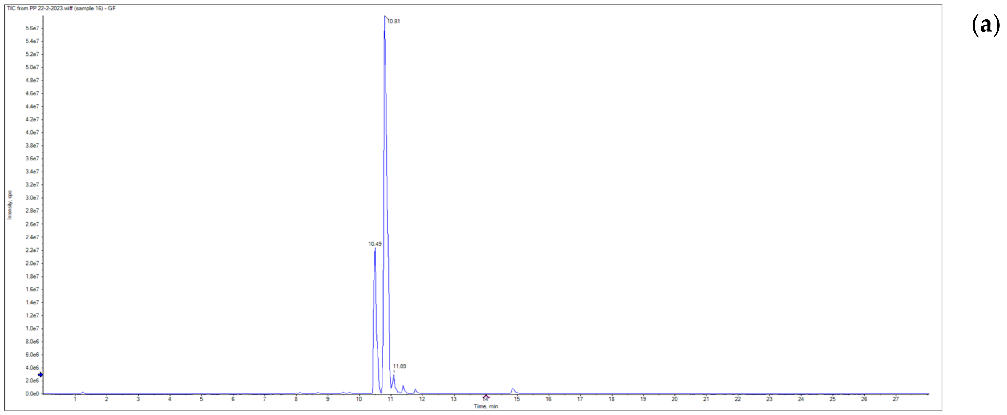
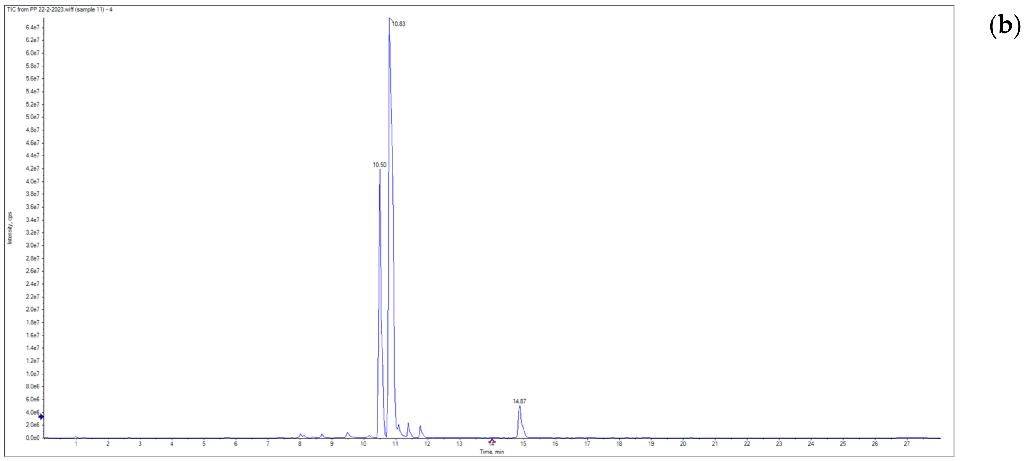
3.1. Grapefruit Essential Oil Composition

The GC-MS analysis of the volatile compounds of the GF-EO is presented in Figure 2. It was possible to identify 35 compounds, accounting for 97.11% of the total peak area (Table 1). The major volatile compounds were identified as D-limonene (24.90%), nootkatone (24.33%), β-pinene (7.71%), γ-Terpinene (7.52%), trans-caryophyllene (4.40%), and α-pinene (3.98%). Nootkatone was the main oxygenated sesquiterpene present in the GF-EO, while limonene, β-pinene, γ-Terpinene, and α-pinene were the main non-oxygenated monoterpenes present. D-limonene was the main prominent compound, which is also reported by other authors [21,42].
D-limonene is the principal component of citrus EOs (orange, lemon, mandarin, lime, and GF) [43]. However, these studies reported a higher quantity of D-limonene, at about 70%. However, a recent study from Ahmed et al. [44] reported that EOs extracted from GF peels cultivated in India, depending on the variety, had 1–15% of limonene. These differences observed between studies in the compound % may be explained by the geographical area of GF cultivation, mainly by the edaphoclimatic factors. Daily sun exposure, air humidity, and soil microbiota are the parameters mostly responsible for the changes observed in secondary metabolites [45]. Additionally, the maturity of the fruit, the harvest season, and the condition of the extraction method can impact the concentrations of the GF-EO. For instance, in Turkish GF-EO, the main compounds present were shown to be the same as those reported in our study [46,47], especially limonene, β-pinene, α-pinene, and nootkatone. However, opposite to our results, in the Turkish GF-EO study, a high concentration of myrcene and sabinene was found.
Figure 2. GC-MS chromatogram of the essential oil extracted from grapefruit (GF-EO).
Applsci 14 02776 g002
Table 1. Composition of essential oils from grapefruit peels.
No.Retention TimeMolecular Weight
(M+)
Base PeakMolecular
Formula
CompoundArea (%)
18.0713693C10H16α-Pinene3.98
28.3313693C10H16Camphene0.44
39.3313693C10H16β-Pinene7.71
410.3712883C8H16O1-Octanal3.40
510.4213693C10H16D-Limonene24.90
612.3213693C10H16γ-Terpinene7.52
713.5815471C10H18OLinalool1.10
814.4915267C10H16OTrans-Limonene oxide0.29
914.9115469C10H18OCitronellal0.61
1015.5915471C10H18OTerpinen-4-ol0.26
1116.0215471C10H18Oα-Terpineol0.81
1216.3815867C10H22ODihydro-citronellol2.65
1316.85152109C10H16OCaveol0.41
1417.1218469C11H20O2Citronellyl formate0.93
1517.3815269C10H16ONeral0.36
1617.4715082C10H14OCarvone0.15
1717.8215469C10H18OGeraniol0.15
1818.1915269C10H16OGeranial (E-citral)1.69
1920.40194109C12H18O2Carvyl acetate0.24
2020.86204161C15H24α-Copaene1.12
2121.1019669C12H20O2Geranyl acetate0.88
2221.23204161C15H24β-Copaene0.86
2321.9920493C15H24Trans-carophyllene4.40
2422.8020493C15H24α-Humulene0.49
2523.47204161C15H24Germacrene-D0.69
2623.86204121C15H24Bicyclogermacrene0.36
2724.51204161C15H24α-Amorphene2.78
2825.13204161C15H24Elemol1.06
2925.8822093C15H24OCaryophllene oxide0.30
3027.52222121C15H26Oα-Cadinol0.32
3127.63222186C15H26OEudesm-7(11)en-4-ol0.63
3228.9722269C15H26OFarnesol0.65
3329.32206135C15H26Nootkatol0.29
3430.41218135C15H22OCurcuphenol0.35
3530.38218146C15H22ONootkatone24.33
Total Area of the Identified Compounds97.11
D-Limonene showed several biological effects, including antioxidant, anti-inflammatory, anticancer activity, and immune modulatory effects [48]. For instance, the study in [49] showed the antioxidant and anti-lipid peroxidation activities of D-limonene (100 mg/kg body weight) for Streptozotocin-induced alterations in male Albino Wistar rats after 45 days of administration. Overall, D-limonene significantly decreased (p < 0.05) the thiobarbituric acid reactive substances, lipid hydroperoxides, and conjugate dienes in the plasma, liver, and kidney and improved (p < 0.05) the tissue antioxidants, namely superoxide dismutase, catalase, and vitamin C. In addition, at a concentration of 0.04%, D-limonene significantly (p < 0.01) decreased nitrite levels and reduced LPS-induced PGE2 production in the murine macrophage cell line RAW 264.7 [50]. Moreover, the D-limonene treatment led to an enhanced protein expression of TNF-α (increased by 30%), IL-1β (increased by 50%), and IL-6 (increased by 20%) compared to the LPS-induced cells left untreated.
Other present compounds, such as nootkatone, are noteworthy, with nootkatone being the most important aromatic organic compound in EO extracted from GF peels [51]. This compound showed, in a mice model, an anti-inflammatory effect, mainly associated with the inhibition of IL1-β and TNF-α production, possibly due to the inhibition of COX-2 activity and antagonism of the histamine receptor type 1 [52]. In addition, this compound showed other beneficial activities, including antimicrobial, antioxidant, cardioprotective, and neuroprotective [53]. Furthermore, the GF-EO and its bioactive properties have scented flavors [54], making them an excellent flavoring agent for different industries. For instance, the cosmetics industry uses EOs for manufacturing gels, shampoos, and creams; in the food industry, they are used as natural flavoring agents to replace the use of synthetic additives [55].

3.2. Grapefruit Polyphenol-Rich Extract Composition

3.2.1. TFC and TPC

Phenolic compounds, found abundantly in plants and fruits like those of the Citrus genus, are recognized for their antioxidant and antimicrobial activities. Recent research has also associated them with additional health benefits, including lower blood sugar and cholesterol levels, as well as reduced inflammation [56]. The TPCs of the GF-EO and hydroethanolic extracts (GF-PE and GF-CE) are presented in Table 2. For the EO, the value was 13 mg GAE/g DE, while for the polyphenol-rich extracts GF-PE and GF-CE, the TPCs were 51 and 208 mg GAE/g DE, respectively. The crude polyphenol-rich extract (GF-CE) showed 4-fold higher values than those of the polyphenol-rich extract obtained after oil extraction (GF-PE) (p < 0.05). The reduction in the TPC value for the GF-PE may be due to the previously high temperature applied during the hydrodistillation process since high temperatures are the most common explanation for the degradation of polyphenols [57]. However, for the TFC, the opposite results are observed. The GF-PE had 29 ± 0.01 mg QE/g DE, while the GF-CE had only 13 ± 0.06 mg QE/g DE (p < 0.05). Flavonoids are sensitive to heat [58]. However, certain extraction methods and parameter combinations may enhance the release or extraction of flavonoids from the plant material because of the breakdown from the cell wall [59].
The TPC and TFC in extracts from GF peels can vary depending on several factors, including the GF variety, ripeness, growing conditions, and the extraction method used. To the best of our knowledge, these TPC and TFC values for the extracts obtained from the fresh GF peels cultivated in Egypt have not been reported in the literature to date. However, for other geographical locations, the value of the TPC of the grapefruit peel extracts can range from approximately 50 to 200 mg GAE/extract [60,61,62,63]. For instance, with 80% ethanolic extraction, the TPC was about of 20 mg GAE/g DE using GF peels cultivated in Pakistan [10]. On the other hand, studies from Bagdatli et al. [64] using ultrasound extraction with ethanol 70% (v/v) demonstrated a TPC of 667 mg GAE/g DE. Another study from Garcia-Castello et al. [62] with GF solid waste from Valencia, Spain reported an optimal TPC extraction of approximately 80 mg/g DE.
Overall, the TPC constituted approximately 20%, 5%, and 1% of the entire extracts of GF-CE, GF-PE, and GF-EO, respectively. Consequently, other compounds, such as sugars, minerals, organic acids, soluble fibers, and proteins, were also extracted, while terpenes were predominant in the GF-EO.

3.2.2. Phenolic Compounds’ Identification and Quantification

The qualitative and quantitative analyses of the phenolic compounds in the GF peels’ polyphenol-rich extracts are crucial for upcycling food ingredients. Understanding the specific BCs present can offer insights into the extract’s bioactivities and elucidate the underlying biological mechanisms involved. The LC/MS chromatograms of each polyphenol-rich extract are presented in Figure 3.
Compared to the commercial standards, sixteen phenolic compounds were identified and quantified in the GF-CE and fourteen in the GF-PE (Table 3). The identification of these compounds led to their distribution into three main structurally related classes, i.e., hydroxycinnamic acids (four compounds), hydroxybenzoic acids (five compounds), and flavonoids (seven compounds).
The presence of such BCs is mainly associated with the antioxidant and antimicrobial activities of GF peels and their derived extracts [65,66,67]. The most abundant compounds found in GF peels’ polyphenol-rich extracts were naringenin, narirutin, and hesperidin, which agrees with the literature available for GF [62,67,68,69]. The values for naringenin, narirutin, and hesperidin ranged from 7.72 to 48, from 15 to 28.7, and from 0.28 to 0.67 mg/g DE, respectively.
An effective extraction technique is essential to achieve a greater yield of BCs, significantly impacting the composition, yield, and bioactivities of the extract. In the food industry, the preference lies in utilizing non-toxic and easily manageable solvents for the extraction of by-products [70]. Water stands out as the most safe and cost-effective environmentally friendly solvent option [71]; it demonstrates effectiveness in extracting polar molecules, whereas for less polar compounds, organic solvents or combination solvent systems are utilized. However, most of the polyphenols are poorly soluble in water. Flavonoid glycosides are usually more water-soluble than aglycones, which have a better solubility in organic solvents [72]. Therefore, to enhance the extraction, mixtures of water–alcohol are used [73]. Typically, insoluble phenolic compounds, such as flavonoids, are in the cell wall, while soluble phenolic compounds are found inside the cell vacuoles [74]. At high temperatures, the integrity of the cell wall is compromised, and this may promote the delivery of cell wall polysaccharides into the solvent and allow the phenolic compounds to diffuse [75,76]; so, their extractability can also increase. Comparing our results (Table 3) to the reported data, it was observed that the concentration of phenolic acids, mainly hydroxycinnamic acids, was higher in the GF-PE obtained after the extraction of the EO by hydrodistillation during 3 h. This process may weaken the cell wall integrity, resulting in an increase in the contents of low-molecular-weight insoluble phenolics and the flavonoid aglycons when extracted by ethanol (80%) [57]. However, certain flavonoid compounds may undergo degradation when exposed to elevated temperatures. For instance, rutin initiates degradation at 75 °C, with its concentration declining progressively as the temperature rises until it becomes non-detectable at 90 °C [77]. On the other hand, gallic acid exhibits instability at higher temperatures. The degradation rates increase as the temperature increases from 60 °C to 100 °C for 4 h [78]. In our results, it was noticed that the complete decomposition of gallic acid was observed in the GF-PE, which may be due to the previous 3 h in hydrodistillation. This explains the decline in the antioxidant, antimicrobial, and anti-inflammatory activities of the GF-PE. The GF-CE was prepared and evaporated at a temperature not exceeding 45 °C, enabling the stability of heat-sensitive compounds, which allows for the synergistic activity with the flavonoid glycosides and phenolic acids present even at a lower concentration than that of the GF-PE (obtained after EO extraction) to perform higher antioxidant, antimicrobial, and anti-inflammatory activities [79,80,81]. Furthermore, it is crucial to account for the total activity of polyphenol-rich extracts, influenced by the synergistic or antagonistic interactions among their constituents. Typically, phytochemicals are recognized for conferring human health benefits, including anti-inflammatory, antimicrobial, antihypertensive, and antidiabetic effects [82,83]. In our results, the GF extracts contained various important flavonoids, such as naringin, quercetin, and hesperidin. Naringenin, the main phenolic compound found in the GF peel extract, is a flavanone aglycone renowned for its multifaceted bioactive effects on human health. These effects include antioxidative, anti-inflammatory, antidiabetic, and anti-neurodegenerative properties, as reported in numerous studies [84,85,86].

3.3. Antioxidant Activity

Certain EOs have been shown to scavenge free radicals that harm the organism and lower the risk of certain diseases brought on by oxidative stress. The peels of GF have been documented to have many phytochemicals, including flavonoids, carotenoids, vitamin C, and other BCs, which have been reported as antioxidant compounds [87]. These BCs promote human health and delay/prevent the incidence of many chronic diseases [13]. To evaluate the antioxidant capacities of GF bioactive extracts, two different methods were used: DPPH and nitric oxide (NO) assays. The antioxidants react with DPPH, transforming it into 1,1-diphenyl-2-picryl hydrazine by swiftly accepting hydrogen, thereby halting the propagation of free radical oxidation chains. This process leads to the creation of stable end products, preventing subsequent lipid oxidation [36]. In the nitric oxide assay, the capacity of GF peel polyphenol-rich extracts to scavenge nitrogen free radicals was evaluated [88]. The antioxidant activities of the GF-EO and polyphenol-rich extracts (GF-CE and GF-PE) were concentration-dependent, which means that the activity increases as the concentration increases (Table 4 and Table 5). Vitamin C, used as a positive control, exhibited the same behavior as the extracts. The IC50 for the GF-EO was 1271 ± 0.85 µg/mL for the DPPH assay and 1656 ± 0.71 µg/mL for the NO assay. Compared to vitamin C, the natural GF-EO showed a lower activity (p < 0.05); however, compared to the results obtained by Deng et al. [21], the Egyptian GF-EO showed 100 times more activity against DPPH radicals than the GF-EO extracted by cold-pressing. Regarding the polyphenol-rich extracts, the IC50 values for the GF-CE were 75.69 ± 0.81 and 113.45 ± 0.71 µg/mL for the DPPH and NO assays, respectively. The IC50 values for the GF-PE were much higher for the DPPH (1069 ± 0.56 µg/mL) and for the NO (791 ± 0.52 µg/mL) assays. Meanwhile, the GF-CE showed a higher activity than the EO at the same concentration (1000 µg/mL) for both assays, while the GF-PE showed a higher activity than the GF-EO only for the NO assay. The variations in antioxidant effectiveness between the polyphenol-rich extracts and the EO could also be related to the levels of ascorbic acid, a water-soluble antioxidant renowned for effectively neutralizing reactive oxygen species [89]. Overall, the polyphenol-rich extracts showed a better antioxidant activity than the EO. Naringenin, the main flavonoid present in both extracts, is a powerful antioxidant that can destroy free radicals and attenuate their formation [90]. However, the GF-EO antioxidant activity could be attributed to the high % of limonene [47]. Citrus polyphenols, which are mainly flavonoids, display exceptional health-promoting protection against oxidative stress-related diseases, including antioxidant, anti-inflammatory, antimicrobial, and other activities [13].

3.4. Antimicrobial Activity

Annually, contaminated food results in 600 million illnesses and 420,000 deaths. Moreover, microbiological spoilage stands as the primary contributor to food waste [91]. Hence, novel and efficient strategies are required to inhibit and eradicate contamination. This research focuses on natural food preservatives endowed with antimicrobial attributes poised as safer substitutes for synthetic counterparts [92]. Therefore, the antimicrobial activities of the GF-EO and GF polyphenol-rich extracts were tested against a panel of several Gram+ and Gram− bacteria and a fungus, specifically selected based on their importance to public health.

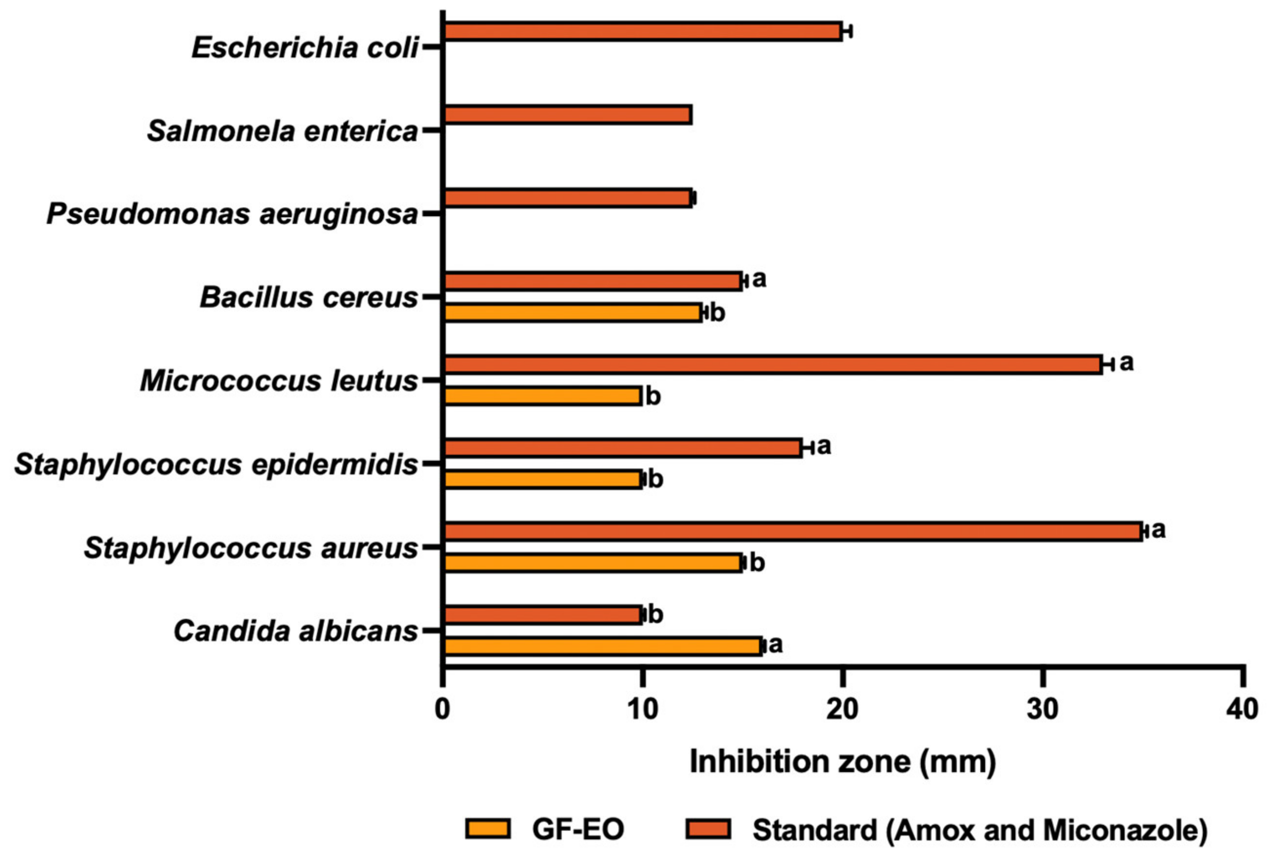
3.4.1. Grapefruit Essential Oil (GF-EO)

Plant-derived EOs are one group of natural food preservatives. The effectiveness of EOs is associated with the presence of multiple volatile compounds with antimicrobial activity reported since ancient times. This results in the effectiveness and specificity of each EO against various microorganisms. The results of the antimicrobial activity measured by the disc diffusion method for the EO extracted from GF and miconazole (standard) are shown in Figure 4. The GF-EO showed a good antibacterial activity against Gm+ bacteria: Bacillus cereus, Micrococcus leutus, Staphylococcus epidermidis, and Staphylococcus aureus. The highest antibacterial activity was demonstrated by the EO against S. aureus (15 mm), followed by the inhibition zone against B. cereus (13 mm). Regarding S. epidermidis and M. leutus, the inhibition zone was 10 mm. S. aureus and S. epidermidis contain teichoic acid in the peptidoglycan layer and are inhibited by different citrus peel EOs and extracts [93]. In addition, the GF-EO also had a great inhibition spectrum toward the pathogenic yeast Candida albicans (16 mm), showing a better potential than amoxicillin (standard). For the Gm− bacteria, there was no inhibition.
Our results align with most of the studies investigating the efficacy of EOs extracted from grapefruit peels against food spoilage organisms and foodborne pathogens. However, some studies reported different findings, mainly regarding Gram− bacteria inhibition. For instance, Deng et al. [94] reported the inhibition for E. coli (26.86 mm) and P. aeruginosa (8.57 mm). In addition, another recent study from Luciardi et al. [95] demonstrated that the GG-EO at 0.1 mg/mL could not inhibit P. aeruginosa growth but inhibited its biofilm production in the range of 52–55%. The differences observed may be related to the method of EO extraction. This study used hydrodistillation, whereas other comparative studies used cold-pressing extraction. However, the previously reported studies agree that EOs are generally slightly more active against Gram+ than Gram− bacteria. Gram+ bacteria typically exhibit a higher susceptibility due to their thick peptidoglycan cell wall layer. Conversely, Gram− bacteria possess a thinner peptidoglycan layer along with an additional outer membrane composed of phospholipids and lipopolysaccharides [96]. The presence of non-oxygenated compounds and oxygenated sesquiterpenes, such as D-Limonene, β-pinene, γ-Terpinene, α-pinene, and nootkatone, contributes significantly to the antimicrobial activity. Although the precise mechanism of action of EOs remains unclear, it is hypothesized that they disrupt cell membrane function and structure, consequently interfering with electron chain transport, enzyme activity, nutrient uptake, and the synthesis of nucleic acids and proteins [80].

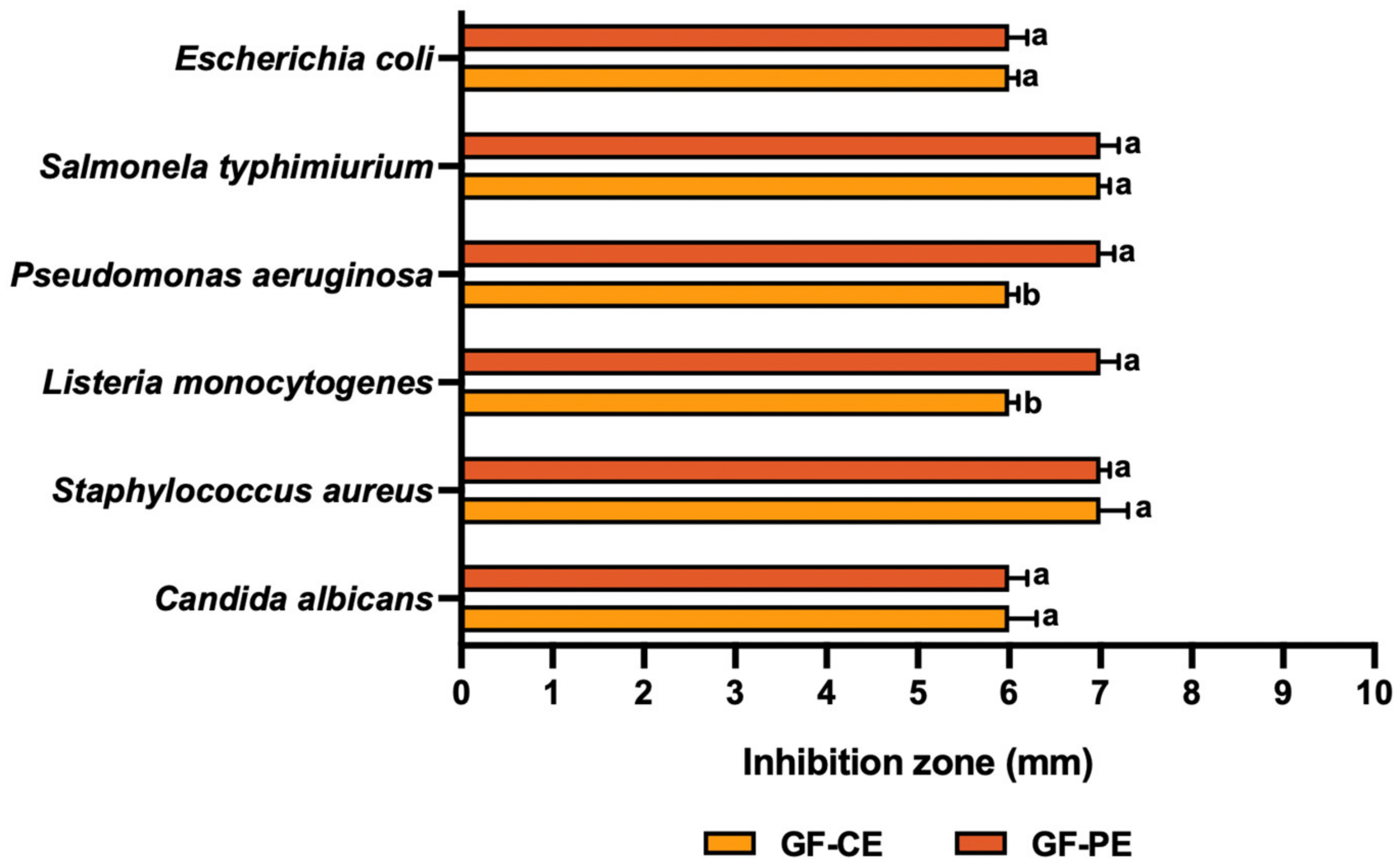
3.4.2. Grapefruit Polyphenol-Rich Extracts (GF-CE and GF-PE)

In addition to GF-EOs, extracts rich in phenolic compounds, another group of secondary metabolites in plants, are important BCs due to their bioactive properties. They demonstrate remarkable effectiveness as inhibitors against numerous foodborne pathogenic and spoilage bacteria. Moreover, they are excellent natural alternatives [97]. The results of the antimicrobial activity measured by the disc diffusion method of the polyphenol-rich extracts (GF-CE and GF-PE) are shown in Figure 5. The results show that the GF peel extract has a good activity against both Gm+ and Gm− and is also active against Candida albicans. However, the GF-PE shows a better activity than the GF-CE against P. aeruginosa (7 mm) and L. monocytogenes (7 mm). Although extracts rich in phenolic compounds can effectively inhibit pathogens, they are less potent than EOs.
To the best of our knowledge, there is limited literature available on the antimicrobial activity of GF peel extracts rich in polyphenols. Generally, GF polyphenol-rich extracts are obtained from seeds. A recent study from Arsène et al. [98] demonstrated that the antibacterial activity of a hydroethanolic extract (80%, v/v) was dose-dependent, and a higher inhibition diameter was observed for the Gm+ bacteria S. aureus (13 mm) and the Gm− bacteria E. coli (11 mm). Overall, the results are in line with those observed in our study. To the best of our knowledge, the antifungal activity of the polyphenol-rich extracts obtained from GF peels against Candida albicans is reported for the first time in this study. However, a study from Yaldiz et al. [99] showed that a treatment with mixtures of polyphenol-rich extracts and EO from GF peels (25:75%) for 8 h had an effective antifungal activity against C. albicans. Therefore, EOs are generally considered to be particularly effective in this context. The research indicates that phenolic compounds, such as rutin, quercetin, and naringenin, interact with bacterial cell membranes, increasing permeability, reducing ATP production, binding to metabolic enzymes, and disrupting membrane integrity, ultimately resulting in bacterial cell membrane destruction [100]. Additionally, flavonoids, a large group of phenolic compounds found in GF, demonstrate the ability to inhibit bacterial metabolism and the synthesis of DNA and RNA in bacteria [101,102].

3.5. Anti-Inflammatory Activity

The relationship between anti-inflammatory activity and the enzymes COX-1 (cyclooxygenase-1) and COX-2 (cyclooxygenase-2) is closely tied to the inflammatory response and the production of prostaglandins [103]. COX-1 and COX-2 are enzymes involved in synthesizing prostaglandins from arachidonic acid, a fatty acid found in cell membranes. COX1 is constitutively expressed in many tissues and maintains normal physiological functions, such as protecting the stomach lining and regulating blood platelets. COX-1 produces prostaglandins that are involved in these housekeeping functions [103]. Meanwhile, COX2 is typically produced during inflammation and is responsible for producing the prostaglandins that contribute to the inflammatory response. COX-2 is often upregulated in response to various stimuli, including injury or infection [103]. Non-steroidal anti-inflammatory drugs (NSAIDs), including indomethacin and celecoxib, exert their anti-inflammatory effects by suppressing the activity of both COX-1 and COX-2 enzymes [104]. Nevertheless, there is considerable interest in natural extracts or compounds with anti-inflammatory properties that may act as inhibitors of COX-1 and COX-2 [105,106,107]. One of these groups may be polyphenols, more precisely, flavonoids [108].
Figure 6 shows the anti-inflammatory effects of the GF peel polyphenol-rich extracts (GF-CE and GF-PE) and GF-EO against the biomarkers COX1 and COX2.
The GF-EO, GF-CE, and GF-PE exhibited activities as COX1 and COX2 inhibitors, with IC50 values of 0.97 ± 0.1, 25 ± 0.1, and 0.67 ± 0.058 ug/mL for COX1 and 3.9 ± 0.1, 0.28 ± 0.0006, and 0.7 ± 0.1 ug/mL for COX2, compared to the reference standards celecoxib and indomethacin, with values of 97.5 ± 0.1 and 6.25 ± 0.002, and 0.31 ± 0.01 and 0.52 ± 0.01, respectively. It was noticed from the results that EO has a weak activity as a COX2 inhibitor and good activity as an inhibitor of COX1, while the GF-CE has selective activity as a COX2 inhibitor and mild inhibitor of COX1, but the GF-PE has a weak activity as a COX2 inhibitor compared to the extract obtained from the crude GF peels and good activity as a COX1 inhibitor. The bioactive extracts obtained from GF peels showed a significantly better activity (p < 0.05) than synthetic drugs. Our results are consistent with recent studies, which demonstrate the inhibitory effect of grapefruit peel extracts on the production of inflammatory mediators, including prostaglandin E2 and nitric oxide (NO), in LPS-activated RAW 264.7 cells. Flavanone glycosides have been identified as significant contributors to the anti-inflammatory activity of citrus peels due to their higher abundance compared to polymethoxyflavones [109]. Furthermore, in vivo investigations have indicated that the EO extracted from grapefruit peels exhibits anti-inflammatory properties compared to the negative control group over the entire 5 h post-induction assessment period [110].
The correlation between inhibiting COX-1 and COX-2 and anti-inflammatory activity is rooted in the role of these enzymes in the inflammatory process. Inflammation involves the production of prostaglandins, which COX-1 and COX-2 synthesize. By inhibiting these enzymes, the production of prostaglandins is reduced, leading to a decreased inflammatory response. The GF-PE had a higher content of valuable phytochemicals, such as naringenin, narirutin, hesperidin, ferulic acid, and chlorogenic acid. These BCs exhibit diverse anti-inflammatory properties by inhibiting various pathways, including regulatory enzyme inhibition, the alteration of arachidonic acid metabolism, the modulation of gene expression, and targeting transcription factors crucial for regulating inflammatory mediators [111,112,113].
Additionally, it can modulate the activity of human macrophages and, in turn, reduce inflammation [114]. Inflammation is a biological response to noxious stimuli, such as pathogens, which cause tissue and cell damage [115]. It may be acute or chronic, depending on the reaction of the body to the stimuli, with an either short or prolonged response.

4. Conclusions

The current study presented a comparative study of the valorization of GF peels to obtain a single BCs (EO or polyphenol-rich extracts) or a multi-cascade BCs from the same by-product. A sustainable and integrated approach using Egyptian GF peel, a by-product from the juice industry, by producing consecutive GF-EO and polyphenol-rich extracts (GF-PE) using the same by-product was assessed. The method of extraction and pretreatment applied before the polyphenol extraction significantly influences the content of phenolic compounds and the biological activity of the final ingredient. The TPC and TFC increased 2-fold, while naringenin, narirutin, and hydroxycinnamic acids increased by 6.2-, 1.9-, and 3.0-fold, respectively. However, the crude GF extract (GF-CE) exhibited a stronger free radical scavenging activity, mainly in DPPH (IC50 = 75.69 ± 0.81 µg/mL), than the GF-PE (113.45 ± 0.85 µg/mL). However, the GF-PE showed a higher anti-inflammatory activity than the GF-CE (0.67 ± 0.058 ug/mL for COX1 and 0.7 ± 0.1 ug/mL for COX2). Lastly, both the GF-CE and GF-PE showed antimicrobial activity (p > 0.05) against Gram− and Gram+ bacteria; however, the GF-PE showed a better inhibitory capacity against P. aeruginosa (7 mm) and L. monocytogenes (7 mm). Nevertheless, the GF-EO demonstrated a higher antimicrobial activity than the polyphenol-rich extracts, mainly against Gram+ bacteria compared to the reference standard (amoxicillin) and a strong activity against the yeast Candida albicans (inhibition zone of 16 mm). The major compounds in the GF-EO included D-limonene (25%), nootkatone (24%), and β-pinene (8%). Although the GF-EO demonstrates anti-inflammatory capacity, its ability to inhibit COX 1 (0.97 ± 0.1 µg/mL) is greater than that to inhibit COX 2 (3.9 ± 0.1 µg/mL).
This study highlights the potential of GF peels as a sustainable source of valuable ingredients/extracts for functional foods and their potential to establish synergies with the cosmetic and pharmaceutical industries. In addition, it proves the efficiency of the consecutive extraction of BCs from GF peels. By using these new upcycled ingredients, we can contribute to the achievement of the SDGs and zero waste, a set of principles focused on waste prevention that encourage finding new uses for resources instead of throwing them away.

Author Contributions

Conceptualization, F.M.I., R.S.M. and E.S.E.H.; methodology, F.M.I., R.S.M., E.A. and W.E.S.A.; software, R.S.M.; validation, R.S.M. and F.M.I.; formal analysis, F.M.I., R.S.M. and E.A.; investigation, F.M.I., R.S.M., E.A. and W.E.S.A.; resources, E.S.E.H.; data curation, F.M.I.; writing—original draft preparation, A.A.V.-B., F.M.I., R.S.M. and E.A.; writing—review and editing, A.A.V.-B., F.M.I. and M.P.; project administration, E.S.E.H. All authors have read and agreed to the published version of the manuscript.

Funding

This research was financial supported by the Science, Technology and Innovation Funding Authority (STDF) from Egypt and PRIMA Program through the MEDISMART project—Mediterranean Citrus innovative soft-processing solutions for S.M.A.R.T (Sustainable, Mediterranean, Agronomically evolved nutritionally enriched Traditional) products (reference number: PRIMA-EU-2019-SECTION2; http://doi.org/10.54499/PRIMA/0014/2019).

Institutional Review Board Statement

Not applicable.

Informed Consent Statement

Not applicable.

Data Availability Statement

Data will be available upon request.

Acknowledgments

The authors gratefully acknowledge the Science, Technology and Innovation Funding Authority (STDF) from Egypt and, the scientific collaboration of the Escola Superior de Biotecnologia—Universidade Católica Portuguesa through CBQF under the FCT project UIDB/50016/2020 and http://doi.org/10.54499/PRIMA/0014/2019. Lastly, the author Ana A. Vilas-Boas would like to acknowledge the FCT for the individual PhD grant (reference: 2020.05655.BD).

Conflicts of Interest

The authors declare no conflicts of interest.

References

  1. Wedamulla, N.E.; Fan, M.; Choi, Y.-J.; Kim, E.-K. Citrus Peel as a Renewable Bioresource: Transforming Waste to Food Additives. J. Funct. Foods 2022, 95, 105163. [Google Scholar] [CrossRef]
  2. Shahbandeh, M. Grapefruit Production Worldwide from 2012/2013 to 2022/2023. Available online: https://www.statista.com/statistics/577836/world-grapefruit-production/ (accessed on 17 March 2024).
  3. UN Food and Agriculture Organization, C.S.D. (FAOSTAT). Grapefruit Production in 2022, Crops/Regions/World List/Production Quantity (Pick Lists). Available online: https://www.fao.org/faostat/en/#data/QCL (accessed on 17 March 2024).
  4. Khalil, M.N.A.; Farghal, H.H.; Farag, M.A. Outgoing and Potential Trends of Composition, Health Benefits, Juice Production and Waste Management of the Multi-Faceted Grapefruit Citrus Χ Paradisi: A Comprehensive Review for Maximizing Its Value. Crit. Rev. Food Sci. Nutr. 2022, 62, 935–956. [Google Scholar] [CrossRef] [PubMed]
  5. Chavan, P.; Singh, A.K.; Kaur, G. Recent Progress in the Utilization of Industrial Waste and By-products of Citrus Fruits: A Review. J. Food Process Eng. 2018, 41, e12895. [Google Scholar] [CrossRef]
  6. Vilas-Boas, A.A.; Gómez-García, R.; Campos, D.A.; Correia, M.; Pintado, M. Integrated Biorefinery Strategy for Orange Juice By-Products Valorization: A Sustainable Protocol to Obtain Bioactive Compounds. In Food Waste Conversion; Springer: Berlin/Heidelberg, Germany, 2023; pp. 113–124. [Google Scholar]
  7. Sharma, H.P.; Patel, H. Sugandha Enzymatic Added Extraction and Clarification of Fruit Juices—A Review. Crit. Rev. Food Sci. Nutr. 2017, 57, 1215–1227. [Google Scholar] [CrossRef]
  8. Saini, R.K.; Ranjit, A.; Sharma, K.; Prasad, P.; Shang, X.; Gowda, K.G.M.; Keum, Y.-S. Bioactive Compounds of Citrus Fruits: A Review of Composition and Health Benefits of Carotenoids, Flavonoids, Limonoids, and Terpenes. Antioxidants 2022, 11, 239. [Google Scholar] [CrossRef] [PubMed]
  9. Sir Elkhatim, K.A.; Elagib, R.A.A.; Hassan, A.B. Content of Phenolic Compounds and Vitamin C and Antioxidant Activity in Wasted Parts of Sudanese Citrus Fruits. Food Sci. Nutr. 2018, 6, 1214–1219. [Google Scholar] [CrossRef] [PubMed]
  10. Saleem, M.; Durani, A.I.; Asari, A.; Ahmed, M.; Ahmad, M.; Yousaf, N.; Muddassar, M. Investigation of Antioxidant and Antibacterial Effects of Citrus Fruits Peels Extracts Using Different Extracting Agents: Phytochemical Analysis with in Silico Studies. Heliyon 2023, 9, e15433. [Google Scholar] [CrossRef]
  11. Alsakhawy, S.A.; Baghdadi, H.H.; El-Shenawy, M.A.; El-Hosseiny, L.S. Comparative Phytochemical Composition and Antimicrobial Activity of Citrus Peel Essential Oils and Phenolic Compounds. Antiinfect. Agents 2023, 21, 57–68. [Google Scholar] [CrossRef]
  12. Liu, N.; Li, X.; Zhao, P.; Zhang, X.; Qiao, O.; Huang, L.; Guo, L.; Gao, W. A Review of Chemical Constituents and Health-Promoting Effects of Citrus Peels. Food Chem. 2021, 365, 130585. [Google Scholar] [CrossRef]
  13. Addi, M.; Elbouzidi, A.; Abid, M.; Tungmunnithum, D.; Elamrani, A.; Hano, C. An Overview of Bioactive Flavonoids from Citrus Fruits. Appl. Sci. 2022, 12, 29. [Google Scholar] [CrossRef]
  14. Kaur, S.; Panesar, P.S.; Chopra, H.K. Citrus Processing By-Products: An Overlooked Repository of Bioactive Compounds. Crit. Rev. Food Sci. Nutr. 2023, 63, 67–86. [Google Scholar] [CrossRef] [PubMed]
  15. Maqbool, Z.; Khalid, W.; Atiq, H.T.; Koraqi, H.; Javaid, Z.; Alhag, S.K.; Al-Shuraym, L.A.; Bader, D.M.D.; Almarzuq, M.; Afifi, M.; et al. Citrus Waste as Source of Bioactive Compounds: Extraction and Utilization in Health and Food Industry. Molecules 2023, 28, 1636. [Google Scholar] [CrossRef] [PubMed]
  16. Jeong, D.; Park, H.; Jang, B.K.; Ju, Y.; Shin, M.H.; Oh, E.J.; Lee, E.J.; Kim, S.R. Recent Advances in the Biological Valorization of Citrus Peel Waste into Fuels and Chemicals. Bioresour. Technol. 2021, 323, 124603. [Google Scholar] [CrossRef] [PubMed]
  17. Deng, Y.; Tu, Y.; Lao, S.; Wu, M.; Yin, H.; Wang, L.; Liao, W. The Role and Mechanism of Citrus Flavonoids in Cardiovascular Diseases Prevention and Treatment. Crit. Rev. Food Sci. Nutr. 2022, 62, 7591–7614. [Google Scholar] [CrossRef]
  18. Razavi, B.M.; Hosseinzadeh, H. A Review of the Effects of Citrus paradisi (Grapefruit) and Its Flavonoids, Naringin, and Naringenin in Metabolic Syndrome. In Bioactive Food as Dietary Interventions for Diabetes, 2nd ed.; Academic Press: Cambridge, MA, USA, 2019; pp. 515–543. [Google Scholar]
  19. Chao, C.-L.; Weng, C.-S.; Chang, N.-C.; Lin, J.-S.; Kao, S.-T.; Ho, F.-M. Naringenin More Effectively Inhibits Inducible Nitric Oxide Synthase and Cyclooxygenase-2 Expression in Macrophages than in Microglia. Nutr. Res. 2010, 30, 858–864. [Google Scholar] [CrossRef] [PubMed]
  20. Nwosu, O.K.; Keskin, M.; Lohani, H.; Egbuna, C.; Haider, S.Z. Bioactive Lead Compounds and Molecular Targets for the Development of Antiinflammatory Drugs. In Phytochemicals as Lead Compounds for New Drug Discovery; Elsevier: Amsterdam, The Netherlands, 2020; pp. 317–331. [Google Scholar]
  21. Deng, W.; Liu, K.; Cao, S.; Sun, J.; Zhong, B.; Chun, J. Chemical Composition, Antimicrobial, Antioxidant, and Antiproliferative Properties of Grapefruit Essential Oil Prepared by Molecular Distillation. Molecules 2020, 25, 217. [Google Scholar] [CrossRef]
  22. Nikolic, D.; Bosco, L.; Moschetti, M.; Tinnirello, V.; Pucci, M.; Corleone, V.; Raimondo, S.; Alessandro, R.; Fontana, S. Anti-inflammatory Properties of an Aldehydes-enriched Fraction of Grapefruit Essential Oil. J. Food Sci. 2023, 88, 1172–1187. [Google Scholar] [CrossRef] [PubMed]
  23. Osaili, T.M.; Dhanasekaran, D.K.; Zeb, F.; Faris, M.E.; Naja, F.; Radwan, H.; Ismail, L.C.; Hasan, H.; Hashim, M.; Obaid, R.S. A Status Review on Health-Promoting Properties and Global Regulation of Essential Oils. Molecules 2023, 28, 1809. [Google Scholar] [CrossRef] [PubMed]
  24. Hassoun, A.; Bekhit, A.E.-D.; Jambrak, A.R.; Regenstein, J.M.; Chemat, F.; Morton, J.D.; Gudjónsdóttir, M.; Carpena, M.; Prieto, M.A.; Varela, P.; et al. The Fourth Industrial Revolution in the Food Industry—Part II: Emerging Food Trends. Crit. Rev. Food Sci. Nutr. 2024, 64, 407–437. [Google Scholar] [CrossRef]
  25. Guo, Y.; Li, Z.; Chen, F.; Chai, Y. Polyphenols in Oral Health: Homeostasis Maintenance, Disease Prevention, and Therapeutic Applications. Nutrients 2023, 15, 4384. [Google Scholar] [CrossRef]
  26. Kim, J.H.; Hong, W.S.; Oh, S.W. Effect of Layer-by-Layer Antimicrobial Edible Coating of Alginate and Chitosan with Grapefruit Seed Extract for Shelf-Life Extension of Shrimp (Litopenaeus vannamei) Stored at 4 °C. Int. J. Biol. Macromol. 2018, 120, 1468–1473. [Google Scholar] [CrossRef] [PubMed]
  27. Durmus, M. The Effects of Nanoemulsions Based on Citrus Essential Oils (Orange, Mandarin, Grapefruit, and Lemon) on the Shelf Life of Rainbow Trout (Oncorhynchus mykiss) Fillets at 4 ± 2 °C. J. Food Saf. 2020, 40, e12718. [Google Scholar] [CrossRef]
  28. Olszewska, M.A.; Gędas, A.; Simões, M. Antimicrobial Polyphenol-Rich Extracts: Applications and Limitations in the Food Industry. Food Res. Int. 2020, 134, 109214. [Google Scholar] [CrossRef] [PubMed]
  29. Cioni, E.; Migone, C.; Ascrizzi, R.; Muscatello, B.; De Leo, M.; Piras, A.M.; Zambito, Y.; Flamini, G.; Pistelli, L. Comparing Metabolomic and Essential Oil Fingerprints of Citrus australasica F. Muell (Finger Lime) Varieties and Their in Vitro Antioxidant Activity. Antioxidants 2022, 11, 2047. [Google Scholar] [CrossRef] [PubMed]
  30. Choudhary, P.; Khade, M.; Savant, S.; Musale, A.; Chelliah, M.S.; Dasgupta, S. Empowering Blue Economy: From Underrated Ecosystem to Sustainable Industry. J. Environ. Manag. 2021, 291, 112697. [Google Scholar] [CrossRef] [PubMed]
  31. Aschemann-Witzel, J.; Asioli, D.; Banovic, M.; Perito, M.A.; Peschel, A.O.; Stancu, V. Defining Upcycled Food: The Dual Role of Upcycling in Reducing Food Loss and Waste. Trends Food Sci. Technol. 2023, 132, 132–137. [Google Scholar] [CrossRef]
  32. Some, S.; Roy, J.; Chatterjee, J.S.; Butt, M.H. Low Demand Mitigation Options for Achieving Sustainable Development Goals: Role of Reduced Food Waste and Sustainable Dietary Choice. J. Clean. Prod. 2022, 369, 133432. [Google Scholar] [CrossRef]
  33. Lemaire, A.; Limbourg, S. How Can Food Loss and Waste Management Achieve Sustainable Development Goals? J. Clean. Prod. 2019, 234, 1221–1234. [Google Scholar] [CrossRef]
  34. Adams, R.P. Identification of Essential Oil Components by Gas Chromatography/Mass Spectrometry, 4.1. ed.; Allured Pub Corp: Carol Stream, IL, USA, 2017; ISBN 978-1-932633-21-4. [Google Scholar]
  35. Farid, M.M.; Ibrahim, F.M.; Ragheb, A.Y.; Mohammed, R.S.; Hegazi, N.M.; Shabrawy, M.O.E.L.; Kawashty, S.A.; Marzouk, M.M. Comprehensive Phytochemical Characterization of Raphanus raphanistrum L.: In Vitro Antioxidant and Antihyperglycemic Evaluation. Sci. Afr. 2022, 16, e01154. [Google Scholar] [CrossRef]
  36. Ibrahim, F.M.; Fouad, R.; EL-Hallouty, S.; Hendawy, S.F.; Omer, E.A.; Mohammed, R.S. Egyptian Myrtus communis L. Essential Oil Potential Role as Invitro Antioxidant, Cytotoxic and α-Amylase Inhibitor. Egypt. J. Chem. 2021, 64, 3005–3017. [Google Scholar] [CrossRef]
  37. Mostafa, F.A.; Abd El Aty, A.A.; Hamed, E.R.; Eid, B.M.; Ibrahim, N.A. Enzymatic, Kinetic and Anti-Microbial Studies on Aspergillus Terreus Culture Filtrate and Allium Cepa Seeds Extract and Their Potent Applications. Biocatal. Agric. Biotechnol. 2016, 5, 116–122. [Google Scholar] [CrossRef]
  38. Visan, D.-C.; Oprea, E.; Radulescu, V.; Voiculescu, I.; Biris, I.-A.; Cotar, A.I.; Saviuc, C.; Chifiriuc, M.C.; Marinas, I.C. Original Contributions to the Chemical Composition, Microbicidal, Virulence-Arresting and Antibiotic-Enhancing Activity of Essential Oils from Four Coniferous Species. Pharmaceuticals 2021, 14, 1159. [Google Scholar] [CrossRef] [PubMed]
  39. McFarland, J. The Nephelometer: An Instrument for Estimating the Number of Bacteria in Suspensions Used for Calculating the Opsonic Index and for Vaccines. J. Am. Med. Assoc. 1907, 49, 1176–1178. [Google Scholar] [CrossRef]
  40. El-Liethy, M.A.; Hemdan, B.A.; El-Taweel, G.E. Phenotyping Using Semi-Automated BIOLOG and Conventional PCR for Identification of Bacillus Isolated from Biofilm of Sink Drainage Pipes. Acta Ecol. Sin. 2018, 38, 334–338. [Google Scholar] [CrossRef]
  41. Blobaum, A.L.; Marnett, L.J. Structural and Functional Basis of Cyclooxygenase Inhibition. J. Med. Chem. 2007, 50, 1425–1441. [Google Scholar] [CrossRef] [PubMed]
  42. Tran, T.H.; Dao, T.P.; Le, X.T.; Huynh, B.L.; Minh, L.T.N. Volatile Compounds of Grapefruit (Citrus grandis (L.) Osbeck) Peel Essential Oil by Cold Pressing and Hydrodistillation Methods. In IOP Conference Series: Earth and Environmental Science; Institute of Physics; IOP Publishing: Bristol, UK, 2023; Volume 1241. [Google Scholar]
  43. Kim, Y.W.; Kim, M.J.; Chung, B.Y.; Bang, D.Y.; Lim, S.K.; Choi, S.M.; Lim, D.S.; Cho, M.C.; Yoon, K.; Kim, H.S. Safety Evaluation and Risk Assessment of D-Limonene. J. Toxicol. Environ. Health Part. B 2013, 16, 17–38. [Google Scholar] [CrossRef] [PubMed]
  44. Ahmed, S.; Rattanpal, H.S.; Gul, K.; Dar, R.A.; Sharma, A. Chemical Composition, Antioxidant Activity and GC-MS Analysis of Juice and Peel Oil of Grapefruit Varieties Cultivated in India. J. Integr. Agric. 2019, 18, 1634–1642. [Google Scholar] [CrossRef]
  45. Matera, R.; Lucchi, E.; Valgimigli, L. Plant Essential Oils as Healthy Functional Ingredients of Nutraceuticals and Diet Supplements: A Review. Molecules 2023, 28, 901. [Google Scholar] [CrossRef]
  46. Kirbaşlar, F.G.; Boz, I. Composition of Turkish Lemon and Grapefruit Peel Oils. J. Essent. Oil Res. 2006, 18, 525–543. [Google Scholar] [CrossRef]
  47. Kirbaşlar, F.G.; Tavman, A.; Dülger, B.; Türker, G. Antimicrobial Activity of Turkish Citrus Peel Oils. Pak. J. Bot. 2009, 41, 3207–3212. [Google Scholar]
  48. Anandakumar, P.; Kamaraj, S.; Vanitha, M.K. D-Limonene: A Multifunctional Compound with Potent Therapeutic Effects. J. Food Biochem. 2021, 45, e13566. [Google Scholar] [CrossRef] [PubMed]
  49. Murali, R.; Karthikeyan, A.; Saravanan, R. Protective Effects of D-limonene on Lipid Peroxidation and Antioxidant Enzymes in Streptozotocin-induced Diabetic Rats. Basic. Clin. Pharmacol. Toxicol. 2013, 112, 175–181. [Google Scholar] [CrossRef] [PubMed]
  50. Yoon, W.-J.; Lee, N.H.; Hyun, C.-G. Limonene Suppresses Lipopolysaccharide-Induced Production of Nitric Oxide, Prostaglandin E2, and pro-Inflammatory Cytokines in RAW 264.7 Macrophages. J. Oleo Sci. 2010, 59, 415–421. [Google Scholar] [CrossRef] [PubMed]
  51. Li, X.; Ren, J.-N.; Fan, G.; Zhang, L.-L.; Pan, S.-Y. Advances on (+)-Nootkatone Microbial Biosynthesis and Its Related Enzymes. J. Ind. Microbiol. Biotechnol. 2021, 48, kuab046. [Google Scholar] [CrossRef] [PubMed]
  52. Bezerra Rodrigues Dantas, L.; Silva, A.L.M.; da Silva Júnior, C.P.; Alcântara, I.S.; de Oliveira, M.R.; Oliveira Brito Pereira Bezerra Martins, A.; Ribeiro-Filho, J.; Coutinho, H.D.M.; Rocha Santos Passos, F.; Quintans-Junior, L.J.; et al. Nootkatone Inhibits Acute and Chronic Inflammatory Responses in Mice. Molecules 2020, 25, 2181. [Google Scholar] [CrossRef] [PubMed]
  53. Zhang, L.-L.; Chen, Y.; Li, Z.-J.; Fan, G.; Li, X. Production, Function, and Applications of the Sesquiterpenes Valencene and Nootkatone: A Comprehensive Review. J. Agric. Food Chem. 2022, 71, 121–142. [Google Scholar] [CrossRef] [PubMed]
  54. Barra, L.; Schulz, B.; Dickschat, J.S. Pogostol Biosynthesis by the Endophytic Fungus Geniculosporium. ChemBioChem 2014, 15, 2379–2383. [Google Scholar] [CrossRef] [PubMed]
  55. Bora, H.; Kamle, M.; Mahato, D.K.; Tiwari, P.; Kumar, P. Citrus Essential Oils (CEOs) and Their Applications in Food: An Overview. Plants 2020, 9, 357. [Google Scholar] [CrossRef] [PubMed]
  56. Singh, B.; Singh, J.P.; Kaur, A.; Singh, N. Phenolic Composition, Antioxidant Potential and Health Benefits of Citrus Peel. Food Res. Int. 2020, 132, 109114. [Google Scholar] [CrossRef]
  57. Antony, A.; Farid, M. Effect of Temperatures on Polyphenols during Extraction. Appl. Sci. 2022, 12, 2107. [Google Scholar] [CrossRef]
  58. Chaaban, H.; Ioannou, I.; Chebil, L.; Slimane, M.; Gérardin, C.; Paris, C.; Charbonnel, C.; Chekir, L.; Ghoul, M. Effect of Heat Processing on Thermal Stability and Antioxidant Activity of Six Flavonoids. J. Food Process Preserv. 2017, 41, e13203. [Google Scholar] [CrossRef]
  59. Chaves, J.O.; De Souza, M.C.; Da Silva, L.C.; Lachos-Perez, D.; Torres-Mayanga, P.C.; Machado, A.P.D.F.; Forster-Carneiro, T.; Vázquez-Espinosa, M.; González-de-Peredo, A.V.; Barbero, G.F.; et al. Extraction of Flavonoids from Natural Sources Using Modern Techniques. Front. Chem. 2020, 8, 507887. [Google Scholar] [CrossRef]
  60. Babaoğlu, A.S.; Ainiwaer, T.; Özkan, H.; Karakaya, M. Grapefruit and Pomelo Peel Extracts as Natural Antioxidants for Improved Storage Stability of Turkey Patties during Refrigerated Storage. J. Food Sci. Technol. 2022, 59, 4067–4074. [Google Scholar] [CrossRef] [PubMed]
  61. Islam, M.; Malakar, S.; Dwivedi, U.; Kumar, N.; Prabakar, P.K.; Kishore, A.; Kumar, A. Impact of Different Drying Techniques on Grapefruit Peels and Subsequent Optimization of Ultrasonic Extraction Conditions for Bioactive Compounds. J. Food Process Eng. 2023, 46, e14331. [Google Scholar] [CrossRef]
  62. Garcia-Castello, E.M.; Rodriguez-Lopez, A.D.; Mayor, L.; Ballesteros, R.; Conidi, C.; Cassano, A. Optimization of Conventional and Ultrasound Assisted Extraction of Flavonoids from Grapefruit (Citrus paradisi L.) Solid Wastes. LWT-Food Sci. Technol. 2015, 64, 1114–1122. [Google Scholar] [CrossRef]
  63. Singh, V.; Chahal, T.S.; Grewal, S.K.; Gill, P.S. Effect of Fruit Development Stages on Antioxidant Properties and Bioactive Compounds in Peel, Pulp and Juice of Grapefruit Varieties. J. Food Meas. Charact. 2021, 15, 2531–2539. [Google Scholar] [CrossRef]
  64. Bağdatli, İ.; Khalily, F. Changes in Total Phenolic Content and Antioxidant Activity of Grapefruit and Mandarin Peels Extracted with Different Solvents. Eurasian J. Food Sci. Technol. 2022, 6, 88–93. [Google Scholar]
  65. Rafiq, S.; Kaul, R.; Sofi, S.A.; Bashir, N.; Nazir, F.; Nayik, G.A. Citrus Peel as a Source of Functional Ingredient: A Review. J. Saudi Soc. Agric. Sci. 2018, 17, 351–358. [Google Scholar] [CrossRef]
  66. Ademosun, A.O.; Oboh, G.; Passamonti, S.; Tramer, F.; Ziberna, L.; Boligon, A.A.; Athayde, M.L. Phenolics from Grapefruit Peels Inhibit HMG-CoA Reductase and Angiotensin-I Converting Enzyme and Show Antioxidative Properties in Endothelial EA. Hy 926 Cells. Food Sci. Hum. Wellness 2015, 4, 80–85. [Google Scholar] [CrossRef]
  67. Castro-Vazquez, L.; Alañón, M.E.; Rodríguez-Robledo, V.; Pérez-Coello, M.S.; Hermosín-Gutierrez, I.; Díaz-Maroto, M.C.; Jordán, J.; Galindo, M.F.; Arroyo-Jimenez, M.D.M. Bioactive Flavonoids, Antioxidant Behaviour, and Cytoprotective Effects of Dried Grapefruit Peels (Citrus paradisi Macf.). Oxidative Med. Cell. Longev. 2016, 2016, 8915729. [Google Scholar] [CrossRef]
  68. Gómez-Mejía, E.; Rosales-Conrado, N.; León-González, M.E.; Madrid, Y. Citrus Peels Waste as a Source of Value-Added Compounds: Extraction and Quantification of Bioactive Polyphenols. Food Chem. 2019, 295, 289–299. [Google Scholar] [CrossRef] [PubMed]
  69. Peng, G.; Gan, J.; Dong, R.; Chen, Y.; Xie, J.; Huang, Z.; Gu, Y.; Huang, D.; Yu, Q. Combined Microwave and Enzymatic Treatment Improve the Release of Insoluble Bound Phenolic Compounds from the Grapefruit Peel Insoluble Dietary Fiber. LWT 2021, 149, 111905. [Google Scholar] [CrossRef]
  70. Awad, A.M.; Kumar, P.; Ismail-Fitry, M.R.; Jusoh, S.; Ab Aziz, M.F.; Sazili, A.Q. Green Extraction of Bioactive Compounds from Plant Biomass and Their Application in Meat as Natural Antioxidant. Antioxidants 2021, 10, 1465. [Google Scholar] [CrossRef] [PubMed]
  71. Lim, K.J.A.; Cabajar, A.A.; Lobarbio, C.F.Y.; Taboada, E.B.; Lacks, D.J. Extraction of Bioactive Compounds from Mango (Mangifera indica L. Var. Carabao) Seed Kernel with Ethanol–Water Binary Solvent Systems. J. Food Sci. Technol. 2019, 56, 2536–2544. [Google Scholar] [CrossRef] [PubMed]
  72. Dibacto, R.E.K.; Tchuente, B.R.T.; Nguedjo, M.W.; Tientcheu, Y.M.T.; Nyobe, E.C.; Edoun, F.L.E.; Kamini, M.F.G.; Dibanda, R.F.; Medoua, G.N. Total Polyphenol and Flavonoid Content and Antioxidant Capacity of Some Varieties of Persea americana Peels Consumed in Cameroon. Sci. World J. 2021, 2021, 8882594. [Google Scholar] [CrossRef] [PubMed]
  73. Strugała, P.; Tronina, T.; Huszcza, E.; Gabrielska, J. Bioactivity in Vitro of Quercetin Glycoside Obtained in Beauveria Bassiana Culture and Its Interaction with Liposome Membranes. Molecules 2017, 22, 1520. [Google Scholar] [CrossRef] [PubMed]
  74. Chiriac, E.R.; Chiţescu, C.L.; Geană, E.-I.; Gird, C.E.; Socoteanu, R.P.; Boscencu, R. Advanced Analytical Approaches for the Analysis of Polyphenols in Plants Matrices—A Review. Separations 2021, 8, 65. [Google Scholar] [CrossRef]
  75. Kubra, I.R.; Jagan Mohan Rao, L. Microwave Drying of Ginger (Zingiber Officinale Roscoe) and Its Effects on Polyphenolic Content and Antioxidant Activity. Int. J. Food Sci. Technol. 2012, 47, 2311–2317. [Google Scholar] [CrossRef]
  76. Mukherjee, S.; Mandal, N.; Dey, A.; Mondal, B. An Approach towards Optimization of the Extraction of Polyphenolic Antioxidants from Ginger (Zingiber Officinale). J. Food Sci. Technol. 2014, 51, 3301–3308. [Google Scholar] [CrossRef]
  77. da Silveira, T.F.F.; Meinhart, A.D.; Ballus, C.A.; Godoy, H.T. The Effect of the Duration of Infusion, Temperature, and Water Volume on the Rutin Content in the Preparation of Mate Tea Beverages: An Optimization Study. Food Res. Int. 2014, 60, 241–245. [Google Scholar] [CrossRef]
  78. Volf, I.; Ignat, I.; Neamtu, M.; Popa, V.I. Thermal Stability, Antioxidant Activity, and Photo-Oxidation of Natural Polyphenols. Chem. Pap. 2014, 68, 121–129. [Google Scholar] [CrossRef]
  79. M’hiri, N.; Ioannou, I.; Ghoul, M.; Mihoubi Boudhrioua, N. Phytochemical Characteristics of Citrus Peel and Effect of Conventional and Nonconventional Processing on Phenolic Compounds: A Review. Food Rev. Int. 2017, 33, 587–619. [Google Scholar] [CrossRef]
  80. Li, X.; Ren, J.-N.; Fan, G.; Pan, S.-Y. Changes of Aroma Compounds and Qualities of Freshly-Squeezed Orange Juice during Storage. J. Food Sci. Technol. 2018, 55, 4530–4543. [Google Scholar] [CrossRef] [PubMed]
  81. Bozkir, H.; Kola, O.; Duran, H.; Şimşek, M.; Kelebek, H. Effect of Thermal Processing on Carotenoids of Some Orange Juices. J. Food Agric. Environ. 2015, 13, 52–57. [Google Scholar]
  82. Oikeh, E.I.; Oriakhi, K.; Omoregie, E.S. Proximate Analysis and Phytochemical Screening of Citrus Sinensis Fruit Wastes. Biosci. J. 2013, 1, 164–170. [Google Scholar]
  83. Oikeh, E.I.; Omoregie, E.S.; Oviasogie, F.E.; Oriakhi, K. Phytochemical, Antimicrobial, and Antioxidant Activities of Different Citrus Juice Concentrates. Food Sci. Nutr. 2016, 4, 103–109. [Google Scholar] [CrossRef]
  84. Moran, E.P.; Wang, Z.; Chen, J.; Sapieha, P.; Smith, L.E.H.; Ma, J. Neurovascular Cross Talk in Diabetic Retinopathy: Pathophysiological Roles and Therapeutic Implications. Am. J. Physiol.-Heart Circ. Physiol. 2016, 311, H738–H749. [Google Scholar] [CrossRef]
  85. Xu, G.; Ye, X.; Chen, J.; Liu, D. Effect of Heat Treatment on the Phenolic Compounds and Antioxidant Capacity of Citrus Peel Extract. J. Agric. Food Chem. 2007, 55, 330–335. [Google Scholar] [CrossRef]
  86. Zhang, M.; Duan, C.; Zang, Y.; Huang, Z.; Liu, G. The Flavonoid Composition of Flavedo and Juice from the Pummelo Cultivar (Citrus grandis (L.) Osbeck) and the Grapefruit Cultivar (Citrus paradisi) from China. Food Chem. 2011, 129, 1530–1536. [Google Scholar] [CrossRef]
  87. Al-Ogaili, N.A.; Yasin, Z. Qualitative and Quantitative Investigation of Iraqi Grapefruit (Citrus padisi) Flavonoids From Peel and Seeds and Comparing Their Aqueous Extracts for Antimicrobial Activity. Iraqi J. Sci. 2016, 57, 2627–2633. [Google Scholar]
  88. Can, Z.; Keskin, B.; Üzer, A.; Apak, R. Detection of Nitric Oxide Radical and Determination of Its Scavenging Activity by Antioxidants Using Spectrophotometric and Spectrofluorometric Methods. Talanta 2022, 238, 122993. [Google Scholar] [CrossRef] [PubMed]
  89. Rekha, C.; Poornima, G.; Manasa, M.; Abhipsa, V.; Devi, J.P.; Kumar, H.T.V.; Kekuda, T.R.P. Ascorbic Acid, Total Phenol Content and Antioxidant Activity of Fresh Juices of Four Ripe and Unripe Citrus Fruits. Chem. Sci. Trans. 2012, 1, 303–310. [Google Scholar] [CrossRef]
  90. Stabrauskiene, J.; Kopustinskiene, D.M.; Lazauskas, R.; Bernatoniene, J. Naringin and Naringenin: Their Mechanisms of Action and the Potential Anticancer Activities. Biomedicines 2022, 10, 1686. [Google Scholar] [CrossRef] [PubMed]
  91. World Health Organization. Food Safety. Available online: https://www.who.int/news-room/fact-sheets/detail/food-safety (accessed on 5 February 2024).
  92. Faustino, M.; Veiga, M.; Sousa, P.; Costa, E.M.; Silva, S.; Pintado, M. Agro-Food Byproducts as a New Source of Natural Food Additives. Molecules 2019, 24, 1056. [Google Scholar] [CrossRef] [PubMed]
  93. Bozkurt, T.; Gülnaz, O.; Kaçar, Y.A. Chemical Composition of the Essential Oils from Some Citrus Species and Evaluation of the Antimicrobial Activity. J. Environ. Sci. Toxicol. Food Technol. 2017, 11, 29–33. [Google Scholar]
  94. Deng, Q.; Penner, M.H.; Zhao, Y. Chemical Composition of Dietary Fiber and Polyphenols of Five Different Varieties of Wine Grape Pomace Skins. Food Res. Int. 2011, 44, 2712–2720. [Google Scholar] [CrossRef]
  95. Luciardi, M.C.; Blázquez, M.A.; Alberto, M.R.; Cartagena, E.; Arena, M.E. Grapefruit Essential Oils Inhibit Quorum Sensing of Pseudomonas Aeruginosa. Food Sci. Technol. Int. 2020, 26, 231–241. [Google Scholar] [CrossRef] [PubMed]
  96. Jackson, M.; Stevens, C.M.; Zhang, L.; Zgurskaya, H.I.; Niederweis, M. Transporters Involved in the Biogenesis and Functionalization of the Mycobacterial Cell Envelope. Chem. Rev. 2020, 121, 5124–5157. [Google Scholar] [CrossRef] [PubMed]
  97. Takó, M.; Kerekes, E.B.; Zambrano, C.; Kotogán, A.; Papp, T.; Krisch, J.; Vágvölgyi, C. Plant Phenolics and Phenolic-Enriched Extracts as Antimicrobial Agents against Food-Contaminating Microorganisms. Antioxidants 2020, 9, 165. [Google Scholar] [CrossRef]
  98. Arsène, M.M.J.; Podoprigora, I.V.; Davares, A.K.L.; Razan, M.; Das, M.S.; Senyagin, A.N. Antibacterial Activity of Grapefruit Peel Extracts and Green-Synthesized Silver Nanoparticles. Vet. World 2021, 14, 1330. [Google Scholar] [CrossRef]
  99. Yaldiz, B.; Saglam-Metiner, P.; Cakmak, B.; Kaya, E.; Deliogullari, B.; Yesil-Celiktas, O. Essential Oil and Supercritical Carbon Dioxide Extract of Grapefruit Peels Formulated for Candida Albicans Infections: Evaluation by an in Vitro Model to Study Fungal–Host Interactions. ACS Omega 2022, 7, 37427–37435. [Google Scholar] [CrossRef] [PubMed]
  100. Côté, H.; Pichette, A.; Simard, F.; Ouellette, M.-E.; Ripoll, L.; Mihoub, M.; Grimard, D.; Legault, J. Balsacone C, a New Antibiotic Targeting Bacterial Cell Membranes, Inhibits Clinical Isolates of Methicillin-Resistant Staphylococcus Aureus (MRSA) without Inducing Resistance. Front. Microbiol. 2019, 10, 2341. [Google Scholar] [CrossRef] [PubMed]
  101. Wen, Q.-H.; Wang, R.; Zhao, S.-Q.; Chen, B.-R.; Zeng, X.-A. Inhibition of Biofilm Formation of Foodborne Staphylococcus Aureus by the Citrus Flavonoid Naringenin. Foods 2021, 10, 2614. [Google Scholar] [CrossRef] [PubMed]
  102. Li, A.-P.; He, Y.-H.; Zhang, S.-Y.; Shi, Y.-P. Antibacterial Activity and Action Mechanism of Flavonoids against Phytopathogenic Bacteria. Pestic. Biochem. Physiol. 2022, 188, 105221. [Google Scholar] [CrossRef] [PubMed]
  103. Ricciotti, E.; FitzGerald, G.A. Prostaglandins and Inflammation. Arterioscler. Thromb. Vasc. Biol. 2011, 31, 986–1000. [Google Scholar] [CrossRef] [PubMed]
  104. Vishwakarma, R.K.; Negi, D.S. The Development of COX-1 and COX-2 Inhibitors: A Review. Int. J. Pharm. Sci. Res. 2020, 11, 3544. [Google Scholar]
  105. Cui, J.; Jia, J. Natural COX-2 Inhibitors as Promising Anti-Inflammatory Agents: An Update. Curr. Med. Chem. 2021, 28, 3622–3646. [Google Scholar] [CrossRef] [PubMed]
  106. Ambati, G.G.; Jachak, S.M. Natural Product Inhibitors of Cyclooxygenase (COX) Enzyme: A Review on Current Status and Future Perspectives. Curr. Med. Chem. 2021, 28, 1877–1905. [Google Scholar] [CrossRef]
  107. Ahmadi, M.; Bekeschus, S.; Weltmann, K.-D.; von Woedtke, T.; Wende, K. Non-Steroidal Anti-Inflammatory Drugs: Recent Advances in the Use of Synthetic COX-2 Inhibitors. RSC Med. Chem. 2022, 13, 471–496. [Google Scholar] [CrossRef]
  108. Ribeiro, D.; Freitas, M.; Tomé, S.M.; Silva, A.M.S.; Laufer, S.; Lima, J.L.F.C.; Fernandes, E. Flavonoids Inhibit COX-1 and COX-2 Enzymes and Cytokine/Chemokine Production in Human Whole Blood. Inflammation 2015, 38, 858–870. [Google Scholar] [CrossRef]
  109. Huang, Y.-S.; Ho, S.-C. Polymethoxy Flavones Are Responsible for the Anti-Inflammatory Activity of Citrus Fruit Peel. Food Chem. 2010, 119, 868–873. [Google Scholar] [CrossRef]
  110. Miya, G.; Nyalambisa, M.; Oyedeji, O.; Gondwe, M.; Oyedeji, A. Chemical Profiling, Toxicity and Anti-Inflammatory Activities of Essential Oils from Three Grapefruit Cultivars from KwaZulu-Natal in South Africa. Molecules 2021, 26, 3387. [Google Scholar] [CrossRef]
  111. Arafah, A.; Rehman, M.U.; Mir, T.M.; Wali, A.F.; Ali, R.; Qamar, W.; Khan, R.; Ahmad, A.; Aga, S.S.; Alqahtani, S. Multi-Therapeutic Potential of Naringenin (4′, 5, 7-Trihydroxyflavonone): Experimental Evidence and Mechanisms. Plants 2020, 9, 1784. [Google Scholar] [CrossRef]
  112. Dayarathne, L.A.; Ranaweera, S.S.; Natraj, P.; Rajan, P.; Lee, Y.J.; Han, C.-H. Restoration of the Adipogenic Gene Expression by Naringenin and Naringin in 3T3-L1 Adipocytes. J. Vet. Sci. 2021, 22, e55. [Google Scholar] [CrossRef]
  113. Kampschulte, N.; Alasmer, A.; Empl, M.T.; Krohn, M.; Steinberg, P.; Schebb, N.H. Dietary Polyphenols Inhibit the Cytochrome P450 Monooxygenase Branch of the Arachidonic Acid Cascade with Remarkable Structure-Dependent Selectivity and Potency. J. Agric. Food Chem. 2020, 68, 9235–9244. [Google Scholar] [CrossRef]
  114. Cheng, L.; Zheng, W.; Li, M.; Huang, J.; Bao, S.; Xu, Q.; Ma, Z. Citrus Fruits Are Rich in Flavonoids for Immunoregulation and Potential Targeting ACE2. Nat. Prod. Bioprospect. 2020, 12, 4. [Google Scholar]
  115. Ferrero-Miliani, L.; Nielsen, O.H.; Andersen, P.S.; Girardin, S. Chronic Inflammation: Importance of NOD2 and NALP3 in Interleukin-1β Generation. Clin. Exp. Immunol. 2007, 147, 227–235. [Google Scholar] [CrossRef]
Figure 1. Diagram of the essential oil (GF-EO) and polyphenol-rich extract (GF-CE and GF-PE) extraction.
Applsci 14 02776 g001
Figure 3. LC/MS chromatograms for (a) GF-CE and (b) GF-PE.
Applsci 14 02776 g003aApplsci 14 02776 g003b
Figure 4. Antimicrobial activities of the GF-EO and the standards (amoxicillin and miconazole) determined by the disc diffusion assay. Values represent the mean ± standard deviation. Different letters mean significant differences between the GF-EO and the standard (p < 0.05).
Applsci 14 02776 g004
Figure 5. Antimicrobial activities of the polyphenol-rich extracts (GF-CE and GF-PE) determined by the disc diffusion assay. Values represent the mean ± standard deviation. Different letters mean significant differences between the polyphenol-rich extracts (p < 0.05).
Applsci 14 02776 g005
Figure 6. Anti-inflammatory activity in vitro for the biomarkers COX1 and COX2 expressed as IC50 (ug/mL) for the polyphenol-rich extracts (GF-CE and GF-PE) and essential oil (GF-EO) obtained from grapefruit peels. Indomethacin and celecoxib were used as the standards. Values represent the mean ± standard deviation. Different letters mean significant differences between the bioactive extracts and standards for each cyclooxygenase (same color in the bar).
Applsci 14 02776 g006
Table 2. Total phenolic content (TPC) and total flavonoid content (TFC) of the grapefruit essential oil (GF-EO) and polyphenol-rich extracts (GF-CE and GF-PE). Values represent the mean ± standard deviation. Different letters mean significant differences between the bioactive extracts (p < 0.05).
TPC
(mg GAE/g DE)
TFC
(mg QE/g DE)
GF-CE20.18 ± 0.02 a13.09 ± 0.06 b
GF-EO13.21 ± 0.00 c--
GF-PE51.27 ± 0.01 b29.31 ± 0.01 a
Table 3. Quantification of the phenolic compounds (µg/g DE) in the GF peels’ polyphenol-rich extracts (GF-CE and GF-PE). Values represent the mean ± standard deviation. Different letters mean significant differences between the extracts for the same phenolic compound (p < 0.05).
Phenolic CompoundGF-CEGF-PE
Hydroxycinnamic acids
Chlorogenic acid3.88 ± 0.21 a11.22 ± 0.09 b
Caffeic acid7.48 ± 0.40 b43.83 ± 0.61 a
p-Coumaric acid12.92 ± 0.52 b52.50 ± 0.45 a
Ferulic acid50.99 ± 1.03 b227.89 ± 2.15 a
Hydroxybenzoic acids
Gallic acid2.86 ± 0.01n.d.
3.4-Dihydroxybenzoic acid11.63 ± 0.18 b24.52 ± 0.08 a
Methyl gallate0.43 ± 0.00 a0.12 ± 0.01 b
Ellagic acid2.32 ± 0.00 b3.15 ± 0.03 a
Saponarin1.29 ± 0.00n.d
Flavonoids
Rutin 6.43 ± 0.24 a3.71 ± 0.23 b
Narirutin15,026.94 ± 6.05 b28,729.79 ± 7.41 a
Hesperidin667.73 ± 1.53 a285.58 ± 1.98 b
Diosmin62.34 ± 0.81 b80.80 ± 0.78 a
Quercetin0.62 ± 0.01 b1.14 ± 0.11 a
Naringenin7716.13 ± 2.36 b48,609.40 ± 9.16 a
Hesperitin3.00 ± 0.02 b4.08 ± 0.31 a
n.d., not detected.
Table 4. Percentage (%) inhibition of the GF-EO extracted from grapefruit peels in terms of DPPH and nitric oxide free radicals at different concentrations (µg/mL) and their IC50 values compared to vitamin C (standard). Values represent the mean ± standard deviation. Different letters mean significant differences between concentrations (p < 0.05).
DPPHNitric Oxide
Concentration (µg/mL)GP-EOVit. CGP-EOVit. C
25021.8 ± 0.69 e23.73 ± 0.53 e9.85 ± 0.047 e43.32 ± 0.32 e
50035.7 ± 0.37 d41.08 ± 0.52 d14.63 ± 0.18 d63.61± 0.42 d
100046.7 ± 1.27 c64.57 ± 0.59 c29.37 ± 0.33 c77.83 ± 0.51 c
150054.8 ± 0.46 b81.85 ± 0.46 b39.86 ± 0.53 b85.25 ± 0.32 b
200065.8 ± 0.28 a96.09 ± 0.14 a64.71 ± 0.52 a92.94 ± 0.28 a
IC501271.24 ± 0.85 b734.42 ± 0.43 c1656.19 ± 0.71 a263.60 ± 0.52 b
Table 5. Percentage inhibition (%) of the polyphenol-rich extracts from GF peels in terms of scavenged DPPH and nitric oxide free radicals at different concentrations (µg/mL) and their IC50 values. Vitamin C was used as the standard. Values represent the mean ± standard deviation. Different letters mean significant differences between concentrations (p < 0.05).
DPPHNitric Oxide
Concentration (µg/mL)GP-CEGF-PEVit. CGP-CEGF-PEVit. C
31.2531.20 ± 0.72 f2.97 ± 0.82 f23.73 ± 0.471 f27 ± 1.31 f2.60 ± 0.25 e43.32 ± 0.32 f
62.546.06 ± 0.53 e13.80 ± 0.76 e41.08 ± 0.68 e45.33 ± 0.88 e9.62 ± 0.71 d63.61 ± 0.42 e
12562.23 ± 0.28 d21.27 ± 0.44 e64.57 ± 0.53 d65.67 ± 0.33 d10.5 ± 0.40 d77.83 ± 0.51 d
25071.13 ± 0.33 c25.83 ± 0.75 c71.85 ± 0.52 c75.33 ± 0.67 c28.86 ± 0.52 c85.25 ±0.32 c
50088.96 ± 0.52 b31.42 ± 1.47 b86.09 ± 0.59 b84.33 ± 0.33 b44.04 ± 0.76 b88.12 ± 0.64 b
100092.26 ± 0.54 a42.87 ± 0.62 a96.09 ± 46 a90 ± 0.58 a55.30 ± 0.51 a94.89 ± 0.78 a
IC5075.69 ± 0.81 e1069.00 ± 0.56 a118.16 ± 0.78 c113.45 ± 0.71 d791.40 ± 0.52 b59.61 ± 0.65 f
Disclaimer/Publisher’s Note: The statements, opinions and data contained in all publications are solely those of the individual author(s) and contributor(s) and not of MDPI and/or the editor(s). MDPI and/or the editor(s) disclaim responsibility for any injury to people or property resulting from any ideas, methods, instructions or products referred to in the content.

