Review

Immuno-PET for Glioma Imaging: An Update

by
Maria Silvia De Feo
1,
Giorgia Maria Granese
1,
Miriam Conte
1,
Barbara Palumbo
2,
Stefano Panareo
3,
Viviana Frantellizzi
1,*,
Giuseppe De Vincentis
1 and
Luca Filippi
4
1
Department of Radiological Sciences, Oncology and Anatomo-Pathology, Sapienza, University of Rome, 00161 Rome, Italy
2
Section of Nuclear Medicine and Health Physics, Department of Medicine and Surgery, University of Perugia, Piazza Lucio Severi 1, 06132 Perugia, Italy
3
Nuclear Medicine Unit, Department of Oncology and Hematology, University Hospital of Modena, Via del Pozzo 71, 41124 Modena, Italy
4
Nuclear Medicine Unit, Department of Oncohaematology, Fondazione PTV, Policlinico Tor Vergata University Hospital, Viale Oxford 81, 00133 Rome, Italy
*
Author to whom correspondence should be addressed.
Appl. Sci. 2024, 14(4), 1391; https://doi.org/10.3390/app14041391
Submission received: 16 January 2024 / Revised: 4 February 2024 / Accepted: 6 February 2024 / Published: 8 February 2024
(This article belongs to the Special Issue Targeting Cellular Key Points in Drug Discovery)

Abstract

:
Despite significant advances in glioma diagnosis and treatment, overall outcomes remain suboptimal. Exploring novel therapeutic avenues show promise in advancing the field. Theranostics, an evolving discipline integrating diagnosis and therapy, emerges as a particularly auspicious approach. However, an unmet need exists for glioma-associated biomarkers as theranostic targets. Immuno-positron emission tomography (Immuno-PET), a pioneering method uniting PET diagnostic precision with antibody specificity, holds potential for identifying cancer-associated biomarkers. This review aims to provide an updated overview of immuno-PET applications in gliomas. Notably, [44Sc]-CHX-A″-DTPA-Cetuximab-Fab targeting Epidermal Growth Factor Receptor (EGFR) has displayed promise in glioma xenografts, enabling potential imaging at 4 h post-injection. Similarly, [89Zr]-bevacizumab targeting vascular endothelial growth factor (VEGF) yielded encouraging results in preclinical models and a pioneering clinical trial for pediatric patients with diffuse intrinsic pontine glioma (DIPG). Several cell differentiation markers, including CD146, indicative of tumor aggressiveness, and CD11b, reflecting tumor-associated myeloid cells (TAMCs), proved effective targets for immuno-PET. Additionally, immuno-PET directed at prostate-specific antigen (PSMA) demonstrated efficacy in imaging glioma-associated neovasculature. While holding promise for precise diagnosis and treatment guidance, challenges persist in achieving target specificity and selecting suitable radionuclides. Further studies are imperative to advance the field and bridge a translational gap from bench to bedside.

1. Introduction

Gliomas represent a challenging and prevalent category of brain tumors with a significant impact on both patients and healthcare systems worldwide [1]. These tumors originate from the glial cells that support and nourish nerve cells within the brain. Their incidence is notable, with gliomas accounting for approximately 80% of all malignant brain tumors. While gliomas can occur in individuals of all ages, they are most frequently diagnosed in adults, particularly those in their 40s and 50s [2]. The prognosis associated with gliomas varies widely, depending on several factors, including the tumor’s grade and location, and the patient’s age and overall health [3]. Gliomas are categorized into different grades (I to IV) based on their aggressiveness and malignancy. Low-grade gliomas (grades I and II) tend to progress more slowly and are associated with a better prognosis, whereas high-grade gliomas (grades III and IV), such as glioblastoma multiforme (GBM), are highly malignant and carry a significantly worse prognosis. GBM, in particular, is notorious for its aggressiveness and resistance to treatment, with a median survival of around 14–16 months after diagnosis, despite the use of aggressive therapies like surgery, radiation, and chemotherapy [4]. The scarcity of viable therapeutic options for gliomas is one of the biggest obstacles to their management. When possible, surgical excision attempts to remove the majority of the tumor. However, total tumor excision is sometimes unattainable due to the infiltrative nature of high-grade gliomas, and tumor persistence/recurrence is frequent. Although they are often used, chemotherapy and radiation therapy, including temozolomide, only slightly improve patient survival [5]. There is optimism for better results derived from research into cutting-edge therapeutic approaches, including targeted molecular medicines and immunotherapy. These strategies seek to target particular genetic alterations inside the tumor cells or to activate the body’s immune system [6,7]. However, challenges related to the blood–brain barrier (BBB) and tumor heterogeneity continue to pose obstacles in the development of effective glioma treatments. In this context, theranostics, an approach that integrates diagnosis and therapy in a unified process, might advance the field [8]. There are two main stages in the field of theranostics: (1) the use of a molecule (radiopharmaceutical) labeled with a radionuclide emitting energy suitable for imaging to identify tumor-associated biomarkers; (2) the administration of the same molecule, or a very similar one, conjugated with radionuclide-emitting particles (beta or alpha) to exert anti-tumoral effects. In this case, there is an unfulfilled need to take into account glioma-associated biomarkers as potential theranostic targets. Through the novel technique known as immuno-PET, cancer-associated biomarkers can be found by combining the high diagnostic performance of positron emission computed tomography (PET/CT) with the increased specificity of antibodies. This may prove beneficial for therapies that target certain molecules. In addition to attempting to describe the necessary steps for its future use in clinical practice, the present research aims to give an updated summary of the literature on the applications of immuno-PET in gliomas.

2. Gliomas

2.1. Classification

In 2021 the World Health Organization (WHO) published the fifth edition of the Classification of Tumors of the Central Nervous System (CNS), which comprises many important data related to molecular features and updates pathologic diagnoses with respect to the previous version [9,10]. In order to provide a more biologically and molecularly defined grouping of those neoplasms, our most recent categorization combines histopathological data with the most recent developments in understanding the molecular etiology of brain tumors. Additionally, the available of more natural history data has led to a major improvement in the most recent classification of CNS tumors [11]. Because histopathological categorization alone is insufficient to predict the clinical course of gliomas, researchers additionally depend on genetic classification to inform clinical management and therapy decisions. Mutations in IDH1 and IDH2 (two very similar genes) represent the majority of lower-grade gliomas in adults and establish a subtype that is associated with a favorable prognosis. In comparison to diffuse gliomas without these changes, lower-grade gliomas that have both an IDH mutation (in either IDH1 or IDH2) and the deletion of chromosome arms 1p and 19q (1p/19q codeletion), which is most common in oligodendrogliomas, respond better to radiochemotherapy and are linked to longer survival. Astrocytomas are more likely to have TP53 and ATRX mutations, which are significant indicators of clinical behavior [12].

2.2. Natural History

Documenting the natural history of diseases is very important; this means collecting information of how a disease affects a person over a lifetime. The rarer the disease is, the more valuable that data are. Diffusely infiltrating Low-Grade Gliomas (DLGGs) were approached as slowly progressing and overall benign lesions, especially if the associated symptoms were tolerable by the patient. Other research, however, has demonstrated that untreated DLGGs may develop into high-grade lesions with aggressive and widespread infiltration that are malignant and incompatible with a surgical strategy intended to be curative. Patients with developing deep brain gelatin globules typically exhibit concomitant symptoms such as headaches, seizures, focal neurologic abnormalities, and cognitive impairments. In the current neuroimaging era, DLGGs that are unintentionally found are becoming common [13]. Unfortunately, owing to their rapid progression, delayed diagnosis, and early surgical intervention, High-Grade Gliomas’ (HGGs’) natural history has not been well documented [14].

2.3. Cinical Management

2.3.1. Surgery

Referring to the clinical therapy of gliomas, surgery is unquestionably essential. Numerous studies have shown that the first-line treatment that most improves survival is the maximally safe extent of tumoral resection (EOR). The current definition of EOR is as follows: “EOR = preoperative tumor volume—postoperative tumor volume\preoperative tumor volume”. This formula provides an accurate and objective calculation of the volume of a residual tumor. The surgical examination is now more detached from the subjective assessment of the surgeon thanks to this novel tumor volume-estimating technique [15]. Maintaining functional integrity while removing the maximum amount of the tumor is the primary objective of surgery for both high-grade and low-grade gliomas. Eloquent cortical areas and subcortical pathways—which are a component of the sophisticated motor and associative functions—are among the crucial brain regions that are most vital for maintaining the patients’ quality of life. Utilizing an intraoperative strategy and anatomo-functional planning, surgical care for each patient must be customized. Frameless navigational systems, intraoperative imaging, navigated transcranial magnetic stimulation (nTMS), functional mapping, intraoperative neurophysiological monitoring, real-time neuropsychological assessment, and awake surgery are all essential tools used by contemporary neurosurgical oncologists [16]. Surgery alone is not curative, and radiotherapy either alone or in combination with chemotherapy is often needed.

2.3.2. Radiotherapy

The role of radiotherapy in gliomas has been long debated. The best way to employ radiation therapy for this particular population has not yet been determined by clinical trials conducted in the last ten years. The important points to be decided by clinicians are: the timing of use of ionizing radiations, their optimal dose and which chemotherapy represents the best concomitant or sequential active treatment. In order to improve life expectancy, radiotherapy is currently considered fundamental in gliomas’ clinical management. Many studies have been carried out and are currently ongoing, seeking to understand which is the best timing in the postoperative setting—either early, immediately following surgery, or later, when the illness recurs. Both early and delayed treatment are appropriate at this time, given the challenging balance between reducing radiation-induced side effects and increasing tumor control by delaying recurrence. Patients with LGG often receive radiation doses of 45–54 Gy in 1.8–2 Gy fractions; these dosages represent a fair trade-off between tumor management and reducing neurological adverse effects [17,18,19]. Moving on to HGG, an initial brachytherapy treatment using local sources of 125I may cause a tumor shrinking that will eventually allow for surgery in those patients where surgical intervention is initially impossible due to the infiltration of key neurological areas. Studies conducted in vitro have demonstrated that dividing the overall dosage of radiation therapy into fractions ten times smaller than the typical 2.0 Gy (also known as ultra-hyper-fractionated radiotherapy, or ultra-hyper FRT) significantly increases the therapeutic efficiency of the radiation towards HGG cells [20].

2.3.3. Chemotherapy

Chemotherapy is the third choice for treating gliomas. Chemotherapy has been shown to increase the survival rates of patients with malignant gliomas, and can be administered as an adjuvant or at the time of recurrence. The most chemosensitive tumors found to date are oligodendrogliomas and anaplastic astocytomas; glioblastomas have demonstrated more resistance. Carmustine, procarbazine, eflornithine, and their combinations, such as procarbazine, vincristine, and lomustine, or thioguanine, dibromodulcitol, procarbazine, lomustine, fluorouracil, and hydroxyurea, are the most active drugs used in chemotherapy [21]. It has become evident that the management of gliomas requires a multidisciplinary strategy involving radiation, chemotherapy, and surgery. Chemotherapeutic drugs are effective when used in treating individuals with newly diagnosed or recurrent gliomas, according to recent studies. Comprehensive clinical investigations have yielded significant insights into the effects of chemotherapy on anaplastic oligodendrogliomas. Randomized trials have shown that chemoradiation is beneficial for glioblastoma patients. More research is currently being done to determine the role of chemotherapy for both high-grade and low-grade gliomas [22].

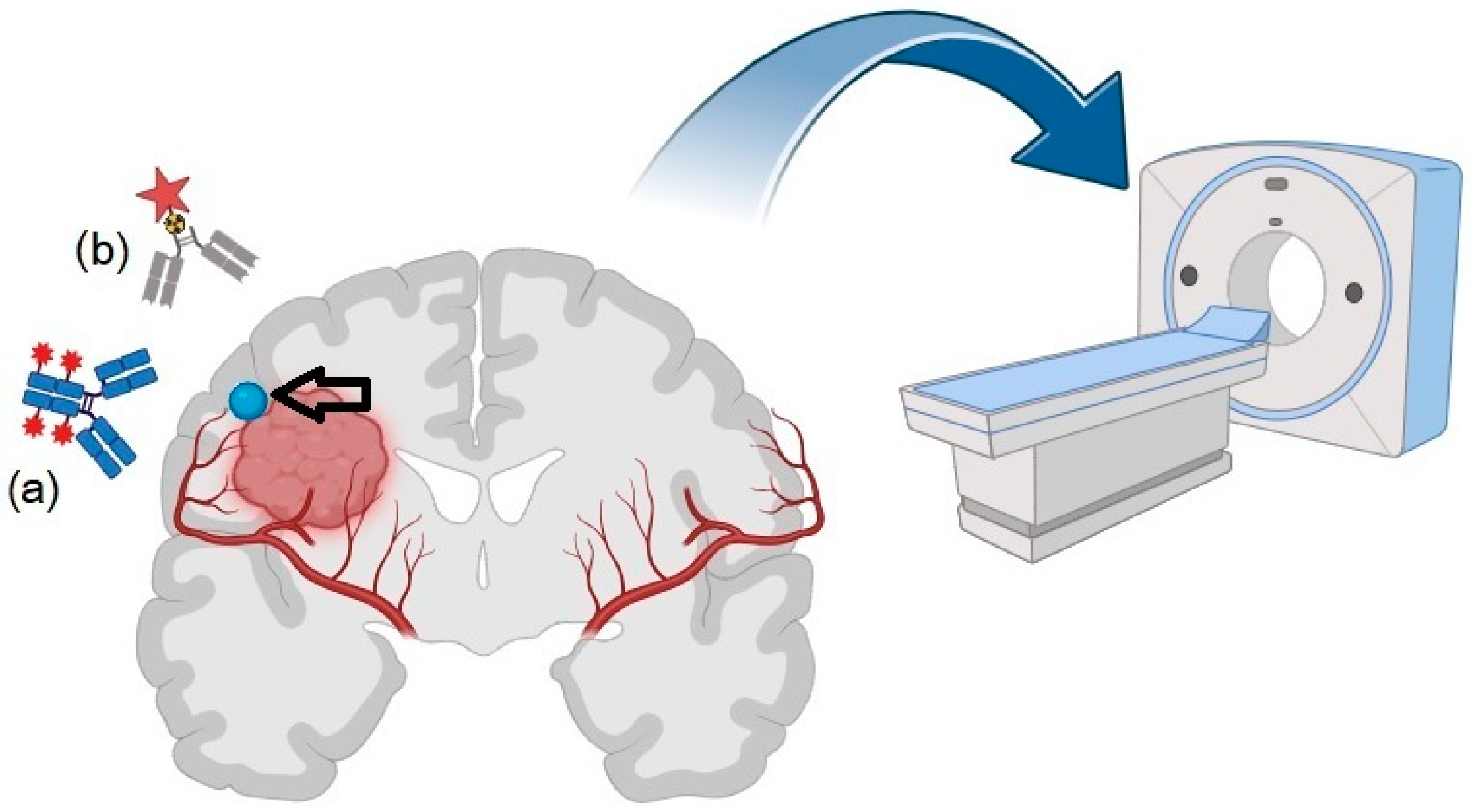
3. Immuno-PET: Basic Principles and Technical Features

The use of radiolabeled antibodies for cancer imaging is not something new. In past years, immuno-scintigraphy, based on the administration of antibodies labeled with gamma-emitting radionuclides (i.e., 99mTc and 123I), has been applied with encouraging results in several clinical settings [23]. As concerns gliomas, monoclonal antibodies (mAbs), specifically those recognizing the type III mutant EGFR (EGFRvIII), were found to specifically accumulate into subcutaneous or intracranial glioma xenografts expressing the EGFRvIII [24]. Nevertheless, the use of immunoscintigraphy has been limited by the low resolution of planar imaging obtained with a gamma-camera, which is only partially overcome by the implementation of hybrid SPECT/CT [25,26]. Immuno-PET is a cutting-edge medical imaging technique that has revolutionized the field of molecular imaging and cancer diagnostics [27,28]. This powerful technology combines the specificity of antibodies with the high-resolution imaging capabilities of PET scans to provide valuable insights into the molecular and cellular processes occurring within the body. Immuno-PET holds immense promise in cancer diagnosis, treatment monitoring, and drug development, as it allows for the visualization and quantification of specific biomarkers and immune responses. The fundamental principle of immuno-PET lies in its ability to target and visualize specific molecules or cells within the body. At its core, immuno-PET involves the use of radiolabeled antibodies, or antibody fragments, which are designed to bind with high affinity to particular biomarkers or antigens present on the surface of cancer cells, immune cells, or other disease-related targets. These radiolabeled antibodies are usually coupled with a positron-emitting radionuclide, such as fluorine-18 (18F) or copper-64 (64Cu), which emits positrons. When injected into the patient’s bloodstream, these radiolabeled antibodies circulate throughout the body, seeking out and binding specifically to their target antigens. However, in the advancement of immuno-PET, a pivotal milestone has been the creation of antibodies tagged with zirconium-89 (89Zr), a positron-emitting element with a physical half-life of 78.4 h [29]. This half-life aligns with the extended presence of antibodies within the body following administration, facilitating their use. A schematic representation of immuno-PET in gliomas is depicted in Figure 1.
Once the radiolabeled antibodies have localized at the target site, the emitted positrons undergo annihilation reactions with nearby electrons, resulting in the release of gamma rays in opposite directions. These gamma rays are then detected by a PET scanner, which is equipped with sensitive detectors arranged in a ring around the patient’s body. By measuring the spatial distribution of these gamma rays, the PET scanner creates a three-dimensional image of the concentration and distribution of the radiolabeled antibodies within the body [30]. This provides a detailed map of the location and extent of the targeted molecules or cells. Because radiolabeled antibodies are highly selective for their target antigens, immuno-PET can detect even low levels of specific biomarkers with great precision. This is particularly valuable in the context of cancer diagnosis, where the early detection of tumor-associated antigens can lead to earlier and more effective treatments. Additionally, Immuno-PET can differentiate between cancerous and non-cancerous tissues, reducing false positives and improving diagnostic accuracy. Another important aspect of immuno-PET is its ability to provide quantitative information. Unlike traditional imaging techniques like CT or MRI, immuno-PET allows for the measurement of biomarker expression levels and changes over time [31]. Among the various PET-derived parameters, maximum and mean standardized uptake values (SUVmax and SUVmean, respectively) have been shown to have a meaningful prognostic impact in oncology, as well as the more recently introduced volumetric data, such as metabolic tumor volume (MTV) or total lesion glycolysis (TLG) [32]. These quantitative data are crucial for monitoring disease progression, evaluating treatment responses, and guiding treatment decisions. More recently, some technological innovations have further boosted the applications of PET in immuno-oncology, such as the implementation of silicon photomultiplier (SiMP)-based detection, referred to as the “digital PET”, and the introduction of long axial field-of-view (LAFOV) PET/CT scanners, capable of covering the entire scan length in a single or a few bed positions, and characterized by exquisite sensitivity [33].

4. Targets for Immuno-PET

A large number of novel positron-emitting radionuclides are being created at the moment. In recent years, there has been an increase in the production and use of high-purity radiometals, which are a key component of Immune-PET imaging probes. Radiometals have traditionally either been eluted from generators or generated using solid targets and cyclotrons. In addition to solid targets, liquid targets (solution targets) can be employed to optimize the production of radiometals. Physical features (e.g., half-life [T1/2] and decay mode), chemical properties, production efficiency, safety profiles, and pricing are all important and must be thoroughly analyzed before they can be used for radiolabeling. The T1/2 of a selected positron emitter must correspond to the biological half-life of the targeted vector. The positron emitter in conjugating immuno-PET imaging probes is usually completed with an inert chelator that is coupled to the targeted antibody. The basic notion is that the final radiopharmaceutical’s binding affinity, stability, and pharmacokinetic properties will cooperate with the antibody [27,34,35].

4.1. EGFR

The overexpression of the EGFR in many malignancies makes it a suitable molecular target for diagnosing and treating different malignancies [36]. Scandium-44/44Sc (with a half-life of 3.9 h) is a relatively new radioisotope with potential applications in clinical PET. Chakravarty et al. demonstrated a ground-breaking achievement for in vivo PET imaging [37]: they generated the Fab fragment of Cetuximab, an mAb known for its strong binding to the EGFR, and radiolabeled it with 44Sc at room temperature. To achieve this, the Fab fragment was conjugated with N-[(R)-2-amino-3-(para-isothiocyanato-phenyl)propyl]. CHX-A″-DTPA stands for -trans-(S,S)-cyclohexane-1,2-diamine-N,N,N′,N″,N″-pentaacetic acid. SDS-PAGE and mass spectrometry were used to thoroughly confirm the high quality and purity of cetuximab-Fab. After labeling with 44Sc, the potential of this bioconjugate for the PET imaging of EGFR expression was assessed in a human glioblastoma (U87MG) tumor-bearing mice model. The PET imaging results show that [44Sc]-CHX-A″-DTPA-Cetuximab-Fab was rapidly absorbed in the tumor, with a peak uptake of around 12% ID/g observed 4 h after injection. Importantly, this uptake showed an outstanding tumor-to-background ratio, implying the possibility of same-day PET imaging in future clinical applications. In addition, immunofluorescence labeling was used to correlate tracer uptake in tumor and normal tissues with EGFR expression. This successful immuno-PET imaging strategy, utilizing [44Sc]-CHX-A″-DTPA-Cetuximab-Fab, has the potential to advance clinical practices by aiding in patient selection for EGFR-targeted therapy, and by facilitating the monitoring of the effectiveness of anti-EGFR treatments.

4.2. VEGF

The vascular endothelial growth factor receptor 2 (VEGFR-2/Flk-1/KDR) is a biomarker that is overexpressed in tumor neo-vasculature and several types of malignancies [38]. Angiogenesis has identified important players as targets, including vascular endothelial growth factors (VEGF), VEGF receptors (VEGFRs), integrins (particularly v3), and matrix metalloproteinases (MMPs). These targets are used to produce both imaging and therapeutic medicines. Several specialized imaging agents, such as HuMV833, VG76e, and bevacizumab, have been developed using radiolabeled VEGF or monoclonal antibodies against VEGF. Furthermore, [18F]-fluciclatide, a radioisotope-labeled short cyclic peptide comprising the arginyl-glycyl-aspartic acid (RGD) tripeptide with great binding affinity to v3 integrins, has shown promise for imaging angiogenesis. Reduced [18F]-fluciclatide absorption was associated with the decreased expressions of three integrins and an early response to antiangiogenic targeted therapies in both experimental and clinical studies. Furthermore, in a preclinical MMP study, 18F-labeled marimastat, an MMP inhibitor, and MMP2/9 substrates were observed to accumulate in tumor locations. As a result, radiolabeling therapeutic antiangiogenic drugs has become a well-established practice in scientific literature [39]. Beyond the mentioned angiogenesis indicators, researchers have identified a minimum of 46 potential therapeutic targets residing on the endothelial cells of tumor blood vessels. These specific markers on tumor endothelial cells, referred to as TEMs (Tumor Endothelial Markers), constitute a cluster of genes participating in angiogenesis regulation. Among these TEMs, TEM8 stands out as it has been observed to exhibit increased expression in tumor vasculature across various tumor types in both human patients and mouse models bearing tumors. Importantly, this upregulation is not observed in the context of normal reparative angiogenesis during processes such as wound healing or ovulation [40]. Bevacizumab is a humanized monoclonal antibody with a distinct capacity to bind exclusively to vascular endothelial growth factor A (VEGF-A). VEGF-A is an overexpressed biomarker in cervical cancer, where it plays a critical role in the beginning and maintenance of tumor-associated neo-angiogenesis, or the development of new blood vessels to support tumor growth. However, the effectiveness of the VEGF-targeted mAb bevacizumab in treating diffuse intrinsic pontine glioma (DIPG) remains uncertain. Jansen et al. investigated how bevacizumab labeled with zirconium-89 (89Zr) behaves in DIPG mouse models regarding biodistribution and uptake [41]. Human E98-FM, U251-FM glioma cells, and HSJD-DIPG-007-FLUC primary DIPG cells were injected into nude mice through subcutaneous, pons, or striatum injection. Tumor development was tracked via bioluminescence imaging and observed using MRI. PET scans were performed 72 to 96 h post [89Zr]-bevacizumab administration, and biodistribution was assessed through ex vivo analysis. The researchers verified elevated VEGF expression in human DIPG using publicly available mRNA data. However, no significant uptake of [89Zr]-bevacizumab was identified in the brain or brain tumors at any disease stage, although significant uptake was observed in subcutaneous tumors. VEGF expression was definitely detectable in the perinecrotic areas of subcutaneous E98-FM tumors, despite the absence of VEGF expression in cerebral tumors via in situ hybridization. The low absorption of [89Zr]-bevacizumab in brain-based xenografts shows that bevacizumab targeting VEGF may be ineffective in treating diffuse infiltrative portions of glial brain tumors in mice. Extrapolating these results to clinical scenarios implies that administering bevacizumab to DIPG patients should only be considered after confirming VEGF targeting through [89Zr]-bevacizumab immuno-PET. Following this, the same research group conducted a preliminary investigation to explore the accessibility of bevacizumab to tumors in children with DIPGs [42]. The study involved measuring the tumor uptake of [89Zr]-bevacizumab and determining the optimal time for PET imaging. Seven children with DIPG participated, including one with spinal cord metastases and another with disease extension to the facial nerve. All subjects had previously undergone radiation therapy in conjunction with gemcitabine or temozolomide. PET scans yielded positive results in five cases and negative results in two. Tumor uptake of the tracer varied among patients, with tumor SUVs ranging from 1.0 to 5.3 at 72 h and from 1.0 to 6.7 at 144 h post-injection. In the case of spinal cord metastases, the PET scan was positive in all metastatic locations. A biodistribution study revealed that tracer uptake was highest in the liver, followed by the kidneys, spleen, lungs, and vertebrae, resulting in an effective dose of 0.9 mSv/MBq. Notably, four out of five tumors exhibited significant [89Zr]-bevacizumab uptake only within contrast-enhanced areas on MRI, indicating substantial variability in tracer uptake among tumors and suggesting differences in local VEGF expression. While these findings were promising and supported the potential utility of immuno-PET with [89Zr]-bevacizumab for assessing VEGF expression in DIPGs in vivo, the limited number of enrolled patients and the absence of pathological validation underscore the need for further studies to establish more robust confirmation.

4.3. Cell Differentiation Antigens

CD146 is a key biomarker in cancer that was first discovered in 1987 in metastatic lesions and advanced original melanoma, with infrequent detection in benign lesions, and was dubbed “MIC18”. CD146’s principal functions are associated with intercellular and cell–matrix adhesion [43]. Nonetheless, its involvement in a variety of other processes, including development, cell motility, signal transmission, stem cell differentiation, immunological response, angiogenesis, and, more recently, the onset of epithelial–mesenchymal transition, has been documented. Yang et al. developed YY146, an mAb -targeting human CD146, using a novel approach for noninvasive CD146 imaging in GBM mice [44]. Their research also studied CD146’s correlation with tumor traits and used a unique immunization approach, identifying YY146 as the top CD146-binding clone, especially in CD146-overexpressing melanoma cells. They compared CD146 levels in U87MG and U251 GBM cell lines, showing higher CD146 expression in U87MG cells. In vivo, immuno-Pet with [64Cu]-NOTA-YY146 effectively located CD146-overexpressing U87MG tumors, with high-contrast PET imaging. Blocking studies confirmed specificity, and biodistribution studies and immunofluorescence validated uptake. Of note, CD146-positive staining correlated with high tumor grade, highlighting [64Cu]-NOTA-YY146’s potential for use in GBM detection and targeted therapy. The antibody YY146 was subsequently labeled with the 89Zr radionuclide, known for its longer half-life (t1/2: 78.4 h), aiding in distribution and kinetics studies. Hernandez and colleagues synthesized [89Zr]-Df-YY146, a new mAb for imaging CD146 expression in GBM mouse models via noninvasive immuno-PET [45]. YY146 was linked to deferoxamine (Df) for 89Zr labeling. In vitro assays on two different cell lines, U87MG and U251 GBM, defined CD146 levels. The CD146-binding affinities of YY146 and Df-YY146 were compared (5%), and their activity was 44 GBq/mol. Longitudinal PET revealed significant and persistent U87MG absorption (14.00 3.28%ID/g at 48 h p.i.). U251 tumors, on the other hand, displayed lesser uptake (5.15 0.99%ID/g at 48 h p.i.) due to lower CD146 expression. CD146-specificity was confirmed by a competitive inhibition experiment that prevented U87MG absorption. PET data from biodistribution were confirmed, and histological investigation associated tracer uptake with CD146 expression. Notably, [89Zr]-Df-YY146 exhibited significant and specific uptake in brain tumors, highlighting its potential for use in the noninvasive PET imaging of CD146 expression. In future clinical applications, this might guide interventions and assess responses to CD146-targeted therapies. In recent years, immunoevasion has emerged as one of the most significant mechanisms underlying tumor growth and proliferation. This is especially important in the setting of GBM, where tumor-associated myeloid cells (TAMCs) account for roughly 40% of the tumor mass. TAMCs play a pivotal role not only in disease progression, but also in therapy resistance, making them promising targets for therapeutic interventions [46,47]. Therefore, it is imperative to develop imaging and molecular biomarkers capable of detecting and monitoring TAMCs. Nigam et al. devised a novel approach by conjugating a human/mouse cross-reactive anti-CD11b antibody with desferrioxamine, incorporating 89Zr for the purpose of detecting TAMC infiltration in GBM models [48]. Immuno-PET imaging was conducted, with and without the administration of a blocking dose of anti-CD11b antibody, 72 h after injecting [89Zr]-anti-CD11b antibody into mice bearing orthotopic syngeneic GL261 gliomas, as well as in non-tumor-bearing mice. Significantly, increased tracer uptake was observed at tumor sites compared to the contralateral brain hemisphere. Notably, the use of a radiocompound with a 10-fold lower specific activity for blocking substantially reduced the SUV in tumor xenografts, providing compelling evidence of binding specificity. It is essential to emphasize that the immuno-PET biodistribution study revealed physiological tracer accumulation in the spleen and lymph nodes, both of which are rich in myeloid cells. Furthermore, the CD11b signal detected in PET images exhibited a strong correlation with biomarker expression, as determined by histochemistry and flow cytometry.

4.4. Prostate-Specific Membrane Antigen (PSMA)

PSMA (prostate-specific membrane antigen) is a type II transmembrane glycoprotein that includes zinc and works as a metal enzyme. It is well-known for use in the molecularly targeted approach to prostate cancer (PC). PSMA has been widely researched throughout the last three decades. Previously, immunoscintigraphy with 111In-capromab pendetide, an mAb-targeting PSMA, was used to image PC recurrence and metastases. However, its low sensitivity and specificity were attributable to the fact that it targeted the PSMA intracellular domain, which is only present in dying or necrotic cells. The discovery of radiolabeled urea-based PSMA inhibitors targeting PSMA’s extracellular enzymatic region was a watershed moment in the field. This innovation cleared the way for the development of a plethora of PSMA inhibitors labeled with radionuclides, appropriate for use in both PET imaging ([68Ga]-PSMA-11 or [18F]-PSMA-1007) and therapy ([177Lu]-PSMA-617). PSMA, despite its name, is not just associated with prostate cancer. Antibodies targeting this biomarker react strongly with neovasculature in a wide range of cancers, including colorectal and breast cancer. Moving on to gliomas, immunohistochemistry has verified PSMA expression in high-grade gliomas. Furthermore, PSMA-PET has shown promise in preliminary investigations on GBM diagnosis and recurrence [49,50]. From this perspective, it is important to note that PSMA-PET conducted using PSMA inhibitors should not be classified as immuno-PET because it does not utilize radiolabeled antibodies. However, in a different approach, a humanized mAb known as huJ591 (J591), which specifically targets the extracellular domain of PSMA, has been conjugated with various radiometals such as 111In and 89Zr. This conjugate has been subject to investigation as a potential imaging agent in a phase I/II clinical study carried out in PC patients [51]. However, scientific data on the potential use of immuno-PET with [89Zr]-J591 for glioma imaging remain limited. Recently, Krebs et al. published a clinical report detailing the detection of grade II oligodendroglioma (1p/19q co-deleted, IDH mutant) using [89Zr]-J591 immuno-PET [52]. The patient, who had undergone multiple treatments (surgery, chemotherapy, radiation therapy, and IDH inhibitor AG-120), showed MRI findings suggestive of disease progression. Serial immuno-PET scans targeting PSMA were conducted on days 1, 2, and 6 post-injection, and these scans were fused with axial post-contrast T1-weighted MRI images. They revealed a nodular enhancing lesion located anterior and inferior to the resection cavity. The authors propose that immuno-PET, owing to the extended physical half-life of 89Zr, not only holds promise for glioma diagnosis, but also offers the potential for precise dosimetric calculations through serial PET/CT scans, with implications for future radioimmunotherapeutic approaches.
Table 1 provides a summary of the key conclusions derived from research articles on the use of immuno-PET in gliomas.

5. Discussion

Despite advances in the field of diagnosis and therapy, primary brain tumors, of which gliomas represent the most common category, remain a diagnostic and therapeutic challenge. In particular, the prognosis for high-grade gliomas is still grim, with a median survival of around 13 months [53]. In addition, the introduction of new therapeutic approaches, such as the combination of radiotherapy with temozolomide, has led to the emergence of atypical patterns in MRI and CT, such as pseudoprogression [54,55]. The latter, which occurs more frequently in the first 3 months after the completion of combined radiotherapy and chemotherapy, is predominantly associated with edema, increased capillary permeability, and inflammatory components. In this context, molecular imaging with PET can provide an important contribution through the use of radiopharmaceuticals targeted towards specific metabolic pathways characteristic of tumors, such as radiolabeled amino acids [56]. The utilization of amino acid radiopharmaceuticals in PET imaging shows promise for diagnosing and grading gliomas. It can serve as a valuable tool for guiding biopsies and planning surgical and radiotherapy procedures. Specifically, integrating PET imaging into the resection process and radiotherapy planning may offer more insightful information compared to standard MRI, potentially leading to improved survival outcomes for patients with gliomas [57]. However, PET with radiolabeled amino-acids exploits a non-specific metabolic mechanism. In the era of personalized medicine, it is imperative to identify biomarkers associated with gliomas that can be used for molecular imaging and potential targeted therapy. In this context, our review of the existing literature has prompted several considerations. First, although several attempts have been made in the field, none among the examined potential targets (i.e., EGFR, VEGF, CD146, CD11b, PSMA) have emerged as predominantly effective in the field, since their expression has been found to be not homogeneous, and strictly depending on the tumor grade and type (i.e., DIPG, GBM, oligodendroglioma etc.). Secondly, other relevant considerations should be made on the various radionuclides and ligands employed for immuno-PET. One of the most challenging aspects of immuno-PET is its reliance on mAbs, or sometimes antibody fragments. Despite their high specificity, these agents are characterized by slow clearance, necessitating delayed imaging (e.g., 48 h p.i.) to achieve an adequate target-to-background ratio [58]. Notably, certain radionuclides traditionally used in clinical practice, such as 68Ga and 18F, have a too-short physical half-life, rendering them unsuitable for delayed imaging. In contrast, 89Zr has shown more promising results due to its longer physical half-life [59]. In this context, it is crucial to emphasize that specific radiochemical approaches merit exploration to facilitate the labeling of mAbs with [68Ga] or [18F], particularly those involving in vivo bio-orthogonal reactions [60]. Initial steps in investigating this methodology were taken by Devaraj et al. in 2012 [61]. The researchers conjugated anti-CD45 mAbs with Trans-CycloOctyne (TCO) and administered them intravenously to mice 24 h prior to in vivo click chemistry. They employed polymer-modified tetrazine (PMT) as a pivotal facilitator. Subsequent to the reaction between TCO and tetrazine, [18F]-PMT was successfully utilized for whole-body PET imaging. This approach holds significant implications and warrants further exploration in the realm of glioma-targeted immuno-PET. Although humanized mAbs have been recently integrated for immuno-PET imaging, careful consideration is warranted for potential host immuno-mediated adverse reactions [62]. Furthermore, the application of mAb in CNS tumors is constrained by the BBB, which impedes the delivery of both small-molecule drugs and therapeutic proteins. It is crucial to note that, while highly aggressive gliomas like GBM lead to significant BBB leakage, facilitating the integration of radiopharmaceuticals into tumors, low-grade gliomas may maintain an intact BBB, obstructing the passage of radiolabeled mAbs. To address challenges associated with BBB permeability, researchers have devised antibody-based carriers that utilize the inherent macromolecule transportation pathway known as receptor-mediated transcytosis (RMT). A substantial portion of existing studies in this area focus on RMT receptors, specifically the transferrin receptor (TfR), insulin receptor (InsR), and FC5 antibody binding receptor [63]. Adopting this approach involves the utilization of bispecific antibodies (BsAbs), which possess the capability to bind to RMT-associated receptors with one arm, facilitating BBB crossing. Once within the brain, the other arm of these BsAbs can target tumor-associated biomarkers. In this context, Schaller and colleagues developed a BsAb that activates cells, demonstrating promising outcomes in orthotopic patient-derived malignant glioma and syngeneic glioblastoma in mice [64]. This innovative BsAb comprises two single-chain antibody fragments (bi-scFvs) that bind to the mutant epidermal growth factor receptor variant III (EGFRvIII), a common mutation GBM, and human CD3ε on T cells. Consequently, the BsAb-based approaches hold considerable potential and merit further exploration to address BBB permeability challenges, especially in the case of low-grade gliomas, thereby expanding the range of applications for immuno-PET in clinical settings. Another potential critique of immuno-PET in gliomas is associated with the mechanisms through which various tracers are assimilated [65]. There is a hypothesis that their incorporation may be more indicative of BBB disruption than relying on specific biomarker-mediated processes. This aspect warrants further investigation in future studies. Shaping the future of immuno-PET in gliomas will depend significantly on the synergy between technology and radiopharmaceutical research. For instance, aptamers, single-stranded oligonucleotide DNA or RNA sequences, exhibit extremely high specificity toward tumor-associated biomarkers without inducing immunogenicity [66]. However, despite their promise, radiolabeled aptamers have seen limited application in PET imaging. Other noteworthy advancements include the incorporation of glioma-associated biomarkers for functionalizing nanoparticles, paving the way for radiotheranostic-based approaches [67,68].
In the near future, the significance and potential integration of immuno-PET in the diagnostic evaluation of gliomas will be enhanced through a comparative analysis with alternative non-radionuclide-based imaging techniques, including fluorescence and bioluminescent imaging. Regarding fluorescence imaging, particularly Near-Infrared Fluorescence (NIRF) has garnered increasing attention due to its safety, detection sensitivity, and resolution [69]. This makes it a valuable tool for the early diagnosis and visualization of various cancer types, given its convenience. Cyanine and its derivatives have been developed to emit visible light when excited by a specific wavelength, rendering them valuable tools for biological imaging [70]. In this context, it is noteworthy to highlight the significance of tumor specific Heptamethine Cyanine Dyes (HMCDs). Originally recognized for their fluorescent and mitochondria-targeting properties, these compounds have demonstrated a distinct preference for tumor up-take both in vitro and in animal models. Furthermore, their ability to cross the BBB positions HMCDs as an appealing platform for drug delivery systems. However, as of now, only indocyanine green (ICG) and methylene blue have received approval from the Food and Drug Administration for use in medical diagnostics as near-infrared fluorescent (NIRF) compounds. Meanwhile, red-fluorescent dyes such as 5-ALA are gaining consideration for their potential application in fluorescent-guided surgery for gliomas [70].
Bioluminescence imaging (BLI) relies on detecting and quantifying light emission resulting from the conversion of chemical energy into photon energy. This phenomenon occurs when a substrate undergoes oxidation by the enzyme luciferase in living cells and tissues [71,72]. Coelenterazine (CLZN), for instance, serves as the substrate for a group of bioluminescent enzymes derived from marine organisms. Various luciferase genes, such as those from fireflies, renilla, gaussia, and most recently vargula hilgen-dorfii, have been cloned and utilized, with firefly luciferase being the most commonly employed. BLI has found extensive use in assessing and optimizing therapeutic strategies for GBM. One of the significant advantages of bioluminescence is its ability to offer real-time, non-invasive evaluation of specific physiological processes. However, conventional BLI is limited to providing two-dimensional (2D) and qualitative information about the tumor region due to the light scattering effect, in contrast to immuno-PET technology. Recognizing these limitations, efforts have been directed towards transitioning from BLI to Bioluminescence Tomography (BLT). Along this path, Gao et al. developed a new reconstruction method that accurately locates the tumor and defines its morphology in three dimensions. This approach was successfully tested in orthotopic glioma mouse models [73]. Further studies are essential to better understand the potential contribution of immuno-PET to glioma research compared to non-radionuclide-based alternatives. Additionally, it is crucial to define its practical implementation in the clinical workup of gliomas.

6. Conclusions

Despite progress in diagnosis and therapy, effectively managing primary brain tumors, especially high-grade gliomas, remains a challenge. Although immuno-PET holds promise for accurate diagnosis and treatment guidance, obstacles persist in attaining target specificity and selecting suitable radionuclides. Further studies are essential to advance the field and bridge the gap from bench to bedside.

Author Contributions

Conceptualization, V.F. and G.D.V.; methodology, M.S.D.F. and G.M.G.; formal analysis, G.D.V.; investigation, G.M.G.; data curation, M.S.D.F. and B.P.; writing—original draft preparation, M.S.D.F., G.M.G., B.P., S.P. and L.F. writing—review and editing, M.C., G.M.G. and B.P.; visualization, S.P.; supervision, V.F. and L.F. All authors have read and agreed to the published version of the manuscript.

Funding

This research received no external funding.

Institutional Review Board Statement

Not applicable.

Informed Consent Statement

Not applicable.

Data Availability Statement

Not applicable.

Conflicts of Interest

The authors declare no conflicts of interest.

References

  1. Ostrom, Q.T.; Price, M.; Neff, C.; Cioffi, G.; Waite, K.A.; Kruchko, C.; Barnholtz-Sloan, J.S. CBTRUS Statistical Report: Primary Brain and Other Central Nervous System Tumors Diagnosed in the United States in 2015–2019. Neuro Oncol. 2022, 24, v1–v95. [Google Scholar] [CrossRef]
  2. Wen, P.Y.; Kesari, S. Malignant Gliomas in Adults. N. Engl. J. Med. 2008, 359, 492–507. [Google Scholar] [CrossRef]
  3. Liang, J.; Lv, X.; Lu, C.; Ye, X.; Chen, X.; Fu, J.; Luo, C.; Zhao, Y. Prognostic Factors of Patients with Gliomas—An Analysis on 335 Patients with Glioblastoma and Other Forms of Gliomas. BMC Cancer 2020, 20, 35. [Google Scholar] [CrossRef] [PubMed]
  4. Frosina, G. Recapitulating the Key Advances in the Diagnosis and Prognosis of High-Grade Gliomas: Second Half of 2021 Update. Int. J. Mol. Sci. 2023, 24, 6375. [Google Scholar] [CrossRef] [PubMed]
  5. Lee, J.H.; Wee, C.W. Treatment of Adult Gliomas: A Current Update. Brain NeuroRehabil. 2022, 15, e24. [Google Scholar] [CrossRef] [PubMed]
  6. Bloch, O. Immunotherapy for Malignant Gliomas. In Cancer Treatment and Research; Springer: Cham, Switzerland, 2015; Volume 163, pp. 143–158. [Google Scholar] [CrossRef]
  7. Rajesh, Y.; Pal, I.; Banik, P.; Chakraborty, S.; Borkar, S.A.; Dey, G.; Mukherjee, A.; Mandal, M. Insights into Molecular Therapy of Glioma: Current Challenges and next Generation Blueprint. Acta Pharmacol. Sin. 2017, 38, 591–613. [Google Scholar] [CrossRef] [PubMed]
  8. Werner, R.A.; Higuchi, T.; Pomper, M.G.; Rowe, S.P. Theranostics in Oncology-Thriving, Now More than Ever. Diagnostics 2021, 11, 805. [Google Scholar] [CrossRef] [PubMed]
  9. Wesseling, P.; Capper, D. WHO 2016 Classification of Gliomas. Neuropathol. Appl. Neurobiol. 2018, 44, 139–150. [Google Scholar] [CrossRef] [PubMed]
  10. Louis, D.N.; Perry, A.; Wesseling, P.; Brat, D.J.; Cree, I.A.; Figarella-Branger, D.; Hawkins, C.; Ng, H.K.; Pfister, S.M.; Reifenberger, G.; et al. The 2021 WHO Classification of Tumors of the Central Nervous System: A Summary. Neuro Oncol. 2021, 23, 1231–1251. [Google Scholar] [CrossRef]
  11. Berger, T.R.; Wen, P.Y.; Lang-Orsini, M.; Chukwueke, U.N. World Health Organization 2021 Classification of Central Nervous System Tumors and Implications for Therapy for Adult-Type Gliomas: A Review. JAMA Oncol. 2022, 8, 1493–1501. [Google Scholar] [CrossRef]
  12. Cancer Genome Atlas Research Network; Brat, D.J.; Verhaak, R.G.W.; Aldape, K.D.; Yung, W.K.A.; Salama, S.R.; Cooper, L.A.D.; Rheinbay, E.; Miller, C.R.; Vitucci, M.; et al. Comprehensive, Integrative Genomic Analysis of Diffuse Lower-Grade Gliomas. N. Engl. J. Med. 2015, 372, 2481–2498. [Google Scholar] [CrossRef]
  13. Opoku-Darko, M.; Eagles, M.E.; Cadieux, M.; Isaacs, A.M.; Kelly, J.J.P. Natural History and Growth Patterns of Incidentally Discovered Diffusely Infiltrating Low-Grade Gliomas: A Volumetric Study. World Neurosurg. 2019, 132, e133–e139. [Google Scholar] [CrossRef]
  14. Altieri, R.; Hirono, S.; Duffau, H.; Ducati, A.; Fontanella, M.M.; La Rocca, G.; Melcarne, A.; Panciani, P.P.; Spena, G.; Garbossa, D. Natural History of de Novo High Grade Glioma: First Description of Growth Parabola. J. Neurosurg. Sci. 2020, 64, 399–403. [Google Scholar] [CrossRef]
  15. Lombardi, G.; Barresi, V.; Castellano, A.; Tabouret, E.; Pasqualetti, F.; Salvalaggio, A.; Cerretti, G.; Caccese, M.; Padovan, M.; Zagonel, V.; et al. Clinical Management of Diffuse Low-Grade Gliomas. Cancers 2020, 12, 3008. [Google Scholar] [CrossRef]
  16. Ius, T.; Angelini, E.; Thiebaut de Schotten, M.; Mandonnet, E.; Duffau, H. Evidence for Potentials and Limitations of Brain Plasticity Using an Atlas of Functional Resectability of WHO Grade II Gliomas: Towards a “Minimal Common Brain”. Neuroimage 2011, 56, 992–1000. [Google Scholar] [CrossRef]
  17. Buckner, J.C.; Shaw, E.G.; Pugh, S.L.; Chakravarti, A.; Gilbert, M.R.; Barger, G.R.; Coons, S.; Ricci, P.; Bullard, D.; Brown, P.D.; et al. Radiation plus Procarbazine, CCNU, and Vincristine in Low-Grade Glioma. N. Engl. J. Med. 2016, 374, 1344–1355. [Google Scholar] [CrossRef] [PubMed]
  18. Shaw, E.; Arusell, R.; Scheithauer, B.; O’Fallon, J.; O’Neill, B.; Dinapoli, R.; Nelson, D.; Earle, J.; Jones, C.; Cascino, T.; et al. Prospective Randomized Trial of Low- versus High-Dose Radiation Therapy in Adults with Supratentorial Low-Grade Glioma: Initial Report of a North Central Cancer Treatment Group/Radiation Therapy Oncology Group/Eastern Cooperative Oncology Group Study. J. Clin. Oncol. 2002, 20, 2267–2276. [Google Scholar] [CrossRef]
  19. Wang, T.J.C.; Mehta, M.P. Low-Grade Glioma Radiotherapy Treatment and Trials. Neurosurg. Clin. N. Am. 2019, 30, 111–118. [Google Scholar] [CrossRef] [PubMed]
  20. Frosina, G. Radiotherapy of High-Grade Gliomas: Dealing with a Stalemate. Crit. Rev. Oncol. Hematol. 2023, 190, 104110. [Google Scholar] [CrossRef] [PubMed]
  21. Kyritsis, A.P. Chemotherapy for Malignant Gliomas. Oncology 1993, 7, 93–100; discussion 103+106. [Google Scholar] [PubMed]
  22. See, S.-J.; Gilbert, M.R. Chemotherapy in Adults with Gliomas. Ann. Acad. Med. Singap. 2007, 36, 364–366. [Google Scholar]
  23. Garousi, J.; Orlova, A.; Frejd, F.Y.; Tolmachev, V. Imaging Using Radiolabelled Targeted Proteins: Radioimmunodetection and Beyond. EJNMMI Radiopharm. Chem. 2020, 5, 16. [Google Scholar] [CrossRef] [PubMed]
  24. Takasu, S.; Takahashi, T.; Okamoto, S.; Oriuchi, N.; Nakayashiki, N.; Okamoto, K.; Muramatsu, H.; Hayashi, T.; Nakahara, N.; Mizuno, M.; et al. Radioimmunoscintigraphy of Intracranial Glioma Xenograft with a Technetium-99m-Labeled Mouse Monoclonal Antibody Specifically Recognizing Type III Mutant Epidermal Growth Factor Receptor. J. Neurooncol. 2003, 63, 247–256. [Google Scholar] [CrossRef] [PubMed]
  25. Filippi, L.; Schillaci, O.; Santoni, R.; Manni, C.; Danieli, R.; Simonetti, G. Usefulness of SPECT/CT with a Hybrid Camera for the Functional Anatomical Mapping of Primary Brain Tumors by [Tc99m] Tetrofosmin. Cancer Biother. Radiopharm. 2006, 21, 41–48. [Google Scholar] [CrossRef] [PubMed]
  26. Schillaci, O.; Danieli, R.; Filippi, L.; Romano, P.; Cossu, E.; Manni, C.; Simonetti, G. Scintimammography with a Hybrid SPECT/CT Imaging System. Anticancer Res. 2007, 27, 557–562. [Google Scholar] [PubMed]
  27. Wei, W.; Rosenkrans, Z.T.; Liu, J.; Huang, G.; Luo, Q.-Y.; Cai, W. ImmunoPET: Concept, Design, and Applications. Chem. Rev. 2020, 120, 3787–3851. [Google Scholar] [CrossRef] [PubMed]
  28. Lugat, A.; Bailly, C.; Chérel, M.; Rousseau, C.; Kraeber-Bodéré, F.; Bodet-Milin, C.; Bourgeois, M. Immuno-PET: Design Options and Clinical Proof-of-Concept. Front. Med. 2022, 9, 1026083. [Google Scholar] [CrossRef] [PubMed]
  29. Yoon, J.-K.; Park, B.-N.; Ryu, E.-K.; An, Y.-S.; Lee, S.-J. Current Perspectives on 89Zr-PET Imaging. Int. J. Mol. Sci. 2020, 21, 4309. [Google Scholar] [CrossRef] [PubMed]
  30. Levin Klausen, T.; Høgild Keller, S.; Vinter Olesen, O.; Aznar, M.; Andersen, F.L. Innovations in PET/CT. Q. J. Nucl. Med. Mol. Imaging 2012, 56, 268–279. [Google Scholar]
  31. Meikle, S.R.; Sossi, V.; Roncali, E.; Cherry, S.R.; Banati, R.; Mankoff, D.; Jones, T.; James, M.; Sutcliffe, J.; Ouyang, J.; et al. Quantitative PET in the 2020s: A Roadmap. Phys. Med. Biol. 2021, 66, 06RM01. [Google Scholar] [CrossRef]
  32. Ling, T.; Zhang, L.; Peng, R.; Yue, C.; Huang, L. Prognostic Value of 18F-FDG PET/CT in Patients with Advanced or Metastatic Non-Small-Cell Lung Cancer Treated with Immune Checkpoint Inhibitors: A Systematic Review and Meta-Analysis. Front. Immunol. 2022, 13, 1014063. [Google Scholar] [CrossRef]
  33. Wang, D.; Qiu, B.; Liu, Q.; Xia, L.; Liu, S.; Zheng, C.; Liu, H.; Mo, Y.; Zhang, X.; Hu, Y.; et al. Patlak-Ki Derived from Ultra-High Sensitivity Dynamic Total Body [18F]FDG PET/CT Correlates with the Response to Induction Immuno-Chemotherapy in Locally Advanced Non-Small Cell Lung Cancer Patients. Eur. J. Nucl. Med. Mol. Imaging 2023, 50, 3400–3413. [Google Scholar] [CrossRef] [PubMed]
  34. Price, E.W.; Orvig, C. Matching Chelators to Radiometals for Radiopharmaceuticals. Chem. Soc. Rev. 2014, 43, 260–290. [Google Scholar] [CrossRef] [PubMed]
  35. Morris, O.; Fairclough, M.; Grigg, J.; Prenant, C.; McMahon, A. A Review of Approaches to 18F Radiolabelling Affinity Peptides and Proteins. J. Labelled Comp. Radiopharm. 2019, 62, 4–23. [Google Scholar] [CrossRef] [PubMed]
  36. Lee, T.S.; Song, I.H.; Shin, J., II; Park, Y.S.; Kim, J.Y.; Kim, K., II; Lee, Y.J.; Kang, J.H. PET Imaging Biomarkers of Anti-EGFR Immunotherapy in Esophageal Squamous Cell Carcinoma Models. Cells 2018, 7, 187. [Google Scholar] [CrossRef] [PubMed]
  37. Chakravarty, R.; Goel, S.; Valdovinos, H.F.; Hernandez, R.; Hong, H.; Nickles, R.J.; Cai, W. Matching the Decay Half-Life with the Biological Half-Life: ImmunoPET Imaging with (44)Sc-Labeled Cetuximab Fab Fragment. Bioconjug. Chem. 2014, 25, 2197–2204. [Google Scholar] [CrossRef] [PubMed]
  38. Köllermann, J.; Helpap, B. Expression of Vascular Endothelial Growth Factor (VEGF) and VEGF Receptor Flk-1 in Benign, Premalignant, and Malignant Prostate Tissue. Am. J. Clin. Pathol. 2001, 116, 115–121. [Google Scholar] [CrossRef] [PubMed]
  39. Kuo, F.; Histed, S.; Xu, B.; Bhadrasetty, V.; Szajek, L.P.; Williams, M.R.; Wong, K.; Wu, H.; Lane, K.; Coble, V.; et al. Immuno-PET Imaging of Tumor Endothelial Marker 8 (TEM8). Mol. Pharm. 2014, 11, 3996–4006. [Google Scholar] [CrossRef] [PubMed]
  40. St Croix, B.; Rago, C.; Velculescu, V.; Traverso, G.; Romans, K.E.; Montgomery, E.; Lal, A.; Riggins, G.J.; Lengauer, C.; Vogelstein, B.; et al. Genes Expressed in Human Tumor Endothelium. Science 2000, 289, 1197–1202. [Google Scholar] [CrossRef]
  41. Jansen, M.H.A.; Lagerweij, T.; Sewing, A.C.P.; Vugts, D.J.; van Vuurden, D.G.; Molthoff, C.F.M.; Caretti, V.; Veringa, S.J.E.; Petersen, N.; Carcaboso, A.M.; et al. Bevacizumab Targeting Diffuse Intrinsic Pontine Glioma: Results of 89Zr-Bevacizumab PET Imaging in Brain Tumor Models. Mol. Cancer Ther. 2016, 15, 2166–2174. [Google Scholar] [CrossRef]
  42. Jansen, M.H.; Veldhuijzen van Zanten, S.E.M.; van Vuurden, D.G.; Huisman, M.C.; Vugts, D.J.; Hoekstra, O.S.; van Dongen, G.A.; Kaspers, G.-J.L. Molecular Drug Imaging: 89Zr-Bevacizumab PET in Children with Diffuse Intrinsic Pontine Glioma. J. Nucl. Med. 2017, 58, 711–716. [Google Scholar] [CrossRef] [PubMed]
  43. Wang, Z.; Xu, Q.; Zhang, N.; Du, X.; Xu, G.; Yan, X. CD146, from a Melanoma Cell Adhesion Molecule to a Signaling Receptor. Signal Transduct. Target Ther. 2020, 5, 148. [Google Scholar] [CrossRef] [PubMed]
  44. Yang, Y.; Hernandez, R.; Rao, J.; Yin, L.; Qu, Y.; Wu, J.; England, C.G.; Graves, S.A.; Lewis, C.M.; Wang, P.; et al. Targeting CD146 with a 64Cu-Labeled Antibody Enables in Vivo ImmunoPET Imaging of High-Grade Gliomas. Proc. Natl. Acad. Sci. USA 2015, 112, E6525–E6534. [Google Scholar] [CrossRef]
  45. Hernandez, R.; Sun, H.; England, C.G.; Valdovinos, H.F.; Barnhart, T.E.; Yang, Y.; Cai, W. ImmunoPET Imaging of CD146 Expression in Malignant Brain Tumors. Mol. Pharm. 2016, 13, 2563–2570. [Google Scholar] [CrossRef] [PubMed]
  46. Gabrusiewicz, K.; Rodriguez, B.; Wei, J.; Hashimoto, Y.; Healy, L.M.; Maiti, S.N.; Thomas, G.; Zhou, S.; Wang, Q.; Elakkad, A.; et al. Glioblastoma-Infiltrated Innate Immune Cells Resemble M0 Macrophage Phenotype. JCI Insight 2016, 1, e85841. [Google Scholar] [CrossRef]
  47. Lapa, C.; Linsenmann, T.; Lückerath, K.; Samnick, S.; Herrmann, K.; Stoffer, C.; Ernestus, R.-I.; Buck, A.K.; Löhr, M.; Monoranu, C.-M. Tumor-Associated Macrophages in Glioblastoma Multiforme-a Suitable Target for Somatostatin Receptor-Based Imaging and Therapy? PLoS ONE 2015, 10, e0122269. [Google Scholar] [CrossRef] [PubMed]
  48. Nigam, S.; McCarl, L.; Kumar, R.; Edinger, R.S.; Kurland, B.F.; Anderson, C.J.; Panigrahy, A.; Kohanbash, G.; Edwards, W.B. Preclinical ImmunoPET Imaging of Glioblastoma-Infiltrating Myeloid Cells Using Zirconium-89 Labeled Anti-CD11b Antibody. Mol. Imaging Biol. 2020, 22, 685–694. [Google Scholar] [CrossRef]
  49. Holzgreve, A.; Biczok, A.; Ruf, V.C.; Liesche-Starnecker, F.; Steiger, K.; Kirchner, M.A.; Unterrainer, M.; Mittlmeier, L.; Herms, J.; Schlegel, J.; et al. PSMA Expression in Glioblastoma as a Basis for Theranostic Approaches: A Retrospective, Correlational Panel Study Including Immunohistochemistry, Clinical Parameters and PET Imaging. Front. Oncol. 2021, 11, 646387. [Google Scholar] [CrossRef]
  50. Muoio, B.; Albano, D.; Dondi, F.; Bertagna, F.; Garibotto, V.; Kunikowska, J.; Piccardo, A.; Annunziata, S.; Espeli, V.; Migliorini, D.; et al. Diagnostic Accuracy of PET/CT or PET/MRI Using PSMA-Targeting Radiopharmaceuticals in High-Grade Gliomas: A Systematic Review and a Bivariate Meta-Analysis. Diagnostics 2022, 12, 1665. [Google Scholar] [CrossRef]
  51. Pandit-Taskar, N.; O’Donoghue, J.A.; Durack, J.C.; Lyashchenko, S.K.; Cheal, S.M.; Beylergil, V.; Lefkowitz, R.A.; Carrasquillo, J.A.; Martinez, D.F.; Fung, A.M.; et al. A Phase I/II Study for Analytic Validation of 89Zr-J591 ImmunoPET as a Molecular Imaging Agent for Metastatic Prostate Cancer. Clin. Cancer Res. 2015, 21, 5277–5285. [Google Scholar] [CrossRef]
  52. Krebs, S.; Grommes, C.; McDevitt, M.R.; Carlin, S.D.; O’Donoghue, J.A.; Graham, M.S.; Young, R.J.; Schöder, H.; Gutin, P.H.; Bander, N.H.; et al. [89Zr]Zr-HuJ591 Immuno-PET Targeting PSMA in IDH Mutant Anaplastic Oligodendroglioma. Eur. J. Nucl. Med. Mol. Imaging 2022, 49, 783–785. [Google Scholar] [CrossRef]
  53. Lenffer, B.; Ruben, J.; Senthi, S.; Millar, J.; Ong, W.L. Management and Outcomes of Glioblastoma: 20-Year Experience in a Single Australian Institution. J. Med. Imaging Radiat. Oncol. 2023. online ahead of print. [Google Scholar] [CrossRef]
  54. Park, J.E.; Kim, H.S.; Kim, N.; Borra, R.; Mouridsen, K.; Hansen, M.B.; Kim, Y.-H.; Hong, C.-K.; Kim, J.H. Prediction of Pseudoprogression in Post-Treatment Glioblastoma Using Dynamic Susceptibility Contrast-Derived Oxygenation and Microvascular Transit Time Heterogeneity Measures. Eur. Radiol. 2023. online ahead of print. [Google Scholar] [CrossRef] [PubMed]
  55. Muoio, B.; Espeli, V.; Treglia, G. Neuro-Oncology and Positron Emission Tomography: “Just Can’t Get Enough”. Cancers 2023, 15, 4739. [Google Scholar] [CrossRef] [PubMed]
  56. Palumbo, B.; Buresta, T.; Nuvoli, S.; Spanu, A.; Schillaci, O.; Fravolini, M.L.; Palumbo, I. SPECT and PET Serve as Molecular Imaging Techniques and in Vivo Biomarkers for Brain Metastases. Int. J. Mol. Sci. 2014, 15, 9878–9893. [Google Scholar] [CrossRef]
  57. De Marco, R.; Pesaresi, A.; Bianconi, A.; Zotta, M.; Deandreis, D.; Morana, G.; Zeppa, P.; Melcarne, A.; Garbossa, D.; Cofano, F. A Systematic Review of Amino Acid PET Imaging in Adult-Type High-Grade Glioma Surgery: A Neurosurgeon’s Perspective. Cancers 2022, 15, 90. [Google Scholar] [CrossRef]
  58. Mulgaonkar, A.; Udayakumar, D.; Yang, Y.; Harris, S.; Öz, O.K.; Ramakrishnan Geethakumari, P.; Sun, X. Current and Potential Roles of Immuno-PET/-SPECT in CAR T-Cell Therapy. Front. Med. 2023, 10, 1199146. [Google Scholar] [CrossRef] [PubMed]
  59. Sharma, S.; Pandey, M.K. Radiometals in Imaging and Therapy: Highlighting Two Decades of Research. Pharmaceuticals 2023, 16, 1460. [Google Scholar] [CrossRef]
  60. Tokumaru, A.M.; Saito, Y.; Murayama, S.; Kazutomi, K.; Sakiyama, Y.; Toyoda, M.; Yamakawa, M.; Terada, H. Imaging-Pathologic Correlation in Corticobasal Degeneration. Am. J. Neuroradiol. 2009, 30, 1884–1892. [Google Scholar] [CrossRef]
  61. Devaraj, N.K.; Thurber, G.M.; Keliher, E.J.; Marinelli, B.; Weissleder, R. Reactive polymer enables efficient in vivo bioorthogonal chemistry. Proc. Natl. Acad. Sci. USA 2012, 109, 4762–4767. [Google Scholar] [CrossRef]
  62. Triumbari, E.K.A.; Morland, D.; Laudicella, R.; Bauckneht, M.; Albano, D.; Annunziata, S. Clinical Applications of Immuno-PET in Lymphoma: A Systematic Review. Cancers 2022, 14, 3488. [Google Scholar] [CrossRef] [PubMed]
  63. Zhao, P.; Zhang, N.; An, Z. Engineering antibody and protein therapeutics to cross the blood-brain barrier. Antib. Ther. 2022, 5, 311–331. [Google Scholar] [CrossRef] [PubMed]
  64. Schaller, T.H.; Snyder, D.J.; Spasojevic, I.; Gedeon, P.C.; Sanchez-Perez, L.; Sampson, J.H. First in human dose calculation of a single-chain bispecific antibody targeting glioma using the MABEL approach. J. Immunother. Cancer 2020, 8, e000213. [Google Scholar] [CrossRef]
  65. Cavaco, M.; Gaspar, D.; Arb Castanho, M.; Neves, V. Antibodies for the Treatment of Brain Metastases, a Dream or a Reality? Pharmaceutics 2020, 12, 62. [Google Scholar] [CrossRef]
  66. Lepareur, N.; Ramée, B.; Mougin-Degraef, M.; Bourgeois, M. Clinical Advances and Perspectives in Targeted Radionuclide Therapy. Pharmaceutics 2023, 15, 1733. [Google Scholar] [CrossRef]
  67. Singh, A.; Patel, A.; Chaudhary, H.; Yadav, K.; Minocha, N. Nanotheranostics: The Fabrication of Theranostics with Nanoparticles and Their Application to Treat the Neurological Disorders. Recent Pat. Nanotechnol. 2023. online ahead of print. [Google Scholar] [CrossRef]
  68. Filippi, L.; Frantellizzi, V.; De Vincentis, G.; Schillaci, O.; Evangelista, L. Clinical Applications of TSPO PET for Glioma Imaging: Current Evidence and Future Perspective-A Systematic Review. Diagnostics 2023, 13, 1813. [Google Scholar] [CrossRef] [PubMed]
  69. Peng, C.L.; Shih, Y.H.; Chiang, P.F.; Chen, C.T.; Chang, M.C. Multifunctional Cyanine-Based Theranostic Probe for Cancer Imaging and Therapy. Int. J. Mol. Sci. 2021, 22, 12214. [Google Scholar] [CrossRef]
  70. Yang, X.; Shi, C.; Tong, R.; Qian, W.; Zhau, H.E.; Wang, R.; Zhu, G.; Cheng, J.; Yang, V.W.; Cheng, T.; et al. Near IR heptamethine cyanine dye-mediated cancer imaging. Clin. Cancer Res. 2010, 16, 2833–2844. [Google Scholar] [CrossRef]
  71. Ntziachristos, V.; Ripoll, J.; Wang, L.V.; Weissleder, R. Looking and listening to light: The evolution of whole-body photonic imaging. Nat. Biotechnol. 2005, 23, 313–320. [Google Scholar] [CrossRef]
  72. Luwor, R.B.; Stylli, S.S.; Kaye, A.H. Using bioluminescence imaging in glioma research. J. Clin. Neurosci. 2015, 22, 779–784. [Google Scholar] [CrossRef] [PubMed]
  73. Gao, Y.; Wang, K.; Jiang, S.; Liu, Y.; Ai, T.; Tian, J. Bioluminescence Tomography Based on Gaussian Weighted Laplace Prior Regularization for In Vivo Morphological Imaging of Glioma. IEEE Trans. Med. Imaging 2017, 36, 2343–2354, Erratum in IEEE Trans. Med. Imaging 2018, 37, 2161. [Google Scholar] [CrossRef] [PubMed]
Figure 1. Schematic representation of immuno-PET basic principles. Monoclonal antibodies (a) or their fragment antigen-binding region (b), labeled with a radioisotope, target a biomarker associated with gliomas (blue sphere, black arrow). Subsequently, the tumor can be detected through PET technology (right side of the image). Figure has been created with Biorender.com.
Applsci 14 01391 g001
Table 1. Main findings of selected papers on the applications of immuno-PET to target cell differentiation antigens.
ReferenceLocation/YearStudyIsotopeHalf-LifeImmuno-PET
Ligand
TargetAnimal/Human Study and Tracer Administration ModalityComparison with Other Imaging ModalitiesComment
Chakravarty et al. [37]USA/2014Pre-clinical44Sc3.97 h44Sc- Cetuximab-FabEGFRhuman glioblastoma (U87MG) tumor-bearing mice, i.v.N.A.44Sc-Cetuximab-Fab in the tumor, with a peak uptake of approximately 12% ID/g observed at 4 h post-injection, potentially suitable for 1 day protocol
Jansen et al. [41]The Netherlands/2016Pre-clinical89Zr78.4 h89Zr-bevacizumabVEGFDIPG mouse model (Human E98-FM, U251-FM glioma cells, and HSJD-DIPG-007-FLUC primary DIPG cells), i.v.N.A.89Zr-bevacizumab immuno-PET might help identify patients affected by DIPG suitable for bevacizumab therapy
Jansen et al. [42]The Netherlands/2017Pilot study (fist-in-humans)89Zr78.4 h89Zr-bevacizumabVEGFDIPG patients, i.v.Differently from MRI, 89Zr-bevacizumab PET imaging is helpful in candidates’ selection for bevacizumab treatment89Zr-bevacizumab immuno-PET may be feasible in children affected by DIPG, potentially useful to assess heterogeneity in VEGF expression in tumors
Yang et al. [44]USA/2015Pre-clinical64Cu12.7 h64Cu-NOTA-YY146CD146mice bearing U87MG and U251 xenografts, i.v.N.A.In animal models, immuno-PET targeting CD146 showed potential for use in GBM detection and targeted therapy
Hernandez et al. [45]USA/2016Pre-clinical89Zr78.4 h89Zr-Df-YY146CD146Mice bearing U87MG and U251 xenografts, i.v.N.A.Immuno-PET showed 89Zr-Df-YY146 in GBM xenografts, peaking at 48 h p.i.
Nigam et al. [48]USA/2020Pre-clinical89Zr78.4 h89Zr-anti-CD11b AbCD11bMice bearing established orthotopic syngeneic GL261 gliomas, i.v.MRI is useful for tumor volume assessment, but it is not capable of quantifying immune cell population status of tumor microenvironmentImmuno-PET showed promising results when used to visualize tumor-associated myeloid cells (TAMCs) in GBM
Krebs et al. [52]USA/2022Case report89Zr78.4 h89Zr-huJ591PSMAHuman study (grade II oligodendroglioma, 1p/19q co-deleted, IDH mutant), i.v.MRI used for fusion imagingPSMA-targeted immuno-PET was able to detect oligodendroglioma-associated neovasculature
EGFR: epidermal growth factor receptor. VEGF: vascular endothelial growth factor. PSMA: prostate specific membrane antigen. GBM: glioblastoma multiforme. PET: positron emission tomography. i.v.: intravenous. N.A.: not applicable.
Disclaimer/Publisher’s Note: The statements, opinions and data contained in all publications are solely those of the individual author(s) and contributor(s) and not of MDPI and/or the editor(s). MDPI and/or the editor(s) disclaim responsibility for any injury to people or property resulting from any ideas, methods, instructions or products referred to in the content.

