Systematic Review

The Diagnostic Accuracy of Overnight Oximetry for Pediatric Obstructive Sleep Apnea: A Systematic Review and Meta-Analysis

by
Serena Incerti Parenti
1,*,
Maria Lavinia Bartolucci
1,
Andrea Fiordelli
2,
Pierangelo Gigola
2,
Corrado Paganelli
2 and
Giulio Alessandri-Bonetti
1
1
Unit of Orthodontics and Sleep Dentistry, Department of Biomedical and Neuromotor Sciences (DIBINEM), University of Bologna, via San Vitale 59, 40125 Bologna, Italy
2
School of Dentistry, University of Brescia, Spedali Civili 1, 25121 Brescia, Italy
*
Author to whom correspondence should be addressed.
Appl. Sci. 2024, 14(22), 10208; https://doi.org/10.3390/app142210208
Submission received: 5 September 2024 / Revised: 1 October 2024 / Accepted: 31 October 2024 / Published: 7 November 2024
(This article belongs to the Section Applied Dentistry and Oral Sciences)

Abstract

:
Polysomnography (PSG) is the gold standard for the diagnosis of pediatric obstructive sleep apnea (OSA); however, high costs and limited availability restrict its use for routine screening. This systematic review and meta-analysis investigated the accuracy of overnight oximetry for the diagnosis of pediatric OSA. Studies evaluating overnight oximetry against PSG-derived apnea-hypopnea index (AHI) in subjects aged ≤18 years were considered in the qualitative analysis and evaluated with the QUADAS-2 tool. Only oximetry parameters adopted by at least four studies using the currently accepted diagnostic thresholds for pediatric OSA (AHI of 1, 5 and 10 events/h) were included for quantitative analyses. A bivariate meta-analysis was used to estimate sensitivity and specificity, as well as to construct summary receiver operator characteristic curves. The positive and negative predictive values were calculated. A total of 28 studies (9122 participants) were included in qualitative analyses. Only 3% oxygen desaturation index (ODI3) was eligible for the quantitative analyses (six studies, 1276 participants). As OSA severity increases sensitivity, specificity and the negative predictive value also increase, reaching values of 79%, 84%, and 89% at AHI ≥ 10, respectively. Oximetry displays a good performance as a screening tool for pediatric OSA, especially with moderate-to-severe disease. ODI3 is particularly effective at ruling out OSA in children who test negative.

1. Introduction

Pediatric obstructive sleep apnea (OSA) is defined as a disorder of breathing during sleep characterized by prolonged partial upper airway obstruction and/or intermittent complete obstruction, which could interfere with normal ventilation and sleep patterns [1,2,3]. Untreated OSA can lead to behavioral problems, impairment of neurocognitive development, and metabolic and cardiovascular disturbances, thus contributing to the increase in healthcare use and associated costs [4]. For these reasons the early assessment of OSA in children with suspected symptoms is of clinical importance.
The gold standard diagnostic tool is in-laboratory polysomnography (PSG), which is a labor-intensive, time-consuming, and expensive test that requires technical and clinical expertise [3]. However, the demand exceeding capacity often results in long waiting times, limiting the applicability of PSG as a systematic screening tool for OSA in children. Additionally, undesired interfering effects may result from sleeping in an unfamiliar environment while wearing many uncomfortable electrodes. For these reasons, the clinical practice guidelines published by the American Academy of Pediatrics have proposed the use of screening tools for pediatric OSA when PSG is not available [3]. So far, the use of validated questionnaires [5,6,7,8], single-channel recordings [9], biomarkers [10], and artificial intelligence-based methods [11] have been discussed. The sleep-related breathing disorder scale of the pediatric sleep questionnaire (SRBD-PSQ) proved to perform well as a screening method for OSA, but the diagnostic accuracy was not good enough to replace PSG [5,6,7]. The SRBD-PSQ has also been recommended together with overnight oximetry when PSG is not available [8]. To date, studies on biomarkers are relatively small in number and show a limited reproducibility. Thus, their overall validity for the diagnosis of pediatric OSA remains unclear [10]. Much emphasis has recently been given to the adoption of machine learning approaches to diagnose pediatric OSA, but greater efforts and larger databases are still needed to optimize their accuracy [11].
Already part of the PSG, overnight oximetry has emerged as a potential standalone method that may quickly and inexpensively suggest the presence of pediatric OSA [4,12,13,14,15,16], thus supporting the risk stratification, identification, and appropriate management of children potentially affected by OSA. However, it is worthy to note that, at present, no consensus exists on which oximetry parameter is the most accurate. Various parameters have been adopted to classify oximetry recordings as abnormal, with additional variability arising from the different cut-offs used to define pediatric OSA [17].
A previous systematic review and meta-analysis has been focused on the diagnostic accuracy of single- or double-channel portable sleep monitors for the screening of pediatric OSA [9]. The study reported a pooled sensitivity of 74% (95% confidence interval [CI]: 66–80%) and a pooled specificity of 90% (95% CI: 85–94%) for oximetry-based statistical classifiers at a PSG-derived apnea-hypopnea index (AHI), cut-off of 5 events/h, concluding that oximetry may provide a simple and effective alternative to PSG for the diagnosis of moderate-severe pediatric OSA [9]. However, there is no systematic review and meta-analysis investigating oximetry compared with PSG for diagnosing mild to severe OSA in children in an attempt to identify a consistent criterion for detecting OSA.
The primary objective of this systematic review and meta-analysis was to provide an up-to-date evaluation of the accuracy of overnight oximetry parameters for the diagnosis of pediatric OSA, as compared to PSG, using AHI cut-offs of 1, 5, and 10 events/h (i.e., the currently accepted diagnostic thresholds for pediatric OSA). Another aim was to recommend key suggestions for conducting and/or reporting future primary studies.

2. Materials and Methods

The protocol of this systematic review and meta-analysis was registered a priori in PROSPERO (registration number: CRD42021285644; available from: https://www.crd.york.ac.uk/prospero/display_record.php?ID=CRD42021285644, accessed on 30 August 2024).
The study was conducted in accordance with the Cochrane Handbook for Systematic Reviews of Diagnostic Test Accuracy [18] and followed the Preferred Reporting Items for a Systematic Review and Meta-Analysis of Diagnostic Test Accuracy studies (PRISMA-DTA) statement [19]. Adherence to the PRISMA statement is reported in Table S1.

2.1. Search Strategy

A comprehensive search of the PubMed, Web of Science, Scopus, and Cochrane Library databases was carried out without limitations for language or publication date. The full search strategies for all electronic databases used are reported in Table S2. We also searched the reference lists of the included studies, key journals, and trial registers to identify further studies eligible for review. The electronic search was performed once again before the final analyses in order to retrieve further studies of relevance to the review (the date of last search is 8 January 2024).

2.2. Eligibility Criteria

Studies were included in the systematic review if they met the following criteria: (1) examination of subjects under 18 years of age, (2) use of overnight oximetry (as a part of the reference PSG, or as a standalone method), and (3) adoption of the PSG-derived AHI as the reference standard for the identification of pediatric OSA. Exclusion criteria were as follows: (1) reviews, editorials, or commentaries, and (2) insufficient data to calculate the diagnostic performance of oximetry.
The studies were further selected for subsequent quantitative analyses according to the following inclusion criteria: (1) number of individuals with true positive, true negative, false positive, and false positive results could be obtained at the AHI thresholds of 1, 5, and 10 events/h, currently recommended by the American Academy of Sleep Medicine for pediatric OSA (1 ≤ AHI < 5 mild OSA, 5 ≤ AHI < 10 moderate OSA, and AHI ≥ 10 severe OSA), [20]; and (2) oximetry parameters were adopted by at least four studies. The exclusion criterion was as follows: studies with high risk of bias.

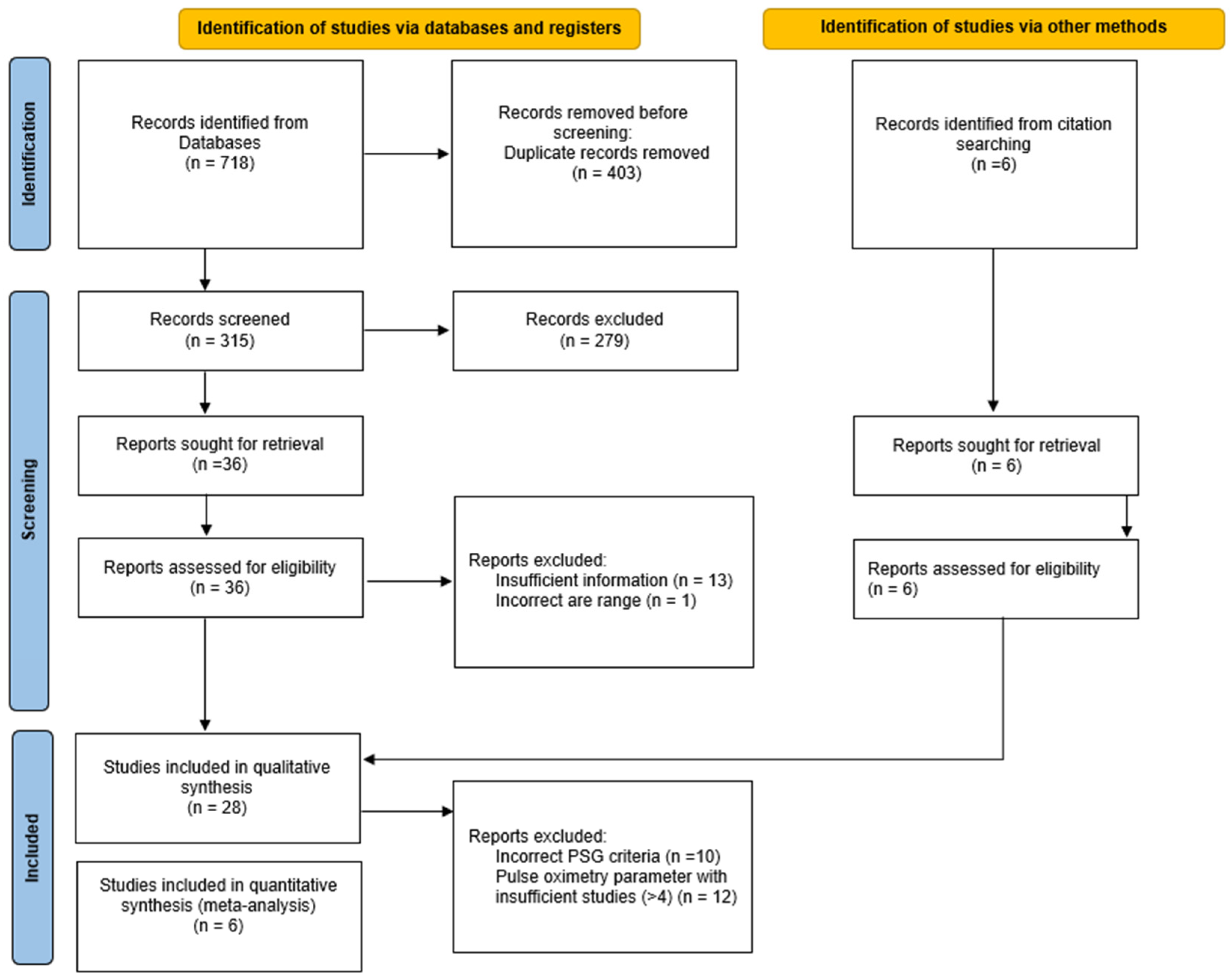
2.3. Study Selection and Data Extraction

Two researchers (M.L.B. and S.I.P.) independently performed all selection, data extraction, and quality assessment steps. Disagreements were resolved by discussion. Where a resolution was not possible, a third reviewer (G.A.B.) was consulted.
Titles and abstracts were screened to exclude duplicates and irrelevant records. The full texts of the remaining studies were evaluated according to the eligibility criteria. The following information were extracted from the full texts included in the systematic review using a predesigned form: study setting, sample baseline characteristics, oximetry device, oximetry parameters, PSG device, PSG criteria and scoring rules for the diagnosis of OSA, prevalence, sensitivity, specificity, positive predictive value (PPV), negative predictive value (NPV), positive likelihood ratio, negative likelihood ratio, true positives, false positives, true negatives and false negatives. Where the required data were not found, we deduced the values whenever the data permitted. The study authors were not contacted about missing data.

2.4. Assessment of Quality of Studies

The quality of the studies included in the systematic review was assessed using the Quality Assessment of Diagnostic Accuracy Studies (QUADAS-2) tool [21], which comprises the four following key domains: patient selection, index test, reference standard, flow and timing.
“Patient selection” describes the included patients (prior testing, presentation, intended use of index test and setting). “Index test” and “reference standard” describe how they were conducted and interpreted. “Flow and timing” describes the time interval and any interventions between index test and reference standard.
All domains are assessed in terms of risk of bias (low, unclear, or high) and the first three domains are assessed in terms of concerns regarding applicability as well (low, unclear, or high).

2.5. Data Synthesis

Bivariate meta-analysis was used to estimate sensitivity and specificity. PPV and NPV were calculated as well. The summary receiver operator characteristic (SROC) curves were plotted, and the 95% confidence region and the 95% prediction region were added.
Stata (StataCorp. 2015. Stata Statistical Software: Release 14.1 College Station, TX, USA: StataCorp LP) and Review Manager 5.3 are the statistical software used for the data process. Stata was used to determine accuracy and SROC parameters while Review Manager 5.3 was used to design forest plots and SROC graphs.

2.6. Investigations of Heterogeneity

The confidence region represents uncertainty in the overall average value due to sampling variability. The prediction region represents variation between study heterogeneity. The 95% prediction region is much larger than the 95% confidence region if heterogeneity is high. No equivalent to the I2 statistic is available for the meta-analysis of the diagnostic test accuracy. To estimate I2 statistics for sensitivity and specificity separately, we overestimated the degree of heterogeneity [22].
Stata (StataCorp. 2015. Stata Statistical Software: Release 14.1 College Station, TX, USA: StataCorp LP) was used for computing the 95% confidence and 95% prediction regions.

3. Results

Our search resulted in 737 total records by searching the electronic databases. After removing duplicates (n = 403) and irrelevant studies (n = 292) through screening titles and abstracts, the full texts of the remaining 42 records were retrieved for more detailed evaluation. Twenty-eight articles were finally included in the systematic review [13,14,15,23,24,25,26,27,28,29,30,31,32,33,34,35,36,37,38,39,40,41,42,43,44,45,46,47] and six in the meta-analysis [24,26,30,33,34,39].
The flow chart showing the selection process for this study is shown in Figure 1.
Reasons for exclusions of full text articles from the systematic review and from the meta-analysis are listed, respectively, in Tables S3 and S4.

3.1. Characteristics of Studies

The detailed characteristics of the 28 studies included in this systematic review are presented in Table 1.
The studies were conducted in 13 different countries including six in the USA [14,24,26,30,34,45]; three each in Spain [23,25,38], China [27,28,37], and UK [13,31,41]; two each in Taiwan [15,33], Italy [29,43], and Belgium [40,46]; and one each in Japan [39], Australia [35], Canada [36], Argentina [42], Thailand [44], and Greece [47]. One study by Hornero et al. [32] included findings from 13 pediatric sleep laboratories around the world.
All the studies compared oximetry and PSG as the reference standard simultaneously, but two studies, those by Alvarez et al. [25] and Jonas et al. [35], had a gap of, respectively, several weeks and 3–4 months between the two tests. A prospective cross-sectional or cohort design was the most commonly adopted design, but ten studies were retrospective [13,15,27,28,33,35,38,40,46,47]. Among the 28 studies included, the mean age of children was 7.47 years. Sample size varied from 32 to 4191 participants. The sample population consisted mainly of children referred to sleep centers for suspected OSA or sleep-disordered breathing; two studies were limited to pediatric patients affected by obesity [23,34], one by Down syndrome [31], and one by laryngomalacia [46]. Fourteen studies excluded patients with serious comorbidities, such as craniofacial abnormalities, neuromuscular disease, syndromic diseases, chronic pulmonary diseases, or congenital heart disease [15,24,25,27,28,29,33,36,38,40,42,43,44,47]. The remaining studies failed to report the type of comorbidities.
Nine different parameters to define abnormal oximetry recordings were found; the most commonly used were a 3% oxygen desaturation index (ODI3) and a McGill score [12] > 1. Twelve out of 28 studies adopted the 2007 rules of the American Academy of Sleep Medicine for the Scoring of Sleep and Associated Events as the scoring criteria.

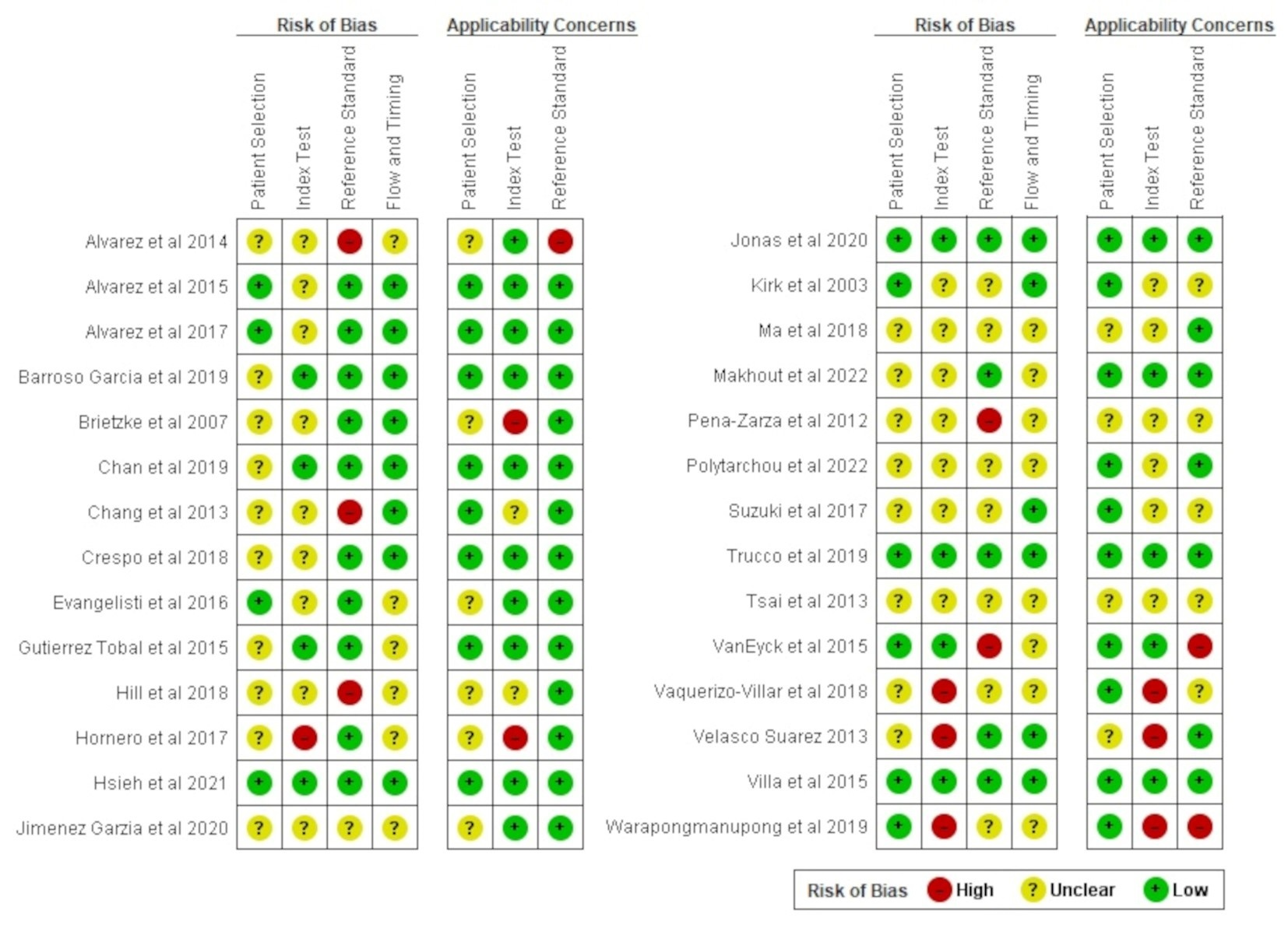
3.2. Assessment of Quality of Studies

The results of the QUADAS-2 assessment are summarized in Figure 2.
In all four domains, nearly half of studies had an unclear risk of bias, reflecting a tendency toward poor reporting. An unclear risk for bias in the patient selection domain was found in almost 60% of studies because it was not clear if an all consecutive or a random sample of eligible children with suspected OSA were collected. There could also be a difference between children included in the primary studies and participants targeted by the review question for the presence of comorbidities, leading to applicability concerns as well.
Approximately 15–20% of the studies were judged as having a high risk of bias in the index test and reference standard domains, mainly due to the lack of blinding to PSG results when interpreting the oximetry recordings (and vice versa). The interpretation of the index test could have also been influenced by the subjectivity of the interpretation and the order of testing for those studies using oximetry as a standalone method (it is not clear if the index test has been always conducted and interpreted prior to the reference standard). Variations in oximetry recording execution or interpretation, together with an unthorough reporting of the criteria for good quality sleep studies may also affect estimates of the diagnostic accuracy, leading to concerns regarding applicability.
Regarding the flow and timing domain, results of the index test and reference standard were mostly collected from the same patient at the same time. Only two studies reported a delay between index test and reference standard, involving a high risk of bias due to potential temporal changes in a chronic disease such as OSA. A verification bias may also have occurred because not all study groups received confirmation of the diagnosis by the same reference standard cut-off, thus leading to an unclear risk of bias in almost 50% of studies.

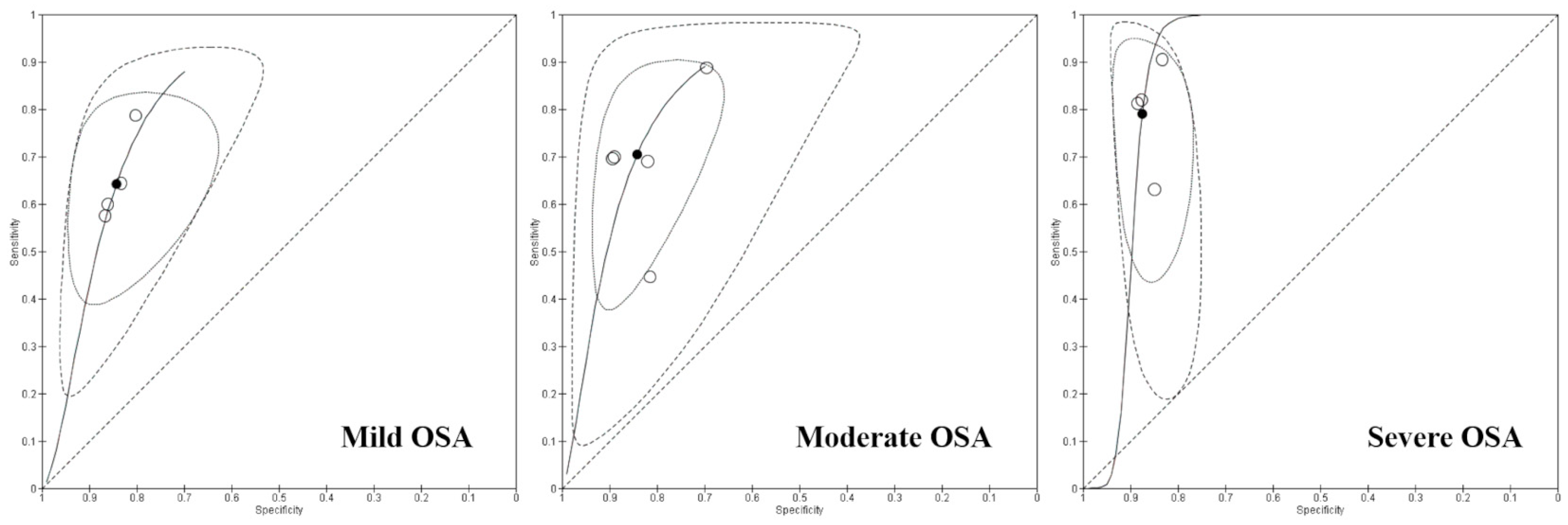
3.3. Data Synthesis and Findings

Only ODI3 were tested in at least four studies and qualified for the meta-analysis for the summary diagnostic accuracy measures (Table 2) [24,26,30,33,34,39]. In four studies, oximetry data were acquired from the reference PSG [24,26,30,34], while in two studies, oximetry data were obtained from home respiratory polygraphy [33,39].
Description of summary estimates for six oximetry-based studies using ODI3 are shown in Table 3. Sensitivity and specificity increase with pathological severity. Forest plots and SROC curves are shown in Figure 3 and Figure 4.

3.4. Heterogeneity of Included Studies

As evidenced by the different sizes of the confidence and prediction regions SROC curves, the heterogeneity of the included studies was high, mostly for moderate OSA (Figure 4). The extensive surface of the prediction regions SROC curves was probably explained by the small number of studies and the large OSA prevalence range in the study populations (ranging between 42.6% and 83% in mild OSA, between 37.4% and 54.8% in moderate OSA, and between 21.3% and 53.8% in severe OSA).

4. Discussion

The combined use of systematic reviews and meta-analyses is increasingly adopted in the field of diagnostic test accuracy. This approach has the advantage of deriving more generalizable summary estimates of the diagnostic accuracy of an index test by combining data from multiple primary studies, which should provide a more robust foundation for guidelines, recommendation, and clinical decision-making. An additional advantage is the ability to identify areas where data are either insufficient or inconsistent, thereby guiding future research efforts to address these gaps. This process facilitates that diagnostic procedures are based on the most robust scientific evidence available.
Already part of the standard PSG, oximetry is a simple, widely available, and non-invasive method of measuring blood oxygen saturation and recording blood volume changes in tissue using the photoplethysmographic signals. Three clusters of desaturation events on an overnight oximetry trend graph have been demonstrated by Brouillette et al. [12] to yield a 97% PPV. Another study by Nixon et al. [48] showed that the McGill score accurately estimates the severity of OSA and can also play an important role in planning for adenotonsillectomy. The McGill score is a pediatric screening tool for OSA, based on oxygen saturation (SpO2) monitoring. It evaluates desaturation events, the desaturation index, and the time spent with SpO2 below critical thresholds. The score ranges from 1 to 4, with higher scores indicating a greater likelihood of OSA. However, these interpretations are complex, time-consuming, and may also vary regarding the exact number of clusters. Another parameter is ODI, i.e., the number of times per hour of sleep that SpO2 falls by a defined level from baseline (e.g., ODI4 indicates a ≥4% reduction in SpO2). Notably, due to the variability in definitions and cutoff values for ODI, there is currently no consensus on which the threshold is the most accurate or on the optimal definition of abnormal values in children. Tsai et al. [15] demonstrated that an ODI4 of >2.05 events/h exhibits strong diagnostic performance, with a sensitivity of 78% and a specificity of 89%. However, it has been observed that this threshold may overlap with ODI values observed in healthy children [49]. Chang et al. [28] recommended the use of a combination of clinical symptoms (such as apnea, mouth breathing, and restlessness) along with the ODI3 as a screening tool for pediatric OSA. Moreover, the standard deviation of SpO2, representing the variability of SpO2 from the baseline, was shown to correlate strongly with the AHI, with a higher standard deviation indicating more severe OSA and a standard deviation greater than 1.06 providing strong evidence of moderate to severe OSA [44]. Conventional oximetry parameters were also recently joined with features from other data elaboration approaches in order to improve their diagnostic performance, including statistical parameters, power spectral density, and nonlinear features [11].
A previous systematic review aimed to qualitatively summarize abnormal conventional oximetry patterns that predict pediatric OSA and to identify nocturnal oximetry abnormalities predicting the response to treatment interventions for OSA and potential treatment complications [17]. The study concluded that at least three clusters of desaturation events, and a minimum of three SpO2 drops below 90% in a nocturnal oximetry recording with a minimum duration of 6 h, are indicative of moderate-to-severe OSA. Furthermore, children without clusters of desaturation events exhibit a low risk of major respiratory complications following adenotonsillectomy [17]. However, the review did not include any quantitative analyses (i.e., meta-analysis).
Another systematic review tried to qualitatively investigate the diagnostic performance of portable single- or two-channel sleep monitors compared with PSG for the diagnosis of pediatric OSA [9]. As a further step of this study, a meta-analysis was also carried out, pooling data from studies using monitoring devices adopted by at least four studies, including oximetry, as a part of the reference PSG or as a standalone method, based on statistical classifier models for the AHI cut-off of 5 events/h. It was concluded that oximetry based on machine learning algorithms may provide a simple and effective alternative to PSG for the diagnosis of moderate-severe pediatric OSA with a pooled sensitivity of 74% (95% CI: 66–80%) and a pooled specificity of 90% (95% CI: 85–94%) [9]. Notably, the meta-analysis was carried out only for the AHI threshold of 5 events/h (i.e., the currently accepted diagnostic threshold for moderate-to-severe OSA in children), as there was an insufficient number of studies reporting data on 1 and 10 events/h.
The present study was specifically focused on the diagnostic accuracy of oximetry, as a part of the reference PSG or as a standalone method, compared to PSG for all the AHI diagnostic thresholds of pediatric OSA currently recommended by the American Academy of Sleep Medicine. To our knowledge, this is the first study to perform a meta-analysis of diagnostic accuracy for each AHI cut-off of 1, 5, and 10 events/h. A better understanding of the diagnostic performance of oximetry parameters could potentially increase accessibility to the diagnosis of pediatric OSA and support the value and usefulness of oximetry as a simplified screening modality. A further advantage could be to streamlining the referral procedure to sleep medicine specialists, reducing long waiting lists by establishing priorities for children already referred to specialists and improving the monitoring of pediatric OSA. Conducting a systematic review and meta-analysis in this field enables us to combine data from different studies (i.e., from a large sample size) in order to identify relevant findings for clinical practice and to appraise the methodological limitations of each primary study in order to guide future research.
The findings of the present study indicate that, as the AHI threshold increases, the sensitivity of ODI3 also increases, with the highest sensitivity being observed at AHI ≥ 10 (79% [95% CI 73; 84]), meaning that 79% of children with severe OSA will have a positive ODI3 at overnight oximetry. This indicates that the ability of the test to correctly identify children with OSA improves as the severity of the condition (measured by PSG-derived AHI) increases. The specificity of ODI3 is consistently high across all AHI thresholds, ranging from 84% at AHI ≥ 1 to 88% at AHI ≥ 10, meaning that 88% of children without severe OSA will have a negative ODI3 at overnight oximetry. This suggests that the test is reliable in correctly classifying children without the disease, particularly at higher AHI thresholds. A highly specific test means that there are few false positive results. In this context, it is important to observe that it would not be desirable to use a test with low specificity for the screening of pediatric OSA, since many children without the disease will screen positive, and potentially undergo unnecessary and expensive diagnostic procedures such as PSG.
A further finding of the present study is that PPV decreases as the AHI threshold increases, with the highest PPV being observed at AHI ≥ 1 (91% [95% CI 88; 93]), meaning that 91% of children who have a positive ODI3 at overnight oximetry will actually have OSA. The PPV decrease indicates that while the test is sensitive at higher AHI thresholds, the chance that children who result positive with the index test really have OSA diminishes slightly as the threshold increases. This could be ascribed to the fact that predictive values are determined by the sensitivity and specificity of the test, but it also depends on the prevalence of the disease in the population being tested, and in the articles included in our study, the prevalence decreases as the OSA severity increases.
Conversely, the NPV increases with higher AHI thresholds, reaching its peak at AHI ≥ 10 (89% [95% CI 87; 91]), meaning that 89% of children who have negative ODI3 at overnight oximetry will not actually have OSA. This trend indicates that a negative test result is increasingly likely to be correct as the severity of OSA increases. This is important to provide an indication of the ability to rule out more severe forms of OSA, reducing unnecessary follow-ups and diagnostic procedures.
Our findings can be considered consistent with the trends observed in the literature, such as in Kaditis et al. [17], who reported a similar range of sensitivity and specificity for ODI4 when assessing moderate-to-severe OSA in children, and in the study by Wu et al. [8], in which oximetry yielded the highest specificity against two validated questionnaires used for the screening of pediatric OSA. However, the pooled sensitivity and specificity in our results appears slightly lower compared to those by Gao et al. [9], which reported higher values for moderate pediatric OSA. This difference might reflect variations in the population characteristics or the specific methodological approaches used across studies.
This comparison suggests that while oximetry parameters, particularly ODI3, are useful for identifying pediatric OSA, sensitivity and specificity can vary significantly depending on the AHI threshold and the study population, highlighting the importance of context in interpreting these diagnostic tools.
There are some limitations in this systematic review that warrant consideration. A substantial proportion of the included primary studies were conducted in sleep centers or involved patients presenting with snoring or respiratory symptoms, which may have inflated the observed prevalence of OSA within the sample. Additionally, the heterogeneity in age, health status, and ethnicity across the primary studies could restrict the generalizability of the findings to the broader pediatric population. Furthermore, many studies did not explicitly report whether consecutive or random samples of eligible children were selected, raising concerns about potential selection biases and the applicability of the results. The lack of blinding between oximetry and PSG results in several studies may also have introduced bias in the interpretation of findings. Other notable limitations include the lack of information regarding the influence of age on the accuracy of overnight oximetry for detecting pediatric OSA, as well as the absence of clear data on how many children were excluded from analysis due to technical issues or other factors such as artifacts. Variability in the implementation and interpretation of oximetry, combined with inconsistent reporting of criteria for high-quality sleep studies, further complicates the precision of the diagnostic estimates. To enhance clinical relevance, future research should focus on standardizing oximetry execution and interpretation methods, while also addressing the methodological shortcomings identified in the primary studies included in this review.

5. Conclusions

Overnight oximetry is a suitable screening test for pediatric OSA due to its low cost, ease of use, and acceptability by both patients and practitioners. The findings of the present systematic review and meta-analysis suggest that ODI3 displays a satisfactory diagnostic performance in detecting pediatric OSA, which increases with the severity of the disease. Notably, the ability to correctly classify children without the disease increases at higher AHI thresholds, thus reducing the need for potentially unnecessary and expensive diagnostic procedures. This suggests that overnight oximetry is particularly effective at ruling out OSA in children who test negative.
Further well-designed studies are necessary to integrate overnight oximetry into evidence-based clinical practice and to establish the optimal thresholds for defining abnormal recordings. Future research should explore the influence of age on the diagnostic accuracy of overnight oximetry for pediatric OSA detection, while also standardizing the reporting of oximetry feasibility. Particular attention should be given to dropout rates due to technical challenges or artifacts, to ensure a more consistent and reliable application in clinical settings.

Supplementary Materials

The following supporting information can be downloaded at: https://www.mdpi.com/article/10.3390/app142210208/s1, Table S1: Adherence to PRISMA statement; Table S2: Combinations of keywords used during electronic literature searches (last updated search 2024/01); Table S3: List of excluded studies with reasons for qualitative synthesis; Table S4: List of excluded studies with reasons for quantitative synthesis. Reference [50] are cited in the supplementary materials.

Author Contributions

Conceptualization, G.A.-B. and S.I.P.; methodology, A.F.; formal analysis, A.F.; investigation, M.L.B. and S.I.P.; writing—original draft preparation, S.I.P.; writing—review and editing, M.L.B.; supervision, P.G. and C.P. All authors have read and agreed to the published version of the manuscript.

Funding

This research received no external funding.

Institutional Review Board Statement

Not applicable.

Informed Consent Statement

Not applicable.

Data Availability Statement

The data presented in this study are available on request from the corresponding author.

Conflicts of Interest

The authors declare no conflicts of interest.

References

  1. American Thoracic Society. Standards and indications for cardiopulmonary sleep studies in children. Am. J. Respir. Crit. Care Med. 1996, 153, 866–878. [Google Scholar] [CrossRef] [PubMed]
  2. Sateia, M.J. International classification of sleep disorders-third edition: Highlights and modifications. Chest 2014, 146, 1387–1394. [Google Scholar] [CrossRef] [PubMed]
  3. Marcus, C.L.; Brooks, L.J.; Ward, S.D.; Draper, K.A.; Gozal, D.; Halbower, A.C.; Jones, J.; Lehmann, C.; Schechter, M.S.; Sheldon, S.; et al. Diagnosis and management of childhood obstructive sleep apnea syndrome. Pediatrics 2012, 130, e714–e755. [Google Scholar] [CrossRef] [PubMed]
  4. Kaditis, A.G.; Alvarez, M.L.A.; Boudewyns, A.; Alexopoulos, E.I.; Ersu, R.; Joosten, K.; Larramona, H.; Miano, S.; Narang, I.; Trang, H.; et al. Obstructive sleep disordered breathing in 2- to 18-year-old children: Diagnosis and management. Eur. Respir. J. 2016, 47, 69–94. [Google Scholar] [CrossRef]
  5. de Luca Canto, G.; Singh, V.; Major, M.P.; Witmans, M.; El-Hakim, H.; Major, P.W.; Flores-Mir, C. Diagnostic capability of questionnaires and clinical examinations to assess sleep-disordered breathing in children: A systematic review and meta-analysis. J. Am. Dent. Assoc. 2014, 145, 165–178. [Google Scholar] [CrossRef]
  6. Michelet, D.; Julien-Marsollier, F.; Vacher, T.; Bellon, M.; Skhiri, A.; Bruneau, B.; Fournier, J.; Diallo, T.; Luce, V.; Brasher, C.; et al. Accuracy of the sleep-related breathing disorder scale to diagnose obstructive sleep apnea in children: A meta-analysis. Sleep Med. 2019, 54, 78–85. [Google Scholar] [CrossRef]
  7. Incerti Parenti, S.; Fiordelli, A.; Bartolucci, M.L.; Martina, S.; D’Antò, V.; Alessandri-Bonetti, G. Diagnostic accuracy of screening questionnaires for obstructive sleep apnea in children: A systematic review and meta-analysis. Sleep Med. Rev. 2021, 57, 101464. [Google Scholar] [CrossRef]
  8. Wu, C.-R.; Tu, Y.-K.; Chuang, L.-P.; Gordon, C.; Chen, N.-H.; Chen, P.-Y.; Hasan, F.; Kurniasari, M.D.; Susanty, S.; Chiu, H.-Y. Diagnostic meta-analysis of the Pediatric Sleep Questionnaire OSA-18 and pulse oximetry in detecting pediatric obstructive sleep apnea syndrome. Sleep Med. Rev. 2020, 54, 101355. [Google Scholar] [CrossRef]
  9. Gao, X.; Li, Y.; Xu, W.; Han, D. Diagnostic accuracy of level IV portable sleep monitors versus polysomnography for pediatric obstructive sleep apnea: A systematic review and meta-analysis. Sleep Med. 2021, 87, 127–137. [Google Scholar] [CrossRef]
  10. de Luca Canto, G.; Pachêco-Pereira, C.; Aydinoz, S.; Major, P.W.; Flores-Mir, C.; Gozal, D. Diagnostic capability of biological markers in assessment of obstructive sleep apnea: A systematic review and meta-analysis. J. Clin. Sleep Med. 2015, 11, 27–36. [Google Scholar] [CrossRef]
  11. Gutiérrez-Tobal, G.C.; Álvarez, D.; Kheirandish-Gozal, L.; del Campo, F.; Gozal, D.; Hornero, R. Reliability of machine learning to diagnose pediatric obstructive sleep apnea: Systematic review and meta-analysis. Pediatr. Pulmonol. 2022, 57, 1931–1943. [Google Scholar] [CrossRef] [PubMed]
  12. Brouillette, R.T.; Morielli, A.; Leimanis, A.; Waters, K.A.; Luciano, R.; Ducharme, F.M. Nocturnal pulse oximetry as an abbreviated testing modality for pediatric obstructive sleep apnea. Pediatrics 2000, 105, 405–412. [Google Scholar] [CrossRef] [PubMed]
  13. Trucco, F.; Rosenthal, M.; Bush, A.; Tan, H.-L. The McGill score as a screening test for obstructive sleep disordered breathing in children with co-morbidities. Sleep Med. 2020, 68, 173–176. [Google Scholar] [CrossRef] [PubMed]
  14. Brietzke, S.E.; Katz, E.S.; Roberson, D.W. Pulse transit time as a screening test for pediatric sleep-related breathing disorders. Arch. Otolaryngol. Head Neck Surg. 2007, 133, 980–984. [Google Scholar] [CrossRef]
  15. Tsai, C.-M.; Kang, C.-H.; Su, M.-C.; Lin, H.-C.; Huang, E.-Y.; Chen, C.-C.; Hung, J.-C.; Niu, C.-K.; Liao, D.-L.; Yu, H.-R. Usefulness of desaturation index for the assessment of obstructive sleep apnea syndrome in children. Int. J. Pediatr. Otorhinolaryngol. 2013, 77, 1286–1290. [Google Scholar] [CrossRef]
  16. Bannink, N.; Mathijssen, I.M.J.; Joosten, K.F.M. Use of ambulatory polysomnography in children with syndromic craniosynostosis. J. Craniofac. Surg. 2010, 21, 1365–1368. [Google Scholar] [CrossRef]
  17. Kaditis, A.; Kheirandish-Gozal, L.; Gozal, D. Pediatric OSAS: Oximetry can provide answers when polysomnography is not available. Sleep Med. Rev. 2016, 27, 96–105. [Google Scholar] [CrossRef]
  18. Deeks, J.J.; Wisniewski, S.; Diagnostic Test Accuracy Working Group; Bhandari, M.; Bossuyt, P.M. Guide to the contents of a Cochrane Diagnostic Test Accuracy Protocol. Cochrane Rev. 2013, 1, 1–15. [Google Scholar]
  19. McInnes, M.D.F.; Moher, D.; Thombs, B.D.; McGrath, T.A.; Bossuyt, P.M.; Clifford, T.; Cohen, J.F.; Deeks, J.J.; Gatsonis, C.; Hooft, L.; et al. Preferred Reporting Items for a Systematic Review and Meta-analysis of Diagnostic Test Accuracy Studies: The PRISMA-DTA Statement. JAMA 2018, 319, 388–396. [Google Scholar] [CrossRef]
  20. Berry, R.B.; Brooks, R.; Gamaldo, C.E.; Harding, S.M.; Lloyd, R.M.; Quan, S.F.; Troester, M.M.; Vaughn, B.V. The AASM Manual for the Scoring of Sleep and Associated Events: Rules, Terminology, and Technical Specifications. Version 2.4; American Academy of Sleep Medicine: Darien, IL, USA, 2017; pp. 1–89. [Google Scholar]
  21. Whiting, P.F.; Rutjes, A.W.S.; Westwood, M.E.; Mallett, S.; Deeks, J.J.; Reitsma, J.B.; Leeflang, M.M.G.; Sterne, J.A.C.; Bossuyt, P.M.M.; QUADAS-2 Group. QUADAS-2: A Revised Tool for the Quality Assessment of Diagnostic Accuracy Studies. Ann. Intern. Med. 2011, 155, 529–536. [Google Scholar] [CrossRef]
  22. Bossuyt, P.; Davenport, C.; Deeks, J.; Hyde, C.; Leeflang, M.; Scholten, R. Cochrane Handbook for Systematic Reviews of Diagnostic Test Accuracy Chapter 11: Interpreting results and drawing conclusions. Cochrane Rev. 2013, 1, 1–31. [Google Scholar]
  23. Álvarez, D.; Gutierrez-Tobal, G.C.; Alonso, M.L.; Teran, J.; del Campo, F.; Hornero, R. Statistical and nonlinear analysis of oximetry from respiratory polygraphy to assist in the diagnosis of Sleep Apnea in children. Annu. Int. Conf. IEEE Eng. Med. Biol. Soc. 2014, 2014, 1860–1863. [Google Scholar] [CrossRef] [PubMed]
  24. Alvarez, D.; Kheirandish-Gozal, L.; Gutierrez-Tobal, G.C.; Crespo, A.; Philby, M.F.; Mohammadi, M.; del Campo, F.; Gozal, D.; Hornero, R. Automated analysis of nocturnal oximetry as screening tool for childhood obstructive sleep apnea-hypopnea syndrome. In Proceedings of the 37th Annual International Conference of the IEEE Engineering in Medicine and Biology Society (EMBC), Milan, Italy, 25–29 August 2015; pp. 2800–2803. [Google Scholar] [CrossRef]
  25. Álvarez, D.; Alonso-Álvarez, M.L.; Gutiérrez-Tobal, G.C.; Crespo, A.; Kheirandish-Gozal, L.; Hornero, R.; Gozal, D.; Terán-Santos, J.; Del Campo, F. Automated Screening of Children With Obstructive Sleep Apnea Using Nocturnal Oximetry: An Alternative to Respiratory Polygraphy in Unattended Settings. J. Clin. Sleep Med. 2017, 13, 693–702. [Google Scholar] [CrossRef] [PubMed]
  26. Barroso-Garcia, V.; Gutierrez-Tobal, G.C.; Kheirandish-Gozal, L.; Alvarez, D.; Vaquerizo-Villar, F.; del Campo, F.; Gozal, D.; Hornero, R. Usefulness of Spectral Analysis of Respiratory Rate Variability to Help in Pediatric Sleep Apnea-Hypopnea Syndrome Diagnosis. In Proceedings of the 41st Annual International Conference of the IEEE Engineering in Medicine & Biology Society (EMBC), Berlin, Germany, 23–27 July 2019; pp. 4580–4583. [Google Scholar] [CrossRef]
  27. Chan, W.-S.; Chan, E.-T.; Ng, D.-K.; Kwok, K.-L.; Yip, A.-F.; Leung, S.-Y. McGill oximetry score to predict risk of obstructive sleep apnea in pediatric patients. Pediatr. Respirol. Crit. Care Med. 2019, 3, 36. [Google Scholar] [CrossRef]
  28. Chang, L.; Wu, J.; Cao, L. Combination of symptoms and oxygen desaturation index in predicting childhood obstructive sleep apnea. Int. J. Pediatr. Otorhinolaryngol. 2013, 77, 365–371. [Google Scholar] [CrossRef]
  29. Evangelisti, M.; Shafiek, H.; Rabasco, J.; Forlani, M.; Montesano, M.; Barreto, M.; Verhulst, S.; Villa, M.P. Oximetry in obese children with sleep-disordered breathing. Sleep Med. 2016, 27–28, 86–91. [Google Scholar] [CrossRef]
  30. Tobal, G.C.G.; Kheirandish-Gozal, L.; Alvarez, D.; Crespo, A.; Philby, M.F.; Mohammadi, M.; Del Campo, F.; Gozal, D.; Hornero, R. Analysis and classification of oximetry recordings to predict obstructive sleep apnea severity in children. In Proceedings of the 37th Annual International Conference of the IEEE Engineering in Medicine and Biology Society (EMBC), Milan, Italy, 25–29 August 2015; pp. 4540–4543. [Google Scholar] [CrossRef]
  31. Hill, C.M.; E Elphick, H.; Farquhar, M.; Gringras, P.; Pickering, R.M.; Kingshott, R.N.; Martin, J.; Reynolds, J.; Joyce, A.; Gavlak, J.C.; et al. Home oximetry to screen for obstructive sleep apnoea in Down syndrome. Arch. Dis. Child. 2018, 103, 962–967. [Google Scholar] [CrossRef]
  32. Hornero, R.; Kheirandish-Gozal, L.; Gutiérrez-Tobal, G.C.; Philby, M.F.; Alonso-Álvarez, M.L.; Álvarez, D.; Dayyat, E.A.; Xu, Z.; Huang, Y.-S.; Kakazu, M.T.; et al. Nocturnal Oximetry-based Evaluation of Habitually Snoring Children. Am. J. Respir. Crit. Care Med. 2017, 196, 1591–1598. [Google Scholar] [CrossRef]
  33. Hsieh, H.-S.; Kang, C.-J.; Chuang, H.-H.; Zhuo, M.-Y.; Lee, G.-S.; Huang, Y.-S.; Chuang, L.-P.; Kuo, T.B.-J.; Yang, C.C.-H.; Lee, L.-A.; et al. Screening Severe Obstructive Sleep Apnea in Children with Snoring. Diagnostics 2021, 11, 1168. [Google Scholar] [CrossRef]
  34. Jiménez-García, J.; Gutiérrez-Tobal, G.C.; García, M.; Kheirandish-Gozal, L.; Martín-Montero, A.; Álvarez, D.; del Campo, F.; Gozal, D.; Hornero, R. Assessment of Airflow and Oximetry Signals to Detect Pediatric Sleep Apnea-Hypopnea Syndrome Using AdaBoost. Entropy 2020, 22, 670. [Google Scholar] [CrossRef]
  35. Jonas, C.; Thavagnanam, S.; Blecher, G.; Thambipillay, G.; Teng, A.Y. Comparison of nocturnal pulse oximetry with polysomnography in children with sleep disordered breathing. Sleep Breath. 2020, 24, 703–707. [Google Scholar] [CrossRef] [PubMed]
  36. Kirk, V.G.; Bohn, S.G.; Flemons, W.W.; Remmers, J.E. Comparison of home oximetry monitoring with laboratory polysomnography in children. Chest 2003, 124, 1702–1708. [Google Scholar] [CrossRef] [PubMed]
  37. Ma, J.-R.; Huang, J.-J.; Chen, Q.; Wu, H.-T.; Xiao, K.-L.; Zhang, Y.-T. Value of pulse oximetry watch for diagnosing pediatric obstructive sleep apnea/hypopnea syndrome. Acta Oto-Laryngol. 2018, 138, 175–179. [Google Scholar] [CrossRef]
  38. Peña-Zarza, J.A.; de Torres, B.O.-R.; Gil-Sanchez, J.A.; Figuerola-Mulet, J. Utility of the pediatric sleep questionnaire and pulse oximetry as screening tools in pediatric patients with suspected obstructive sleep apnea syndrome. Sleep Disord. 2012, 2012, 819035. [Google Scholar] [CrossRef]
  39. Suzuki, M.; Furukawa, T.; Sugimoto, A.; Kotani, R.; Hosogaya, R. Comparison of diagnostic reliability of out-of-center sleep tests for obstructive sleep apnea between adults and children. Int. J. Pediatr. Otorhinolaryngol. 2017, 94, 54–58. [Google Scholar] [CrossRef]
  40. Van Eyck, A.; Lambrechts, C.; Vanheeswijck, L.; Van Hoorenbeeck, K.; Haentjens, D.; Boudewyns, A.; De Winter, B.Y.; Van Gaal, L.; De Backer, W.; Verhulst, S.L. The role of nocturnal pulse oximetry in the screening for obstructive sleep apnea in obese children and adolescents. Sleep Med. 2015, 16, 1409–1412. [Google Scholar] [CrossRef]
  41. Vaquerizo-Villar, F.; Alvarez, D.; Kheirandish-Gozal, L.; Gutierrez-Tobal, G.C.; Barroso-Garcia, V.; Crespo, A.; del Campo, F.; Gozal, D.; Hornero, R.; Vaquerizo-Villar, F.; et al. Utility of bispectrum in the screening of pediatric sleep apnea-hypopnea syndrome using oximetry recordings. Comput. Methods Programs Biomed. 2018, 156, 141–149. [Google Scholar] [CrossRef]
  42. Suárez, C.T.V.; Turienzo, J.M.F.; Len, F.; Mansilla, E. Pulse oximetry recording in children with adenotonsillar hypertrophy: Usefulness in the diagnosis of obstructive sleep apnea syndrome. Arch. Argent. Pediatr. 2013, 111, 196–201. [Google Scholar] [CrossRef]
  43. Villa, M.P.; Pietropaoli, N.; Supino, M.C.; Vitelli, O.; Rabasco, J.; Evangelisti, M.; Del Pozzo, M.; Kaditis, A.G. Diagnosis of Pediatric Obstructive Sleep Apnea Syndrome in Settings With Limited Resources. JAMA Otolaryngol. Head Neck Surg. 2015, 141, 990–996. [Google Scholar] [CrossRef]
  44. Warapongmanupong, S.; Preutthipan, A. Can standard deviation of overnight pulse oximetry be used to screen childhood obstructive sleep apnea. Int. J. Pediatr. Otorhinolaryngol. 2019, 119, 27–31. [Google Scholar] [CrossRef]
  45. Crespo, A.; Álvarez, D.; Kheirandish-Gozal, L.; Gutiérrez-Tobal, G.C.; Cerezo-Hernández, A.; Gozal, D.; Hornero, R.; del Campo, F. Assessment of oximetry-based statistical classifiers as simplified screening tools in the management of childhood obstructive sleep apnea. Sleep Breath. 2018, 22, 1063–1073. [Google Scholar] [CrossRef] [PubMed]
  46. Makhout, S.; Boudewyns, A.; Van Hoorenbeeck, K.; Verhulst, S.; Van Eyck, A. Nocturnal pulse oximetry as a possible screening method for obstructive sleep apnea in infants with laryngomalacia. Sleep Med. 2022, 90, 91–95. [Google Scholar] [CrossRef] [PubMed]
  47. Polytarchou, A.; Ohler, A.; Moudaki, A.; Koltsida, G.; Kanaka-Gantenbein, C.; Kheirandish-Gozal, L.; Gozal, D.; Kaditis, A.G. Nocturnal oximetry parameters as predictors of sleep apnea severity in resource-limited settings. J. Sleep Res. 2023, 32, e13638. [Google Scholar] [CrossRef]
  48. Nixon, G.M.; Kermack, A.S.; Davis, G.M.; Manoukian, J.J.; Brown, K.A.; Brouillette, R.T. Planning adenotonsillectomy in children with obstructive sleep apnea: The role of overnight oximetry. Pediatrics 2004, 113, e19–e25. [Google Scholar] [CrossRef]
  49. Urschitz, M.S.; Wolff, J.; Von Einem, V.; Urschitz-Duprat, P.M.; Schlaud, M.; Poets, C.F. Reference values for nocturnal home pulse oximetry during sleep in primary school children. Chest 2003, 123, 96–101. [Google Scholar] [CrossRef]
  50. Page, M.J.; McKenzie, J.E.; Bossuyt, P.M.; Boutron, I.; Hoffmann, T.C.; Mulrow, C.D.; Shamseer, L.; Tetzlaff, J.M.; Akl, E.A.; Brennan, S.E.; et al. The PRISMA 2020 statement: An updated guideline for reporting systematic reviews. BMJ 2021, 372, n71. [Google Scholar] [CrossRef]
Figure 1. Flowchart of the study selection process.
Applsci 14 10208 g001
Figure 2. Quality appraisal of the studies included in the systematic review using the QUADAS-2 tool [13,14,15,23,24,25,26,27,28,29,30,31,32,33,34,35,36,37,38,39,40,41,42,43,44,45,46,47].
Applsci 14 10208 g002
Figure 3. Forest plot of studies adopting ODI3 as parameter to define abnormal oximetry recordings. CI, confidence interval; FN, false negative; FP, false positive; TN, true negative; TP, true positive [24,26,30,33,34,39].
Applsci 14 10208 g003
Figure 4. SROC curves of studies based on ODI3 as oximetry parameter: 95% confidence region (full line area), 95% prediction region (dashed line area), studies (circles), summary point (black dot) and summary curve (full line).
Applsci 14 10208 g004
Table 1. Characteristics of studies included in this systematic review.
NFirst Author, Year of PublicationAgePopulationSample
Size
PO DevicePO CriteriaPSG DevicePSG Criteria
(Scoring Rules)
1Alvarez et al., 2014 [23]5.30 ± 2.55 ySuspected OSA50Part of at-home RP (eXim Apnea Polygraph, Bitmed, Sibel S.A., Barcelona, Spain)Avg SpO2
Min SpO2
ODI3
CT
Statistical features
Nonlinear features
Attended PSG (Deltamed Coherence
3NT version 3.0; Diagniscan, S.A.U., Group Werfen, Paris, France)
AHI ≥ 3/h
(AASM 2007)
2Alvarez et al., 2015 [24]6.95 ± 3.55 ySuspected OSA176Part of reference PSGODI3
Statistical features
Nonlinear features
Spectral features
Digital PSG (Polysmith; Nihon Kohden
America Inc., Irvine, CA, USA)
AHI ≥ 1/h
AHI ≥ 3/h
AHI ≥ 5/h
(AASM 2007)
3Alvarez et al., 2017 [25]3–13 ySuspected OSA50Part of at-home RP (eXim Apnea Polygraph, Bitmed, Sibel S.A., Barcelona, Spain)Avg SpO2
Min SpO2
ODI3
CT
Statistical features
Attended PSG (Deltamed Coherence
3NT version 3.0; Diagniscan, S.A.U., Group Werfen, Paris, France)
OAHI ≥ 1/h
OAHI ≥ 3/h
OAHI ≥ 5/h
(AASM 2007)
4BarrosoGarcia et al., 2019 [26]6 ySuspected OSA376Part of reference PSGODI3
Spectral features
Digital PSG
(Polysmith; Nihon Kohden America Inc., Irvine, CA, USA)
AHI ≥ 1/h
AHI ≥ 5/h
AHI ≥ 10/h
(AASM 2007)
5Brietzke et al., 2007 [14]2–16 ySleep-related breathing disorders59Ambulatory device (Stowood Scientific Instruments, Oxford, England)PTTPSG (Bio-logic
Systems Corporation, Mundelein, IL, USA)
AHI > 1/h
AHI > 3/h
AHI > 5/h
(NIH Manual 1968)
6Chan et al., 2019 [27]10.86 ± 4.22 yHabitual snoring573Part of reference
PSG
McGill
Nadir SpO2
Ambulatory PSG (Siesta,
Compumedics)
AHI > 1/h
AHI > 5/h
(AASM 2007)
7Chang et al., 2013 [28]1.8–12.8 ySuspected OSA141Part of reference
PSG
ODI3Standard PSG (Alice 5, Philips,
Respironics, USA)
AHI > 5/h
(AASM 2007)
8Crespo et al., 2018 [45]1–13 ySuspected OSA176Part of reference
PSG
Statistical features ODI3Digital PSG (Polysmith; Nihon Kohden
America Inc., CA, USA)
AHI ≥ 1/h
AHI ≥ 3/h
AHI ≥ 5/h
(AASM 2012)
9Evangelisti et al., 2016 [29]5.54–10.33 y
4.71–9 y
Suspected OSA (obese)
Suspected OSA (non-obese)
128
120
Home nocturnal PO (Nonin Medical, Plymouth, MN, USA)McGill
ODI3
Laboratory PSG
(Grass Heritage polygraph; Natus Neurology IncorporatedeGrass
Products, Warwick, RI, USA)
AHI ≥ 1/h
1/h ≤ AHI < 10/h
AHI ≥ 5/h
(AASM 2007)
10Gutierrez-Tobal et al., 2015 [30]7.0 ± 3.6 ySuspected OSA176Part of reference
PSG
ODI3
MLP
Overnight PSGAHI ≥ 1/h
AHI ≥ 5/h
(AASM 2012)
11Hill et al., 2018 [31]0.5–6 yDown syndrome161Home nocturnal PO Masimo Radical 7 device (Masimo, Irvine, CA, USA)ODI3
Avg SpO2
Min SpO2
CT
Home CP
(SOMNOtouch
device; Somnomedics, Randersacker, Germany)
OAHI ≥ 5
(AASM 2012)
12Hornero et al., 2017 [32]6.7 ± 4.4 yHabitual snoring4191Part of reference
PSG
Neural network analysisNocturnal PSGAHI ≥ 1/h
AHI ≥ 5/h
(AASM 2012)
13Hsieh et al., 2021 [33]6–10 yOSA39Home nocturnal PO (3100WristOx, Nonin Medical, Inc., Minneapolis, MN, USA)ODI3Attended night PSG (Nicolet
Biomedical Inc., Madison, WI, USA)
AHI ≥ 2/h
(AASM 2012)
14Jimenez-Garzia et al., 2020 [34]3–9 ySuspected OSA390Part of reference
PSG
ODI3
Nonlinear analysis
Spectral analysis
Digital PSG
(Nihon Kohden America Inc., Irvine, CA, USA)
AHI ≥ 1/h
AHI ≥ 5/h
AHI ≥ 10/h
(AASM 2007)
15Jonas et al., 2020 [35]4.75–38.5 moSuspected OSA110Hospital or at home POMcGillLaboratory PSG (Compumedics, Melbourne, Australia)MOAHI ≥ 1
MOAHI ≥ 5
(NR)
16Kirk et al., 2003 [36]4–18 ySuspected OSA58At home portable monitor (SnoreSat, SagaTech Electronics, Calgary, AB, Canada)DI
Laboratory PSG
(Sandman NT; Nellcor Puritan Bennett; Ottawa, ON, Canada)
AHI > 5/h
(ATS 1996)
17Ma et al., 2018 [37]4–16 ySnoring32PO Watch
(CloudCare Healthcare Co., Ltd.,
Chengdu, China)
ODI4Laboratory PSGAHI > 1/h
AHI > 5/h
AHI > 10/h
AHI > 15/h
AHI > 20/h
(AASM 2012)
18Makhout et al., 2022 [46]3.72 ± 0.26 yLaryngomalacia53Nocturnal PO during PSGMcGillLaboratory PSGOAHI ≥ 2/h
(AASM 2007)
19PenaZarza et al., 2012 [38]2–15 ySuspected OSA98Home nocturnal PO (3DI Pulsox Minolta)McGillHome Nocturnal Polygraphy (Sleepscreen, Viasys Healthcare GmBH, Hoechberg, Leibnizstr, Germany)AHI ≥ 3/h
AHI ≥ 5/h
AHI ≥ 10/h
(AASM 2007)
20Polytarchou et al., 2022 [47]3.9–9.1 ySuspected OSA98Part of reference
PSG
ODI3
McGill
Laboratory PSG (EMBLA S4500 System)AHI ≥ 5/h
(AASM 2012)
21Suzuki et al., 2017 [39]7 ± 2.6 ySuspected OSA119Home nocturnal PO (PMP-200GplusX, Philips Respironics, Pittsburgh,
PA, USA)
ODI3Type 1 laboratory overnight
PSG (Alice 6, Philips Respironics, Pittsburgh, PA, USA)
AHI < 1/h
AHI < 5/h
AHI < 10/h
(AASM 2016)
22Trucco et al., 2019 [13]2.4–7.9 ySuspected OSA312Transcutaneous monitoring (CombiM monitor, Radiometer, Copenhagen, DenmarkMcGillCP (SOMNOScreen™ plus,
SOMNOmedics, Germany)
OAHI ≥ 1/h
OAHI ≥ 5/h
(NR)
23Tsai et al., 2013 [15]7.18 ± 2.57 ySuspected OSA148Part of reference
PSG
DIOvernight laboratory PSG (Sandman
EliteTM, Nellcor Puritan Bennett [Melville] Ltd., Canada)
AHI ≥ 1/h
AHI ≥ 5/h
AHI ≥ 10/h
(AASM 2007)
24VanEyck et al., 2015 [40]6–17 ySuspected OSA (obese)130Part of reference
PSG
(Xpod, Nonin, MN, USA)
ODIOvernight laboratory PSGOAHI ≥ 2/h
(AASM 2007)
25Vaquerizo-Villar et al., 2018 [41]0–13 ySuspected OSA298Part of reference
PSG
Bispectrum analysisOvernight laboratory PSG
(Polysmith; Nihon Kohden America Inc., CA, USA).
AHI ≥ 5/h
AHI ≥ 10/h
(NR)
26Velasco Suarez 2013 [42]2–16 ySuspected OSA167Part of reference
PSG. PO (NONIN 8008JFW)
Visual analysisDigital PSG (Akonic Neurotrace)AHI ≥ 1/h
(NR)
27Villa et al., 2015 [43]5.93 ± 2.97Suspected OSA (SCR-positive)236First night PO (Nonin 2500A; NoninMedical)McGillSecond night PSG (GrassHeritage; GrassTechnologies, Fort Myers, FL, USA)1/h ≤ AHI ≤ 5/h
AHI > 5/h
(AASM 2007)
28Warapongmanupong et al., 2019 [44]6.7 ± 3.2 ySnoring adenotonsillar hypertrophy457Part of reference
PSG (Masimo SET Radical-7)
SpO2 SDOvernight laboratory PSG
(Grael system)
AHI ≥ 1.5/h
AHI ≥ 5/h
(AASM 2012)
AASM, American Academy of Sleep Medicine; ATS, American Thoracic Society; Avg SpO2, average saturation; CP, cardiorespiratory polygraphy; CT, cumulative time; DI, desaturation index; Min SpO2, minimum saturation; MLP, multi-layer perceptron; NIH, National Institutes of Health; ODI3, oxygen desaturation index ≥ 3%; ODI4, oxygen desaturation index ≥ 4%; PO, pulse oximetry; PSG, polysomnography; PTT, pulse transit time; RP, respiratory polygraphy; SD, standard deviation; SpO2, oxygen saturation.
Table 2. Description of studies included in the quantitative analysis.
NFirst Author, Year of PublicationSample SizePrevalenceSeverityTrue PositiveFalse PositiveFalse NegativeTrue Negative
1Alvarez et al., 2015 [24]17683.0Mild OSA9455225
40.3Moderate OSA6332873
2Barroso Garcia et al., 2019 [26]37680.9Mild OSA1821012262
37.5Moderate OSA982543210
21.3Severe OSA653415262
3Gutierrez-Tobal et al., 2015 [30]17642.6Mild OSA59201681
40.3Moderate OSA49192286
4Hsieh et al., 2021 [33]3953.8Severe OSA193215
5Jimenez-Garzia et al., 2020 [34]39080.8Mild OSA1811013465
37.4Moderate OSA1022744217
21.3Severe OSA683815269
6Suzuki et al., 2017 [39]11954.8Moderate OSA30103643
38.5Severe OSA29111762
Table 3. Description of summary estimates for ODI3.
Number of StudiesNumber of ParticipantsSensitivity (95% Confidence Interval)Specificity (95% Confidence Interval)Positive Predictive Value (95% Confidence Interval)Negative Predictive Value (95% Confidence Interval)
Mild OSA4111861% (58–65%)84% (79–88%)91% (88–93%)46% (44–49%)
Moderate OSA5123769% (65–73%)85% (82–87%)77% (73–80%)79% (77–81%)
Severe OSA492479% (73–84%)88% (85–90%)76% (72–80%)89% (87–91%)
Disclaimer/Publisher’s Note: The statements, opinions and data contained in all publications are solely those of the individual author(s) and contributor(s) and not of MDPI and/or the editor(s). MDPI and/or the editor(s) disclaim responsibility for any injury to people or property resulting from any ideas, methods, instructions or products referred to in the content.

