Abstract
The efficiency of photopolymerisation significantly impacts achieving a high degree of conversion and, consequently, determines the success and strength of resin-based composite (RBC) restorations. The study aimed to measure the light irradiance of selected LED curing lamps, taking into account various exposure modes and the increased distance of the light source from the radiometer surface. The study material consisted of 21 LED polymerisation lamps of a single type (Woodpecker Medical Instrument Co., Guilin, China) with three exposure modes: standard, soft start, and pulse. During the measurement, the distance was increased from 0 mm to 8 mm, every 2 mm. Light irradiance measurements were made with a Bluephase Meter II photometer (Ivoclar Vivadent, Opfikon, Switzerland). Increasing the distance affected the soft mode the most, causing a significant drop in light irradiance on the photometer. Standard mode coped best with distance. Even at a distance of 0 mm, the soft start mode does not reach the power of the standard and pulse modes. The standard mode seems to be the most clinically effective, especially if it is planned to polymerise a material in a deep cavity. The soft start mode, as the least resistant to increasing distance, is recommended for use in front teeth or the cervical area.
1. Introduction
Resin-based composites are widely used as materials for direct restoration of teeth [1,2]. Their advantages, however, such as easy handling and excellent aesthetics, are accompanied by significant disadvantages, namely setting shrinkage and release of potentially toxic and allergenic monomers. For many years, research has been conducted to analyse the stability of the polymer in difficult oral conditions [3,4,5]. Researchers are increasingly paying attention to the fact that the physicochemical properties of composite materials are determined not only by the composition of the material itself but also by the handling and setting techniques [6,7]. These materials are highly sensitive to the restoration technique and potential cascade of errors in the clinical procedure, which ultimately leads to a filling of poor quality and an increased risk of pulp injury [8,9,10].
The final quality and durability of a composite filling are determined by many factors, such as the selection of the composite material for a given clinical situation, proper cavity preparation, adhesion procedure, polymerisation, and polishing [11,12,13]. Proper polymerisation is of critical importance. The efficiency of photopolymerisation significantly impacts a high degree of conversion and, consequently, determines the strength and longevity of the restoration [6,14]. Studies show that the conversion rate of composite used for direct fillings is 60–75%, which leads to chemically and mechanically unstable filling [15,16]. This may result in secondary caries, release of monomers and degradation products, pulp injury, and may ultimately lead to tooth destruction [11,17,18]. One of the best methods to measure the degree of composite conversion is Raman microscopy analysis [19,20].
Since the advent of composite materials, many lamps have been used, such as quartz tungsten halogen (QTH), xenon plasma (PAC), and argon lasers. Still, in the last few years, the most popular have become light-emitting diode (LED) lights [14,21,22]. They are efficient, durable, and do not emit excessive heat [23]. An interesting approach to improving the quality of in vivo polymerisation seems to be the introduction of various exposure modes, such as soft-start activation mode and pulse mode. Soft start mode involves a gradual increase in radiation irradiance over time. According to the literature, this makes it possible to reduce shrinkage stresses (compared to the classic mode). This limits the formation of a marginal gap and affects the tightness and strength of the filling. Many studies have shown it is the best mode for polymerising the composite [24,25]. The pulse mode involves a cyclical increase and decrease in radiation intensity over time. Research shows that using this mode leads to a much smaller increase in temperature in the tooth chamber, regardless of the thickness of the dentine [26]. For this reason, this mode is recommended for deep cavities [27].
A crucial measurable feature of the curing lamp is the irradiance value (measured in mW/cm2). It determines the amount of light reaching the surface of a composite filling [28]. Irradiance value depends on many factors, e.g., distance from the composite surface [29,30], the degree of wear, or battery level [31,32,33]. The operator’s position and hand preference (right or left) are also significant, especially in the case of posterior teeth [34]. The risk of uncontrolled movement of the light-curing unit (LCU) also increases in the case of exposure with the so-called free hand. Sometimes the dentist is not able to properly support the hand with the lamp, and the LCU changes position, usually moving away from the cavity.
Numerous studies prove that the amount of light reaching the composite surface during exposure in vivo is insufficient for proper polymerisation [35,36]. Extending the polymerisation time and increasing the lamp power do not yield the expected results. This is believed to be due to operator-related factors and morphological features (cusp height or steepness and the cavity floor) affecting the radiant exposure [6,34].
The recommended minimum irradiance is now 500–550 mW/cm2 with a wavelength bandwidth of 400–515 nm on the tip of the light curing device [37]. In practice, irradiance level values above 400 mW/cm2 are considered sufficient with an exposure time of 40–60 s. Currently, however, many dental manufacturers recommend a shorter exposure time; therefore, an LCU should produce a light irradiance of at least 500 mW/cm2 [31,38].
The exposure distance is one of the most frequently analysed variables influencing polymerisation in the literature. For a point-source spherical wave, the irradiance is inversely proportional to the square of the distance. As the distance of the light-curing tip from the material surface increases, the light intensity decreases [29,30]. Irradiance figures described by manufacturers are usually inaccurate because their measurements are made at zero source distance, which is rarely seen in clinical practice [22]. There are recommendations in the literature regarding the optimal distance of the lamp from the surface of the composite filling. Polymerization must be effective if the tip of the light-curing device is at least 2 mm from the resin area and no further than 6 mm. Therefore, dentists in their daily clinical work should polymerise the resin composite from as short a distance as possible [39].
However, there is no data in the literature on the kinetics of the lamp’s light irradiance (power curve shape) for the exposure modes proposed by manufacturers, including the increasing distance of the light tip from the composite material surface. It seems that our study can be an excellent complement to previously performed analyses and provide clinicians with clear guidelines for selecting the optimal polymerisation mode.
2. Materials and Methods
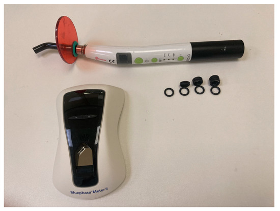
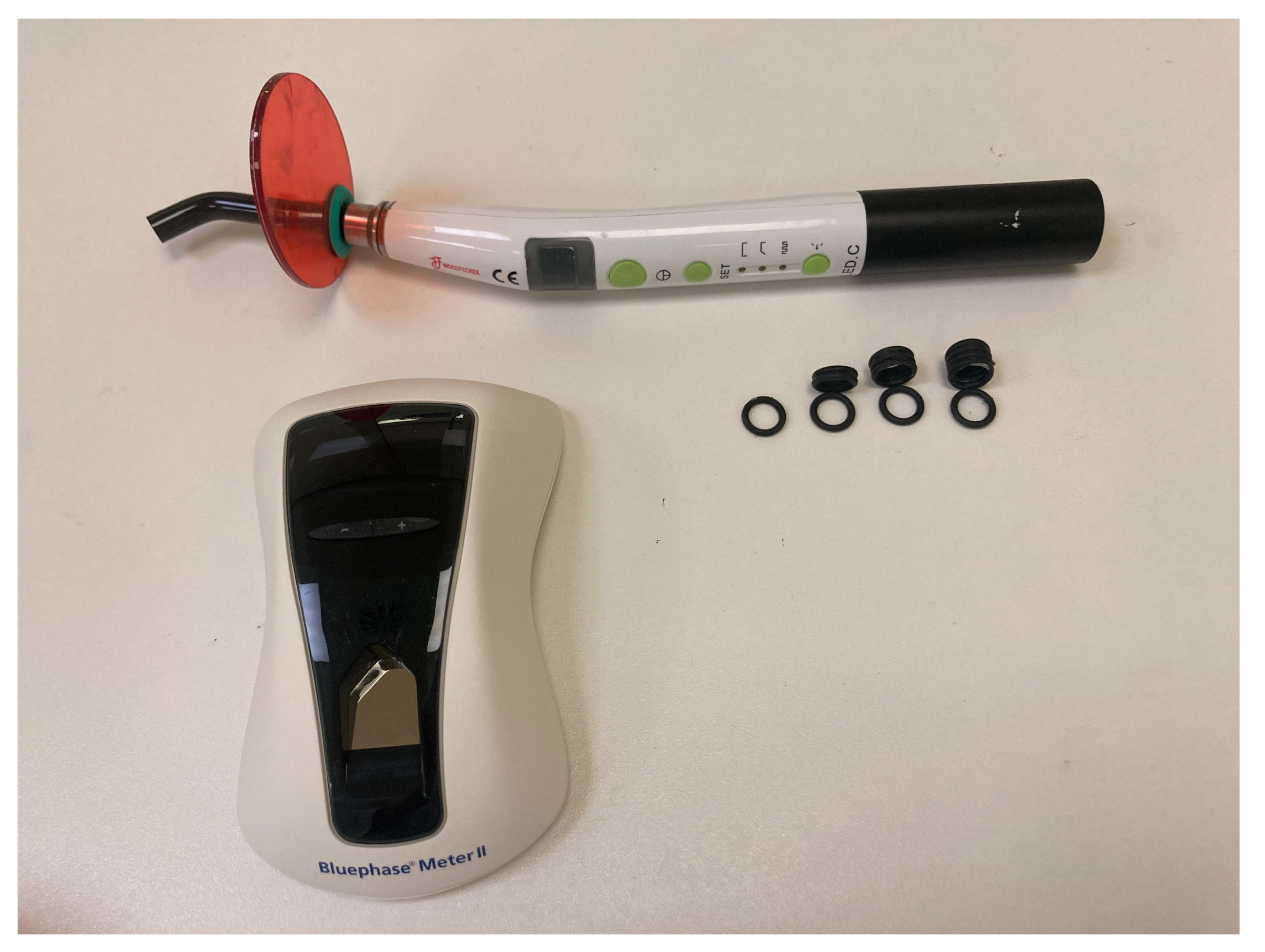
The study material was 21 LED polymerisation lamps with 8 mm tip diameter (Woodpecker LED.H, Guilin Woodpecker Medical Instrument Co., Guilin, China) used in the Department of Conservative Dentistry and Endodontics in Poznan University of Medical Sciences, with three exposure modes: standard, soft start, and pulse mode. In standard mode, the light irradiance is the same through the entire exposure time; in the soft start mode, the light irradiance increases and is recorded after reaching the maximum (top pick); in pulse mode, the highest appearing score was recorded. Each measurement usually lasted circa 5–10 s and was performed three times for each lamp, and the average was taken for further analysis. During the light irradiance measurement, the LCU was placed in a holder, and the distance to the photometer was increased to imitate the clinical situation in which the dentist moved the lamp tip away from the irradiated surface. To increase the distance between the LCU and the radiometer, spacer rings were used. The distance was increased from 0 to 8 mm in 2 mm steps. The lamps were fully charged and placed on the charger to maintain the maximum charge. Light irradiance measurements were made using a photometer, Bluephase Meter II (Ivoclar Vivadent, Opfikon, Switzerland), with a measurement range from 300 to 4000 mW/cm2. A total of 945 measurements were performed; for every 50 measurements, the batteries in the photometer were replaced with new ones. The experimental set-up is presented in Figure 1.

Figure 1.
Experimental set-up.
Statistical Analysis
The light irradiances were compared using the Friedman ANOVA test with the post-hoc test due to non-compliance with the normal distribution. The significance level was defined as α = 0.05. The statistical analysis was performed using Statistica 13.3 (Statsoft, Cracow, Poland).
3. Results
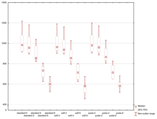
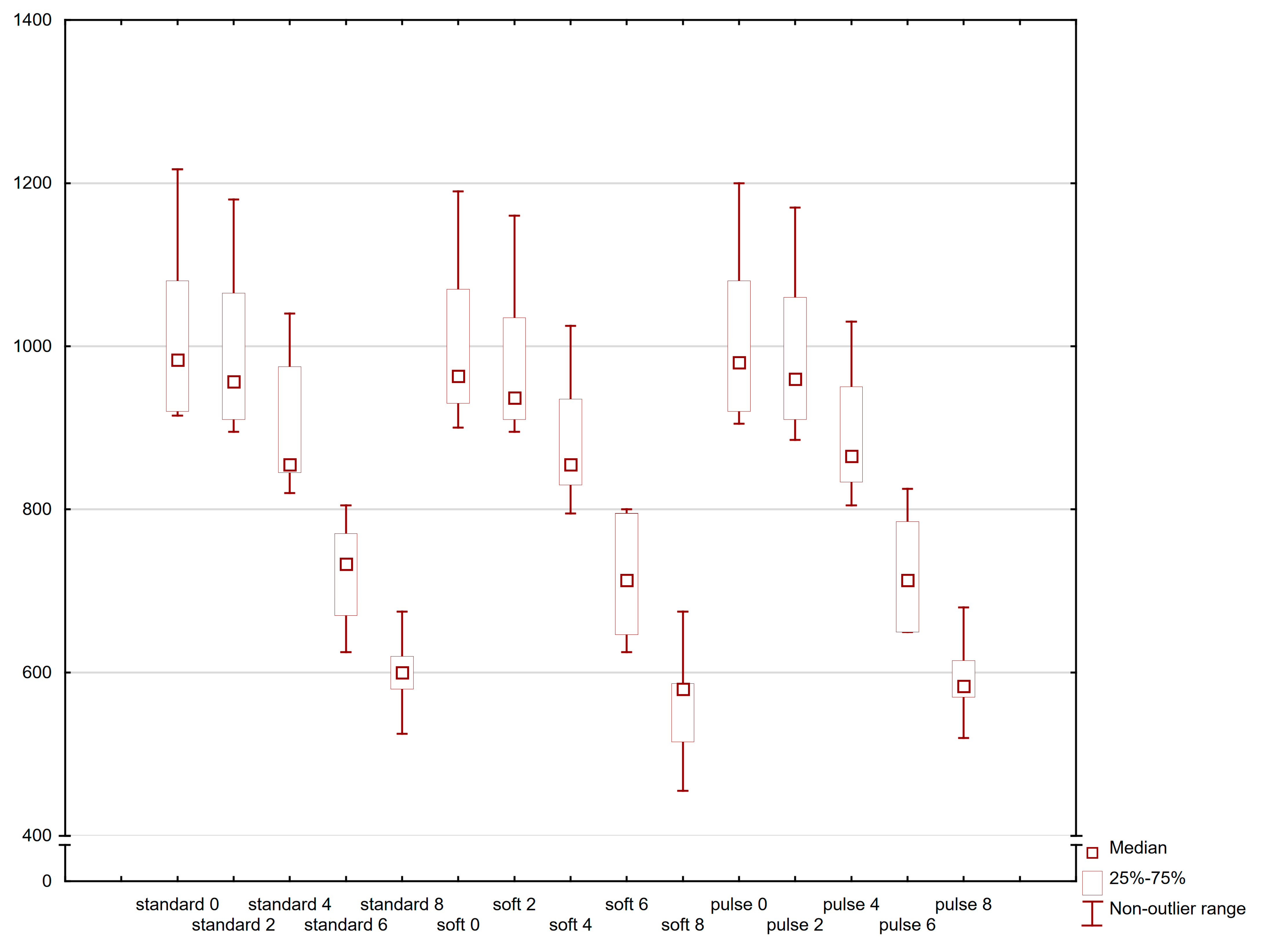
For each mode, a significant decrease in light irradiance was observed comparing zero distance with distances above 4 mm, 2 mm distance with distances above 6 mm, and 4 mm with distances above 8 mm—Figure 2. The greatest power loss with increasing distance was observed in the soft mode, the irradiance of which was statistically significantly lower at each distance than in the other modes, and at a distance of 8 mm, it did not even reach 600 mW/cm2. The standard mode achieved the highest values during the entire experiment; statistically significant differences were found at all tested distances compared to the other modes—Table 1. The irradiance values obtained when testing the pulse mode were like those in the standard mode.

Figure 2.
The box plot presents the changes in light irradiance (mW/cm2) depending on polymerisation mode and light curing distance.

Table 1.
The comparisons of irradiance values for different polymerisation modes and light curing distances (significant differences for the post-hoc Friedman ANOVA test).
4. Discussion
To our knowledge, this study was the first to analyse changes in light irradiance caused by increasing distance in different exposure modes. The standard soft start and pulse modes, which many lamp manufacturers widely use, were tested [40,41]. Monowave LED-LCUs were selected for the study because, in our clinic, only composites with camphorquinone are used, which polymerises better in this type of light [24,42]. A Bluephase Meter II radiometer (Ivoclar Vivadent, Opfikon, Switzerland) was used to measure the lamp power. Although it does not match the accuracy of laboratory instruments, it has undoubted advantages [43]. It is easily accessible and enables quick and simple measurement of irradiance power. Previous research has shown great usefulness of this radiometer [28,44]; according to the manufacturer, it can measure the radiant power output from 380–550 nm with an accuracy of ±10% [45]. However, several studies have been found in the literature showing the limitations of this measuring device [28,37].
Evaluation of the lamp irradiance with an increasing distance from 0 to 8 mm seems clinically useful. When restoring anterior teeth, the dentist can bring the LED LCU close to the composite surface, but in posterior teeth, the difficulty is the depth of the cavity and the height of the matrix band. The literature shows that increasing the distance to 10 mm or more results in insufficient superficial polymerisation or even no polymerisation [46,47]. Many publications indicate a distance of 4 mm as critical for the quality of polymerisation [48,49]. Our study showed statistically significant differences in light irradiance between values 0 and 2 mm versus 6 and 8, which may correspond to the studies of other researchers [49,50].
The light irradiance values at distances 0 and 2 were around 1000 mW/cm2, which many researchers consider optimal for polymerisation in clinical conditions [48]. An increase in the distance to 4 mm resulted in an estimated decrease in intensity of 100–150 mW/cm2. Previous studies have shown a regularity of 20% loss of lamp output with each 1 mm distance [37]. The range and light output of the lamp seem to be more stable now. Our results indicate an estimated loss of approximately 10% of light power/intensity with each 2 mm added distance. Also, Cacciafaesta et al. revealed the high sensitivity of the LED LCU to increasing distance. Their research showed the advantages of the halogen lamp over the LED lamp [49]. The explanation for this phenomenon may be the high thermal energy generated by halogen lamps, which has a positive effect on the polymerisation process.
Researchers also indicate that of the many factors influencing the polymerisation process of the composite, the lamp intensity is the most crucial. Interestingly, according to some authors, it turns out to be much more important than layer thickness and exposure time [51]. It is also known that incomplete polymerisation results in the increased release of monomers and degradation products, as excellently demonstrated by Kardas et al. in their study. It has been shown that even at 4 mm, the amount of monomer released is significant [52]. This confirms the observations from our experiment that 4 mm is a somewhat critical distance.
Increasing the distance of the LCU LED from the measuring device showed interesting differences between the tested modes. The most significant power loss was observed in the soft start mode. It seems that the potential advantages of this mode may be offset by its low resistance to distance increases. It’s worth remembering that the rationale behind the effects of soft start and pulse modes is that using low irradiance in the first phase of curing slows the rate of resin polymerisation. As a result, this may allow polymer chains to slide on each other and prolong the pre-gel stage, yielding low polymerisation contraction stress and good marginal adaptation. Some studies have shown that modulated polymerisation modes, especially soft start, result in better polymerisation dynamics and consequently lower shrinkage [53]. Final high irradiance would complete polymerisation, leading to a convenient degree of conversion and reaching the desired physical and mechanical properties [40]. According to Lim et al., a high degree of conversion (DC) of composite polymerisation is a prerequisite for obtaining the optimal physical and mechanical properties, biocompatibility, and improved success of composite restoration [54]. Many clinical studies indicate the advantage of the soft start mode over others; however, this is true only at a distance of 0 mm. Unfortunately, 0 mm distance is seldom possible in clinical conditions, so to maintain the mentioned advantages of this mode, the exposure time should be significantly extended, even up to 1 min [40,55].
The slight differences in lamp power observed in standard and pulse modes correspond to the results of Carvalho et al., who examined the physicochemical properties of composites polymerised in various ways. The characteristics of the polymer exposed to standard and pulse modes were very similar [56]. The differences in modulated polymerisation modes are very subtle and difficult to detect, especially in clinical conditions, and require further analysis.
As already mentioned, proper polymerisation is directly related to the degree of composite conversion. The authors suspect that in the case of increasing the distance in the pulsating and soft start modes, the degree of conversion may not even reach 50%. The next step of the research will be to combine the exposure modes, increasing the distance, and a more advanced (using a Raman microscope) measurement of the degree of conversion of the polymerised material to prove the thesis even more precisely.
Dental composite is a highly aesthetic material for tooth restoration, but it causes dentists many problems and generates numerous complications [57]. Such many articles on composite complications encourage authors to search for new teaching methods and create algorithms for polymerisation procedures.
Introducing dental radiometers to assess the light irradiance into everyday practice could be helpful for clinicians to evaluate and control the ability of LCUs to polymerise [14,35]. This would have a real impact on the quality and stability of fillings and, thus, the effectiveness of treatment. One limitation of this study is that the Bluephase Meter II was brand new. However, the manufacturer guarantees proper device operation for up to 3 years as well as high measuring accuracy with a tolerance of only ± 10% compared to the measurement with an integrating sphere. Moreover, the LCUs were not new, but they were undamaged and in good working condition; they were recharged after every five exposures. Thus, they represented what may be found in many dental offices. The positioning of the lamp in the holder relative to the radiometer also may be a limitation. In the next analyses, it is planned to use an optical bench to fasten the correct position.
This article provides dental clinicians with a portion of knowledge and simple clinical tips that can be used in every dental office during the most performed procedure—direct tooth restoration using a composite material.
5. Conclusions
Within the limitations of this study, the following conclusions can be made:
- If there is a significant risk of the LCU tip being placed at a distance from the material surface bigger than 4 mm, e.g., in case of deep cavities or limited access caused by a high matrix, or accidentally being moved away during exposure, it is recommended to use the standard curing mode.
- The pulse mode, even in the case of increased distance from the irradiated surface, can still provide high light intensity enabling sufficient polymerisation, so it is worth using it in deep cavities with vital pulp, as this form of exposure does not risk overheating of the pulp.
- The soft start mode, as it is least resistant to increasing distance, is recommended for use in front teeth or the cervical area, where high aesthetics is required, and the lamp can be easily brought closer to the restoration surface.
Our study only determines the amount of light energy reaching the surface of the radiometer. It is known that in dental practice the thickness of the filling is usually 1–2 mm. As already mentioned, the next research stage will be the analysis of the irradiated samples in a Raman microscope, which will allow us to look deep into the sample and create a three-dimensional map of the light propagation.
Author Contributions
Conceptualization, A.L.; methodology, A.L. and K.N.; formal analysis, K.N.; investigation, A.L. and M.M.; data curation, A.L. and K.N.; writing—original draft preparation, A.L., M.M. and F.P.; writing—review and editing, A.L., K.N. and B.C.; visualization, K.N. and F.P.; supervision, A.S. All authors have read and agreed to the published version of the manuscript.
Funding
This research received no external funding.
Institutional Review Board Statement
Not applicable.
Informed Consent Statement
Not applicable.
Data Availability Statement
The original contributions presented in the study are included in the article; further enquiries can be directed to the corresponding authors.
Conflicts of Interest
The authors declare no conflicts of interest.
References
- Yadav, R.; Kumar, M. Dental Restorative Composite Materials: A Review. J. Oral Biosci. 2019, 61, 78–83. [Google Scholar] [CrossRef] [PubMed]
- Wysokińska-Miszczuk, J.; Piotrowska, K.; Paulo, M.; Madej, M. Composite Materials Used for Dental Fillings. Materials 2024, 17, 4936. [Google Scholar] [CrossRef] [PubMed]
- De Angelis, F.; Sarteur, N.; Buonvivere, M.; Vadini, M.; Šteffl, M.; D’Arcangelo, C. Meta-Analytical Analysis on Components Released from Resin-Based Dental Materials. Clin. Oral. Invest. 2022, 26, 6015–6041. [Google Scholar] [CrossRef] [PubMed]
- Duruk, G.; Akküç, S.; Uğur, Y. Evaluation of Residual Monomer Release after Polymerization of Different Restorative Materials Used in Pediatric Dentistry. BMC Oral Health 2022, 22, 232. [Google Scholar] [CrossRef]
- Lehmann, A.; Nijakowski, K.; Nowakowska, M.; Woś, P.; Misiaszek, M.; Surdacka, A. Influence of Selected Restorative Materials on the Environmental pH: In Vitro Comparative Study. Appl. Sci. 2021, 11, 11975. [Google Scholar] [CrossRef]
- Maktabi, H.; Balhaddad, A.A.; Alkhubaizi, Q.; Strassler, H.; Melo, M.A.S. Factors Influencing Success of Radiant Exposure in Light-Curing Posterior Dental Composite in the Clinical Setting. Am. J. Dent. 2018, 31, 320–328. [Google Scholar]
- Opdam, N.; Frankenberger, R.; Magne, P. From ‘Direct Versus Indirect’ Toward an Integrated Restorative Concept in the Posterior Dentition. Oper. Dent. 2016, 41, S27–S34. [Google Scholar] [CrossRef]
- Tkáčiková, S.; Sabo, J. Release of Monomers from Dental Composite Materials into Saliva and the Possibility of Reducing the Toxic Risk for the Patient. Medicina 2023, 59, 1204. [Google Scholar] [CrossRef]
- Schneider, T.R.; Hakami-Tafreshi, R.; Tomasino-Perez, A.; Tayebi, L.; Lobner, D. Effects of Dental Composite Resin Monomers on Dental Pulp Cells. Dent. Mater. J. 2019, 38, 579–583. [Google Scholar] [CrossRef]
- Nijakowski, K.; Ortarzewska, M.; Jankowski, J.; Lehmann, A.; Surdacka, A. The Role of Cellular Metabolism in Maintaining the Function of the Dentine-Pulp Complex: A Narrative Review. Metabolites 2023, 13, 520. [Google Scholar] [CrossRef]
- Elgezawi, M.; Haridy, R.; Abdalla, M.A.; Heck, K.; Draenert, M.; Kaisarly, D. Current Strategies to Control Recurrent and Residual Caries with Resin Composite Restorations: Operator- and Material-Related Factors. J. Clin. Med. 2022, 11, 6591. [Google Scholar] [CrossRef] [PubMed]
- Montagner, A.F.; Sande, F.H.V.D.; Müller, C.; Cenci, M.S.; Susin, A.H. Survival, Reasons for Failure and Clinical Characteristics of Anterior/Posterior Composites: 8-Year Findings. Braz. Dent. J. 2018, 29, 547–554. [Google Scholar] [CrossRef] [PubMed]
- Lehmann, A.; Nijakowski, K.; Potempa, N.; Sieradzki, P.; Król, M.; Czyż, O.; Radziszewska, A.; Surdacka, A. Press-On Force Effect on the Efficiency of Composite Restorations Final Polishing—Preliminary In Vitro Study. Coatings 2021, 11, 705. [Google Scholar] [CrossRef]
- Assaf, C.; Fahd, J.-C.; Sabbagh, J. Assessing Dental Light-Curing Units’ Output Using Radiometers: A Narrative Review. J. Int. Soc. Prevent. Community Dent. 2020, 10, 1. [Google Scholar] [CrossRef] [PubMed]
- Abed, Y.A.; Sabry, H.A.; Alrobeigy, N.A. Degree of Conversion and Surface Hardness of Bulk-Fill Composite versus Incremental-Fill Composite. Tanta Dent. J. 2015, 12, 71–80. [Google Scholar] [CrossRef]
- Galvão, M.R.; Caldas, S.G.F.R.; Bagnato, V.S.; de Souza Rastelli, A.N.; de Andrade, M.F. Evaluation of Degree of Conversion and Hardness of Dental Composites Photo-Activated with Different Light Guide Tips. Eur. J. Dent. 2013, 7, 86–93. [Google Scholar]
- Bourbia, M.; Finer, Y. Biochemical Stability and Interactions of Dental Resin Composites and Adhesives with Host and Bacteria in the Oral Cavity: A Review. J. Can. Dent. Assoc. 2018, 84, i1. [Google Scholar]
- Lehmann, A.; Nijakowski, K.; Drożdżyńska, A.; Przybylak, M.; Woś, P.; Surdacka, A. Influence of the Polymerization Modes on the Methacrylic Acid Release from Dental Light-Cured Materials—In Vitro Study. Materials 2022, 15, 8976. [Google Scholar] [CrossRef]
- Iordache, S.-M.; Iordache, A.-M.; Gatin, D.I.; Grigorescu, C.E.A.; Ilici, R.R.; Luculescu, C.-R.; Gatin, E. Performance Assessment of Three Similar Dental Restorative Composite Materials via Raman Spectroscopy Supported by Complementary Methods Such as Hardness and Density Measurements. Polymers 2024, 16, 466. [Google Scholar] [CrossRef]
- Par, M.; Gamulin, O.; Spanovic, N.; Bjelovucic, R.; Tarle, Z. The Effect of Excitation Laser Power in Raman Spectroscopic Measurements of the Degree of Conversion of Resin Composites. Dent. Mater. 2019, 35, 1227–1237. [Google Scholar] [CrossRef]
- Jandt, K.D.; Mills, R.W. A Brief History of LED Photopolymerization. Dent. Mater. 2013, 29, 605–617. [Google Scholar] [CrossRef] [PubMed]
- Shortall, A.C.; Price, R.B.; MacKenzie, L.; Burke, F.J.T. Guidelines for the Selection, Use, and Maintenance of LED Light-Curing Units-Part 1. Br. Dent. J. 2016, 221, 453–460. [Google Scholar] [CrossRef] [PubMed]
- Hasanain, F.A.; Nassar, H.M. Utilizing Light Cure Units: A Concise Narrative Review. Polymers 2021, 13, 1596. [Google Scholar] [CrossRef] [PubMed]
- Dos Santos Sousa, G.; Guimarães, G.F.; Marcelino, E.; Rodokas, J.E.P.; De Oliveira Júnior, A.J.; Cesarino, I.; Leão, A.L.; Dos Santos Riccardi, C.; Arjmand, M.; Simões, R.P. Shrinkage Stress and Temperature Variation in Resin Composites Cured via Different Photoactivation Methods: Insights for Standardisation of the Photopolymerisation. Polymers 2021, 13, 2065. [Google Scholar] [CrossRef] [PubMed]
- Poggio, C.; Lombardini, M.; Gaviati, S.; Chiesa, M. Evaluation of Vickers Hardness and Depth of Cure of Six Composite Resins Photo-Activated with Different Polymerization Modes. J. Conserv. Dent. 2012, 15, 237. [Google Scholar] [CrossRef]
- Hubbezoglu, I.; Dogan, A.; Dogan, O.M.; Bolayir, G.; Bek, B. Effects of Light Curing Modes and Resin Composites on Temperature Rise under Human Dentin: An in Vitro Study. Dent. Mater. J. 2008, 27, 581–589. [Google Scholar] [CrossRef][Green Version]
- Jabbour, O.; Alfares, R. Effect of Light Curing Modes of High-Powered LEDs on Temperature Rise under Primary Teeth Dentin (an in Vitro Study). J. Oral Dent. Health 2022, 6, 144–152. [Google Scholar]
- Shimokawa, C.A.K.; Harlow, J.E.; Turbino, M.L.; Price, R.B. Ability of Four Dental Radiometers to Measure the Light Output from Nine Curing Lights. J. Dent. 2016, 54, 48–55. [Google Scholar] [CrossRef]
- Price, R.B.; Ferracane, J.L.; Shortall, A.C. Light-Curing Units: A Review of What We Need to Know. J. Dent. Res. 2015, 94, 1179–1186. [Google Scholar] [CrossRef]
- Price, R.B.; Ferracane, J.L.; Hickel, R.; Sullivan, B. The Light-Curing Unit: An Essential Piece of Dental Equipment. Int. Dent. J. 2020, 70, 407–417. [Google Scholar] [CrossRef]
- Prochnow, F.H.O.; Kunz, P.V.M.; Correr, G.M.; Kaizer, M.D.R.; Gonzaga, C.C. Relationship between Battery Level and Irradiance of Light-Curing Units and Their Effects on the Hardness of a Bulk-Fill Composite Resin. Restor. Dent. Endod. 2022, 47, e45. [Google Scholar] [CrossRef] [PubMed]
- Tongtaksin, A.; Leevailoj, C. Battery Charge Affects the Stability of Light Intensity from Light-Emitting Diode Light-Curing Units. Oper. Dent. 2017, 42, 497–504. [Google Scholar] [CrossRef] [PubMed]
- Cardoso, I.; Machado, A.; Teixeira, D.; Basílio, F.; Marletta, A.; Soares, P. Influence of Different Cordless Light-Emitting-Diode Units and Battery Levels on Chemical, Mechanical, and Physical Properties of Composite Resin. Oper. Dent. 2020, 45, 377–386. [Google Scholar] [CrossRef] [PubMed]
- Soares, C.J.; Bragança, G.F.D.; Pereira, R.A.D.S.; Rodrigues, M.D.P.; Braga, S.S.L.; Oliveira, L.R.S.; Giannini, M.; Price, R.B. Irradiance and Radiant Exposures Delivered by LED Light-Curing Units Used by a Left and Right-Handed Operator. Braz. Dent. J. 2018, 29, 282–289. [Google Scholar] [CrossRef] [PubMed][Green Version]
- Altaie, A.; Hadis, M.; Wilson, V.; German, M.; Nattress, B.; Wood, D.; Palin, W. An Evaluation of the Efficacy of LED Light Curing Units in Primary and Secondary Dental Settings in the United Kingdom. Oper. Dent. 2021, 46, 271–282. [Google Scholar] [CrossRef]
- Omidi, B.-R.; Gosili, A.; Jaber-Ansari, M.; Mahdkhah, A. Intensity Output and Effectiveness of Light Curing Units in Dental Offices. J. Clin. Exp. Dent. 2018, 10, e555–e560. [Google Scholar] [CrossRef]
- Maucoski, C.; Price, R.B.; Arrais, C.A.; Sullivan, B. Power Output from 12 Brands of Contemporary LED Light-Curing Units Measured Using 2 Brands of Radiometers. PLoS ONE 2022, 17, e0267359. [Google Scholar] [CrossRef]
- Maucoski, C.; Price, R.B.; Arrais, C.A.G. Irradiance from 12 LED Light Curing Units Measured Using 5 Brands of Dental Radiometers. J. Esthet. Restor. Dent. 2023, 35, 968–979. [Google Scholar] [CrossRef]
- Segal, P.; Lugassy, D.; Mijiritsky, E.; Dekel, M.; Ben-Amar, A.; Ormianer, Z.; Matalon, S. The Effect of the Light Intensity and Light Distances of LED and QTH Curing Devices on the Hardness of Two Light-Cured Nano-Resin Composites. Mater. Sci. Appl. 2015, 06, 1071–1083. [Google Scholar] [CrossRef]
- Najjar, Y.M.; Burhan, A.S.; Hajeer, M.Y.; Nawaya, F.R. Effects of the Conventional, Soft Start, and Pulse Delay Modes Produced by Light-Emitting Diode Device on Microleakage beneath Metal Brackets: An in Vitro Comparative Study. Int. Orthod. 2023, 21, 100718. [Google Scholar] [CrossRef]
- Asmussen, E.; Peutzfeld, A. Influence of Pulse-Delay Curing on Softening of Polymer Structures. J. Dent. Res. 2001, 80, 1570–1573. [Google Scholar] [CrossRef] [PubMed]
- Lima, R.B.W.; Melo, A.M.D.S.; Dias, J.D.N.; Barbosa, L.M.M.; Santos, J.V.D.N.; Souza, G.M.D.; Andrade, A.K.M.; Assunção, I.V.D.; Borges, B.C.D. Are Polywave Light-Emitting Diodes More Effective than Monowave Ones in the Photoactivation of Resin-Based Materials Containing Alternative Photoinitiators? A Systematic Review. J. Mech. Behav. Biomed. Mater. 2023, 143, 105905. [Google Scholar] [CrossRef] [PubMed]
- Giannini, M.; André, C.B.; Gobbo, V.C.; Rueggeberg, F.A. Accuracy of Irradiance and Power of Light-Curing Units Measured with Handheld or Laboratory Grade Radiometers. Braz. Dent. J. 2019, 30, 397–403. [Google Scholar] [CrossRef] [PubMed]
- Price, R.B.; Labrie, D.; Kazmi, S.; Fahey, J.; Felix, C.M. Intra- and Inter-Brand Accuracy of Four Dental Radiometers. Clin. Oral Investig. 2012, 16, 707–717. [Google Scholar] [CrossRef] [PubMed]
- Bluephase Meter II|Dental Radiometer|Ivoclar: Ivoclar. Available online: https://www.ivoclar.com/en_nordic/products/equipment/bluephase-meter-ii (accessed on 22 October 2023).
- Al-Zain, A.O.; Eckert, G.J.; Platt, J.A. The Influence of Distance on Radiant Exposure and Degree of Conversion Using Different Light-Emitting-Diode Curing Units. Oper. Dent. 2019, 44, E133–E144. [Google Scholar] [CrossRef]
- Diab, R.A.; Yap, A.U.; Gonzalez, M.A.G.; Yahya, N.A. Impact of Light-Curing Distance on the Effectiveness of Cure of Bulk-Fill Resin-Based Composites. Saudi Dent. J. 2021, 33, 1184–1189. [Google Scholar] [CrossRef]
- Luca, B.-I.; Ilie, N. Estimation of the Tolerance Threshold for the Irradiance of Modern LED Curing Units When Simulating Clinically Relevant Polymerization Conditions. Dent. Mater. J. 2021, 40, 750–757. [Google Scholar] [CrossRef]
- Cacciafesta, V.; Sfondrini, M.F.; Scribante, A.; Boehme, A.; Jost-Brinkmann, P.-G. Effect of Light-Tip Distance on the Shear Bond Strengths of Composite Resin. Angle Orthod. 2005, 75, 386–391. [Google Scholar] [CrossRef]
- Felix, C.A.; Price, R.B.T. The Effect of Distance from Light Source on Light Intensity from Curing Lights. J. Adhes. Dent. 2003, 5, 283–291. [Google Scholar]
- Maximov, J.; Dikova, T.; Duncheva, G.; Georgiev, G. Influence of Factors in the Photopolymerization Process on Dental Composites Microhardness. Materials 2022, 15, 6459. [Google Scholar] [CrossRef]
- Karadas, M.; Hatipoglu, O.; Er, H.; Akyüz Turumtay, E. Influence of Different Light-Curing Units on Monomer Elution from Bulk Fill Composites. J. Adhes. Sci. Technol. 2018, 32, 2631–2646. [Google Scholar] [CrossRef]
- Lopes, L.G.; Franco, E.B.; Pereira, J.C.; Mondelli, R.F.L. Effect of Light-Curing Units and Activation Mode on Polymerization Shrinkage and Shrinkage Stress of Composite Resins. J. Appl. Oral Sci. 2008, 16, 35–42. [Google Scholar] [CrossRef] [PubMed]
- Lim, H.-K.; Keerthana, S.; Song, S.-Y.; Li, C.; Shim, J.S.; Ryu, J.J. Effect of Light Irradiance and Curing Duration on Degree of Conversion of Dual-Cure Resin Core in Various Cavities with Different Depths and Diameters. Materials 2024, 17, 4342. [Google Scholar] [CrossRef]
- Ilie, N.; Jelen, E.; Hickel, R. Is the Soft-Start Polymerisation Concept Still Relevant for Modern Curing Units? Clin. Oral Investig. 2011, 15, 21–29. [Google Scholar] [CrossRef] [PubMed]
- Carvalho, A.A.; Moreira, F.D.C.L.; Fonseca, R.B.; Soares, C.J.; Franco, E.B.; Souza, J.B.D.; Lopes, L.G. Effect of Light Sources and Curing Mode Techniques on Sorption, Solubility and Biaxial Flexural Strength of a Composite Resin. J. Appl. Oral Sci. 2012, 20, 246–252. [Google Scholar] [CrossRef]
- Lehmann, A.; Nijakowski, K.; Jankowski, J.; Donnermeyer, D.; Palma, P.J.; Drobac, M.; Martins, J.F.B.; Pertek Hatipoğlu, F.; Tulegenova, I.; Javed, M.Q.; et al. Awareness of Possible Complications Associated with Direct Composite Restorations: A Multinational Survey among Dentists from 13 Countries with Meta-Analysis. J. Dent. 2024, 145, 105009. [Google Scholar] [CrossRef]
Disclaimer/Publisher’s Note: The statements, opinions and data contained in all publications are solely those of the individual author(s) and contributor(s) and not of MDPI and/or the editor(s). MDPI and/or the editor(s) disclaim responsibility for any injury to people or property resulting from any ideas, methods, instructions or products referred to in the content. |
© 2024 by the authors. Licensee MDPI, Basel, Switzerland. This article is an open access article distributed under the terms and conditions of the Creative Commons Attribution (CC BY) license (https://creativecommons.org/licenses/by/4.0/).


