Article

The Role of Salivary Biomarkers in Monitoring Oral Health in Patients with Implants and Periodontitis

by
Pia López-Jornet
1,*,
Joonas Nikolai Hynninen
1,
Francisco Parra-Perez
1,
Camila Peres-Rubio
2,
Eduardo Pons-Fuster
3 and
Asta Tvarijonaviciute
2
1
Faculty of Medicine and Odontology, Hospital Morales Meseguer, Clínica Odontológica, Marqués de los Vélez s/n, 30008 Murcia, Spain
2
Interdisciplinary Laboratory of Clinical Analysis (Interlab-UMU), Veterinary School, Regional Campus of International Excellence Mare Nostrum, University of Murcia, Campus de Espinardo s/n, Espinardo, 30100 Murcia, Spain
3
Departamento de Anatomía Humana y Psicobiología, Faculty of Medicine and Odontology, Biomedical Research Institute (IMIB-Arrixaca), University of Murcia Spain, 30100 Murcia, Spain
*
Author to whom correspondence should be addressed.
Appl. Sci. 2024, 14(2), 927; https://doi.org/10.3390/app14020927
Submission received: 22 December 2023 / Revised: 15 January 2024 / Accepted: 20 January 2024 / Published: 22 January 2024

Abstract

:
Oxidative stress, a physiological process that can damage cells, is known to affect various aspects of oral health. Oxidative stress can influence dental implant longevity and health. Assessing biomarkers of oxidative stress in saliva is beneficial for diagnosing and tracking the progression of oral diseases. A study is made of salivary oxidative stress in patients with dental implants with or without periodontitis. The study consisted of the following groups: Group1 (healthy without dental implants); Group 2 (subjects undergoing periodontal maintenance without dental implants); Group 3 (healthy patients with implants older than six months); and Group 4 (patients undergoing periodontal maintenance with implants older than six months). A complete examination of the oral cavity was made in each patient and a questionnaire was used to assess habits of hygiene, quality of life, and information about the implants. The following parameters were recorded in unstimulated whole saliva: ferric reducing antioxidant power (FRAP), Trolox equivalent antioxidant capacity (TEAC), cupric reducing antioxidant capacity (CUPRAC), advanced oxidation protein products (AOPP), and total proteins (TP). A total of 160 patients were studied, with 40 patients per group. The mean oxidative stress biomarker values obtained in the patients without implants and with implants were FRAP 0.590 ± 0.514 and 0.588 ± 0.334 mmol/L (p = 0.974); TEAC 0.320 ± 0.223 and 0.315 ± 0.172 mmol/L (p = 0.879); CUPRAC 0.286 ± 0.216 and 0.288 ± 0.151 mmol/L (p = 0.956); AOPP 456.04 ± 789.75 and 430.65 ± 752.05 µmol/L (p = 0.838); and TP 73.90 ± 50.83 and 70.36 ± 56.93 mg/dL (p = 0.684), respectively. No substantial variations were noted in the salivary oxidative stress biomarker levels between patients with controlled periodontal disease and/or dental implants compared to healthy individuals.

1. Introduction

In recent years, the insertion of dental implants as a substitute for absent teeth has evolved into a widespread procedure, encompassing even those patients afflicted with periodontal disease [1,2]. Oxidative stress occurs when there is a disproportion between the generation of reactive oxygen species (ROS) and the body’s antioxidant mechanisms’ ability to neutralize these ROS. The latter include free radicals that can damage the cells and tissues [2,3,4,5,6,7,8,9]. Periodontitis, a chronic inflammatory disorder, is distinguished by symptoms in the oral cavity and substantial systemic effects. This condition significantly influences the support tissues within the mouth. Factors contributing to periodontitis include inadequate dental hygiene, smoking, the oral microbiota’s composition, and an individual’s genetic susceptibility. A pivotal aspect in the progression of periodontitis is oxidative damage to tissues, which is crucial in the disease’s advancement [2,3,4,5,6,7,8,9]
The analysis of salivary biomarkers of oxidative stress has been applied to the diagnosis of various diseases of the oral cavity, as they may be involved in the occurrence and/or development of periodontitis, caries, potentially malignant oral diseases, cancer, inflammation, and fungal diseases [10]. Salivary oxidative stress biomarkers have become a tool for analyzing the pathogenesis and conducting follow-up of oral disorders, and their correct analysis could, in future, lead to individualized diagnosis and treatment for each patient, since their determination is noninvasive, simple, safe, rapid, and painless [11,12]. The study of saliva is an easy and reproducible method for determining salivary oxidant activity and antioxidant capacity [11].
Periodontitis is characterized by an increase in ROS at the periodontal level, resulting in oxidant imbalance [13,14,15,16]. Chen et al. [9] observed notable changes in the biochemical markers of patients with periodontitis. Their study indicated a marked reduction in the overall antioxidant capacity. Concurrently, there was an observable elevation in several compounds: malondialdehyde (MDA), nitric oxide, total oxidant status (TOS), and 8-hydroxydeoxyguanosine, all of which were measured in the saliva of these patients. Conventional periodontal treatment exerts beneficial effects upon the antioxidant marker levels; in this regard, several studies have found nonsurgical treatment to significantly modify the MDA concentrations to levels comparable to those observed in periodontally healthy individuals [17].
It is important that patients with a history of periodontitis who are considering dental implant rehabilitation receive complete and comprehensive information. Factors such as oxidative stress and chronic inflammation are known to have the potential to trigger complications, including peri-implantitis [18,19,20,21,22]. There are data suggesting an increased risk of peri-implantitis in people with periodontitis. This risk is exacerbated by inadequate management of dental plaque control and the absence of regular checkups after implant placement [21,22,23].
A study conducted by Sgolastra et al. [23] highlighted that the presence of periodontal disease increases the likelihood of peri-implantitis. Following this, Stacchi et al. [24] investigated the role of periodontitis in the development of peri-implantitis. Their findings revealed a markedly increased risk (odds ratio [OR] 0.23, 95% confidence interval [95%CI] 0.11–0.46) of peri-implantitis.
Implant biocompatibility has been studied; in this regard, titanium (Ti) is the predominant material of choice in the field. Damage to the TiO2 surface layer of dental implants can give rise to corrosion, thereby producing an inflammatory reaction. This in turn results in loss of osseointegration, peri-implantitis, and damage and/or contamination of the peri-implant tissues [8,17].
There is controversy about the role that ROS may play in osseointegrations [8,21]. The key intervention of ROS in the coupling of angiogenesis-osteogenesis may influence the efficacy of implant osseointegration. The quality and amount of available bone are important factors for correct osseointegration. In this regard, bone metabolic disorders related to patient age, smoking, hygiene, medication, osteoporosis, and diabetes mellitus can give rise to bone healing problems and dental implant failure [25,26,27,28,29,30].
In sum, oxidative stress can influence dental implant longevity and health. It is therefore important to address this problem and minimize the factors contributing to oxidative stress in order to ensure long-term implant rehabilitation success [4,10]. Given the variability between patients and the broad range of host response modulating factors, the identification of a unifying pathogenic mechanism could contribute to improve our understanding of the progression of the disorder and develop new therapeutic options [17,18,31].
Although oxidative stress is known to be important in many other disorders, including for example chronic periodontal disease [6,7], its role in relation to dental implants is still unclear [23]. The objective is to analyze salivary biomarkers in patients with dental implants with and without periodontitis.

2. Material and Methods

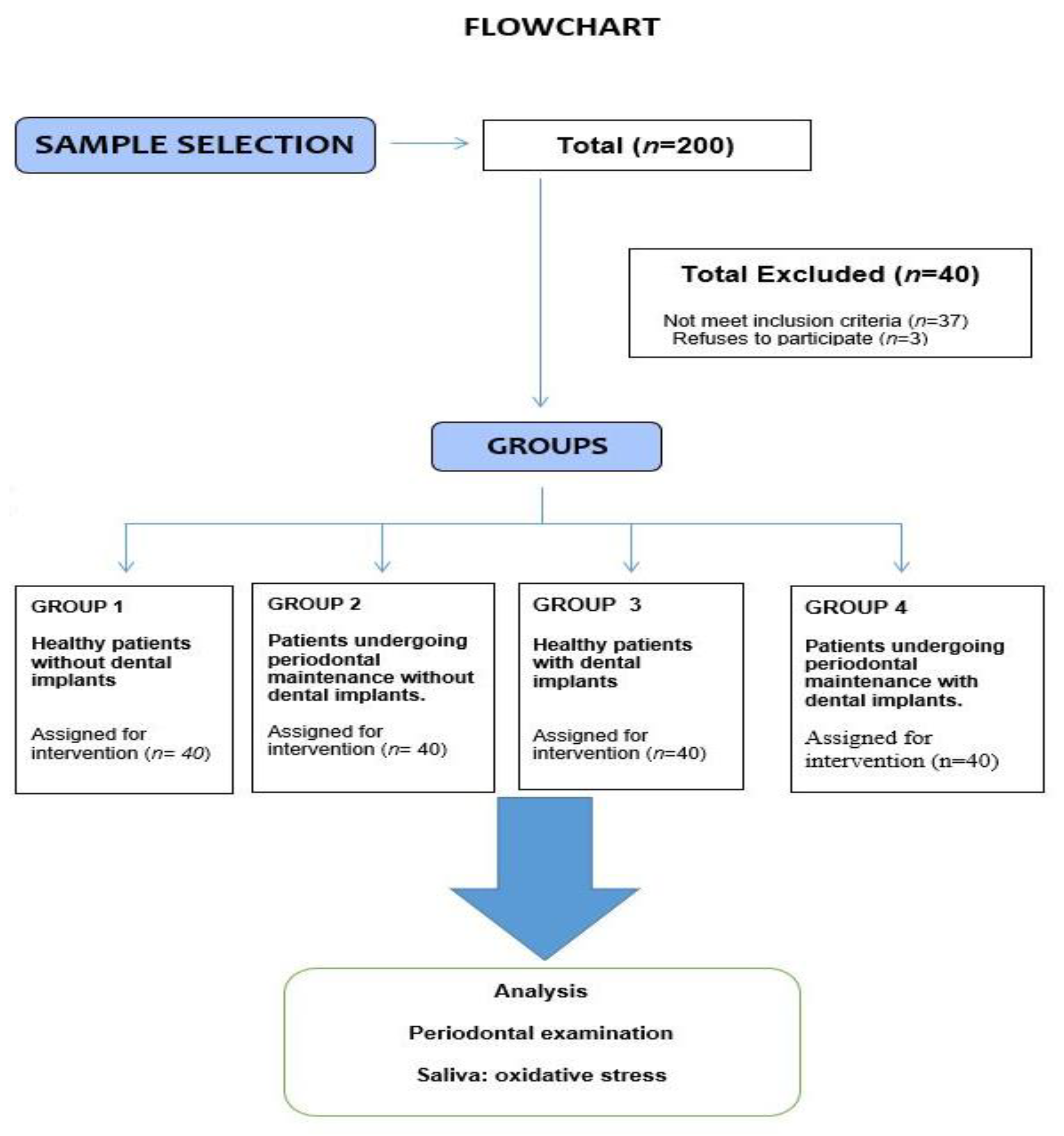
2.1. Sample Selection

The study comprised a total of 160 patients with dental implants consecutively enrolled in a duly certified private clinic in the region of Murcia (Spain) and has been authorized by the Ethics Committee of the University of Murcia (reference: 446/2021). The methodology followed the principles of the Declaration of Helsinki, and participants gave their written informed consent prior to their involvement in the study. This research was carried out in alignment with the STROBE (Strengthening the Reporting of Observational Studies in Epidemiology). The inclusion criteria were healthy individuals (without implants or with implants in place for at least 6 months) and patients with periodontal disease in maintenance. The exclusion criteria were patients with decompensated systemic disease, the use of antioxidant supplements, pregnant or nursing women, pediatric patients, and failure to sign the corresponding informed consent.
A cross-sectional observational design was adopted comparing clinical and salivary outcome variables in the different groups of patients (Figure 1). The study consisted of the following groups:
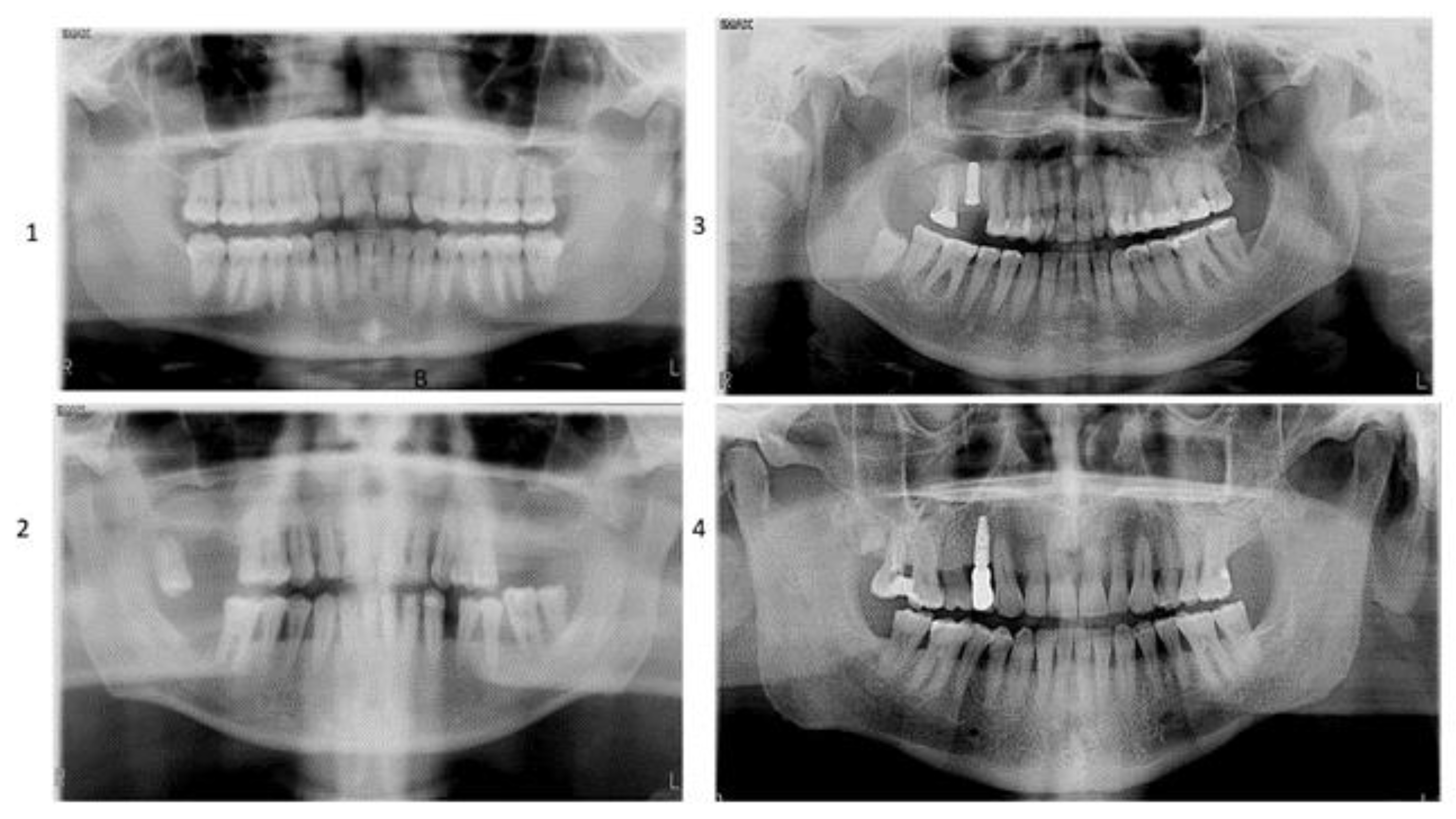
  • Group 1: Healthy patients without implants (n = 40)
  • Group 2: Patients undergoing periodontal maintenance without dental implants (n = 40)
  • Group 3: Healthy patients with implants older than six months (n = 40)
  • Group 4: Patients undergoing periodontal maintenance with implants older than six months (n = 40)

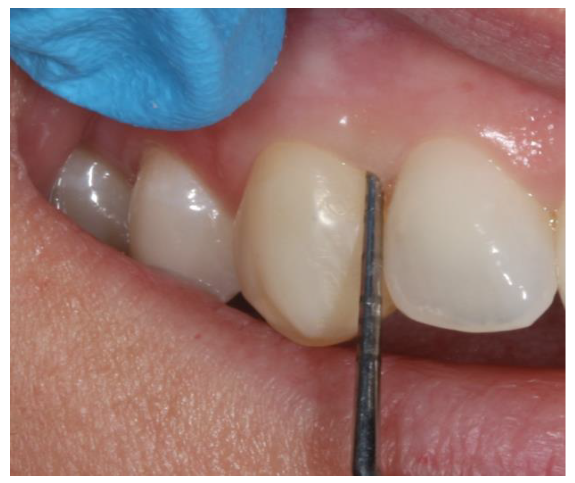
2.2. Clinical Variables

Patient case history was compiled, and demographic data were recorded along with buccodental health (odontogram and periodontal conditions) general health (diseases, Medications). Patients underwent Body Mass Index (BMI), habits of hygiene, and smoking and alcohol intake surveys. Likewise, we recorded the type, diameter, length, and location of the dental implants, the implant material prosthesis, and type of implant-prosthesis connection.
Panoramic radiographs (Figure 2) and periapical series were obtained and a complete oral examination, odontogram, and evaluation of periodontal status (probing of teeth and/or implants) were performed.
All the probing measurements were performed by a single investigator (J.H.) and were calibrated using 10 patients not forming part of the study and who presented moderate to severe periodontitis (Figure 3). Correlation coefficients were used for measurement of both probing depth and attachment loss—the values being found to be >0.75 for both parameters. A sterile exploration kit was used for measurement, together with a universal periodontal probe.
The parameters described below were evaluated in all teeth. Third molars were excluded. Probing was performed at six different points of each tooth. Periodontal condition in turn was assessed based on the updated 2017 guidelines [21].
Patient level of hygiene was evaluated using the O’Leary plaque index, which assesses hygiene of the smooth surfaces of the teeth. The percentage of stained smooth surfaces (indicating caries) is reported with respect to the total dental surface. Four surfaces were recorded for each tooth (mesial interproximal, buccal, distal interproximal, palatine/lingual).
The standards for determining the success and control of dental implants were established on several key factors: no mobility of the implant, no signs of inflammation, no radiographic indications of failure, no pain or related infections, and minimal bone loss within a year (specifically, not exceeding one-third of the implant embedded in bone—0.2 mm) [19].
The Oral Health Impact Profile (OHIP)-14 survey tool for the assessment of oral quality of life was used. This 14-item questionnaire explores the seven dimensions of Locker’s model of oral health, and higher scores indicate worse oral quality of life.

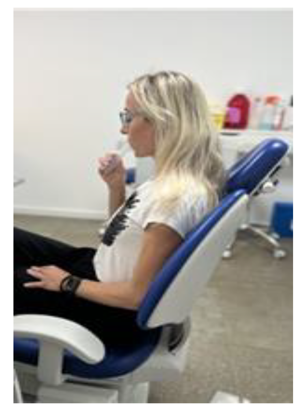
2.3. Sampling of Saliva

Saliva was collected before carrying out any kind of intraoral intervention. Unstimulated whole saliva was always sampled in the morning based on the standard protocols and using the drainage technique described by Navazesh and Kumar [32] for 5 min (Figure 4).

2.4. Measurement of Salivary Oxidative Stress

A total of five samples were excluded due to contamination. The operator analyzing the saliva was blinded as to which study group the patient belonged. Of the different tests performed, those recording plasma ferric reducing antioxidant power (FRAP), Trolox equivalent antioxidant capacity (TEAC), and cupric reducing antioxidant capacity (CUPRAC) are spectrophotometric techniques based on single-electron transfer methods [33,34].
FRAP test: The method of Benzie and Strain [35] was used, with some modifications. In this test, the antioxidants of the sample reduce the ferric tropyridyltriazine (Fe3+-TPTZ) complex to ferrous tropyridyltriazine (Fe2+-TPTZ), which has a bluish color. The intensity of this color is directly proportional to the reducing activity of the antioxidants.
CUPRAC test: This test was carried out based on the method of Campos et al. [36]. It involves the reduction of Cu2+ to Cu1+ mediated by the non-enzymatic antioxidants of the sample. The oxidizing compound is Cu2+ bathocuproinedisulfonic acid (Cu2+-BCS), which reacts with the antioxidants of the sample and reduces to Cu1+ bathocuproinedisulfonic acid (Cu1+-BCS)—a stable compound with maximum absorbance at a wavelength of 490 nm; the antioxidant activity is proportional to the formation of the Cu1+-BCS complex. The test was performed using 0.25 mmol/L of the disodium salt of bathocuproinedisulfonic acid (reagent 1) and 0.5 mmol/L of CuSO4 (reagent 2).
TEAC test: This test was carried out based on the procedure described by Arnao et al. [37]. ABTS (2.2′-azino-bis-3-ethylbenzothiazoline-6-sulfonic acid) is a peroxidase compound that generates a cation radical when oxidized in the presence of hydrogen peroxide (peroxidation). This radical produces a blue-green color, and the antioxidants in the sample suppress this color in proportion to their own concentrations.
Advanced oxidation protein products (AOPP) test: This test was used to evaluate salivary protein oxidation status based on the method of Witko-Sarsat et al. [38].
Total protein (TP) test: Total protein was determined using the procedure of Weichselbaum [39] (Beckman Coulter OSR6132; Beckman Coulter Inc. 250 S. Kraemer Blvd, Brea, CA 92821, USA). The Cu2+ ions in an alkaline solution react with the proteins and polypeptides that contain at least two peptide bonds, generating a violet color.

2.5. Statistical Analysis

A descriptive statistical analysis of the qualitative variables was performed. Comparisons between independent groups were made using the Student’s t-test or the nonparametric Mann–Whitney U-test in the absence of a normal data distribution, as determined by the Shapiro–Wilk test. Homogeneity of variance was assessed with the Levene test. In the case of qualitative variables, comparisons between groups were made with the chi-square test. In the presence of more than two categories and where the chi-square test proved statistically significant, two-by-two comparisons were made with Bonferroni correction. In order to determine the influence of the demographic and clinical variables and salivary markers upon the evolution of periodontitis and peri-implantitis, univariate and multivariate logistic regression models were used (entering those variables found to be statistically significant in the univariate analysis). Statistical significance was considered for p < 0.05. The SPSS version 27.0 statistical package for MS Windows was used throughout.

3. Results

The research sample comprised 160 participants of which 61 were men (38.1%) and 99 women (61.9%). The mean age was 52.7 ± 17.2 years (range 20–88). The descriptive and comparative results referred to the demographic variables and patient habits are reported in Table 1.
With respect to clinical measures (Table 2) the number of amalgam fillings, the number of metal-ceramic bridges and the plaque index scores were significantly greater among the patients with periodontitis.
When it comes to the periodontal variables and implant characteristics (Table 3 and Table 4), the percentage of patients with stage I periodontitis was significantly greater in the patients with dental implants (21.3%) than in those without implants (3.8%). On the other hand, the percentage of patients without implants that showed bleeding (25%) was significantly greater than among the patients with implants (8.8%).
In reference to the salivary markers (Table 5), there were no notable differences observed between the study groups. Specifically, the mean oxidative stress biomarker values obtained in patients without implants and with dental implants, respectively, were: FRAP 0.590 ± 0.514 compared to 0.588 ± 0.334 mmol/L (p = 0.974); TEAC 0.320 ± 0.223 compared to 0.315 ± 0.172 mmol/L (p = 0.879); CUPRAC 0.286 ± 0.216 compared to 0.288 ± 0.151 mmol/L (p = 0.956); AOPP 456.04 ± 789.75 compared to 430.65 ± 752.05 µmol/L (p = 0.838); and TP 73.90 ± 50.83 compared to 70.36 ± 56.93 mg/dL (p = 0.684).
Regarding the overall quality of life scores documented using the OHIP-14, the patients with implants but without periodontal disease yielded better scores, though statistical significance was not reached between the two groups (p = 0.05). However. on examining the different domains of the patients without periodontal disease, significant differences were found in relation to aesthetic satisfaction (9.15 compared to 8.53) (p = 0.005) and chewing satisfaction (9.98 versus 9.35) (p = 0.044). In the univariate analysis, the parameters with a significant influence upon periodontal health were seen to be patient age, medical treatment, smoking, plaque index (with increasing percentage plaque being associated to a greater probability of periodontitis), and the frequency of brushing. In the multivariate analysis, significance was retained for patient age (the probability of periodontitis increasing with age) and smoking (the probability of periodontitis in smokers being 2.87 times more likely than among non-smokers) (Table 6).
Of the total 241 implants analyzed in the study, only 7% presented some degree of peri-implantitis. In the univariate analysis, the following results in relation to the presence of peri-implantitis were recorded: absence or presence of periodontal disease (odds ratio [OR] 0.89. 95% confidence interval [95%CI] 0.33–2.39) (p = 0.815); implant location in the maxilla or mandible (OR 0.76. 95%CI 0.28–2.03) (p = 0.58); years of implant in place (OR 1.14. 95%CI 0.95–1.38) (p = 0.168) (Table 7).

4. Discussion

In our sample of 160 patients with 241 dental implants, we evaluated a number of salivary oxidative stress biomarkers (CUPRAC, FRAP, TEACH, AOPP, and TP) and found no significant changes between the groups studied. Statistical analysis revealed no substantial differences between the groups studied. Nevertheless, it must be noted that the review of the literature yielded few studies on the correlation between clinical parameters and oxidative stress in patients of this kind.
Saliva contains a variety of substances that can indicate the presence of periodontal disease in its early stages. Oxidative stress, identified as a major factor in the likelihood of developing periodontal issues, is indispensable the progression of this condition through a variety of mechanisms, many of which are still in the process of being fully understood. This relationship has led to the use of salivary oxidative stress biomarker analysis as a valuable diagnostic tool for identifying not only periodontal disease but also a range of systemic and local disorders. Significantly, a well-documented relationship has been established linking periodontal disease to various comorbidities, including diabetes, neurological disorders, rheumatological conditions, cardiovascular diseases, and metabolic syndrome. This link underscores the importance of a comprehensive understanding of oral health in relation to an individual’s health and overall well-being [2,4,40,41,42]
Su et al. [43] analyzed 292 subjects (58 with periodontal disease and 234 healthy individuals) to assess the oxidation of DNA, lipids, and proteins. These authors measured 8-OHdG (hydroxyl radical-mediated damage to DNA), 8-epi-PGF2alfa (lipid peroxidation), protein carboxyl group content (protein oxidation), and TAC (total antioxidant capacity), and recorded a notable augmentation in all of these parameters in the participants with periodontal conditions versus the healthy individuals.
Banasovà et al. [15] analyzed the saliva of 42 patients (19 patients with periodontal disease and 23 healthy), and found that lipid peroxidation in periodontitis appears to be caused by an augmented production of reactive oxygen species (ROS) in males and a decrease in antioxidant status in females. The research of Mohideen et al. [14] identified the mean levels of malondialdehyde (MDA), a lipid peroxidation biomarker, to differ significantly between healthy patients and those with periodontitis, with confirmation of increased ROS production on the part of the inflammatory cells in periodontitis.
Wei et al. [44] reported that non-surgical therapies can rehabilitate and regulate the patient’s antioxidant capacity by modifying salivary oxidative stress markers such as MDA, total oxidative stress, and superoxide dismutase (SOD). Karim et al. [45] likewise found that the decrease in inflammatory response following periodontal treatment improves host antioxidant capacity measured both in saliva and in gingival crevicular fluid. Dede et al. [46] indicated a notable decrease in 8-OHdG levels in individuals with chronic periodontitis following initial periodontal treatment. Guentsch et al. [47] found that adequate periodontal treatment exerts a regulatory effect upon the oxidative stress levels as measured by the markers MDA and glutathione peroxidase (GSHPx). In our research, all individuals with periodontal disease were part of a periodontal maintenance program. Therefore, we believe that the lack of significant differences in salivary biomarker levels can be attributed to the fact that these individuals were not experiencing an acute or uncontrolled phase of the disease.
On the other hand, increased oxidative stress levels are one of the causes underlying peri-implant disease, and treatment strategies designed to reduce oxidative stress potentially could contribute to managing peri-implant disease. Martins-Gomes et al. [27] observed that patients undergoing regular periodontal and implant maintenance were less likely to suffer peri-implantitis than individuals without such maintenance. Liskmann et al. [29] in turn found salivary total antioxidant status (TAS) and the concentrations of uric acid and ascorbate were reduced significantly in individuals with peri-implant disease. This might suggest that the elevated generation production of ROS in peri-implant disease leads to a situation of excessive oxidative stress, which may be an important contributor to peri-implant tissue destruction. Song et al. [8] measured the SOD and GSHPx levels in healthy subjects and with peri-implantitis and found both markers to be markedly reduced in the individuals with peri-implantitis, evidencing a lesser capacity to combat cellular oxidative stress in these individuals.
Of the 241 implants analyzed in the present study, only 7% presented some degree of peri-implant disease, and we found no changes in the salivary oxidative stress (CUPRAC, FRAP, TEACH, AOPP, and TP) between groups. This finding aligns with the results found by Jazi et al. [22] who measured SOD, MDA, and TAC levels in crevicular fluid and concluded that the concentrations of these three oxidative stress markers were unable to differentiate between health and peri-implant disease. The significant variability in current studies prevents us from drawing solid and definitive conclusions regarding the effectiveness of salivary biomarkers as diagnostic tools. Therefore, it is essential to enhance and conduct more research in this field.
To date, no individual biomarker or conjunction of biomarkers can effectively evidence tissue destruction in the context of periodontitis and peri-implantitis, and it is still necessary to objectively identify new promising biomarkers. Until then, clinical assessment remains the method of choice. Periodontitis is a chronic disorder, and affected patients must prioritize oral hygiene. The likelihood of recurrence remains very elevated, and strict adherence on the part of the patients is necessary [19,20,21]. In the patients with periodontal disease, the plaque index values were significantly greater than in the healthy individuals. Adequate hygiene among patients with periodontal problems is complicated by the presence of subgingival bone defects and by the existence of enlarged interdental spaces [20]. On the other hand, our data evidence a significantly greater prevalence of smoking among patients with periodontitis (37.5%) versus healthy patients (17.5%). i.e., smokers were 2.31 times more likely to suffer periodontal disease. Smoking significantly influences bone loss around dental implants, a key risk factor. There is a debate in the scientific literature about how smoking affects early implant complications, such as postoperative infection and bone loss at the implant site. Additionally, smoking can alter the colonization of biofilms on implants, increasing the risk of peri-implantitis. Smokers should be informed and provided with appropriate counseling [2,21].
In general, and in line with the observations of other studies [1,28], the patients expressed high satisfaction with the implant-supported prostheses, regarding both their functionality and aesthetic results. Quality of life, as reflected by aesthetic satisfaction and chewing satisfaction, was significantly better in the healthy individuals with implants than in the patients with implants and periodontal disease. Furthermore, the study emphasized the crucial need for consistent oral hygiene practices and routine dental examinations, particularly for individuals suffering from periodontal disease, as a means to ensure the enduring effectiveness and advantages of the implant-supported prostheses.
The heterogeneity of the available evidence complicates comparison and review of the existing studies. The analysis of biomarkers depends on many factors, including the sample collection system used, individual salivary flow, stimulated or unstimulated saliva sampling, the timing of sample collection, sample centrifugation, and processing [33]. Biomarker analysis is a process influenced by a multitude of factors. Individual variations in salivary flow also play a key role as they can affect the concentration of biomarkers in saliva. In addition, the timing of sample collection is crucial, as biomarker levels may fluctuate throughout the day due to circadian rhythms or external factors such as food intake or stress. Therefore, standardizing the timing of collection to always collect in the morning is essential to obtain reliable data.
The procedures followed for centrifugation and sample processing are equally important. Sample storage is another critical aspect, ensuring that samples are stored under optimal conditions.
Finally, the detection method used to identify and quantify biomarkers is an important factor. The sensitivity, specificity, and overall reliability of the detection method directly affect the accuracy and usefulness of biomarker analysis.
The current research comes with several limitations. It involves a cross-sectional exploratory design with sample collection and analysis at a given moment in time. In this regard, studies involving follow-up analysis over different time periods are needed. Furthermore, only patients complying with regular maintenance visits were included in the study, and in this regard those subjects that were invited to participate but did not undergo follow-up were presumably less interested and would likely have presented poorer conditions than those patients that were effectively included in the present analysis.
At present, salivary biomarkers are only used as a complement to regular clinical examination. In this regard, standardization of the saliva sampling protocols is important. In order to ensure the long-term success of dental implant rehabilitation, it is essential to maintain a healthy oral environment, including correct oral hygiene, regular dental control visits, and the avoidance of adverse factors such as smoking. Adequate planning, the control of disease conditions, and the observation of good oral hygiene are crucial for success [20,31].
As a future line of development, it is essential to expand the application of salivary biomarkers in clinical practice. This involves not only integrating the use of existing biomarkers into standard diagnostic and monitoring protocols but also actively promoting research to discover new salivary biomarkers. These efforts could provide more accurate and efficient tools for assessing oral health and related conditions. Furthermore, it is crucial to implement a robust tracking system to assess the impact of these new strategies and practices. This system should be capable of continuously collecting and analyzing data, thereby providing valuable insights into the effectiveness of salivary biomarkers and allowing timely adjustments in treatment and prevention methods.

5. Conclusions

In the analysis of unstimulated whole saliva samples, we observed that parameters such as FRAP, TEAC, CUPRAC, AOPP, and total proteins did not exhibit significant differences among the various groups studied. This includes patients with controlled periodontal disease and/or dental implants compared to healthy individuals.
Furthermore, in the subjects in this study, an increase in age and tobacco consumption was found to elevate the likelihood of developing periodontitis. This finding underscores the importance of considering these risk factors in the assessment and management PLJ, of periodontal health.
Additional research is required to identify metabolites produced under oxidative stress conditions in saliva. This is crucial for facilitating early detection of periodontitis and peri-implant diseases.

Author Contributions

Conceptualization, P.L.-J., J.N.H., F.P.-P. and A.T.; methodology, P.L.-J., F.P.-P. and A.T.; software, J.N.H. and F.P.-P.; validation, P.L.-J. and A.T.; formal analysis, J.N.H., E.P.-F. and C.P.-R.; investigation, J.N.H.; resources, P.L.-J. and A.T.; data J.N.H., F.P.-P. and C.P.-R.; writing—original draft preparation, P.L.-J. and E.P.-F.; writing—review and editing, E.P.-F.; visualization, J.N.H.; supervision, P.L.-J., F.P.-P. and A.T.; project administration, P.L.-J. All authors have read and agreed to the published version of the manuscript.

Funding

This research received no external funding.

Institutional Review Board Statement

The study was conducted in accordance with the Declaration of Helsinki, and the protocol was approved by the Ethics Committee of the University of Murcia (Spain) (reference: 446/2021).

Informed Consent Statement

All subjects gave their informed consent for inclusion before they participated in the study.

Data Availability Statement

Data are reported within this article.

Conflicts of Interest

The authors declare no conflict of interest.

References

  1. Elani, H.W.; Starr, J.R.; Da Silva, J.D.; Gallucci, G.O. Trends in Dental Implant Use in the U.S., 1999–2016, and Projections to 2026. J. Dent. Res. 2018, 97, 1424–1430. [Google Scholar] [CrossRef] [PubMed]
  2. Slots, J. Primer on Etiology and Treatment of Progressive/Severe Periodontitis: A Systemic Health Perspective. Periodontol. 2020, 83, 272–276. [Google Scholar] [CrossRef] [PubMed]
  3. Nazir, M.; Al-Ansari, A.; Al-Khalifa, K.; Alhareky, M.; Gaffar, B.; Almas, K. Global Prevalence of Periodontal Disease and Lack of Its Surveillance. Sci. World J. 2020, 2020, 2146160. [Google Scholar] [CrossRef] [PubMed]
  4. Sczepanik, F.S.C.; Grossi, M.L.; Casati, M.; Goldberg, M.; Glogauer, M.; Fine, N.; Tenenbaum, H.C. Periodontitis Is an Inflammatory Disease of Oxidative Stress: We Should Treat It That Way. Periodontol. 2020, 84, 45–68. [Google Scholar] [CrossRef]
  5. Waddington, R.J.; Moseley, R.; Embery, G. Reactive Oxygen Species: A Potential Role in the Pathogenesis of Periodontal Diseases. Oral Dis. 2000, 6, 138–151. [Google Scholar] [CrossRef]
  6. Chapple, I.L.; Matthews, J.B. The Role of Reactive Oxygen and Antioxidant Species in Periodontal Tissue Destruction. Periodontol. 2007, 43, 160–232. [Google Scholar] [CrossRef]
  7. Wang, Y.; Andrukhov, O.; Rausch-Fan, X. Oxidative Stress and Antioxidant System in Periodontitis. Front. Physiol. 2017, 8, 910. [Google Scholar] [CrossRef]
  8. Song, Z.; Weigl, P.; Wang, B. Correlations of inflammatory cytokines, oxidative stress markers, and matrix metallo-proteinases in gingival crevicular fluid with peri-implantitis. Eur. J. Inflamm. 2019, 17, 2058739219845542. [Google Scholar] [CrossRef]
  9. Chen, M.; Cai, W.; Zhao, S.; Shi, L.; Chen, Y.; Li, X.; Sun, X.; Mao, Y.; He, B.; Hou, Y.; et al. Oxidative Stress-Related Biomarkers in Saliva and Gingival Crevicular Fluid Associated with Chronic Peri-odontitis: A Systematic Review and Meta-Analysis. J. Clin. Periodontol. 2019, 46, 608–622. [Google Scholar] [CrossRef]
  10. Tóthová, L.; Kamodyová, N.; Červenka, T.; Celec, P. Salivary Markers of Oxidative Stress in Oral Diseases. Front. Cell. Infect. Microbiol. 2015, 5, 73. [Google Scholar] [CrossRef]
  11. Čižmárová, B.; Tomečková, V.; Hubková, B.; Hurajtová, A.; Ohlasová, J.; Birková, A. Salivary Redox Homeostasis in Human Health and Disease. Int. J. Mol. Sci. 2022, 23, 10076. [Google Scholar] [CrossRef] [PubMed]
  12. Viglianisi, G.; Tartaglia, G.M.; Santonocito, S.; Amato, M.; Polizzi, A.; Mascitti, M.; Isola, G. The Emerging Role of Salivary Oxidative Stress Biomarkers as Prognostic Markers of Periodontitis: New Insights for a Personalized Approach in Dentistry. J. Pers. Med. 2023, 13, 166. [Google Scholar] [CrossRef] [PubMed]
  13. Baltacioğlu, E.; Akalin, F.A.; Alver, A.; Balaban, F.; Unsal, M.; Karabulut, E. Total Antioxidant Capacity and Su-peroxide Dismutase Activity Levels in Serum and Gingival Crevicular Fluid in Post-Menopausal Women with Chronic Periodontitis. J. Clin. Periodontol. 2006, 33, 385–392. [Google Scholar] [CrossRef] [PubMed]
  14. Mohideen, K.; Chandrasekar, K.; Ramsridhar, S.; Rajkumar, C.; Ghosh, S.; Dhungel, S. Assessment of Oxidative Stress by the Estimation of Lipid Peroxidation Marker Malondialdehyde (MDA) in Patients with Chronic Periodontitis: A Systematic Review and Meta-Analysis. Int. J. Dent. 2023, 2023, 6014706. [Google Scholar] [CrossRef] [PubMed]
  15. Baňasová, L.; Kamodyová, N.; Janšáková, K.; Tóthová, Ľ.; Stanko, P.; Turňa, J.; Celec, P. Salivary DNA and markers of oxidative stress in patients with chronic periodontitis. Clin. Oral Investig. 2015, 19, 201–207. [Google Scholar] [CrossRef]
  16. Tamaki, N.; Tomofuji, T.; Maruyama, T.; Ekuni, D.; Yamanaka, R.; Takeuchi, N.; Yamamoto, T. Relationship between periodontal condition and plasma reactive oxygen metabolites in patients in the maintenance phase of periodontal treatment. J. Periodontol. 2008, 79, 2136–2142. [Google Scholar] [CrossRef] [PubMed]
  17. Bressan, E.; Ferroni, L.; Gardin, C.; Bellin, G.; Sbricoli, L.; Sivolella, S.; Brunello, G.; Schwartz-Arad, D.; Mijiritsky, E.; Penarrocha, M.; et al. Metal Nanoparticles Released from Dental Implant Surfaces: Potential Contribution to Chronic Inflammation and Peri-Implant Bone Loss. Materials 2019, 12, 2036. [Google Scholar] [CrossRef]
  18. Schwarz, F.; Derks, J.; Monje, A.; Wang, H.L. Peri-implantitis. J. Periodontol. 2018, 89 (Suppl. S1), S267–S290. [Google Scholar] [CrossRef]
  19. Aguirre-Zorzano, L.A.; Estefanía-Fresco, R.; Telletxea, O.; Bravo, M. Prevalence of peri-implant inflammatory disease in patients with a history of periodontal disease who receive supportive periodontal therapy. Clin. Oral Implant. Res. 2015, 26, 1338–1344. [Google Scholar] [CrossRef]
  20. Chrcanovic, B.R.; Albrektsson, T.; Wennerberg, A. Periodontally compromised vs. periodontally healthy pa-tients and dental implants: A systematic review and meta-analysis. J. Dent. 2014, 42, 1509–1527. [Google Scholar] [CrossRef] [PubMed]
  21. Chapple, I.L.C.; Mealey, B.L.; Van Dyke, T.E.; Bartold, P.M.; Dommisch, H.; Eickholz, P.; Geisinger, M.L.; Genco, R.J.; Glogauer, M.; Goldstein, M.; et al. Periodontal health and gingival diseases and conditions on an intact and a reduced periodontium: Consensus report of workgroup 1 of the 2017 World Workshop on the Classification of Peri-odontal and Peri-Implant Diseases and Conditions. J. Clin. Periodontol. 2018, 45 (Suppl. S20), S68–S77. [Google Scholar] [CrossRef] [PubMed]
  22. Jazi, M.M.; Rodsari, H.R.S.P.; Mirmiran, F. Level of oxidative stress markers in peri-implant crevicular fluid and their correlation with clinical parameters. J. Dent. 2015, 12, 340. [Google Scholar]
  23. Sgolastra, F.; Petrucci, A.; Severino, M.; Gatto, R.; Monaco, A. Periodontitis, implant loss and peri-implantitis: A meta-analysis. Clin. Oral Implant. Res. 2015, 26, e8–e16. [Google Scholar] [CrossRef] [PubMed]
  24. Stacchi, C.; Berton, F.; Perinetti, G.; Frassetto, A.; Lombardi, T.; Khoury, A.; Andolsek, F.; Di Lenarda, R. Risk factors for peri-implantitis: Effect of history of periodontal dis-ease and smoking habits. A systematic review and meta-analysis. J. Oral Maxillofac. Res. 2016, 7, e3. [Google Scholar] [PubMed]
  25. Derks, J.; Tomasi, C. Peri-implant health and disease. A systematic review of current epidemiology. J. Clin. Periodontol. 2015, 42 (Suppl. S16), S158–S171. [Google Scholar] [CrossRef] [PubMed]
  26. Zitzmann, N.U.; Berglundh, T. Definition and prevalence of peri-implant diseases. J. Clin. Periodontol. 2008, 35 (Suppl. S8), 286–291. [Google Scholar] [CrossRef]
  27. Gomes, A.M.; Douglas-de-Oliveira, D.W.; Oliveira Costa, F. Could the biomarker levels in saliva help distin-guish between healthy implants and implants with peri-implant disease? A systematic review. Arch. Oral Biol. 2018, 96, 216–222. [Google Scholar] [CrossRef]
  28. Roccuzzo, A.; Imber, J.C.; Marruganti, C.; Salvi, G.E.; Ramieri, G.; Roccuzzo, M. Clinical outcomes of dental implants in patients with and without history of periodontitis: A 20-year prospective study. J. Clin. Periodontol. 2022, 49, 1346–1356. [Google Scholar] [CrossRef]
  29. Liskmann, S.; Zilmer, M.; Vihalemm, T.; Salum, O.; Fischer, K. Correlation of peri-implant health and myeloperoxidase levels: A cross-sectional clinical study. Clin. Oral Implant. Res. 2004, 15, 546–552. [Google Scholar] [CrossRef]
  30. Yang, J.; Liu, C.; Sun, H.; Liu, Y.; Liu, Z.; Zhang, D.; Zhao, G.; Wang, Q.; Yang, D. The progress in titanium al-loys used as biomedical implants: From the view of reactive oxygen species. Front. Bioeng. Biotechnol. 2022, 10, 1092916. [Google Scholar] [CrossRef]
  31. Leone, F.D.; Blasi, G.; Amerio, E.; Valles, C.; Nart, J.; Monje, A. Influence of the Level of Compliance with Pre-ventive Maintenance Therapy upon the Prevalence of Peri-Implant Diseases: A cross-sectional study. J. Periodontol. 2023. Epub ahead of print. [Google Scholar] [CrossRef] [PubMed]
  32. Navazesh, M.; Kumar, S.K.S. Measuring salivary flow: Challenges and opportunities. J. Am. Dent. Assoc. 2008, 139 (Suppl. S2), 35S–40S. [Google Scholar] [CrossRef] [PubMed]
  33. Barranco, T.; Rubio, C.P.; Tvarijonaviciute, A.; Rubio, M.; Damia, E.; Lamy, E.; Cugat, R.; Cerón, J.J.; Tecles, F.; Escribano, D. Changes of salivary biomarkers under different storage conditions: Effects of temperature and length of storage. Biochem. Med. 2019, 29, 010706. [Google Scholar] [CrossRef] [PubMed]
  34. Peres Rubio, C.; Hernández-Ruiz, J.; Martínez-Sabiela, S.; Tvarijonaviciute, A.; Ceron, J.J. Spectrophotometric as-says for total antioxidant capacity (TAC) in dog serum: An update. BMC Vet. Res. 2016, 12, 166. [Google Scholar]
  35. Benzie, I.F.; Strain, J.J. The ferric reducing ability (FRAP) as a measure of ‘’antioxidant power’’: The FRAP as-say. Anal. Biochem. 1996, 239, 70–76. [Google Scholar] [CrossRef]
  36. Campos, C.; Guzmán, R.; López-Fernández, E.; Casado, A. Evaluation of the copper(II) reduction assay using bathocuproinedisulfonic acid disodium salt for the total antioxidant capacity assessment: The CUPRAC-BCS assay. Anal. Biochem. 2009, 392, 37–44. [Google Scholar] [CrossRef]
  37. Arnao, M.B.; Cano, A.; Acosta, M. Total antioxidant activity in plant material and its interest in food technology. Recent Res. Devel. Agric. Food Chem. 1998, 2, 893–905. [Google Scholar]
  38. Witko-Sarsat, V.; Friedlander, M.; Capeillère-Blandin, C.; Nguyen-Khoa, T.; Nguyen, A.T.; Zingraff, J.; Jungers, P.; Descamps-Latscha, B. Advanced oxidation protein products as a novel marker of oxidative stress in uremia. Kidney Int. 1996, 49, 1304–1313. [Google Scholar] [CrossRef]
  39. Weichselbaum, T.E. An accurate and rapid method for the determination of proteins in small amounts of blood se-rum and plasma. Am. J. Clin. Pathol. 1946, 10, 40–49. [Google Scholar] [CrossRef]
  40. de Sire, A.; Invernizzi, M.; Ferrillo, M.; Gimigliano, F.; Baricich, A.; Cisari, C.; De Marchi, F.; Foglio Bonda, P.L.; Mazzini, L.; Migliario, M. Functional status and oral health in patients with amyotrophic lateral sclerosis: A cross-sectional study. NeuroRehabilitation 2021, 48, 49–57. [Google Scholar] [CrossRef]
  41. de Sire, A.; Baricich, A.; Ferrillo, M.; Migliario, M.; Cisari, C.; Invernizzi, M. Buccal hemineglect: Is it useful to evalu-ate the differences between the two halves of the oral cavity for the multidisciplinary rehabilitative management of right brain stroke survivors? A cross-sectional study. Top. Stroke Rehabil. 2020, 27, 208–214. [Google Scholar] [CrossRef] [PubMed]
  42. Nakayama, R.; Nishiyama, A.; Matsuda, C.; Nakayama, Y.; Hakuta, C.; Shimada, M. Oral health status of hospitalized amyotrophic lateral sclerosis patients: A single-centre observational study. Acta Odontol. Scand. 2018, 76, 294–298. [Google Scholar] [CrossRef] [PubMed]
  43. Su, H.; Gornitsky, M.; Velly, A.M.; Yu, H.; Benarroch, M.; Schipper, H.M. Salivary DNA, lipid, and protein oxidation in nonsmokers with periodontal disease. Free. Radic. Biol. Med. 2009, 46, 914–921. [Google Scholar] [CrossRef] [PubMed]
  44. Wei, D.; Zhang, X.L.; Wang, Y.Z.; Yang, C.X.; Chen, G. Lipid peroxidation levels, total oxidant status and superox-ide dismutase in serum, saliva and gingival crevicular fluid in chronic periodontitis patients before and after per-iodontal therapy. Aust. Dent. J. 2010, 55, 70–78. [Google Scholar] [CrossRef] [PubMed]
  45. Karim, S.; Pratibha, P.K.; Kamath, S.; Bhat, G.S.; Kamath, U.; Dutta, B.; Sharma, N.; Archana, B.; Bhat, K.M.; Guddattu, V. Superoxide dismutase enzyme and thiol antioxidants in gingival crevicular fluid and saliva. Dent. Res. J. 2012, 9, 266–272. [Google Scholar]
  46. Dede, O.F.; Ozden, F.O.; Avci, B. 8-hydroxy-deoxyguanosine levels in gingival crevicular fluid and saliva in pa-tients with chronic periodontitis after initial periodontal treatment. J. Periodontol. 2013, 84, 821–828. [Google Scholar] [CrossRef]
  47. Guentsch, A.; Preshaw, P.M.; Bremer-Streck, S.; Klinger, G.; Glockmann, E.; Sigusch, B.W. Lipid peroxidation and antioxidant activity in saliva of periodontitis patients: Effect of smoking and periodontal treatment. Clin. Oral Investig. 2008, 12, 345–352. [Google Scholar] [CrossRef]
Figure 1. Diagram flowchart.
Applsci 14 00927 g001
Figure 2. Panoramic radiographic image 1: Healthy patients without implants; 2: Patients undergoing periodontal maintenance without dental implant; 3: Healthy patients with dental implants; 4: Patients undergoing periodontal maintenance with dental implants.
Applsci 14 00927 g002
Figure 3. Periodontal Examination.
Applsci 14 00927 g003
Figure 4. Unstimulated whole saliva.
Applsci 14 00927 g004
Table 1. Demographic data between healthy patients and patients with periodontal disease in maintenance, with and without dental implants.
No Implant
Healthy
No Implant
Periodontitis p Value
Healthy ImplantPeriodontiti Implantp-Value
Gender. n (%) 0.822 0.630
Male17 (42.5)18 (45) 12 (30)14 (35)
Female23 (57.5)22 (55) 28 (70)26 (65)
Age. average36.9 (14.9)57.2 (15.3) 55.43 (15.80)61.20 (11.77)0.068
Medical Treatment n (%) 0.007 0.108
No36 (90)26 (65) 28 (70)21 (52.5)
Yes4 (10)14 (35) 12 (30)19 (47.5)
Tobacco consumption. n (%) 0.045 0.160
No33 (82.5)25 (62.5) 29 (72.5)23 (57.5)
Yes7 (17.5)15 (37.5) 11 (27.5)17 (42.5)
Alcohol consumption. n (%) 0.217 0.644
No31 (77.5)26 (65) 26 (65)24 (60)
Yes9 (22.5)14 (35) 14 (35)16 (40)
BMI. average 23.59 (3.06)24.83 (4.29)0.1424.92 (3.66)24.47 (2.65)0.528
Table 2. Clinical variables between healthy patients and patients with controlled periodontal disease, both with and without dental implants.
Clinical VariablesHealthy + No ImplantPeriodontitis + No Implantp-ValueHealthy + ImplantPeriodontitis + Implantp-Value
Amalgam Fillings. Median0 (0–0)0 (0–0)0.5523 (0–7)2 (0–7)0.496
Metal-Ceramic Crowns. n (%) 0.003 0.883
- 038 (95%)28 (70%) 28 (70%)27 (67.5%)
- 12 (5%)6 (15%) 7 (17.5%)8 (20%)
- 2 3 (7.5%) 3 (7.5%)5 (12.5%)
- 3 1 (2.5%) 1 (2.5%)
- 4 1 (2.5%) 1 (2.5%)
- 5 1 (2.5%) 2 (5%)
Metal-Ceramic Bridges. n (%) 0.098 0.961
- 039 (97.5%)35 (87.5%) 32 (80%)32 (80%)
- 1 3 (7.5%) 5 (12.5%)4 (10%)
- 21 (2.5%)2 (5%) 2 (5%)3 (7.5%)
- 3 1 (2.5%)1 (2.5%)
% O’Leary’s Plaque Index Median8.3 (7.05–11.6)17.8 (11.3–24.55)<0.00112.5 (10.4–17.7)16.3 (11–21.7)0.037
Last Dental Visit. n (%) 0.001 0.062
- <1 year30 a (75%)13 b (32.5%) 12 (30%)22 (55%)
- 1 year9 a (22.5%)21 b (52.5%) 24 (60%)14 (35%)
- >1 year1 a (2.5%)6 b (15%) 4 (10%)4 (10%)
Brushing Frequency. n (%) 0.001 0.595
- 1/day 1 a (2.5%) 1 (2.5%)
- 2/day18 a (45%)33 b (82.5%) 27 (67.5%)27 (67.5%)
- 3 or more/day22 a (55%)6 b (15%) 13 (32.5%)12 (30%)
a, b: Two-by-two comparison (Bonferroni correction).
Table 3. Descriptive and comparative periodontal variables in patients with and without implants.
Dental ImplantsTestp-Value
NoYes
Periodontitis Stage χ2(3) = 13.4670.004
None40 a (50)40 a (50)
I3 a (3.8)17 b (21.3)
II21 a (26.3)15 a (18.8)
III16 a (20)8 a (10)
Periodontitis Extention χ2(2) = 0.6720.715
None40 (50)40 (50)
Localized7 (8.8)10 (12.5)
Generalized33 (41.3)30 (37.5)
Bleeding χ2(1) = 7.530.006
No60 (75)73 (91.3)
Yes20 (25)7 (8.8)
Pain 1 *
No80 (100)79 (98.8)
Yes 1 (1.3)
Mobility 0.245 *
No80 (100)77 (96.3)
Yes 3 (3.8)
a, b: two-by-two comparisons (Bonferroni correction). * Fisher’s exact test.
Table 4. Descriptive and comparative implantology variables between healthy patients and patients with controlled periodontal disease, both with implants.
GroupsTestp-Value
Healthy + ImplantPeriodontitis + Implant
Implant Width (mm)3.96 (0.28)3.85 (0.41) 0.176
Implant Length (mm)10.49 (1.00)10.24 (1.42) 0.366
Type of connection 0.281 *
Internal119 (98.3)115 (95.8)
External2 (1.7)5 (4.2)
Abutment χ2(5) = 29.943< 0.001
161 (50.4)70 (58.3)
223 (19)31 (25.8)
35 (4.1)9 (7.5)
42 (1.7)
56 (5)10 (8.3)
624 (19.8)
Implant Material χ2(1) = 1.4110.235
Titanium102 (84.3)94 (78.3)
Titanium + zirconium19 (15.7)26 (21.7)
Regeneration χ2(2) = 7.8460.02
None69 a (57)89 b (74.2)
Guided Bone Regeneration45 a (37.2)27 b (22.5)
Sinus lift7 a (5.8)4 a (3.3)
Implant-Prosthetic Connection 0.722 *
Screwed118 (97.5)116 (96.7)
Cemented3 (2.5)4 (3.3)
Implant bleeding on probing χ2(1) = 0.3340.564
No114 (94.2)115 (95.8)
Yes7 (5.8)5 (4.2)
Peri-implant inflammation 0.281
No119 (98.3)115 (95.8)
Yes2 (1.7)5 (4.2)
Average probing epth (mm)3.94 (0.54)5.14 (0.76) <0.001
Average insertion level (mm)4.27 (0.57)5.57 (0.96) <0.001
a, b: two-by-two comparisons (Bonferroni correction). * Fisher’s exact test.
Table 5. Descriptive and comparative salivary markers between healthy patients and patients with controlled periodontal disease, with and without implants.
Healthy
No Implant
Periodontitis
No Implant
p-ValueHealthy
Implant
Periodontitis
Implant
p-Value
CUPRAC (mmol/L)0.29 (0.25)0.28 (0.17)0.9030.27 (0.16)0.30 (0.14)0.352
FRAP (mmol/L)0.60 (0.61)0.58 (0.39)0.8610.57 (0.35)0.60 (0.32)0.695
TEACH (mmol/L)0.32 (0.26)0.32 (0.18)0.9390.31 (0.18)0.32 (0.17)0.653
AOPP (µmol/L)318.68 (308.81)608.67 (1.088.45)0.11331.85 (346.96)531.97 (1.008.24)0.239
PT (mg/dL)65.74 (46.82)82.97 (54.15)0.14166.33 (54.23)74.49 (60.00)0.528
Table 6. Effect of demographic and clinical variables and salivary markers on the course of periodontitis.
Univariant Logistic RegressionMultivariant Logistic Regression
OR (IC 95%)p-ValorOR (IC 95%)p-Value
Gender
Male1
Female0.85 (0.45–1.62)0.625
Age2.05 (1.83–4.97)<0.0012.03 (1.72–4.86)<0.001
Medical Treatment
No1 1
Yes2.81 (1.39–5.69)0.0041.24 (0.45–3.43)0.682
Tabacco consumption
No1
Yes2.30 (1.15–4.58)0.0182.87 (1.18–6.95)0.020
Alcohol consumption
No1
Yes1.49 (0.77–2.89)0.241
Body Mass Index1.03 (0.95–1.13)0.473
% O’LEARY’s plaque index1.95 (1.18–3.22)<0.0011.07 (0.99–1.15)0.094
Brushing Frequency
2/day1 1
3 or more/day0.39 (0.19–0.77)0.0070.86 (0.35–2.10)0.732
Bleeding
No
Yes
CUPRAC (mmol/L)1.48 (0.27–8.25)0.655
FRAP (mmol/L)1.03 (0.49–2.14)0.943
TEACH (mmol/L)1.20 (0.24–5.89)0.827
AOPP (µmol/L)1.00 (1.00–1.00)0.099
PT (mg/dL)1.00 (1.00–1.01)0.151
Table 7. Results in relation to the presence of peri-implantitis.
Risk of Implant Failure Univariant Logistic Regression
NoYesOR (IC 95%)p-Value
Group. n (%)
Healthy112 (92.6)9 (7.4)
Periodontitis112 (93.3)8 (6.7)0.89 (0.33–2.39)0.815
Location. n (%)
Mandibula103 (92)9 (8)
Maxilla121 (93.8)8 (6.2)0.76 (0.28–2.03)0.580
Years implant placement. mean (St)2.8 (2.1)3.6 (3.5)1.14 (0.95–1.38)0.168
Width (mm). mean (St)3.85 (0.38)3.8 (0.37)0.71 (0.19–2.62)0.603
Length (mm). mean (St)10.2 (1.1)10 (1)0.84 (0.52–1.34)0.461
Mesiovestibular probing. mean (St) 2 (1)2 (1)1.35 (0.91–2.02)0.138
Bleeding. n (%)
No214 (93.4)15 (6.6)
Yes10 (83.3)2 (16.7)2.85 (0.57–14.22)0.201
St = standard deviation.
Disclaimer/Publisher’s Note: The statements, opinions and data contained in all publications are solely those of the individual author(s) and contributor(s) and not of MDPI and/or the editor(s). MDPI and/or the editor(s) disclaim responsibility for any injury to people or property resulting from any ideas, methods, instructions or products referred to in the content.

