

Cone-Beam Computed Tomography Analysis of the Root Canal Morphology of Lower Second Molars in the Croatian Subpopulation


Abstract
1. Introduction
2. Materials and Methods
3. Results
4. Discussion
5. Conclusions
Author Contributions
Funding
Institutional Review Board Statement
Informed Consent Statement
Data Availability Statement
Conflicts of Interest
References
- Ng, Y.L.; Mann, V.; Rahbaran, S.; Lewsey, J.; Gulabivala, K. Outcome of primary root canal treatment: Systematic review of the literature—Part 2. Influence of clinical factors. Int. Endod. J. 2008, 41, 6–31. [Google Scholar] [CrossRef] [PubMed]
- American Endodontic Association (AEA). Endodontic Case Difficulty Assessment and Referral. Endodontics: Colleagues for Excellence, Spring/Summer 2005. Available online: https://www.aae.org/specialty/wp-content/uploads/sites/2/2017/07/ss05ecfe.pdf (accessed on 12 October 2023).
- Zeichner-David, M.; Oishi, K.; Su, Z.; Zakartchenko, V.; Chen, L.S.; Arzate, H.; Bringas, P., Jr. Role of Hertwig’s epithelial root sheath cells in tooth root development. Dev. Dyn. 2003, 28, 651–663. [Google Scholar] [CrossRef] [PubMed]
- Ahmed, H.M.A.; Versiani, M.A.; De-Deus, G.; Dummer, P.M.H. A new system for classifying root and root canal morphology. Int. Endod. J. 2017, 50, 761–770. [Google Scholar] [CrossRef] [PubMed]
- Shemesh, A.; Levin, A.; Katzenell, V.; Itzhak, J.B.; Levinson, O.; Avraham, Z.; Solomonov, M. C-shaped canals-prevalence and root canal configuration by cone beam computed tomography evaluation in first and second mandibular molars-a cross-sectional study. Clin. Oral Investig. 2017, 21, 2039–2044. [Google Scholar] [CrossRef] [PubMed]
- Kim, S.Y.; Kim, B.S.; Kim, Y. Mandibular second molar root canal morphology and variants in a Korean subpopulation. Int. Endod. J. 2016, 49, 136–144. [Google Scholar] [CrossRef] [PubMed]
- Martins, J.N.R.; Marques, D.; Silva, E.J.N.L.; Caramês, J.; Mata, A.; Versiani, M.A. Prevalence of C-shaped canal morphology using cone beam computed tomography—A systematic review with meta-analysis. Int. Endod. J. 2019, 52, 1556–1572. [Google Scholar] [CrossRef] [PubMed]
- Martins, J.N.; Mata, A.; Marques, D.; Caramês, J. Prevalence of Root Fusions and Main Root Canal Merging in Human Upper and Lower Molars: A Cone-beam Computed Tomography In Vivo Study. J. Endod. 2016, 42, 900–908. [Google Scholar] [CrossRef] [PubMed]
- Raisingani, D.; Gupta, S.; Mital, P.; Khullar, P. Anatomic and diagnostic challenges of C-shaped root canal system. Int. J. Clin. Pediatr. Dent. 2014, 7, 5–9. [Google Scholar]
- Jafarzadeh, H.; Wu, Y.N. The C-shaped root canal configuration: A review. J. Endod. 2007, 33, 517–523. [Google Scholar] [CrossRef]
- Fernandes, M.; de Ataide, I.; Wagle, R. C-shaped root canal configuration: A review of literature. J. Conserv. Dent. 2014, 17, 312–319. [Google Scholar] [CrossRef]
- Fan, B.; Cheung, G.-S.; Fan, M.; Gutmann, J.L.; Bian, Z. C-shaped canal system in mandibular second molars: Part I—Anatomical features. J. Endod. 2004, 30, 899–903. [Google Scholar] [CrossRef] [PubMed]
- Gulabivala, K.; Opasanon, A.; Ng, Y.L.; Alavi, A. Root and canal morphology of Thai mandibular molars. Int. Endod. J. 2002, 35, 56–62. [Google Scholar] [CrossRef] [PubMed]
- Haddad, G.Y.; Nehme, W.B.; Ounsi, H.F. Diagnosis, classification, and frequency of C-shaped canals in mandibular second molars in the Lebanese population. J. Endod. 1999, 25, 268–271. [Google Scholar] [CrossRef] [PubMed]
- Al-Fouzan, K.S. C-shaped root canals in mandibular second molars in a Saudi Arabian population. Int. Endod. J. 2002, 35, 499–504. [Google Scholar] [CrossRef] [PubMed]
- Mashyakhy, M.; Gambarini, G. Root and Root Canal Morphology Differences Between Genders: A Comprehensive in-vivo CBCT Study in a Saudi Population. Acta Stomatol. Croat. 2019, 53, 231–246. [Google Scholar] [CrossRef] [PubMed]
- Pawar, A.M.; Pawar, M.; Kfir, A.; Singh, S.; Salve, P.; Thakur, B.; Neelakantan, P. Root canal morphology and variations in mandibular second molar teeth of an Indian population: An in vivo cone-beam computed tomography analysis. Clin. Oral Investig. 2017, 21, 2801–2809. [Google Scholar] [CrossRef] [PubMed]
- Xia, Y.; Qiao, X.; Huang, Y.J.; Li, Y.H.; Zhou, Z. Root Anatomy and Root Canal Morphology of Maxillary Second Permanent Molars in a Chongqing Population: A Cone-Beam Computed Tomography Study. Med. Sci. Monit. 2020, 26, e922794. [Google Scholar] [CrossRef]
- Yang, S.E.; Lee, T.Y.; Kim, K.J. Prevalence and Morphology of C-Shaped Canals: A CBCT Analysis in a Korean Population. Scanning 2021, 2021, 9152004. [Google Scholar] [CrossRef]
- Shemesh, A.; Levin, A.; Katzenell, V.; Itzhak, J.B.; Levinson, O.; Zini, A.; Solomonov, M. Prevalence of 3- and 4-rooted first and second mandibular molars in the Israeli population. J. Endod. 2015, 41, 338–342. [Google Scholar] [CrossRef]
- Šutalo, J.; Simeon, P.; Tarle, Z.; Prskalo, K.; Pevalek, J.; Staničić, T.; Udovicić, M. C-shaped canal configuration of mandibular second permanent molar. Coll. Antropol. 1998, 22, 179–186. [Google Scholar]
- Melton, D.C.; Krell, K.V.; Fuller, M.W. Anatomical and histological features of C-shaped canals in mandibular second molars. J. Endod. 1991, 17, 384–388. [Google Scholar] [CrossRef] [PubMed]
- Vertucci, F.; Seelig, A.; Gillis, R. Root canal morphology of the human maxillary second premolar. Oral Surg. Oral Med. Oral Pathol. Oral Radiol. 1974, 38, 456–464. [Google Scholar] [CrossRef] [PubMed]
- Van der Linden, W.; Cleaton-Jones, P.; Lownie, M. Diseases and lesions associated with third molars. Review of 1001 cases. Oral Surg. Oral Med. Oral Pathol. Oral Radiol. 1995, 79, 142–145. [Google Scholar]
- Altiparmak, N.; Oguz, Y.; Neto, R.S.; Bayram, B.; Aydin, U. Prevalence of distal caries in mandibular second molars adjacent to impacted third molars: A retrospective study using panoramic radiography. J. Dent. Health Oral Disord. Ther. 2017, 8, 641–645. [Google Scholar] [CrossRef]
- Al-Ani, A.H.; Antoun, J.S.; Thomson, W.M.; Merriman, T.R.; Farella, M. Hypodontia: An Update on Its Etiology, Classification, and Clinical Management. BioMed Res. Int. 2017, 2017, 9378325. [Google Scholar] [CrossRef] [PubMed]
- Ladeira, D.B.; Cruz, A.D.; Freitas, D.Q.; Almeida, S.M. Prevalence of C-shaped root canal in a Brazilian subpopulation: A cone-beam computed tomography analysis. Braz. Oral. Res. 2014, 28, 39–45. [Google Scholar] [CrossRef] [PubMed]
- Wang, Y.; Guo, J.; Yang, H.B.; Han, X.; Yu, Y. Incidence of C-shaped root canal systems in mandibular second molars in the native Chinese population by analysis of clinical methods. Int. J. Oral Sci. 2012, 4, 161–165. [Google Scholar] [CrossRef]
- Mashyakhy, M.; AlTuwaijri, N.; Alessa, R.; Alazzam, N.; Alotaibi, B.; Almutairi, R.; Alroomy, R.; Thota, G.; Abu Melha, A.; Alkahtany, M.F.; et al. Anatomical Evaluation of Root and Root Canal Morphology of Permanent Mandibular Dentition among the Saudi Arabian Population: A Systematic Review. BioMed Res. Int. 2022, 2022, 2400314. [Google Scholar] [CrossRef]
- Nasseh, I.; Al-Rawi, W. Cone Beam Computed Tomography. Dent. Clin. N. Am. 2018, 62, 361–391. [Google Scholar] [CrossRef]
- Von Zuben, M.; Martins, J.N.R.; Berti, L.; Cassim, I.; Flynn, D.; Gonzalez, J.A.; Gu, Y.; Kottoor, J.; Monroe, A.; Aguilar, R.R. Worldwide prevalence of mandibular second molar C-shaped morphologies evaluated by cone-beam computed tomography. J. Endod. 2017, 43, 1442–1447. [Google Scholar] [CrossRef]
- Martins, J.N.R.; Kishen, A.; Marques, D.; Nogueira Leal Silva, E.J.; Caramês, J.; Mata, A.; Versiani, M.A. Preferred Reporting Items for Epidemiologic Cross-sectional Studies on Root and Root Canal Anatomy Using Cone-beam Computed Tomographic Technology: A Systematized Assessment. J. Endod. 2020, 46, 915–935. [Google Scholar] [CrossRef] [PubMed]
- Živanović, S.; Papić, M.; Radović, M.; Mišić, M.; Živić, M.; Popović, M. Prevalence of C-shaped mandibular second molar canals in the population of central Serbia: A cone-beam computed tomography study. Vojnosanit. Pregl. 2021, 78, 9–15. [Google Scholar] [CrossRef]
- Gomez, F.; Brea, G.; Gomez-Sosa, J.F. Root canal morphology and variations in mandibular second molars: An in vivo cone-beam computed tomography analysis. BMC Oral Health 2021, 21, 424. [Google Scholar] [CrossRef] [PubMed]
- Wadhwani, S.; Singh, M.P.; Agarwal, M.; Somasundaram, P.; Rawtiya, M.; Wadhwani, P.K. Prevalence of C-shaped canals in mandibular second and third molars in a central India population: A cone beam computed tomography analysis. J. Conserv. Dent. 2017, 20, 351–354. [Google Scholar] [CrossRef] [PubMed]
- Silva, E.J.; Nejaim, Y.; Silva, A.V.; Haiter-Neto, F.; Cohenca, N. Evaluation of root canal configuration of mandibular molars in a Brazilian population by using cone-beam computed tomography: An in vivo study. J. Endod. 2013, 39, 849–852. [Google Scholar] [CrossRef] [PubMed]
- Martins, J.N.R.; Gu, Y.; Marques, D.; Francisco, H.; Carames, J. Differences on the root and root canal morphologies between Asian and white ethnic groups analyzed bycone-beam computed tomography. J. Endod. 2018, 44, 1096–1104. [Google Scholar] [CrossRef] [PubMed]
- Zheng, Q.; Zhang, L.; Zhou, X.; Wang, Q.; Wang, Y.; Tang, L.; Song, F.; Huang, D. C-shaped root canal system in mandibular second molars in a Chinese population evaluated by cone-beam computed tomography. Int. Endod. J. 2011, 44, 857–862. [Google Scholar] [CrossRef]
- Janani, M.; Rahimi, S.; Jafari, F.; Johari, M.; Nikniaz, S.; Ghasemi, N. Anatomic Features of C-shaped Mandibular Second Molars in a Selected Iranian Population Using CBCT. Iran. Endod J. 2018, 13, 120–125. [Google Scholar]
- Al Omari, T.; AlKhader, M.; Ateş, A.A.; Wahjuningrum, D.A.; Dkmak, A.; Khaled, W.; Alzenate, H. A CBCT based cross sectional study on the prevalence and anatomical feature of C shaped molar among Jordanian. Sci. Rep. 2022, 12, 17137. [Google Scholar] [CrossRef]
- Saber, S.M.; Seoud, M.A.E.; Sadat, S.M.A.E.; Nawer, N.N. Root and canal morphology of mandibular second molars in an Egyptian subpopulation: A cone-beam computed tomography study. BMC Oral Health 2023, 23, 217. [Google Scholar] [CrossRef]
- Ulfat, H.; Ahmed, A.; Javed, M.Q.; Hanif, F. Mandibular second molars’ C-shaped canal frequency in the Pakistani subpopulation: A retrospective cone-beam computed tomography clinical study. Saudi Endod. J. 2021, 11, 383–387. [Google Scholar]
- Patel, S.; Brown, J.; Pimentel, T.; Kelly, R.D.; Abella, F.; Durack, C. Cone Beam Computed Tomography in Endodontics—A Review of the Literature. Int. Endod. J. 2019, 52, 1138–1152. [Google Scholar] [CrossRef]
- Lambrianidis, T.; Lyroudia, K.; Pandelidou, O.; Nicolaou, A. Evaluation of periapical radiographs in the recognition of C-shaped mandibular second molars. Int. Endod. J. 2001, 34, 458–462. [Google Scholar] [CrossRef] [PubMed]
- Jung, H.J.; Lee, S.S.; Huh, K.H.; Yi, W.J.; Heo, M.S.; Choi, S.C. Predicting the configuration of a C-shaped canal system from panoramic radiographs. Oral Surg. Oral Med. Oral Pathol. Oral Radiol. 2010, 109, e37–e41. [Google Scholar] [CrossRef] [PubMed]
- Giuseppe, C.; Elio, B.; Arnaldo, C. Missed anatomy: Frequency and clinical impact. Endod. Top. 2009, 15, 3–31. [Google Scholar]
- Ferraz, J.A.; Pécora, J.D. Three-rooted mandibular molars in patients of Mongolian, Caucasian and Negro origin. Braz. Dent. J. 1993, 3, 113–117. [Google Scholar] [PubMed]
- Calberson, F.L.; De Moor, R.J.; Deroose, C.A. The radix entomolaris and paramolaris: Clinical approach in endodontics. J. Endod. 2007, 33, 58–63. [Google Scholar] [CrossRef]
- Karobari, M.I.; Parveen, A.; Mirza, M.B.; Makandar, S.D.; Nik Abdul Ghani, N.R.; Noorani, T.Y.; Marya, A. Root and Root Canal Morphology Classification Systems. Int. J. Dent. 2021, 2021, 6682189. [Google Scholar] [CrossRef]
- Khawaja, S.; Alharbi, N.; Chaudhry, J.; Khamis, A.H.; El Abed, R.; Ghoneima, A.; Jamal, M. The C-shaped root canal systems in mandibular second molars in an Emirati population. Sci. Rep. 2021, 11, 23863. [Google Scholar] [CrossRef]
- Neelakantan, P.; Subbarao, C.; Subbarao, C.V.; Ravindranath, M. Root and canal morphology of mandibular second molars in an Indian population. J. Endod. 2010, 36, 1319–1322. [Google Scholar] [CrossRef]
- Fan, B.; Cheung, G.S.; Fan, M.; Gutmann, J.L.; Fan, W. C-shaped canal system in mandibular second molars: Part II–Radio- graphic features. J. Endod. 2004, 30, 904–908. [Google Scholar] [CrossRef] [PubMed]
- Jin, G.C.; Lee, S.J.; Rob, B.D. Anatomical study of C-shaped canals in mandibular second molars by analysis of computed tomography. J. Endod. 2006, 32, 10–13. [Google Scholar] [CrossRef] [PubMed]
- Gao, Y.; Fan, B.; Cheung, G.S.; Gutmann, J.L.; Fan, M. C-shaped canal system in mandibular second molars part IV: 3-D morphological analysis and transverse measurement. J. Endod. 2006, 32, 1062–1065. [Google Scholar] [CrossRef] [PubMed]
- Lim, S.S.; Stock, C.J.R. The risk of perforation in the curved canal: Anticurvature filing compared with the stepback technique. Int. Endod. J. 1987, 20, 33–39. [Google Scholar] [CrossRef]
- Kim, Y.; Lee, D.; Kim, D.V.; Kim, S.Y. Analysis of cause of endodontic failure of C-shaped root canals. Scanning 2018, 2018, 2516832. [Google Scholar] [CrossRef]

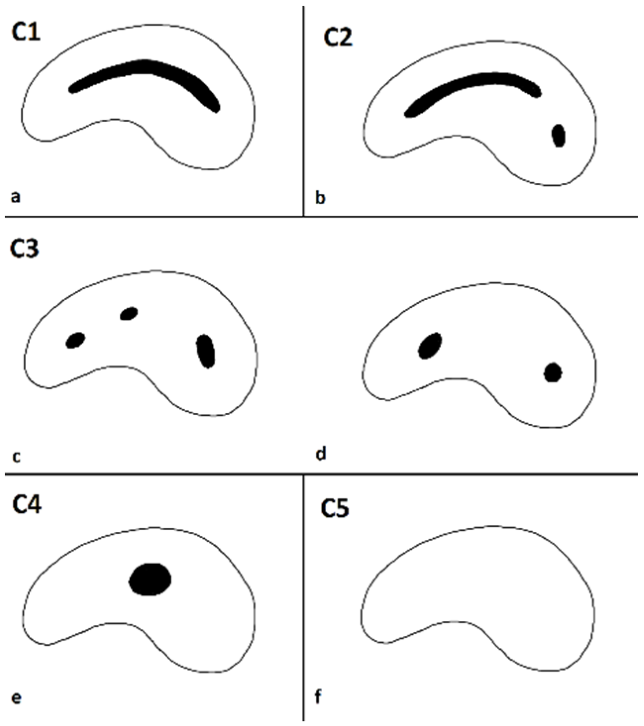
| Root Canal Cross-Section | Root Canal Type According Melton’s Classification | ||||
|---|---|---|---|---|---|
| C1 | C2 | C3c | C3d | C4 | |
| Coronal third | 14.1% | 24.4% | 35.9% | 15.4% | 10.3% |
| Middle third | 6.4% | 21.8% | 44.9% | 19.2% | 7.7% |
| Apical third | 10.3% | 14.1% | 33.3% | 26.9% | 15.4% |
| Vertucci Type | Type I | Type II | Type III | Type IV | Type V | Type VI | Type VII | Type VIII |
|---|---|---|---|---|---|---|---|---|
| Frequency | 12.0% | 41.0% | 0.8% | 45.1% | 0.4% | 0.5% | 0% | 0.1% |
Disclaimer/Publisher’s Note: The statements, opinions and data contained in all publications are solely those of the individual author(s) and contributor(s) and not of MDPI and/or the editor(s). MDPI and/or the editor(s) disclaim responsibility for any injury to people or property resulting from any ideas, methods, instructions or products referred to in the content. |
© 2024 by the authors. Licensee MDPI, Basel, Switzerland. This article is an open access article distributed under the terms and conditions of the Creative Commons Attribution (CC BY) license (https://creativecommons.org/licenses/by/4.0/).
Share and Cite
Mimica, S.; Simeon, P.; Miletić, I.; Baraba, A.; Jukić Krmek, S. Cone-Beam Computed Tomography Analysis of the Root Canal Morphology of Lower Second Molars in the Croatian Subpopulation. Appl. Sci. 2024, 14, 871. https://doi.org/10.3390/app14020871
Mimica S, Simeon P, Miletić I, Baraba A, Jukić Krmek S. Cone-Beam Computed Tomography Analysis of the Root Canal Morphology of Lower Second Molars in the Croatian Subpopulation. Applied Sciences. 2024; 14(2):871. https://doi.org/10.3390/app14020871
Chicago/Turabian StyleMimica, Sarah, Paris Simeon, Ivana Miletić, Anja Baraba, and Silvana Jukić Krmek. 2024. "Cone-Beam Computed Tomography Analysis of the Root Canal Morphology of Lower Second Molars in the Croatian Subpopulation" Applied Sciences 14, no. 2: 871. https://doi.org/10.3390/app14020871
APA StyleMimica, S., Simeon, P., Miletić, I., Baraba, A., & Jukić Krmek, S. (2024). Cone-Beam Computed Tomography Analysis of the Root Canal Morphology of Lower Second Molars in the Croatian Subpopulation. Applied Sciences, 14(2), 871. https://doi.org/10.3390/app14020871