
Differences in Soft Tissue Wound Healing between Immediate and Delayed Implant Placement: An Experimental Preclinical In Vivo Investigation
 ,
,
Abstract
1. Introduction
2. Materials and Methods
2.1. Experimental Animals, Housing and Husbandry
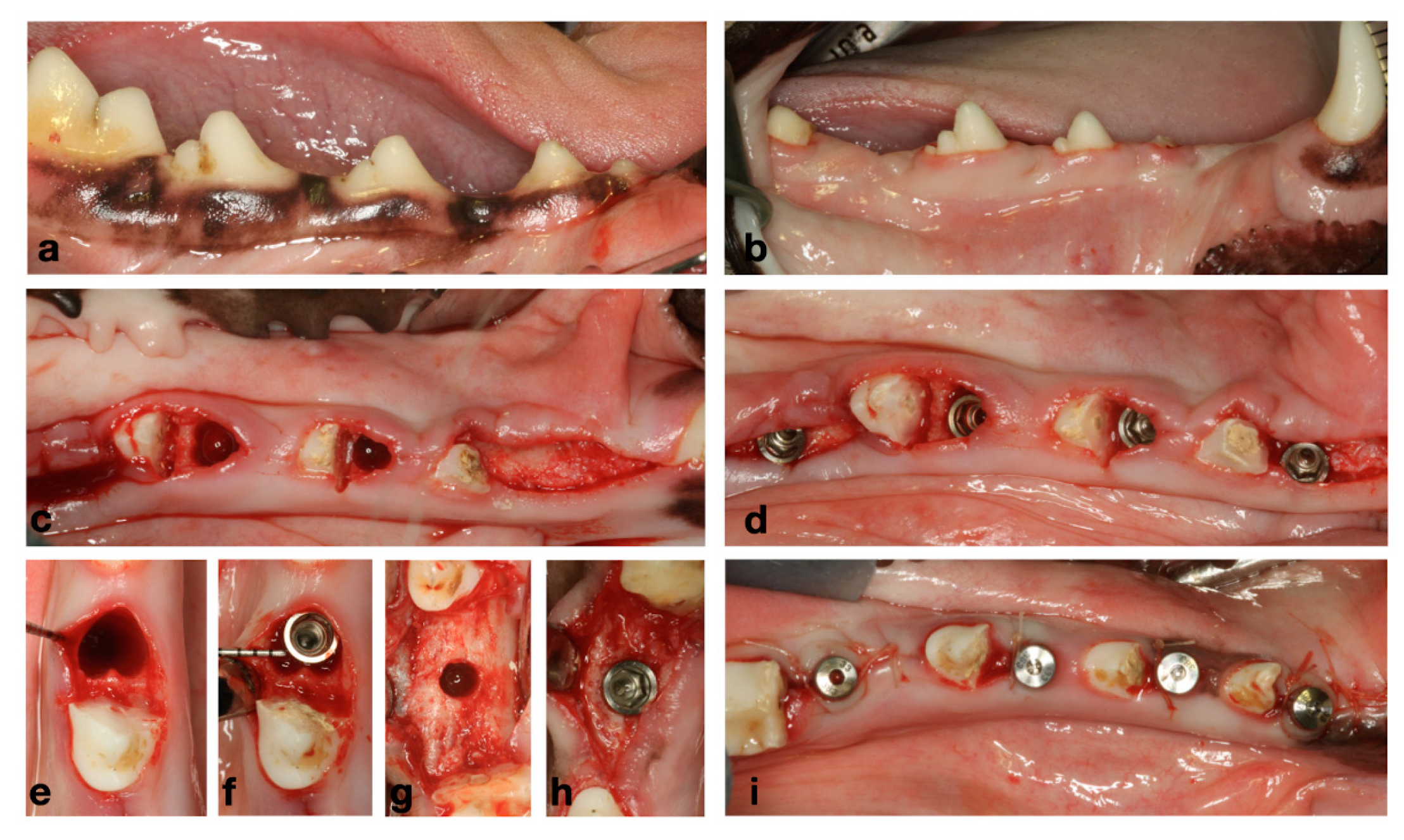
2.2. Experimental Design and Experiments
2.3. Materials
2.4. Veterinarian Care and Postoperative Protocol
2.5. Euthanasia
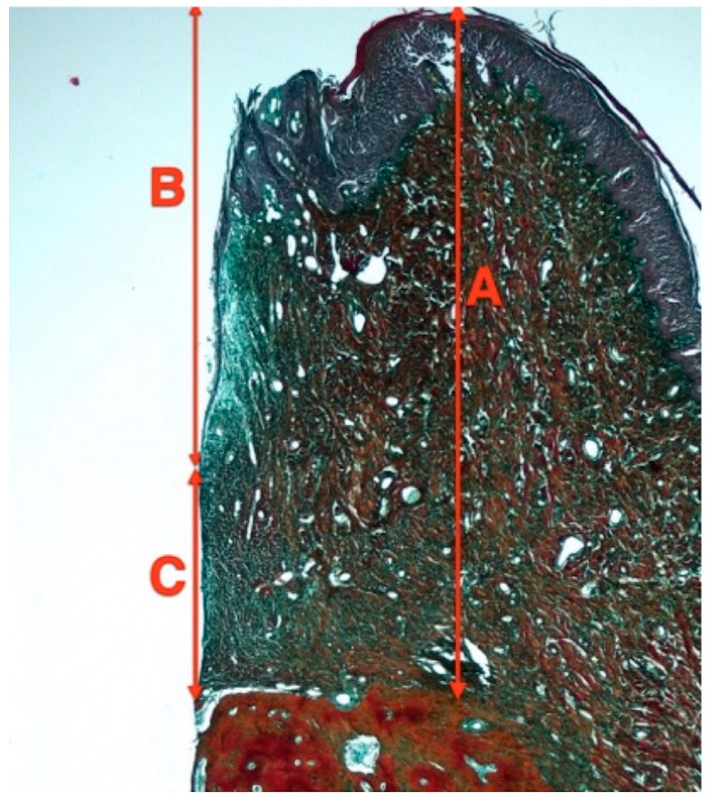
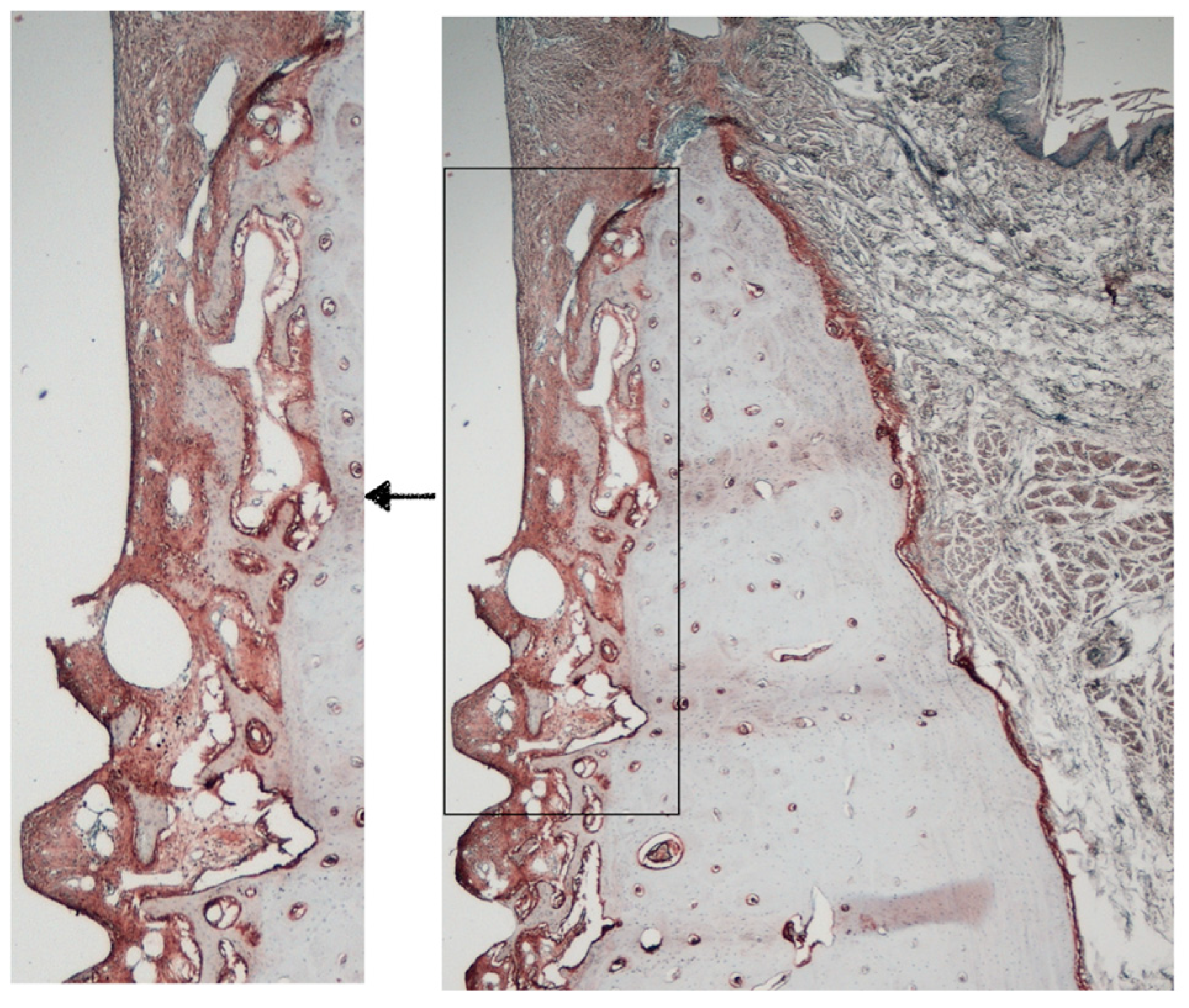
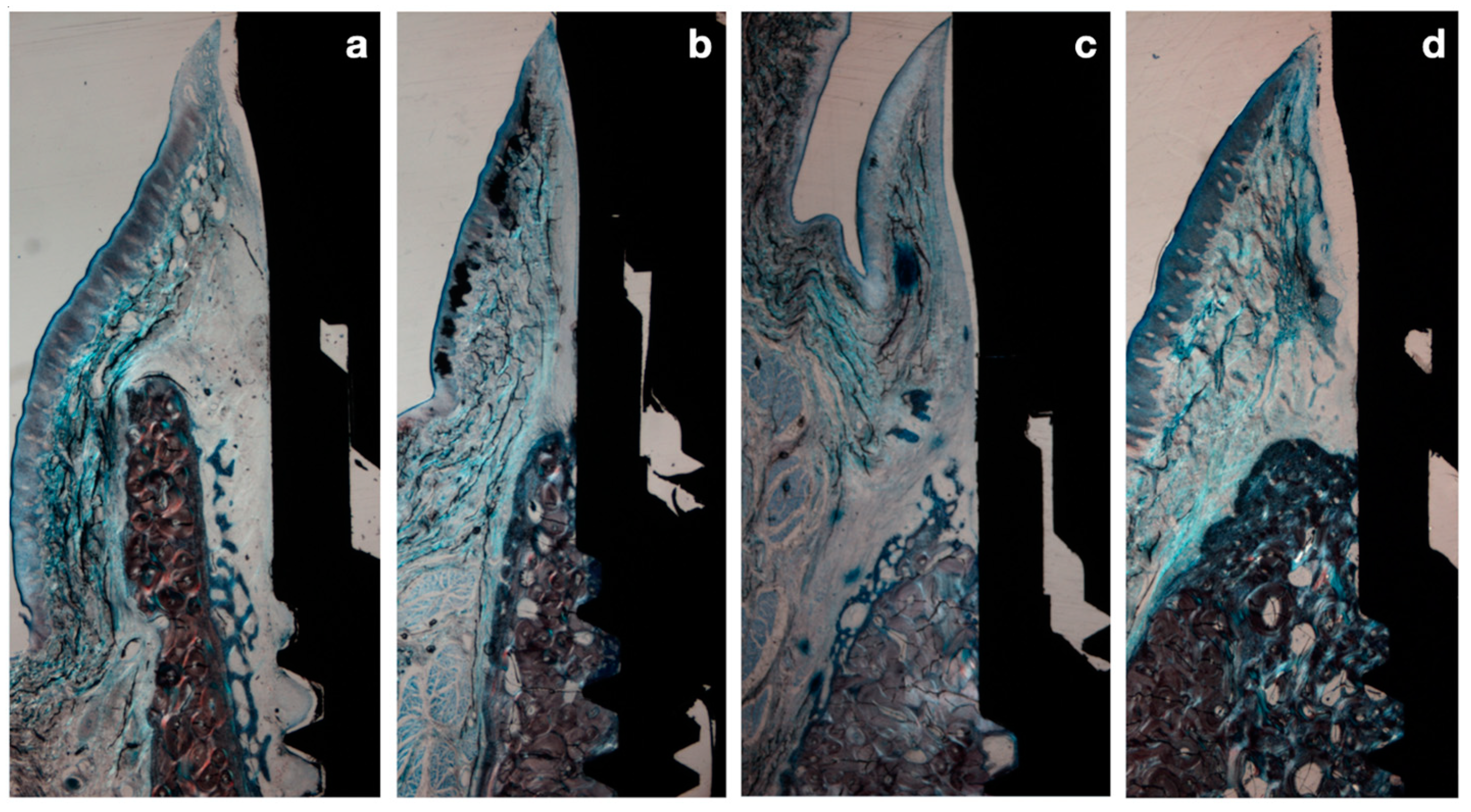
2.6. Histological Preparation and Histomorphometry Analysis
- Height of the Peri-implant mucosa (PM-B)
- Height of the supra-crestal soft tissues (PM-Bc)
- Height of the barrier epithelium (PM-aJE)
- Height of the connective tissue (aJE-B)
- Shoulder of the implant (I)
- Most coronal bone to implant contact (B)
- Most coronal bone crest (Bc)
- Margin of the peri-implant mucosa (PM)
- Apical border of the junctional epithelium (aJE)
2.7. Immunohistochemical Analysis
- Bone Metabolism: Alkaline phosphatase (ALP), Osteopontin (OPN), Matrix Metalloproteinase 2 (MMP2) and tartrate-resistant acid phosphatase (TRAP)
- Immune response: CD4 and Myeloperoxidase (MPO)
- Angiogenesis: Vascular Endothelial Growth Factor (VEGF)
2.8. Statistical Analysis
3. Results
3.1. Preclinical Observations
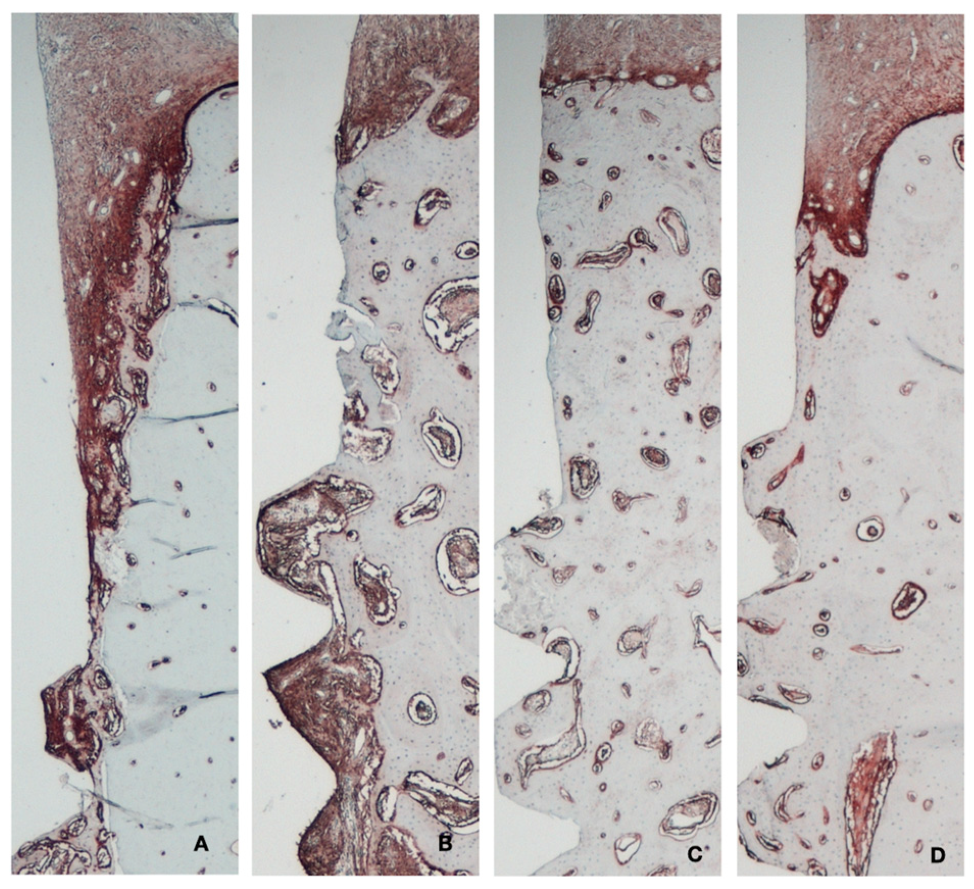
3.2. Histology and Histomorphometry
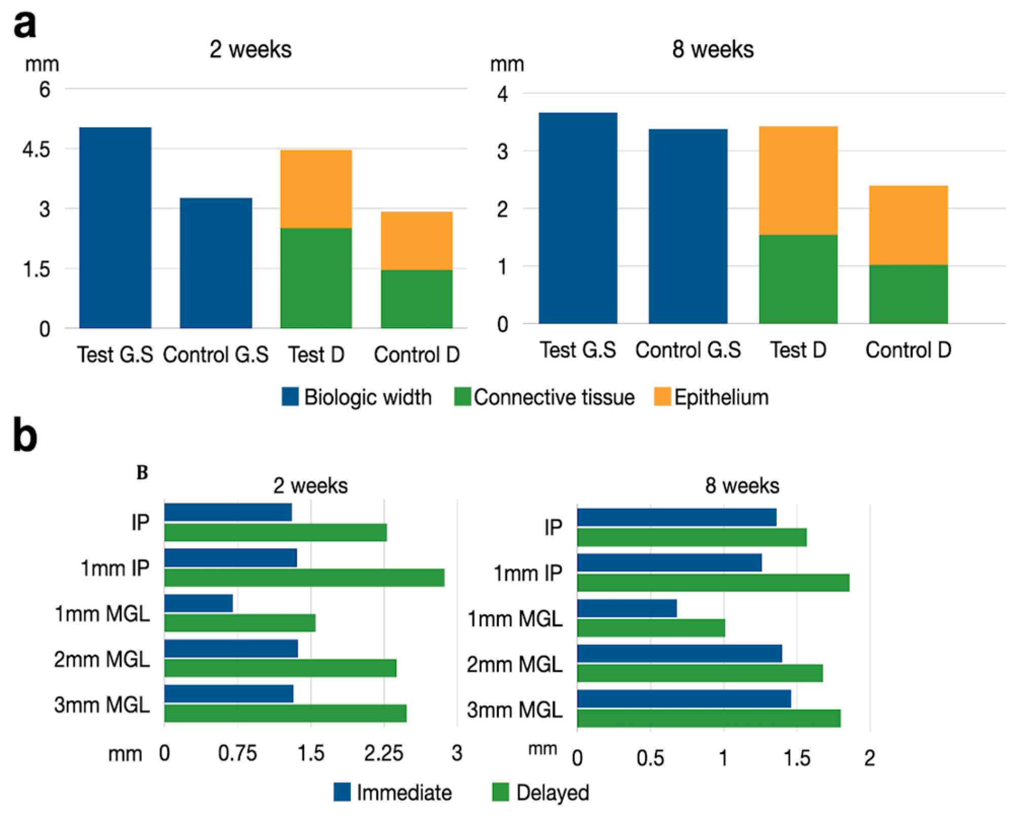
3.3. Histometric Results
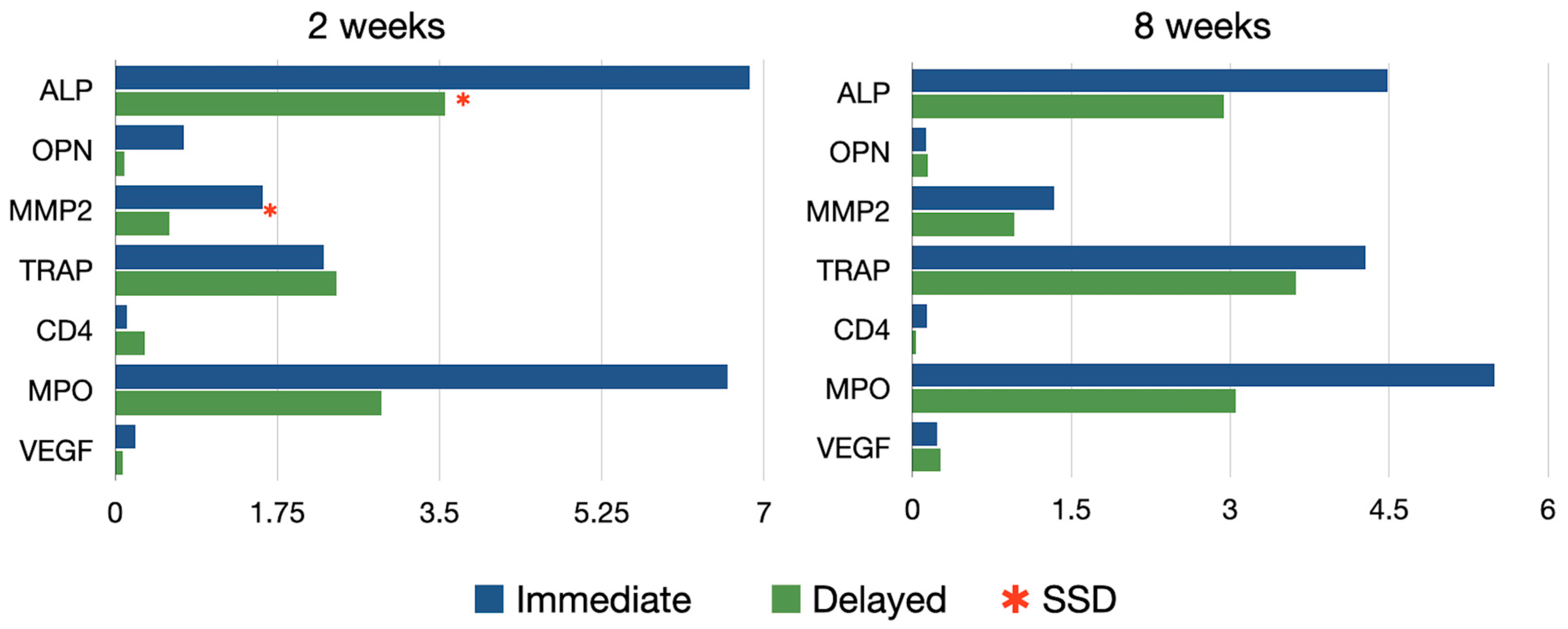
3.4. Immunohistochemical Results
3.5. Bone Metabolism
3.6. Inflammatory Reaction
3.7. Angiogenesis
4. Discussion
5. Conclusions
Author Contributions
Funding
Institutional Review Board Statement
Informed Consent Statement
Data Availability Statement
Conflicts of Interest
Appendix A

| Time | Group | Alkaline Phosphatase | Matrix Metalloproteinase 2 | Osteopontin | Tartrate Resistant Acid Phosphatase | Vascular Endothelial Growth Factor | Lymphocites CD4 | Myeloperoxidase |
|---|---|---|---|---|---|---|---|---|
| 2 wk | Test | 6.85 ± 5.17 * | 1.59 ± 1.64 * | 0.11 ± 0.18 | 2.25 ± 1.99 | 0.22 ± 0.46 | 0.13 ± 0.18 | 6.61 ± 7.31 |
| 2 wk | Control | 3.56 ± 2.31 | 0.58 ± 0.48 | 0.09 ± 0.14 | 2.39 ± 2.05 | 0.08 ± 0.14 | 0.31 ± 0.64 | 2.87 ± 1.24 |
| 8 wk | Test | 4.48 ± 3.31 | 1.34 ± 1.85 | 0.13 ± 0.33 | 4.28 ± 1.55 | 0.23 ± 0.42 | 0.13 ± 0.27 | 5.49 ± 3.9 |
| 8 wk | Control | 2.93 ± 1.38 | 0.95 ± 1.48 | 0.14 ± 0.33 | 3.62 ± 2.21 | 0.26 ± 0.29 | 0.03 ± 0.04 | 3.05 ± 2.33 |
References
- Araujo, M.G.; Lindhe, J. Dimensional ridge alterations following tooth extraction. An experimental study in the dog. J. Clin. Periodontol. 2005, 32, 212–218. [Google Scholar] [CrossRef] [PubMed]
- Araujo, M.G.; Sukekava, F.; Wennstrom, J.L.; Lindhe, J. Ridge alterations following implant placement in fresh extraction sockets: An experimental study in the dog. J. Clin. Periodontol. 2005, 32, 645–652. [Google Scholar] [CrossRef] [PubMed]
- Vina-Almunia, J.; Candel-Marti, M.E.; Cervera-Ballester, J.; Garcia-Mira, B.; Calvo-Guirado, J.L.; Penarrocha-Oltra, D.; Penarrocha-Diago, M. Buccal bone crest dynamics after immediate implant placement and ridge preservation techniques: Review of morphometric studies in animals. Implant. Dent. 2013, 22, 155–160. [Google Scholar] [CrossRef] [PubMed]
- Matarasso, S.; Salvi, G.E.; Iorio Siciliano, V.; Cafiero, C.; Blasi, A.; Lang, N.P. Dimensional ridge alterations following immediate implant placement in molar extraction sites: A six-month prospective cohort study with surgical re-entry. Clin. Oral. Implant. Res. 2009, 20, 1092–1098. [Google Scholar] [CrossRef] [PubMed]
- Vignoletti, F.; Sanz, M. Immediate implants at fresh extraction sockets: From myth to reality. Periodontol. 2000 2014, 66, 132–152. [Google Scholar] [CrossRef]
- De Rouck, T.; Collys, K.; Cosyn, J. Immediate single-tooth implants in the anterior maxilla: A 1-year case cohort study on hard and soft tissue response. J. Clin. Periodontol. 2008, 35, 649–657. [Google Scholar] [CrossRef]
- Tonetti, M.S.; Cortellini, P.; Graziani, F.; Cairo, F.; Lang, N.P.; Abundo, R.; Conforti, G.P.; Marquardt, S.; Rasperini, G.; Silvestri, M.; et al. Immediate versus delayed implant placement after anterior single tooth extraction: The timing randomized controlled clinical trial. J. Clin. Periodontol. 2017, 44, 215–224. [Google Scholar] [CrossRef]
- Blanco, J.; Alves, C.C.; Nunez, V.; Aracil, L.; Munoz, F.; Ramos, I. Biological width following immediate implant placement in the dog: Flap vs. flapless surgery. Clin. Oral. Implant. Res. 2010, 21, 624–631. [Google Scholar] [CrossRef]
- Vignoletti, F.; de Sanctis, M.; Berglundh, T.; Abrahamsson, I.; Sanz, M. Early healing of implants placed into fresh extraction sockets: An experimental study in the beagle dog. III: Soft tissue findings. J. Clin. Periodontol. 2009, 36, 1059–1066. [Google Scholar] [CrossRef]
- Rimondini, L.; Bruschi, G.B.; Scipioni, A.; Carrassi, A.; Nicoli-Aldini, N.; Giavaresi, G.; Fini, M.; Mortellaro, C.; Giardino, R. Tissue healing in implants immediately placed into postextraction sockets: A pilot study in a mini-pig model. Oral. Surg. Oral. Med. Oral. Pathol. Oral. Radiol. Endod. 2005, 100, e43–e50. [Google Scholar] [CrossRef]
- Mair, B.; Fuerst, G.; Kubitzky, P.; Tangl, S.; Bergmeister, H.; Losert, U.; Watzek, G.; Gruber, R. The anti-angiogenic substance TNP-470 impairs peri-implant bone formation: A pilot study in the rabbit metaphysis model. Clin. Oral. Implant. Res. 2007, 18, 370–375. [Google Scholar] [CrossRef] [PubMed]
- Ortiz-Vigon, A.; Martinez-Villa, S.; Suarez, I.; Vignoletti, F.; Sanz, M. Histomorphometric and immunohistochemical evaluation of collagen containing xenogeneic bone blocks used for lateral bone augmentation in staged implant placement. Int. J. Implant. Dent. 2017, 3, 24. [Google Scholar] [CrossRef] [PubMed]
- Hauschka, P.V.; Lian, J.B.; Cole, D.E.; Gundberg, C.M. Osteocalcin and matrix Gla protein: Vitamin K-dependent proteins in bone. Physiol. Rev. 1989, 69, 990–1047. [Google Scholar] [CrossRef] [PubMed]
- Thieu, M.K.L.; Stoetzel, S.; Rahmati, M.; El Khassawna, T.; Verket, A.; Sanz-Esporrin, J.; Sanz, M.; Ellingsen, J.E.; Haugen, H.J. Immunohistochemical comparison of lateral bone augmentation using a synthetic TiO2 block or a xenogeneic graft in chronic alveolar defects. Clin. Implant. Dent. Relat. Res. 2023, 25, 57–67. [Google Scholar] [CrossRef] [PubMed]
- Vignoletti, F.; Abrahamsson, I. Quality of reporting of experimental research in implant dentistry. Critical aspects in design, outcome assessment and model validation. J. Clin. Periodontol. 2012, 39 (Suppl. S12), 6–27. [Google Scholar] [CrossRef]
- Vignoletti, F.; Sanz-Esporrin, J.; Sanz-Martin, I.; Nunez, J.; Luengo, F.; Sanz, M. Ridge alterations after implant placement in fresh extraction sockets or in healed crests: An experimental in vivo investigation. Clin. Oral. Implant. Res. 2019, 30, 353–363. [Google Scholar] [CrossRef]
- Sanz-Martin, I.; Vignoletti, F.; Nunez, J.; Permuy, M.; Munoz, F.; Sanz-Esporrin, J.; Fierravanti, L.; Shapira, L.; Sanz, M. Hard and soft tissue integration of immediate and delayed implants with a modified coronal macrodesign: Histological, micro-CT and volumetric soft tissue changes from a pre-clinical in vivo study. J. Clin. Periodontol. 2017, 44, 842–853. [Google Scholar] [CrossRef]
- Berglundh, T.; Lindhe, J.; Jonsson, K.; Ericsson, I. The topography of the vascular systems in the periodontal and peri-implant tissues in the dog. J. Clin. Periodontol. 1994, 21, 189–193. [Google Scholar] [CrossRef]
- Schroeder, H.E. Ultrastructure of the junctional epithelium of the human gingiva. Helv. Odontol. Acta 1969, 13, 65–83. [Google Scholar]
- Cha, J.K.; Pla, R.; Vignoletti, F.; Jung, U.W.; Sanz-Esporrin, J.; Sanz, M. Immunohistochemical characteristics of lateral bone augmentation using different biomaterials around chronic peri-implant dehiscence defects: An experimental in vivo study. Clin. Oral. Implant. Res. 2021, 32, 569–580. [Google Scholar] [CrossRef]
- Cardaropoli, G.; Araujo, M.; Lindhe, J. Dynamics of bone tissue formation in tooth extraction sites. An experimental study in dogs. J. Clin. Periodontol. 2003, 30, 809–818. [Google Scholar] [CrossRef] [PubMed]
- Berglundh, T.; Abrahamsson, I.; Welander, M.; Lang, N.P.; Lindhe, J. Morphogenesis of the peri-implant mucosa: An experimental study in dogs. Clin. Oral. Implant. Res. 2007, 18, 1–8. [Google Scholar] [CrossRef] [PubMed]
- Hermann, J.S.; Buser, D.; Schenk, R.K.; Cochran, D.L. Crestal bone changes around titanium implants. A histometric evaluation of unloaded non-submerged and submerged implants in the canine mandible. J. Periodontol. 2000, 71, 1412–1424. [Google Scholar] [CrossRef]
- Blanco, J.; Nunez, V.; Aracil, L.; Munoz, F.; Ramos, I. Ridge alterations following immediate implant placement in the dog: Flap versus flapless surgery. J. Clin. Periodontol. 2008, 35, 640–648. [Google Scholar] [CrossRef]
- Monje, A.; Chappuis, V.; Monje, F.; Munoz, F.; Wang, H.L.; Urban, I.A.; Buser, D. The Critical Peri-implant Buccal Bone Wall Thickness Revisited: An Experimental Study in the Beagle Dog. Int. J. Oral. Maxillofac. Implant. 2019, 34, 1328–1336. [Google Scholar] [CrossRef]
- Ellis, R.; Chen, S.; Davies, H.; Fitzgerald, W.; Xu, J.; Darby, I. Primary stability and healing outcomes of apically tapered and straight implants placed into fresh extraction sockets. A pre-clinical in vivo study. Clin. Oral. Implant. Res. 2020, 31, 705–714. [Google Scholar] [CrossRef]
- Zhang, C.; Zhao, X.; Qiao, S.; Zhang, X.; Lai, H.; Gu, Y. Peri-implant tissue alteration around tissue-level and bone-level implants in fresh extraction sockets: A histomorphometric study in dogs. Ann. Transl. Med. 2021, 9, 335. [Google Scholar] [CrossRef] [PubMed]
- Becker, J.; Ferrari, D.; Herten, M.; Kirsch, A.; Schaer, A.; Schwarz, F. Influence of platform switching on crestal bone changes at non-submerged titanium implants: A histomorphometrical study in dogs. J. Clin. Periodontol. 2007, 34, 1089–1096. [Google Scholar] [CrossRef]
- Caneva, M.; Salata, L.A.; de Souza, S.S.; Baffone, G.; Lang, N.P.; Botticelli, D. Influence of implant positioning in extraction sockets on osseointegration: Histomorphometric analyses in dogs. Clin. Oral. Implant. Res. 2010, 21, 43–49. [Google Scholar] [CrossRef]
- Chappuis, V.; Engel, O.; Shahim, K.; Reyes, M.; Katsaros, C.; Buser, D. Soft Tissue Alterations in Esthetic Postextraction Sites: A 3-Dimensional Analysis. J. Dent. Res. 2015, 94, 187S–193S. [Google Scholar] [CrossRef]
- Halling Linder, C.; Ek-Rylander, B.; Krumpel, M.; Norgard, M.; Narisawa, S.; Millan, J.L.; Andersson, G.; Magnusson, P. Bone Alkaline Phosphatase and Tartrate-Resistant Acid Phosphatase: Potential Co-regulators of Bone Mineralization. Calcif. Tissue Int. 2017, 101, 92–101. [Google Scholar] [CrossRef] [PubMed]
- Stucki, U.; Schmid, J.; Hammerle, C.F.; Lang, N.P. Temporal and local appearance of alkaline phosphatase activity in early stages of guided bone regeneration. A descriptive histochemical study in humans. Clin. Oral. Implant. Res. 2001, 12, 121–127. [Google Scholar] [CrossRef]
- Sela, J.; Gross, U.M.; Kohavi, D.; Shani, J.; Dean, D.D.; Boyan, B.D.; Schwartz, Z. Primary mineralization at the surfaces of implants. Crit. Rev. Oral. Biol. Med. 2000, 11, 423–436. [Google Scholar] [CrossRef] [PubMed]
- Li, X.; Jin, L.; Tan, Y. Different roles of matrix metalloproteinase 2 in osteolysis of skeletal dysplasia and bone metastasis (Review). Mol. Med. Rep. 2020, 23, 70. [Google Scholar] [CrossRef]
- Dew, G.; Murphy, G.; Stanton, H.; Vallon, R.; Angel, P.; Reynolds, J.J.; Hembry, R.M. Localisation of matrix metalloproteinases and TIMP-2 in resorbing mouse bone. Cell Tissue Res. 2000, 299, 385–394. [Google Scholar] [CrossRef]
- Accorsi-Mendonca, T.; Paiva, K.B.; Zambuzzi, W.F.; Cestari, T.M.; Lara, V.S.; Sogayar, M.C.; Taga, R.; Granjeiro, J.M. Expression of matrix metalloproteinases-2 and -9 and RECK during alveolar bone regeneration in rat. J. Mol. Histol. 2008, 39, 201–208. [Google Scholar] [CrossRef] [PubMed]
- Leeming, D.J.; Alexandersen, P.; Karsdal, M.A.; Qvist, P.; Schaller, S.; Tankó, L.B. An update on biomarkers of bone turnover and their utility in biomedical research and clinical practice. Eur. J. Clin. Pharmacol. 2006, 62, 781–792. [Google Scholar] [CrossRef] [PubMed]
- Yi, H.Y.; Park, Y.S.; Pippenger, B.E.; Lee, B.; Miron, R.J.; Dard, M. Dimensional Changes Following Immediate and Delayed Implant Placement: A Histomorphometric Study in the Canine. Int. J. Oral. Maxillofac. Implant. 2017, 32, 541–546. [Google Scholar] [CrossRef]
- Pei, X.; Wang, L.; Chen, C.; Yuan, X.; Wan, Q.; Helms, J.A. Contribution of the PDL to Osteotomy Repair and Implant Osseointegration. J. Dent. Res. 2017, 96, 909–916. [Google Scholar] [CrossRef]
- Yuan, X.; Pei, X.; Zhao, Y.; Li, Z.; Chen, C.H.; Tulu, U.S.; Liu, B.; Van Brunt, L.A.; Brunski, J.B.; Helms, J.A. Biomechanics of Immediate Postextraction Implant Osseointegration. J. Dent. Res. 2018, 97, 987–994. [Google Scholar] [CrossRef]
- Schwarz, F.; Rothamel, D.; Herten, M.; Ferrari, D.; Sager, M.; Becker, J. Lateral ridge augmentation using particulated or block bone substitutes biocoated with rhGDF-5 and rhBMP-2: An immunohistochemical study in dogs. Clin. Oral. Implant. Res. 2008, 19, 642–652. [Google Scholar] [CrossRef]


| Time | Group |
|---|---|
| Alkaline phosphatase * | (1:200) |
| Osteopontin * | (1:150) |
| Matrix Metalloproteinase 2 * | (1:200) |
| Tartrate-resistant acid phosphatase † | Direct use |
| Lymphocites CD4 * | (1:250) |
| Myeloperoxidase * | (1:150) |
| Vascular endothelial growth factor * | (1:100) |
| BW (PM-B) Grounds 2 Weeks | BW (PM-BC) Decalcified 2 Weeks | Epithelium (PM-aJE) 2 Weeks | Connective Tissue (aJE-B) 2 Weeks | BW (PM-B) Grounds 8 Weeks | BW (PM-BC) Decalcified 8 Weeks | Epithelium (PM-aJE) 8 Weeks | Connective Tissue (aJE-B) 8 Weeks | |
|---|---|---|---|---|---|---|---|---|
| Test | 5.034 ± 1.125 | 4.466 ± 0.779 * | 1.951 ± 0.522 | 2.518 ± 0.675 | 3.665 ± 0.995 | 3.433 ± 0.838 | 1.887 ± 0.559 | 1.543 ± 0.753 |
| Control | 3.265 ± 0.637 | 2.927 ± 0.351 | 1.463 ± 0.511 | 1.464 ± 0.325 | 3.38 ± 0.34 | 2.4 ± 0.378 | 1.374 ± 0.232 | 1.026 ± 0.269 |
| I–J | 1.058 p = 0.203 | 1.54 p = 0.028 | 0.488 p = 0.981 | 1.003 p = 0.316 | 0.165 p = 1.000 | 1.033 p = 0.088 | 0.513 p = 0.420 | 0.517 p = 0.880 |
| Implant Platform 2 Weeks | 1 mm Apical to IP 2 Weeks | 1 mm Apical to FGM 2 Weeks | 2 mm Apical to FGM 2 Weeks | 3 mm Apical to FGM 2 Weeks | Implant Platform 8 Weeks | 1 mm Apical to IP 8 Weeks | 1 mm Apical to FGM 8 Weeks | 2 mm Apical to FGM 8 Weeks | 3 mm Apical to FGM 8 Weeks | |
|---|---|---|---|---|---|---|---|---|---|---|
| Test | 1.305 ± 0.323 | 1.360 ± 0.360 | 0.704 ± 0.195 | 1.375 ± 0.346 | 1.321 ± 0.386 | 1.362 ± 0.622 | 1.263 ± 0.182 | 0.680 ± 0.222 | 1.402 ± 0.685 | 1.468 ± 0.445 |
| Control | 2.280 * ± 0.432 | 2.870 * ± 0.316 | 1.558 * ± 0.382 | 2.388 ± 0.409 | 2.488 * ± 0.565 | 1.573 ± 0.608 | 1.867 ± 0.309 | 1.013 ± 0.240 | 1.68 ± 0.496 | 1.887 ± 0.493 |
| I–J | −0.975 p = 0.019 | −1.510 p = 0.000 | −0.599 p = 0.002 | −0.691 p = 0.238 | −1.116 p = 0.002 | −0.205 p = 1.000 | −0.603 p = 0.019 | −0.279 p = 0.502 | −0.220 p = 1.000 | −0.367 p = 0.961 |
Disclaimer/Publisher’s Note: The statements, opinions and data contained in all publications are solely those of the individual author(s) and contributor(s) and not of MDPI and/or the editor(s). MDPI and/or the editor(s) disclaim responsibility for any injury to people or property resulting from any ideas, methods, instructions or products referred to in the content. |
© 2024 by the authors. Licensee MDPI, Basel, Switzerland. This article is an open access article distributed under the terms and conditions of the Creative Commons Attribution (CC BY) license (https://creativecommons.org/licenses/by/4.0/).
Share and Cite
Pla, R.; Sanz-Esporrin, J.; Palombo, D.; Vignoletti, F.; Luengo, F.; Sanz, M. Differences in Soft Tissue Wound Healing between Immediate and Delayed Implant Placement: An Experimental Preclinical In Vivo Investigation. Appl. Sci. 2024, 14, 8469. https://doi.org/10.3390/app14188469
Pla R, Sanz-Esporrin J, Palombo D, Vignoletti F, Luengo F, Sanz M. Differences in Soft Tissue Wound Healing between Immediate and Delayed Implant Placement: An Experimental Preclinical In Vivo Investigation. Applied Sciences. 2024; 14(18):8469. https://doi.org/10.3390/app14188469
Chicago/Turabian StylePla, Rafael, Javier Sanz-Esporrin, David Palombo, Fabio Vignoletti, Fernando Luengo, and Mariano Sanz. 2024. "Differences in Soft Tissue Wound Healing between Immediate and Delayed Implant Placement: An Experimental Preclinical In Vivo Investigation" Applied Sciences 14, no. 18: 8469. https://doi.org/10.3390/app14188469
APA StylePla, R., Sanz-Esporrin, J., Palombo, D., Vignoletti, F., Luengo, F., & Sanz, M. (2024). Differences in Soft Tissue Wound Healing between Immediate and Delayed Implant Placement: An Experimental Preclinical In Vivo Investigation. Applied Sciences, 14(18), 8469. https://doi.org/10.3390/app14188469