Article

Study on the Anti-Interference Performance of Substrate-Free PEDOT:PSS ECG Electrodes

School of Mechanical Engineering, Guangxi University, Nanning 53004, China
*
Author to whom correspondence should be addressed.
Appl. Sci. 2024, 14(14), 6367; https://doi.org/10.3390/app14146367
Submission received: 28 May 2024 / Revised: 19 July 2024 / Accepted: 19 July 2024 / Published: 22 July 2024
(This article belongs to the Section Biomedical Engineering)

Abstract

Substrate-free electrodes are promising dry electrodes for long-term physiological electrical signal monitoring due to their ultra-thinness, conformal contact, and stable skin–electrode impedance. However, the response of substrate-free electrodes to various disturbances during electrocardiogram (ECG) monitoring and the corresponding optimization needs to be investigated. This paper investigates the specific effects of various influencing factors on skin–electrode impedance and ECG during electrocardiogram (ECG) detection. The research utilizes substrate-free poly(3,4-ethylenedioxythiophene)/poly(styrene-sulfonate) (PEDOT:PSS) electrodes. The investigation employs several methods, including skin–electrode impedance comparison, ECG waveform analysis, spectrum analysis, and signal-to-noise ratio (SNR) evaluation. To avoid the impact of physiological state differences in subjects at different times, relevant data were only compared with the same group of experiments conducted in the same period. The results demonstrate that the substrate-free conformal contact PEDOT:PSS electrode has more stable skin–electrode impedance and could obtain a more stable ECG than partial contact electrodes (the SNR of the partial contact and conformal contact electrodes are 1.2768 ± 4.0299 dB and 7.2637 ± 1.4897 dB, respectively). Furthermore, the ECG signal quality of the substrate-free conformal contact PEDOT:PSS electrode was independent of the electrode area and shape (the SNRs of the large, medium, and small electrodes are 4.0447 ± 0.4616 dB, 3.9115 ± 0.5885 dB, and 4.1556 ± 0.5557 dB, respectively; the SNRs of the circular, square, and triangular electrodes are 9.2649 ± 0.6326 dB, 9.2471 ± 0.6806 dB, and 9.1514 ± 0.6875 dB, respectively), showing high signal acquisition capability that is the same as microneedle electrodes and better than fabric electrodes. The results of clothing friction effects show that skin–electrode impedance stability was important for ECG stability, while the impedance value was not (the SNRs of friction and non-friction electrodes are 2.4128 ± 7.0784 dB and 9.2164 ± 0.6696 dB, respectively). Moreover, the skin–electrode impedance maintains stability even at a high breathing frequency, but the ECG signal fluctuates at a high breathing frequency. This experiment demonstrates that even when the skin–electrode impedance remains stable, the ECG signal can still be susceptible to interference from other factors. This study suggests that substrate-free PEDOT:PSS that could form conformal contact with the skin has higher skin–electrode impedance stability and could measure a high ECG signal even with a small electrode area, demonstrating its potential as dry ECG electrodes, but the interference from other physiological electrical signals may require better circuit design.

1. Introduction

Bioelectrical monitoring often utilizes various types of electrodes, each with distinct characteristics and applications. Wet electrodes, which require the use of electrolyte gels, are commonly used due to their low impedance and reliable signal quality. However, the long-term use of wet electrodes can lead to skin irritation and performance degradation. Dry electrodes, which do not include gels, include several subtypes such as metal dry electrodes, microneedle array dry electrodes, and substrate-free dry electrodes [1]. Each type offers unique advantages and challenges in terms of comfort, stability, and signal quality. Specifically, substrate-free dry electrodes stand out due to their ultra-thinness, lightweight, and skin compatibility, showing great potential in bioelectrical monitoring, especially for long-term applications such as electrocardiogram (ECG) monitoring [2,3,4,5,6]. Compared to traditional wet electrodes, substrate-free dry electrodes do not require electrolyte gels, which helps prevent skin irritation and long-term property degradation, which makes them more suitable for extended ECG monitoring sessions. Substrate-free poly(3,4-ethylenedioxythiophene)/poly(styrene-sulfonate) (PEDOT:PSS) electrodes can achieve long-term conformal contact with the skin, ensuring comfortability and a stable skin–electrode interface [7]. Several recent studies have demonstrated the superior performance of substrate-free electrodes in applications of heart rate/ECG monitoring [8,9,10,11].
ECG monitoring encompasses interferences including baseline wander, power line interference, electromyographic (EMG) artifacts, and motion artifacts, among others [12,13,14]. Traditionally, low and stable skin–electrode impedance is considered to be crucial for obtaining high-quality ECG signals [15,16,17]. Vargas Lunas [18] discovered that skin–electrode impedance can directly reflect changes at the skin–electrode interface and ECG interference. E. Huigen [19] found that ECG interference during wet-electrode ECG monitoring primarily originates from the electrolyte–skin interface. Theoretically, a larger electrode area corresponds to a lower electrode impedance, suggesting that a larger electrode area can enhance signal quality. Lee et al. [20] corroborated this finding in their study involving ECG monitoring with fabric electrodes. However, some studies have found that it is the stability of the impedance, rather than the absolute value, that affects ECG quality. Beckmann [21] found that the stability of the impedance was more critical to obtaining a high-quality ECG signal than the absolute value of the impedance. Lempka et al. [22] found that the signal quality of biological surface potentials measured by microneedle array dry electrodes was independent of electrode size but related to the skin–electrode interface. Substrate-free PEDOT:PSS dry electrodes offer superior breathability and comfort, but their working principle for ECG monitoring is not entirely the same as that of wet electrodes and metal dry electrodes [7,23]. Studies on the response of substrate-free electrodes to various disturbances during ECG monitoring and the relationship between impedance changes and ECG signal quality would be beneficial for further understanding the substrate-free dry electrode and the corresponding performance improvement.
In this paper, the anti-interference performance of substrate-free PEDOT:PSS ECG electrodes was systematically studied. The influencing factors for ECG detection were classified into static influencing factors (including the contact state of skin–electrode interface, electrode size, and electrode shape) and dynamic influencing factors (including breathing frequency and the friction between electrode and cloth). The relevant skin–electrode impedance measurements and ECG detection experiments were designed for each of these factors, and the results were analyzed by using impedance, waveforms, spectrograms, and SNRs. The results were analyzed to determine how each factor affects skin–electrode impedance and ECG. The results reveal that unstable skin–electrode contact leads to impedance fluctuations, resulting in ECG waveform distortion and interference. While electrode size affects the impedance value, its impact on the ECG signal is small. Clothing friction, however, significantly affects the dynamic impedance, causing ECG interference. Furthermore, different breathing frequencies, although not influencing impedance, result in ECG interference, which may be related to other physiological electrical signals, especially electromyography.

2. Materials and Methods

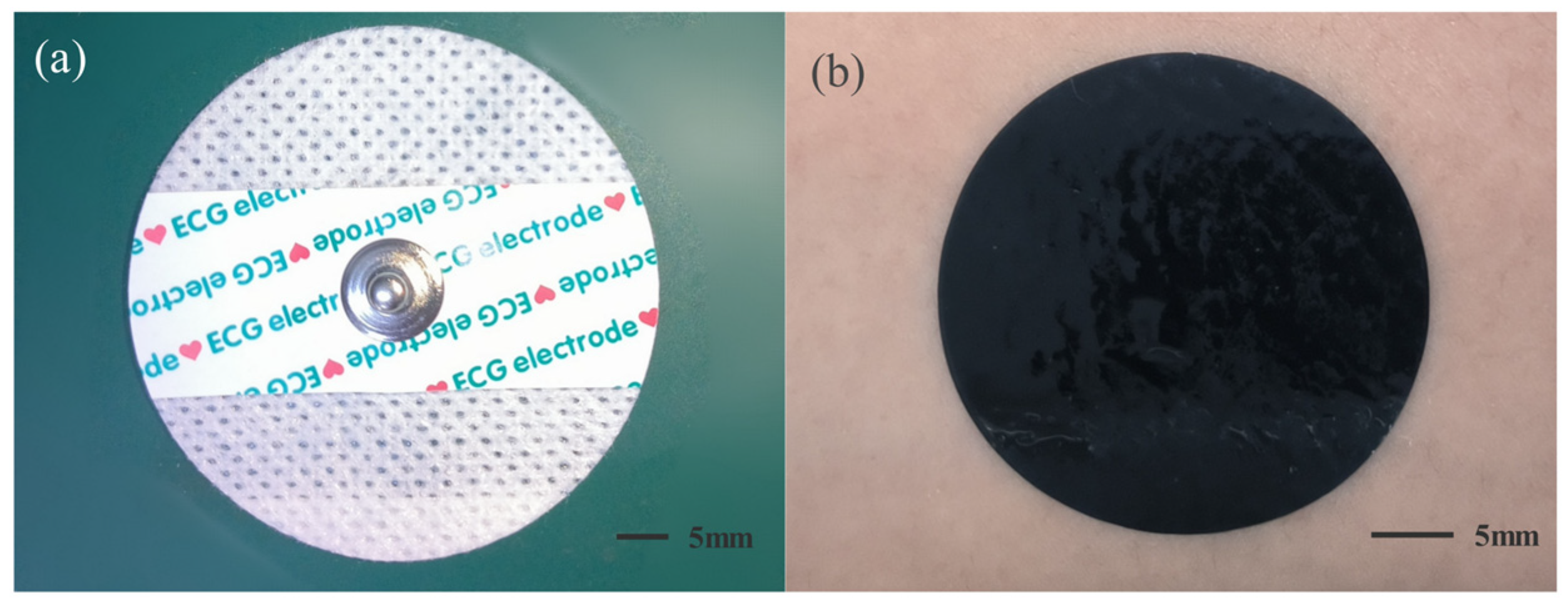
2.1. Experimental Electrodes

The experimental electrodes used in this paper are the substrate-free PEDOT:PSS electrode and commercial Ag/AgCl wet electrodes as reference, as shown in Figure 1. The method of connecting the substrate-free dry electrodes is as follows: Using the preparation method in the previous study to prepare a sheet of PEDOT:PSS film [7], the film was cut into electrodes of the appropriate size and shape, as well as an auxiliary connecting film, using a programmable cutter (SILH-Cameo4 Silhouette). The PEDOT:PSS film electrodes were transferred to the skin with the help of DI water, during which process the electrode would turn into a gel and then dry on the skin again [24]; the electrodes were connected to the wires with the help of connecting film. To prevent the connecting film from contacting the skin, non-conductive medical tape was used for isolation.

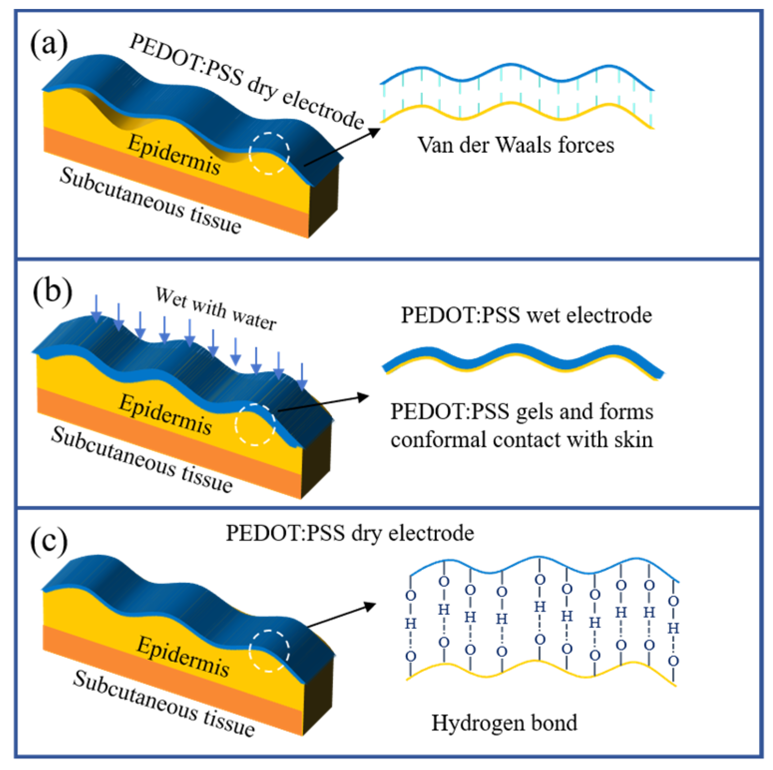
2.2. Electrode Adhesion Mechanism

When PEDOT:PSS dry electrodes are attached to the skin surface, they make partial contact with skin primarily through van der Waals forces, as illustrated in Figure 2a. Upon hydration and gelation, as depicted in Figure 2b, the PEDOT:PSS electrode turns into soft hydrogel and the modulus increase to a low level, enabling conformal contact with the skin. Following the redrying of the PEDOT:PSS electrode on the skin surface, shown in Figure 2c, it regains its original Young’s modulus and thickness. Because the gelation and drying processes of the PEDOT:PSS electrode occur predominantly in the thickness dimension, the redried electrode could maintain conformal contact with the skin. In addition, during the rehydration and subsequent drying of the PEDOT:PSS electrode, water molecules facilitate the formation of intermolecular hydrogen bonds between the electrode and the skin surface. Since hydrogen bonds are stronger molecular forces compared to van der Waals forces, the adhesion strength between the PEDOT:PSS electrode and the skin significantly enhances, enabling self-adhesion. Moreover, this process increases the contact area with the skin, thereby further boosting the adhesion strength between the PEDOT:PSS electrode and the skin [7].

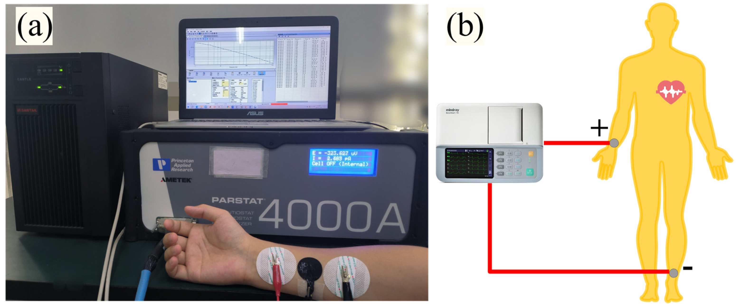
2.3. Impedance Mesurement and ECG Monitoring

The skin–electrode impedance was measured using an electrochemical workstation (Parstat 4000A, Ametek Ltd., Berwyn, PA, USA) with a three-electrode measurement system, in which the counter electrode (CE) and the working electrode (WE) carry the current, the working electrode and the reference electrode (RE) measure the voltage, and the sensing electrode (SE) and the working electrode (WE) are clamped together. Skin–electrode impedance was measured on the right arm of a healthy male, and the skin under test needed to be cleaned and wiped with alcohol before the experiment to prevent the possible presence of oils and grease from affecting the results. As shown in Figure 3a, the subject was seated in front of the electrochemical workstation in a relaxed state to prevent skin tightness, the test electrodes were attached to the inner side of the right arm, where two Ag/AgCl wet electrodes were connected to the reference and counter electrodes in the three-electrode system, with the center distance between the two sets at about 8 cm, and the substrate-free PEDOT:PSS dry electrodes were attached in the middle and connected with the working electrodes and the sensing electrodes for the EIS measurements. The signal amplitude (Vrms) was set to 10 mV, and the frequency scanning range was set to 1–1000 Hz in the static influence factor experiment. Since most of the energy of the ECG was concentrated in 0.1–35 Hz in the dynamic influence factor experiment, the EIS frequency points were set to 1, 10, and 30 Hz for the experiments.
For ECG measurements, as shown in Figure 3b, the substrate-free PEDOT:PSS electrodes were employed as the sensing electrodes, affixed to the inner side of the subject’s right wrist and left ankle skin surface and connected to a digital multi-lead ECG machine (BeneHeart R3, Mindray, Shenzhen, China) for ECG signal acquisition. To better investigate the effects of interference factors on ECG, the ECG machine was set to enable only a fixed 50 Hz power line interference filter, while all other filters or processing functions were disabled.

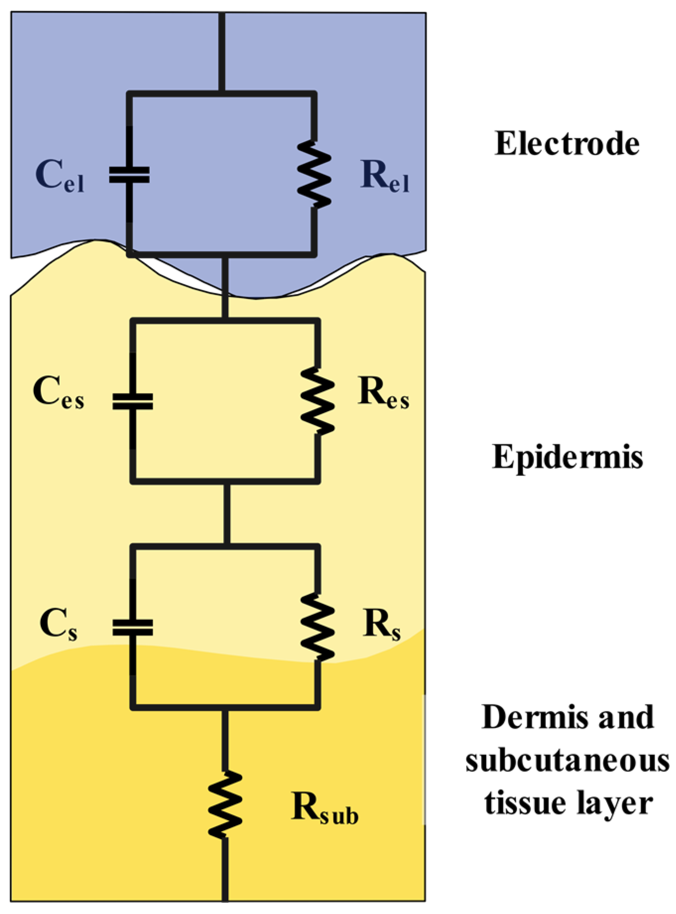
2.4. Skin–Electrode Impedance Equivalent Circuit Model

When a dry electrode is attached to the skin, a typical equivalent circuit model of skin–electrode impedance could be developed as shown in Figure 4. The skin is divided into three layers: the epidermis, the dermis, and the subcutaneous tissue, which can be represented by the equivalent capacitance Cs. The many channels, sweat glands, and hair follicles within the skin connecting the layers can be represented by the equivalent resistance Rs. The subcutaneous tissue layer is well supplied with blood and can therefore be represented by the equivalent resistance Rsub [21].
The capacitance formed by the bubbles or gaps between the dry electrode and the skin, the double-layer capacitance caused by sweat as the electrolyte, and the contact capacitance between the dry electrode and the skin could be expressed by a comprehensive equivalent capacitance Ces [25]. The resistance of electron transfer between the electrode and the skin, as well as the resistance of sweat, can be expressed as the equivalent resistance Res. The capacitance of the substrate-free PEDOT:PSS electrode can be expressed by the equivalent capacitance Cel [23], and its resistivity can be expressed by the equivalent resistance Rel. Combined with the equivalent circuit model analysis, it can be obtained that Rs, Cs, and Rsub are related to human health and the skin state; Rel and Cel are mainly related to the electrode state; Res and Ces are affected by both the electrode and the skin and are most prone to change during the ECG detection process.

2.5. Experimental Design

The static influence factors include the contact state, electrode size, and electrode shape. The substrate-free PEDOT:PSS electrodes that go through water transfer printing can adjust their bending energy and the interface contact energy by gelation and redrying, thereby achieving conformal contact with the skin [7]. The PEDOT:PSS electrodes that do not go through the water transfer printing mainly rely on the van der Waals force to form partial contact with skin. Using this feature, PEDOT:PSS electrodes forming conformal contact and partial contact to skin were fabricated. To study the electrode size effect, three different sizes of circular PEDOT:PSS electrodes with diameters of 10 mm, 20 mm, and 30 mm were prepared for skin–electrode impedance measurements and ECG detection analyses. The thickness of the electrodes used for these studies is 10 μm.
The dynamic influencing factors include friction between clothing on the electrodes and breathing frequency. For the study of dynamic influencing factors, the impedance measurements were carried out at frequencies of 1, 10, and 30 Hz and the number of scanning points was 30. To simulate the impact of clothing on the electrodes in daily life, insulating fabric was utilized to replicate typical friction during everyday activities. This includes applying light back-and-forth friction at a frequency of 2 Hz and an intensity of approximately 0.05 N to the electrodes during the ECG detection process. The electrodes’ positions were marked to enable accurate assessment of any potential movement induced by the friction. The breathing frequency of adults at rest is usually around 12 to 20 breaths per minute. During exercise, sometimes breathing frequency was more than 40 beats per minute. To study the breathing frequency effect, two different breathing rates, slow breathing of about 20 times/min and fast breathing of about 40 times/min, were studied.

2.6. Signal-to-Noise Ratio

To further determine the degree of influence of different factors on ECG, in addition to spectral analysis, signal-to-noise ratio (SNR) was calculated. In this paper, 0.5 Hz high-pass filtering was performed on the original ECG data to filter out the baseline drift [26,27]. Next, a low-pass filter of 35 Hz was applied to filter out high-frequency interference [28]. Then, wavelet variation thresholding was performed using DB8 wavelet type for noise reduction [29] and finally the waveform was smoothed to obtain the ECG data after most of the interference was filtered out and the SNR was calculated for the signal data before and after. The larger SNR indicates that the interference to the ECG is smaller. Due to the differences in the body, skin condition, and electrode attachment position of the subjects in different groups of experiments, only the data of the same group were compared. The SNR calculation equation [30] is as follows:
S N R = 10 l g i = 1 n y i 2 i = 1 n x i y i 2
where x i is the original ECG data, y i is the denoised ECG data, and n is the number of ECG sampling points.

3. Results and Discussion

3.1. Effect of Static Factors on Impedance and ECG

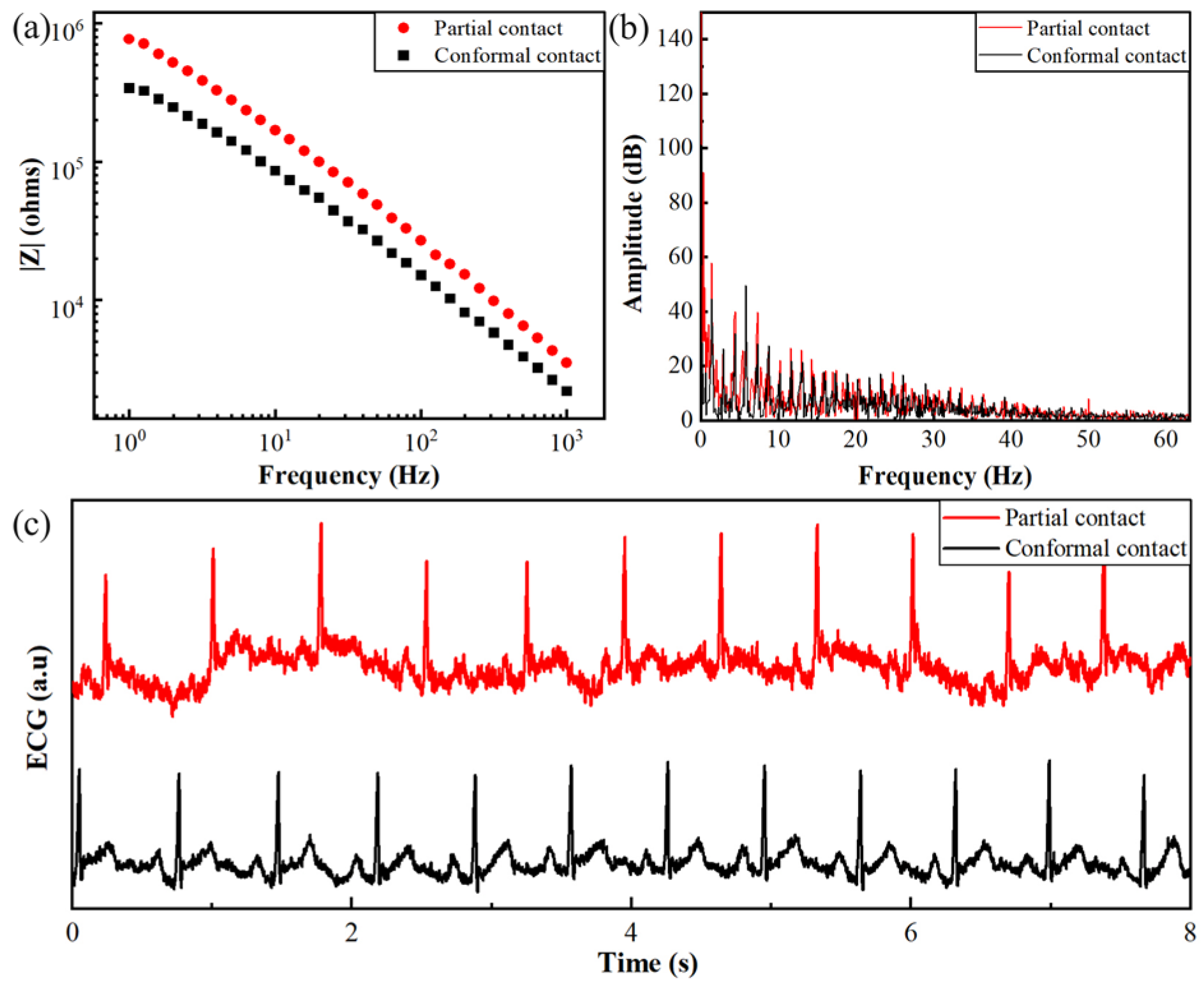
3.1.1. Influence of the Contact State

In the conformal contact state, where the electrode fully conforms to the skin, the electrode–skin interface exhibits stable impedance that does not change when subjected to interference. At the conformal state, the equivalent capacitance (Ces) in the equivalent circuit diagram, as described by Equation (2), is maximized due to the full effective contact area (S) and small spacing (d) between the conductive surfaces.
In contrast to the conformal state, the skin–electrode interface in a partial contact state experiences a reduced contact area and increased gap distance. Consequently, according to Equation (2), the equivalent capacitance Ces decreases as the capacitive area S decreases and the spacing d increases compared to the conformal contact conditions. Simultaneously, the decrease in the contact area leads to an increase in the corresponding electron transfer resistance Res. Since impedance comprises both capacitive and resistive components, the reduction in capacitance and increase in resistance result in a higher overall impedance at the skin–electrode interface in the partial contact state.
C e s = ɛ S d
Figure 5 shows the impedance and ECG results of PEDOT:PSS electrodes at different contact states. As shown in Figure 5a, the skin–electrode impedance was higher for partial contact than conformal contact for the full frequency range. As the frequency increased, the impedance difference between partial contact and conformal contact decreased. At a frequency of 1 Hz, the impedance difference between partial contact and conformal contact was 427.120 kΩ, and at a frequency of 1000 Hz, the impedance difference between partial contact and conformal contact was 1.340 kΩ, indicating that the contact status has more influence on low-frequency impedance, mainly owing to the decrease in Ces. From Figure 5c, it could be seen that the partial contact electrode was more susceptible to interference, and the interference was mainly baseline drift and white noise. As shown in Figure 5b, the interference of the partial contact state was mainly concentrated in the low-frequency band. Comparing the spectral diagrams of the two states in Figure 5b, it could be seen that the partial contact condition exhibited a higher amplitude across the entire frequency range compared to the full conformal contact. In the low-frequency band, the partial contact condition displayed a significantly higher amplitude, indicating its greater susceptibility to baseline drift and low-frequency interference, which aligned with the results shown in Figure 5c. As the frequency increased, the amplitude gradually decreased, but partial contact still exhibited a higher amplitude, with notable peaks at specific frequency points.
The SNR of the partial contact electrode averaged at 1.27675 ± 4.0299 dB, indicating a high variability and lower overall signal quality. This high standard error suggested inconsistent contact quality, likely resulting in significant fluctuations in the recorded signals. On the other hand, the conformal contact electrode exhibited a more stable and higher average SNR of 7.26365 ± 1.4897 dB, reflecting better and more consistent electrode–skin contact, leading to improved signal quality and reliability.
The contact status result shows that the ECG characteristics of the partial contact electrode were much less pronounced and contained more ECG interference, while the conformal contact electrode was insensitive to interference. It was found that the full conformal contact substrate-free PEDOT:PSS electrode could measure the ECG signal more stably.

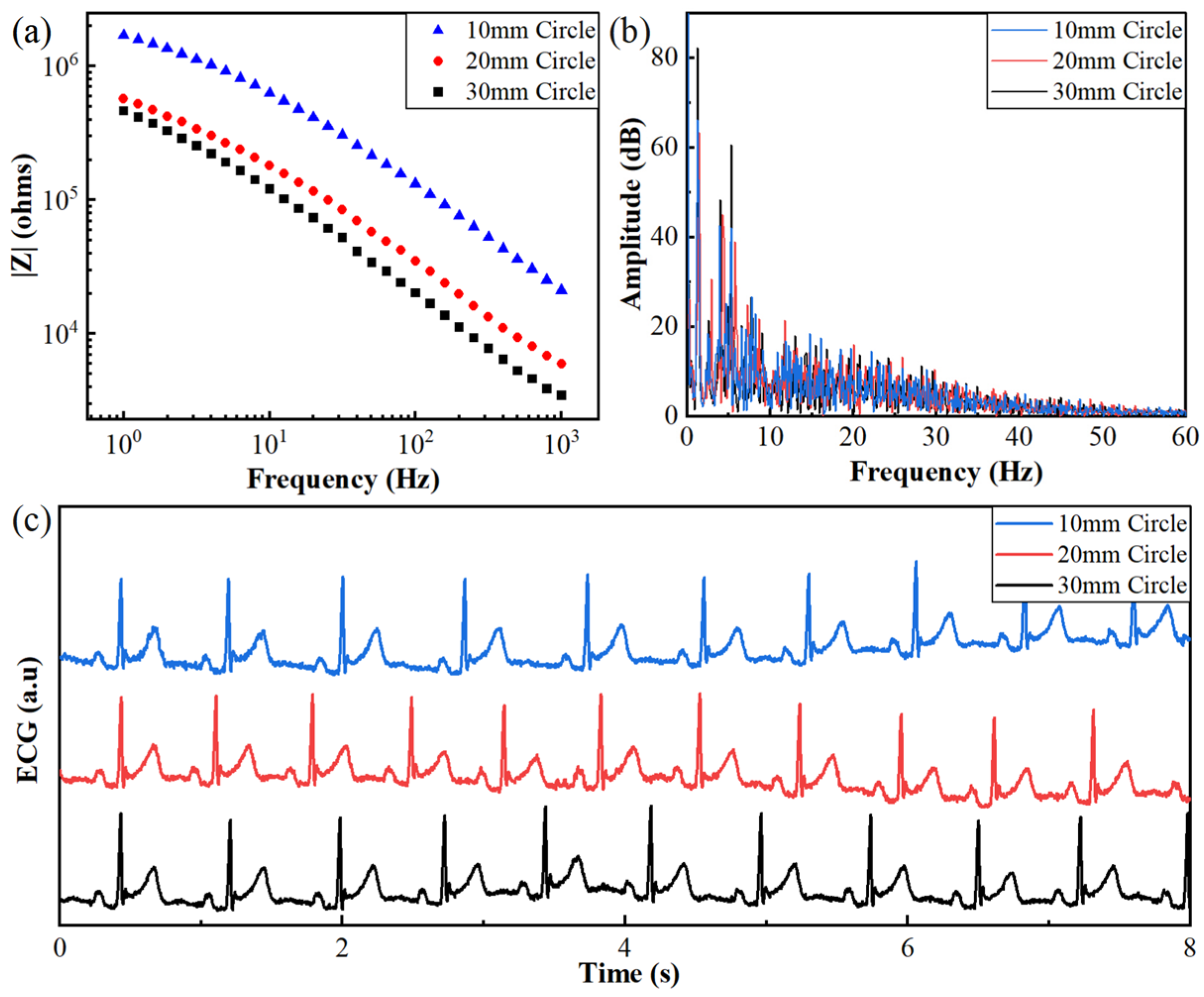
3.1.2. Effect of Electrode Area

Here, electrodes of different sizes are attached to the skin as full conformal contact electrodes. As shown in the equivalent circuit model shown in Figure 4, the electrode size mainly affects the interface resistance Res, the interface capacitance Ces, the electrode resistance Rel, and the electrode capacitance Cel. The smaller the area, the larger the resistance Res, the smaller the Ces, the larger the electrode resistance Rel, and the smaller the electrode capacitance Cel. Therefore, the smaller the electrode area is, the larger the skin–electrode impedance is.
Figure 6 shows the impedance and ECG signal results for different sizes of electrodes. From Figure 6a, it could be seen that the skin–electrode impedance decreased as the electrode area increased, consistent with the results analyzed by the equivalent circuit model. The spectrogram in Figure 6b reveals that the ECG amplitudes for different electrode sizes were similar, with no significant differences. The original ECG waveforms in Figure 6c also demonstrate that ECG waveforms from different electrode sizes exhibited complete waveform characteristics, and the waveform features were consistent across different sizes.
The SNRs were 4.0447 ± 0.4616 dB, 3.9115 ± 0.5885 dB, and 4.1556 ± 0.5557 dB for round electrodes with diameters of 30 mm, 20 mm, and 10 mm, respectively. Analyzing these SNR results in comparison to previous studies offers valuable insights. Previous research by Scott [23] and Syaidah [31] investigated the impact of electrode size on ECG signal quality, with varying conclusions. Scott’s microneedle electrode study suggested that electrode size had a minimal effect on ECG signal quality, while Syaidah’s fabric electrode study found that decreasing electrode size led to decreased ECG signal quality. In this study, a similar trend to Scott’s microneedle electrode findings was observed. Despite the differences in electrode size, the SNR values remained relatively consistent among the electrodes. This suggests that for the full conformal contact substrate-free PEDOT:PSS electrode, electrode size has little effect on ECG signal quality. Even with smaller electrode sizes, high-quality ECG signals were maintained, indicating the robust performance of the electrode design. This consistency in SNRs across varying electrode sizes underscores the importance of achieving full conformal contact and stable skin–electrode impedance, highlighting the effectiveness of the substrate-free PEDOT:PSS electrodes in capturing reliable ECG signals regardless of electrode size.

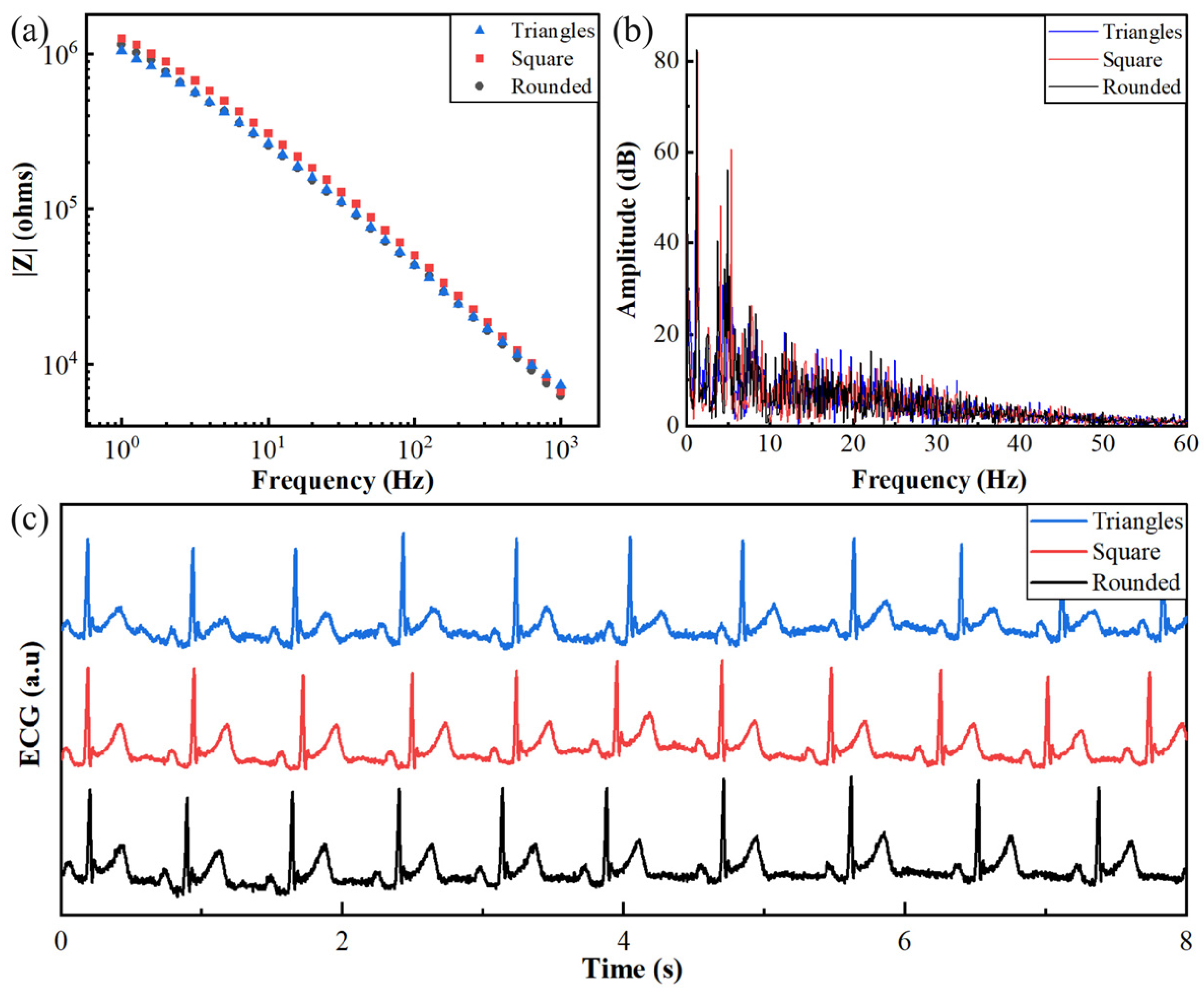
3.1.3. Effect of Electrode Shape

According to the equivalent circuit model shown in Figure 4, for electrodes with different shapes but the same areas, the equivalent resistance Rel, the equivalent capacitance Cel, the electron transfer resistance Res, and the equivalent capacitance Ces between the electrode and skin should be the same. Therefore, for electrodes with different shapes but the same areas, the skin–electrode impedance should be the same and have little effect on ECG.
Figure 7 shows the experimental results of three electrodes of the same area with different shapes. From Figure 7a, it could be seen that the skin–electrode impedance for different shapes of PEDOT:PSS electrodes was similar, which was consistent with the equivalent circuit model analysis. From the spectrum in Figure 7b and the ECG signal in Figure 7c, it can be seen that PEDOT electrodes with different shapes but the same area have no significant impact on the frequency amplitude and ECG waveform.
The SNRs were 9.2649 ± 0.6326 dB, 9.2471 ± 0.6806 dB, and 9.1514 ± 0.6875 dB for the circular electrode, square electrode and triangular electrode, respectively. These results show that the electrode shape of the substrate-free PEDOT:PSS electrode had little effect on the skin–electrode impedance and ECG. Unlike the anisotropic fabric electrodes whose shape had a great impact on skin–electrode impedance and ECG quality [31], the performance of the substrate-free PEDOT:PSS electrodes was independent of the electrode shape and was better than fabric electrodes.

3.2. Effect of Dynamic Factors on Impedance and ECG

3.2.1. Effect of Friction between the Fabric and the Electrode

The friction between the fabric and the electrode dynamically influences the partial gap d between the electrode and the skin and mildly stimulates the skin’s nerves. This friction simulates a real-life scenario, occurring at 2 Hz with an approximate force of 0.05 N in a back-and-forth motion. According to an equivalent circuit model analysis, the friction will mainly affect the equivalent capacitance Ces and will mildly affect the resistance Rs of the skin, causing the skin–electrode impedance to dynamically change with the friction. In addition, based on the marked electrode positions, it was observed that the experimental friction between the fabric and the electrode did not result in electrode displacement.
Figure 8 compares the impedance, ECG waveforms, and spectra of the electrodes under friction and non-friction conditions. Figure 8a–c show the impedance measurements at 1 Hz, 10 Hz, and 30 Hz. The data demonstrate that the effect of friction on impedance increased as the frequency decreased. At low frequencies, the skin–electrode impedance of the electrode subjected to friction exhibited a significant increase compared to the electrode without friction, indicating a more pronounced effect of friction on low-frequency impedance, consistent with the theoretical analysis. Figure 8d,e present the ECG spectra and waveforms, respectively. It could be observed that when the PEDOT:PSS electrode was subjected to friction from insulating fabric, the ECG interference was primarily concentrated in the low-frequency components, reflected on the waveform as baseline interference. In the spectrum of Figure 8d, it could be seen that the signal amplitude for the electrode that suffered friction was higher than that without friction, especially in the low-frequency band. Figure 8e also clearly demonstrates that the ECG signal with friction exhibited more baseline interference.
A quantitative analysis revealed that under friction conditions, the SNR was 2.4128 ± 7.0784 dB, while without friction, it was 9.2164 ± 0.6696 dB, indicating that friction degraded the ECG signal quality. Consequently, during daily wearable ECG monitoring, the friction between clothing and the electrode primarily affected the low-frequency skin–electrode impedance, leading to ECG baseline interference.

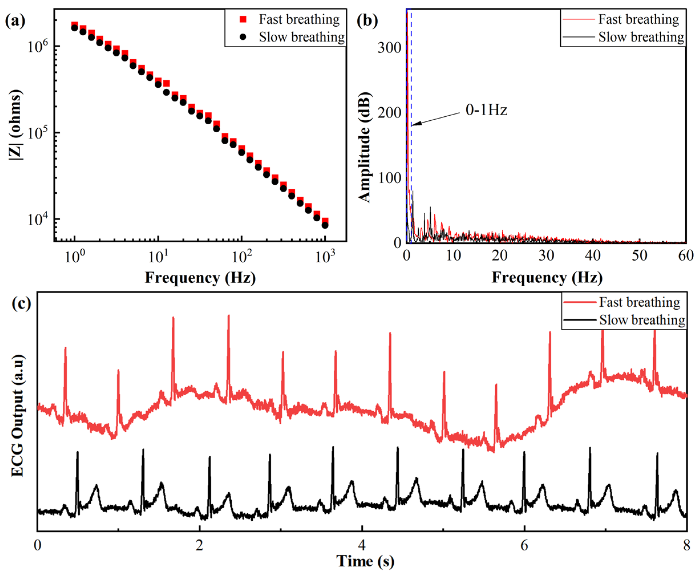
3.2.2. Effect of Breathing Frequency

In this study, two sets of experiments with different breathing frequencies, normal breathing frequency (about 20 breaths/min) and accelerated deeper breathing during exercise (about 40 breaths/min), were conducted to investigate the effects of breathing frequency on skin–electrode impedance and ECG signal. The results are shown in Figure 9. From Figure 9a, it could be observed that the skin–electrode impedance did not differ significantly between the two breathing frequencies. However, the ECG spectrogram analysis in Figure 9b shows that fast breathing led to higher amplitudes at frequencies lower than 1 Hz, indicating more severe baseline interference. Furthermore, the ECG waveform in Figure 9c demonstrates that fast breathing induced an approximate sinusoidal baseline drift, and the characteristics of the P and T waves were not evident.
A quantitative analysis revealed that for slow breathing, the SNR was 5.3715 ± 1.0996 dB, while for fast breathing, it was −7.1862 ± 3.1288 dB. A negative SNR indicates that the noise level is higher than the signal level. This indicates that fast breathing significantly degraded the ECG signal quality compared to slow breathing. These results indicate that different respiration frequencies have no significant effect on the skin–electrode impedance but significantly increase the ECG baseline interference. This also suggests that ECG interference is not solely caused by changes in the skin–electrode impedance, but is also affected by other factors, especially EMG [32,33,34]. Additionally, the results show that even under fast breathing conditions, the substrate-free PEDOT:PSS electrode can maintain a stable skin–electrode interface.

4. Conclusions

This study systematically investigated factors affecting skin–electrode impedance and ECG signals of substrate-free PEDOT:PSS electrodes. The results show that skin–electrode contact status and electrode size significantly influence the skin–electrode impedance. Partial contact leads to poor performance in both impedance stability and ECG signal quality. Conversely, when electrodes form conformal contact with the skin, neither size nor shape significantly affects ECG signals, emphasizing the importance of impedance stability. Among dynamic factors, friction between fabric and electrode caused impedance changes and baseline interference, while breathing frequency did not significantly affect impedance and fast breathing resulted in baseline interference and ECG waveform disruption. The study demonstrates that ECG interference is not solely caused by skin–electrode interface impedance, but that it is also affected by chest and abdominal movements during breathing.
The results of this study provide a theoretical basis for the further optimization of substrate-free electrodes, toward the realization of high-quality, long-term, and comfortable physiological signal monitoring. This research highlights the importance of skin–electrode impedance stability and the impact of dynamic factors like friction between fabric and electrodes and breathing on ECG signal quality. Ongoing improvements in the reliability and durability of substrate-free electrodes are necessary to meet the higher demands of future wearable health-monitoring applications. Future research should focus on correlations between impedance and physiological signals, impedance changes related to human activities, and developing improved interference filtering circuits.

Author Contributions

Conceptualization, Y.C.; methodology, C.L.; formal analysis, C.L.; investigation, C.L.; resources, Y.C.; data curation, Y.C. and C.L.; writing—original draft preparation, C.L.; writing—review and editing, Y.C., C.L., and K.X.; supervision, Y.C.; project administration, C.L.; funding acquisition, Y.C. All authors have read and agreed to the published version of the manuscript.

Funding

This research was funded by the National Natural Science Foundation of China (grant number 52005115).

Institutional Review Board Statement

Not applicable.

Informed Consent Statement

Not applicable.

Data Availability Statement

Data are contained within the article.

Conflicts of Interest

The authors declare no conflicts of interest.

References

  1. Laghlimi, C.; Moutcine, A.; Chtaini, A.; Isaad, J.; Soufi, A.; Ziat, Y.; Amhamdi, H.; Belkhanchi, H. Recent advances in electrochemical sensors and biosensors for monitoring drugs and metabolites in pharmaceutical and biological samples. Admet Dmpk 2023, 11, 151–173. [Google Scholar] [CrossRef] [PubMed]
  2. Bihar, E.; Roberts, T.; Ismailova, E.; Saadaoui, M.; Isik, M.; Sanchez-Sanchez, A.; Mecerreyes, D.; Hervé, T.; De Graaf, J.B.; Malliaras, G.G. Fully Printed Electrodes on Stretchable Textiles for Long-Term Electrophysiology. Adv. Mater. Technol. 2017, 2, 1600251. [Google Scholar] [CrossRef]
  3. Bihar, E.; Roberts, T.; Zhang, Y.; Ismailova, E.; Hervé, T.; Malliaras, G.G.; De Graaf, J.B.; Inal, S.; Saadaoui, M. Fully printed all-polymer tattoo/textile electronics for electromyography. Flex. Print. Electron. 2018, 3, 034004. [Google Scholar] [CrossRef]
  4. Casson, A.J.; Saunders, R.; Batchelor, J.C. Five Day Attachment ECG Electrodes for Longitudinal Bio-Sensing Using Conformal Tattoo Substrates. IEEE Sens. J. 2017, 17, 2205–2214. [Google Scholar] [CrossRef]
  5. Deng, J.; Yuk, H.; Wu, J.; Varela, C.E.; Chen, X.; Roche, E.T.; Guo, C.F.; Zhao, X. Electrical bioadhesive interface for bioelectronics. Nat. Mater. 2020, 20, 229–236. [Google Scholar] [CrossRef]
  6. Wang, Y.; Yin, L.; Bai, Y.; Liu, S.; Wang, L.; Zhou, Y.; Hou, C.; Yang, Z.; Wu, H.; Ma, J.; et al. Electrically compensated, tattoo-like electrodes for epidermal electrophysiology at scale. Sci. Adv. 2020, 6, eabd0996. [Google Scholar] [CrossRef] [PubMed]
  7. Chen, Y.; Zhou, G.; Yuan, X.; Li, C.; Liu, L.; You, H. Substrate-free, ultra-conformable PEDOT: PSS E-tattoo achieved by energy regulation on skin. Biosens. Bioelectron. 2022, 206, 114118. [Google Scholar] [CrossRef]
  8. Jeong, J.-W.; Shin, G.; Park, S.I.; Yu, K.J.; Xu, L.; Rogers, J.A. Soft Materials in Neuroengineering for Hard Problems in Neuroscience. Neuron 2015, 86, 175–186. [Google Scholar] [CrossRef]
  9. Qiao, Y.; Li, X.; Jian, J.; Wu, Q.; Wei, Y.; Shuai, H.; Hirtz, T.; Zhi, Y.; Deng, G.; Wang, Y.; et al. Substrate-Free Multilayer Graphene Electronic Skin for Intelligent Diagnosis. ACS Appl. Mater. Interfaces 2020, 12, 49945–49956. [Google Scholar] [CrossRef]
  10. Aguzin, A.; Dominguez-Alfaro, A.; Criado-Gonzalez, M.; Velasco-Bosom, S.; Picchio, M.L.; Casado, N.; Mitoudi-Vagourdi, E.; Minari, R.J.; Malliaras, G.G.; Mecerreyes, D. Direct ink writing of PEDOT eutectogels as substrate-free dry electrodes for electromyography. Mater. Horiz. 2023, 10, 2516–2524. [Google Scholar] [CrossRef]
  11. Ferrari, L.M.; Sudha, S.; Tarantino, S.; Esposti, R.; Bolzoni, F.; Cavallari, P.; Cipriani, C.; Mattoli, V.; Greco, F. Ultraconformable Temporary Tattoo Electrodes for Electrophysiology. Adv. Sci. 2018, 5, 1700771. [Google Scholar] [CrossRef] [PubMed]
  12. Singh, O.; Sunkaria, R.K. ECG signal denoising via empirical wavelet transform. Australas. Phys. Eng. Sci. Med. 2016, 40, 219–229. [Google Scholar] [CrossRef] [PubMed]
  13. Singh, O.; Sunkaria, R.K. Powerline interference reduction in ECG signals using empirical wavelet transform and adaptive filtering. J. Med. Eng. Technol. 2014, 39, 60–68. [Google Scholar] [CrossRef] [PubMed]
  14. Kalra, A.; Lowe, A.; Al-Jumaily, A. Critical review of electrocardiography measurement systems and technology. Meas. Sci. Technol. 2018, 30, 012001. [Google Scholar] [CrossRef]
  15. Taji, B.; Shirmohammadi, S.; Groza, V.; Batkin, I. Impact of Skin–Electrode Interface on Electrocardiogram Measurements Using Conductive Textile Electrodes. IEEE Trans. Instrum. Meas. 2013, 63, 1412–1422. [Google Scholar] [CrossRef]
  16. Oliveira, C.C.; Machado Da Silva, J.; Trindade, I.G.; Martins, F. Characterization of the electrode-skin impedance of textile electrodes. In Proceedings of the Design of Circuits and Integrated Systems, Madrid, Spain, 26–28 November 2014; pp. 1–6. [Google Scholar]
  17. Gan, Y.; Rahajandraibe, W.; Vauche, R.; Ravelo, B.; Lorriere, N.; Bouchakour, R. A new method to reduce motion artifact in electrocardiogram based on an innovative skin-electrode impedance model. Biomed. Signal Proces. 2022, 76, 103640. [Google Scholar] [CrossRef]
  18. Luna, J.L.V.; Krenn, M.; Ramírez, J.A.C.; Mayr, W. Dynamic Impedance Model of the Skin-Electrode Interface for Transcutaneous Electrical Stimulation. PLoS ONE 2015, 10, e0125609. [Google Scholar] [CrossRef]
  19. Huigen, E.; Peper, A.; Grimbergen, C.A. Investigation into the origin of the noise of surface electrodes. Med. Biol. Eng. Comput. 2002, 40, 332–338. [Google Scholar] [CrossRef] [PubMed]
  20. Lee, S.M.; Sim, K.S.; Kim, K.K.; Lim, Y.G.; Park, K.S. Thin and flexible active electrodes with shield for capacitive electrocardiogram measurement. Med. Biol. Eng. Comput. 2010, 48, 447–457. [Google Scholar] [CrossRef]
  21. Beckmann, L.; Neuhaus, C.; Medrano, G.; Jungbecker, N.; Walter, M.; Gries, T.; Leonhardt, S. Characterization of textile electrodes and conductors using standardized measurement setups. Physiol. Meas. 2010, 31, 233–247. [Google Scholar] [CrossRef]
  22. Lempka, S.F.; Johnson, M.D.; Moffitt, M.A.; Otto, K.J.; Kipke, D.R.; McIntyre, C.C. Theoretical analysis of intracortical microelectrode recordings. J. Neural Eng. 2011, 8, 045006. [Google Scholar] [CrossRef] [PubMed]
  23. Berggren, M.; Malliaras, G.G. How conducting polymer electrodes operate. Science 2019, 364, 233–234. [Google Scholar] [CrossRef]
  24. Lu, B.; Yuk, H.; Lin, S.; Jian, N.; Qu, K.; Xu, J.; Zhao, X. Pure PEDOT:PSS hydrogels. Nat. Commun. 2019, 10, 1043. [Google Scholar] [CrossRef]
  25. Li, G.; Wang, S.; Duan, Y.Y. Towards conductive-gel-free electrodes: Understanding the wet electrode, semi-dry electrode and dry electrode-skin interface impedance using electrochemical impedance spectroscopy fitting. Sens. Actuators B Chem. 2018, 277, 250–260. [Google Scholar] [CrossRef]
  26. Kania, M.; Rix, H.; Fereniec, M.; Zavala-Fernandez, H.; Janusek, D.; Mroczka, T.; Stix, G.; Maniewski, R. The effect of precordial lead displacement on ECG morphology. Med. Biol. Eng. Comput. 2013, 52, 109–119. [Google Scholar] [CrossRef]
  27. Fasano, A.; Villani, V. ECG baseline wander removal and impact on beat morphology: A comparative analysis. In Proceedings of the Computing in Cardiology, Zaragoza, Spain, 22–25 September 2013; pp. 1167–1170. [Google Scholar]
  28. Appathurai, A.; Carol, J.J.; Raja, C.; Kumar, S.; Daniel, A.V.; Malar, A.J.G.; Fred, A.L.; Krishnamoorthy, S. A study on ECG signal characterization and practical implementation of some ECG characterization techniques. Measurement 2019, 147, 106384. [Google Scholar] [CrossRef]
  29. Aqil, M.; Jbari, A.; Bourouhou, A. ECG Signal Denoising by Discrete Wavelet Transform. Int. J. Online Eng. 2017, 13, 51–68. [Google Scholar] [CrossRef]
  30. Mani, R.; Oppenheim, A.V.; Willsky, A.S.; Nawab, S.H. Solutions Manual, Signals & Systems, 2nd ed.; Prentice Hall: Upper Saddle River, NJ, USA, 1997. [Google Scholar]
  31. Saleh, S.M.; Jusob, S.M.; Harun, F.K.C.; Yuliati, L.; Wicaksono, D.H.B. Optimization of Reduced GO-Based Cotton Electrodes for Wearable Electrocardiography. IEEE Sens. J. 2020, 20, 7774–7782. [Google Scholar] [CrossRef]
  32. Devi, R.; Tyagi, H.K.; Kumar, D. Performance Comparison and Applications of Sparsity Based Techniques for Denoising of ECG Signal. In Proceedings of the 2019 6th International Conference on Signal Processing and Integrated Networks (SPIN), Noida, India, 7–8 March 2019; pp. 346–351. [Google Scholar]
  33. Mateo, J.; Sánchez-Morla, E.; Santos, J. A new method for removal of powerline interference in ECG and EEG recordings. Comput. Electr. Eng. 2015, 45, 235–248. [Google Scholar] [CrossRef]
  34. Kuzilek, J.; Kremen, V.; Soucek, F.; Lhotska, L. Independent Component Analysis and Decision Trees for ECG Holter Recording De-Noising. PLoS ONE 2014, 9, e98450. [Google Scholar] [CrossRef]
Figure 1. (a) Ag/AgCl electrode; (b) PEDOT:PSS electrode.
Applsci 14 06367 g001
Figure 2. (a) PEDOT:PSS dry electrode was attached to the skin surface, dominated by van der Waals force; (b) PEDOT:PSS was wetted and gelled on skin; (c) Conformal contact of PEDOT:PSS with skin, dominated by intermolecular hydrogen bonds.
Applsci 14 06367 g002
Figure 3. (a) Skin–electrode impedance measurement; (b) Schematic of ECG detection.
Applsci 14 06367 g003
Figure 4. Schematic diagram of an equivalent circuit model for the skin–electrode impedance.
Applsci 14 06367 g004
Figure 5. Results of different contact states (a) Bode plot of skin–electrode impedance; (b) Spectrogram of different contact states; (c) ECG of different contact states.
Applsci 14 06367 g005
Figure 6. Results for different electrode sizes (a) Bode plot of skin–electrode impedance; (b) Spectrogram of different electrode sizes; (c) ECG waveforms.
Applsci 14 06367 g006
Figure 7. Results of different electrode shapes with the same area (a) Bode plot of skin–electrode impedance; (b) Spectrogram of ECG; (c) ECG waveforms of different shapes.
Applsci 14 06367 g007
Figure 8. (a) Skin–electrode impedance modulus at 1 Hz frequency; (b) Impedance modulus at 10 Hz frequency; (c) Impedance modulus at 30 Hz frequency; (d) Spectrogram of different electrode states; (e) ECG waveforms of different electrode states.
Applsci 14 06367 g008
Figure 9. Experimental results for different breathing frequencies. (a) Bode plot of skin–electrode impedance; (b) Spectrogram of ECG; (c) Waveform of ECG.
Applsci 14 06367 g009
Disclaimer/Publisher’s Note: The statements, opinions and data contained in all publications are solely those of the individual author(s) and contributor(s) and not of MDPI and/or the editor(s). MDPI and/or the editor(s) disclaim responsibility for any injury to people or property resulting from any ideas, methods, instructions or products referred to in the content.

