
Experimental Study to Visualize a Methane Leak of 0.25 mL/min by Direct Absorption Spectroscopy and Mid-Infrared Imaging

 , , , and
Abstract
1. Introduction
2. Materials and Methods
2.1. Experimental Setup
2.2. Image Processing
2.2.1. Preprocessing for DAS
2.2.2. Application of Different DAS Methods (DAS-F, DAS-f and DAS-2f)
- DAS-F method:
- DAS-f method:
- DAS-2f method:
3. Results
3.1. Single Concentration Image of the 0.25 mL/min Methane Leak Scenario
3.2. Spatiotemporal Concentration Image Sequence of the 0.25 mL/min Methane Leak Scenario
4. Discussion
5. Conclusions
Author Contributions
Funding
Data Availability Statement
Conflicts of Interest
Appendix A

Appendix B
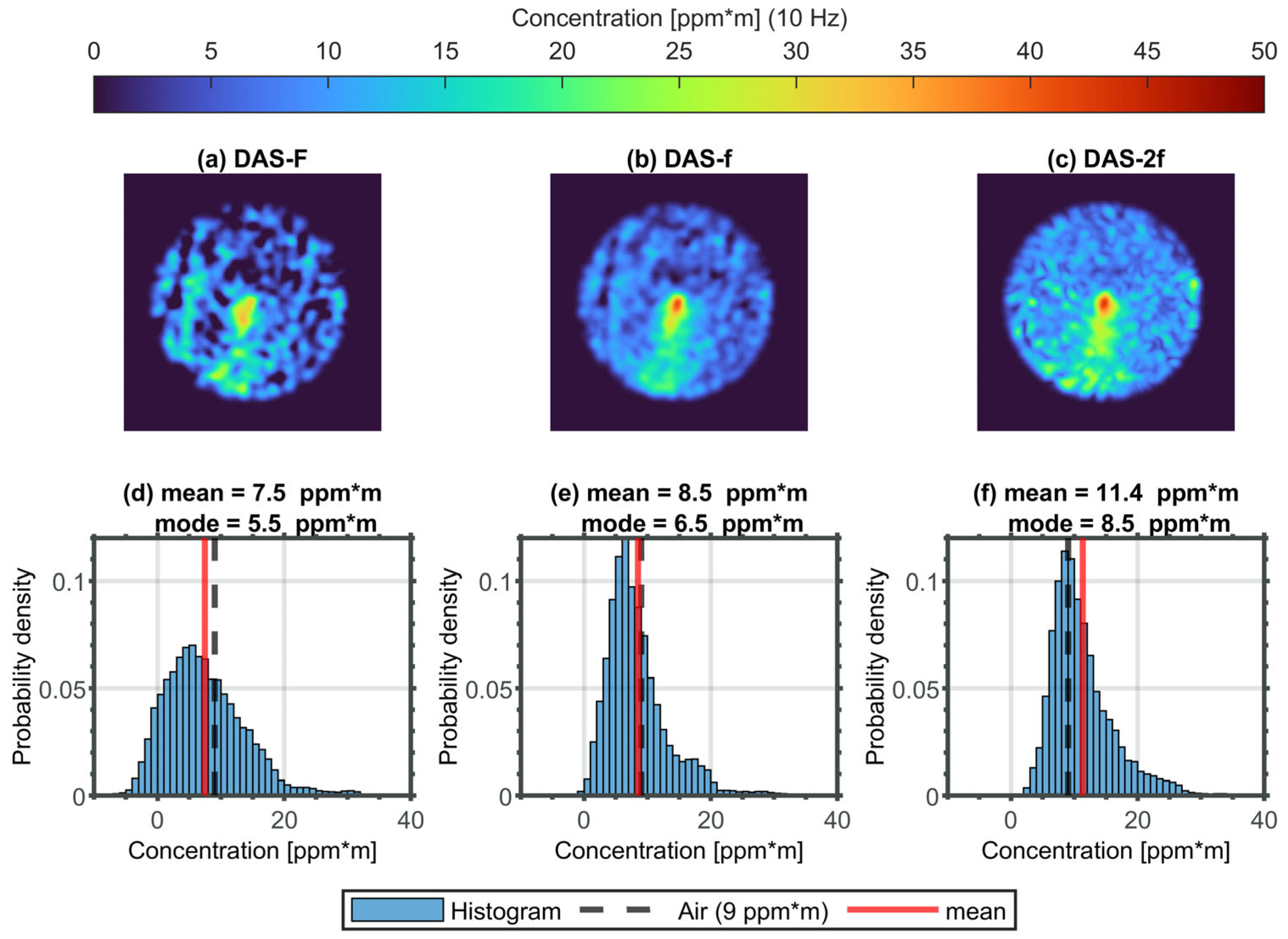
- DAS-f method:As illustrated in Figure 4b, the one-dimensional smoothed absorbance (DAS-f) is in line with the simulation based on the spectroscopic parameters provided by HITRAN. Furthermore, the maximum absorbance (see Figure 4b) is a suitable measure for determining the concentration that can be simply and efficiently transferred to the image sequence. The pixelwise smoothed peak absorbance can be estimated by finding the maximum image along the wavenumber dimension of the DAS-f sequence in Figure 4a. This maximum image is shown in Figure A2a. The transfer function of the smoothed peak absorbance as the DAS-f feature (e.g., ) into a concentration value in ppm*m can be easily derived by further simulation, as described above. The transfer function or smoothed peak absorbance as a function of the concentration shows a linear behavior in the investigated concentration range between 0 and 50 ppm*m, as illustrated in Figure A2b. By means of this function, the feature image of DAS-f can be straightforwardly transferred into the corresponding concentration image.
- DAS-2f method:The DAS-2f concentration is derived in a similar fashion as described for DAS-f.In this case, the peak–dip–peak measure is a suitable measure for determining the concentration that can be simply and efficiently transferred to the image sequence (cf. Figure 4c,d). The peak–dip–peak feature or image will be calculated by a maximum image (first row), minimum image (second row) and maximum image (third row) of the DAS-2f sequence (e.g., ). This DAS-2f feature image is shown in Figure A2c. The corresponding transfer function based on the peak–dip–peak feature () of the smoothed second derivative of the absorbance as a function of the concentration is documented in Figure A2d. This DAS-2f feature also demonstrates a good linearity in the concentration between 0 and 50 ppm*m, such that a methane concentration image can be calculated straightforwardly.

Appendix C


References
- Curl, R.F.; Tittel, F.K. 7 Tunable infrared laser spectroscopy. Annu. Rep. Prog. Chem. Sect. C Phys. Chem. 2002, 98, 219–272. [Google Scholar] [CrossRef]
- Hodgkinson, J.; Tatam, R.P. Optical gas sensing: A review. Meas. Sci. Technol. 2013, 24, 12004. [Google Scholar] [CrossRef]
- Li, J.; Yu, Z.; Du, Z.; Ji, Y.; Liu, C. Standoff Chemical Detection Using Laser Absorption Spectroscopy: A Review. Remote Sens. 2020, 12, 2771. [Google Scholar] [CrossRef]
- Kwaśny, M.; Bombalska, A. Optical Methods of Methane Detection. Sensors 2023, 23, 2834. [Google Scholar] [CrossRef] [PubMed]
- Rothman, L.S. History of the HITRAN Database. Nat. Rev. Phys. 2021, 3, 302–304. [Google Scholar] [CrossRef]
- Gordon, I.E.; Rothman, L.S.; Hargreaves, R.J.; Hashemi, R.; Karlovets, E.V.; Skinner, F.M.; Conway, E.K.; Hill, C.; Kochanov, R.V.; Tan, Y.; et al. The HITRAN2020 molecular spectroscopic database. J. Quant. Spectrosc. Radiat. Transf. 2022, 277, 107949. [Google Scholar] [CrossRef]
- Zeng, Y.; Morris, J. Detection limits of optical gas imagers as a function of temperature differential and distance. J. Air Waste Manag. Assoc. 2019, 69, 351–361. [Google Scholar] [CrossRef] [PubMed]
- Alvarez, R.A.; Zavala-Araiza, D.; Lyon, D.R.; Allen, D.T.; Barkley, Z.R.; Brandt, A.R.; Davis, K.J.; Herndon, S.C.; Jacob, D.J.; Karion, A.; et al. Assessment of methane emissions from the U.S. oil and gas supply chain. Science 2018, 361, 186–188. [Google Scholar] [CrossRef]
- Ravikumar, A.P.; Wang, J.; Brandt, A.R. Are Optical Gas Imaging Technologies Effective For Methane Leak Detection? Environ. Sci. Technol. 2017, 51, 718–724. [Google Scholar] [CrossRef]
- Kemp, C.E.; Ravikumar, A.P.; Brandt, A.R. Comparing Natural Gas Leakage Detection Technologies Using an Open-Source “Virtual Gas Field” Simulator. Environ. Sci. Technol. 2016, 50, 4546–4553. [Google Scholar] [CrossRef]
- Zimmerle, D.; Vaughn, T.; Bell, C.; Bennett, K.; Deshmukh, P.; Thoma, E. Detection Limits of Optical Gas Imaging for Natural Gas Leak Detection in Realistic Controlled Conditions. Environ. Sci. Technol. 2020, 54, 11506–11514. [Google Scholar] [CrossRef]
- Nutt, K.J.; Hempler, N.; Maker, G.T.; Malcolm, G.P.A.; Padgett, M.J.; Gibson, G.M. Developing a portable gas imaging camera using highly tunable active-illumination and computer vision. Opt. Express 2020, 28, 18566–18576. [Google Scholar] [CrossRef]
- Voumard, T.; Wildi, T.; Brasch, V.; Álvarez, R.G.; Ogando, G.V.; Herr, T. AI-enabled real-time dual-comb molecular fingerprint imaging. Opt. Lett. 2020, 45, 6583–6586. [Google Scholar] [CrossRef] [PubMed]
- Strahl, T.; Herbst, J.; Lambrecht, A.; Maier, E.; Steinebrunner, J.; Wöllenstein, J. Methane leak detection by tunable laser spectroscopy and mid-infrared imaging. Appl. Opt. 2021, 60, C68–C75. [Google Scholar] [CrossRef] [PubMed]
- Bergau, M.; Strahl, T.; Scherer, B.; Wöllenstein, J. Real-time active-gas imaging of small gas leaks. J. Sens. Sens. Syst. 2023, 12, 61–68. [Google Scholar] [CrossRef]
- Bergau, M.; Strahl, T.; Ludlum, K.; Scherer, B.; Wöllenstein, J. Flow rate quantification of small methane leaks using laser spectroscopy and deep learning. Process Saf. Environ. Prot. 2024, 182, 752–759. [Google Scholar] [CrossRef]
- Ullah Khan, F.; Guarnizo, G.; Martín-Mateos, P. Direct hyperspectral dual-comb gas imaging in the mid-infrared. Opt. Lett. 2020, 45, 5335–5338. [Google Scholar] [CrossRef]
- Klein, A.; Witzel, O.; Ebert, V. Rapid, time-division multiplexed, direct absorption- and wavelength modulation-spectroscopy. Sensors 2014, 14, 21497–21513. [Google Scholar] [CrossRef]
- Lins, B.; Zinn, P.; Engelbrecht, R.; Schmauss, B. Simulation-based comparison of noise effects in wavelength modulation spectroscopy and direct absorption TDLAS. Appl. Phys. B 2010, 100, 367–376. [Google Scholar] [CrossRef]
- Masiyano, D.; Hodgkinson, J.; Schilt, S.; Tatam, R.P. Self-mixing interference effects in tunable diode laser absorption spectroscopy. Appl. Phys. B 2009, 96, 863–874. [Google Scholar] [CrossRef]
- Werle, P. Accuracy and precision of laser spectrometers for trace gas sensing in the presence of optical fringes and atmospheric turbulence. Appl. Phys. B 2011, 102, 313–329. [Google Scholar] [CrossRef]
- Yan, G.; Zhang, L.; Zheng, C.; Zhang, M.; Zheng, K.; Song, F.; Ye, W.; Zhang, Y.; Wang, Y.; Tittel, F.K. Mobile Vehicle Measurement of Urban Atmospheric CH4/C2H6 Using a Midinfrared Dual-Gas Sensor System Based on Interband Cascade Laser Absorption Spectroscopy. IEEE Trans. Instrum. Meas. 2022, 71, 9509411. [Google Scholar] [CrossRef]


Disclaimer/Publisher’s Note: The statements, opinions and data contained in all publications are solely those of the individual author(s) and contributor(s) and not of MDPI and/or the editor(s). MDPI and/or the editor(s) disclaim responsibility for any injury to people or property resulting from any ideas, methods, instructions or products referred to in the content. |
© 2024 by the authors. Licensee MDPI, Basel, Switzerland. This article is an open access article distributed under the terms and conditions of the Creative Commons Attribution (CC BY) license (https://creativecommons.org/licenses/by/4.0/).
Share and Cite
Strahl, T.; Bergau, M.; Maier, E.; Herbst, J.; Rademacher, S.; Wöllenstein, J.; Schmitt, K. Experimental Study to Visualize a Methane Leak of 0.25 mL/min by Direct Absorption Spectroscopy and Mid-Infrared Imaging. Appl. Sci. 2024, 14, 5988. https://doi.org/10.3390/app14145988
Strahl T, Bergau M, Maier E, Herbst J, Rademacher S, Wöllenstein J, Schmitt K. Experimental Study to Visualize a Methane Leak of 0.25 mL/min by Direct Absorption Spectroscopy and Mid-Infrared Imaging. Applied Sciences. 2024; 14(14):5988. https://doi.org/10.3390/app14145988
Chicago/Turabian StyleStrahl, Thomas, Max Bergau, Eric Maier, Johannes Herbst, Sven Rademacher, Jürgen Wöllenstein, and Katrin Schmitt. 2024. "Experimental Study to Visualize a Methane Leak of 0.25 mL/min by Direct Absorption Spectroscopy and Mid-Infrared Imaging" Applied Sciences 14, no. 14: 5988. https://doi.org/10.3390/app14145988
APA StyleStrahl, T., Bergau, M., Maier, E., Herbst, J., Rademacher, S., Wöllenstein, J., & Schmitt, K. (2024). Experimental Study to Visualize a Methane Leak of 0.25 mL/min by Direct Absorption Spectroscopy and Mid-Infrared Imaging. Applied Sciences, 14(14), 5988. https://doi.org/10.3390/app14145988