Article

Experimental Study to Visualize a Methane Leak of 0.25 mL/min by Direct Absorption Spectroscopy and Mid-Infrared Imaging

by
Thomas Strahl
1,2,*,
Max Bergau
2,3,
Eric Maier
1,
Johannes Herbst
1,
Sven Rademacher
1,
Jürgen Wöllenstein
1,2 and
Katrin Schmitt
1,2
1
Gas and Process Technology Department, Fraunhofer Institute for Physical Measurement Techniques IPM, Georges-Köhler-Allee 301, 79110 Freiburg, Germany
2
Laboratory for Gas Sensors, Department of Microsystems Engineering—IMTEK, University of Freiburg, Georges-Köhler-Allee 102, 79110 Freiburg, Germany
3
Sensors Automation Lab, Endress+ Hauser Process Solutions (DE) GmbH, 79110 Freiburg, Germany
*
Author to whom correspondence should be addressed.
Appl. Sci. 2024, 14(14), 5988; https://doi.org/10.3390/app14145988
Submission received: 2 May 2024 / Revised: 28 June 2024 / Accepted: 8 July 2024 / Published: 9 July 2024
(This article belongs to the Special Issue Novel Laser-Based Spectroscopic Techniques and Applications)

Abstract

:
Tunable laser spectroscopy (TLS) with infrared (IR) imaging is a powerful tool for gas leak detection. This study focuses on direct absorption spectroscopy (DAS) that utilizes wavelength modulation to extract gas information. A tunable interband cascade laser (ICL) with an optical power of 5 mW is periodically modulated by a sawtooth injection current at 10 Hz across the methane absorption around 3271 nm. A fast and sensitive thermal imaging camera for the mid-infrared range between 3 and 5.7 µm is operated at a frame rate of 470 Hz. Offline processing of image stacks is performed using different algorithms (DAS-F, DAS-f and DAS-2f) based on the Lambert–Beer law and the HITRAN database. These algorithms analyze various features of gas absorption, such as area (F), peak (f) and second derivative (2f) of the absorbance. The methane concentration in ppm*m is determined on a pixel-by-pixel analysis without calibration. Leak localization for methane leak rates as low as 0.25 mL/min is accurately displayed in a single concentration image with pixelwise sensitivities of approximately 1 ppm*m in a laboratory environment. Concentration image sequences represent the spatiotemporal dynamics of a gas plume with high contrast. The DAS-2f concept demonstrates promising characteristics, including accuracy, precision, 1/f noise rejection, simplicity and computational efficiency, expanding the applications of DAS.

1. Introduction

The early identification, localization and quantification of gas leaks is important from various points of view, such as health, safety or environmental aspects. Legal and economic considerations also play a role in ensuring the safety of industrial processes and products. Highly sensitive, handheld gas sensors are often used to detect leaks. However, these systems often require local sampling, which can be a time-consuming, error-prone manual task or risky in terms of toxic or explosive gases. For rapid monitoring, a remote gas detection solution without sampling is preferred. Optical and, in particular, laser–optical measurement methods in the near- and mid-infrared (NIR and MIR) are often well suited for determining gas concentrations [1,2,3,4]. Another great wish for many applications (e.g., leak detection) is the visualization of invisible molecular species as an image or video. Since many gases show an infrared (IR) activity [5,6], IR imaging can be used to visualize (invisible) molecular species. Well established, optical gas imaging (OGI) cameras can be described by a thermal camera modified by an optical bandpass filter in front of the quantum detector. The detector and IR filter are cooled to cryogenic temperatures around 77 K to increase the sensitivity (thermal noise reduction). The concept is based on the identification of thermal radiation differences between the target gas and the background. The difference is generated by gas absorption or emission, but only gases are detected that show MIR activity in the wavelength range of the bandpass filter. Since the spectral width of these filters is around 100 cm−1 and more (larger than 3 THz), these passive OGI systems only provide a certain selectivity in terms of molecular species. A typical absorption line has a spectral width (full-width half-maximum, FWHM) around 0.1 cm−1 (or 3 GHz). In consequence, any molecular lines absorbing within the range of the optical filter will be detected in an indistinguishable way, and an accurate gas concentration image is practically not feasible. This particularly holds for small molecules that show discrete absorption lines in contrast to larger molecules where the absorption lines form a broad and continuous absorption feature. These passive OGI concepts are well suited to identify comparably large natural gas leaks [7,8,9,10,11].
More powerful tools for spectroscopic imaging in terms of the selectivity and accuracy of gas concentration estimations (typically in units of ppm*m) have recently been demonstrated by active OGI concepts. In this context, active means the use of a specific illumination source. In these experiments, artificial gas leaks or releases have been illuminated by a tunable laser [12] and optical frequency combs [13] in the infrared wavelength range. The diffusely reflected or backscattered radiation has been detected by an IR camera.
Tunable laser spectroscopy (TLS) imaging was used to demonstrate the detection of methane leakage at a leakage rate of a few milliliters per minute. In this case, the methane concentration images at frame rates up to 125 Hz were extracted by only two wavelengths [14]. Furthermore, the real-time capability [15] and quantitative leak rate determination via a deep learning approach [16] were demonstrated. However, the gas concentration images are based on a simple TLS modulation concept by modulating the laser wavelength on and off the gas absorption feature (two wavelengths) for each alternate frame. A higher spectral resolution (around 30 wavelengths) to resolve the narrow absorption lines of gases has been shown by dual-comb hyperspectral imaging at a frame rate of 10 Hz [17].
In this work, we aimed to investigate a TLS variant including a greater amount of spectral information from a specific methane absorption around 3271 nm compared to the two-wavelength approach described above. Traditionally, there are two main variants of TLS: wavelength modulation spectroscopy (WMS) and direct absorption spectroscopy (DAS) [18,19]. For DAS, a monochromatic laser light is tuned across an absorption line of the target gas, and the attenuation of the laser light by the absorption (transmission) is analyzed directly, which enables a calibration-free determination of the gas concentration in consideration of spectroscopic parameters from the HITRAN database [6]. In conventional WMS, the laser wavelength is relatively slowly scanned across the absorption feature in the range of 10 to 100 Hz and additionally modulated with a sine wave in the range of 1 to 100 kHz. This enables phase-sensitive detection of the transmission signal in its second harmonic (WMS-2f, lock-in detection). A calibration, a reference gas cell or a reference spectrum is generally used to determine the target gas concentration.
Considering the limited frame rate of sensitive mid-infrared cameras and the potential of image processing methods, DAS was applied and investigated in this work. The goal was to show the spectral and spatiotemporal resolution of released methane in a controlled leak scenario at a small leakage rate of 0.25 mL/min in a laboratory setup. A gas concentration image was calculated using the Beer–Lambert law (BLL) and the HITRAN database in a calibration-free manner. A baseline-free DAS analysis approach—called DAS-2f—is introduced and applied with respect to the wavelength and intensity modulation of the laser, as well as the absorption line shape of the gas.

2. Materials and Methods

2.1. Experimental Setup

The artificial leak scenario consists of a shot-blasted aluminum target (10 × 10 cm2) with a central hole as the “leak” (diameter, 0.3 mm). Motivated by Lambert’s cosine law, this target ensures sufficient diffuse reflection of the incident laser radiation to mimic a so-called non-cooperative target of a real application in the vicinity of the leak. The leakage gas is 100% methane (CH4) and released through the hole or drill as the leak by a flow rate of 0.25 mL/min. As illustrated in Figure 1, a gas supply hose is connected at the back side of the leak plate. The flow rate and the leakage gas are set by a calibrated gas mixing station (HovaCAL digital 922 SP flow controller, IAS GmbH, Oberursel, Germany). Distributed feedback (DFB) lasers for the infrared wavelength regime are the best-suited choice for gas sensing. Unique technical characteristics such as continuous wave (cw), single mode, narrow line width (e.g., 5 MHz) emission and tunability over a few wavenumbers (up to 5 cm−1) are required for selective and accurate gas sensing. In this context, an interband cascade laser (ICL, Nanoplus, Meiningen, Germany) was chosen for the illumination of the leak scenario. The emission wavelength of the ICL targets a comparably strong methane absorption line around 3271 nm with an optical power of nearly 5 mW. The laser light is directed at the leakage plate at a distance of approximately 2.25 m, and the beam diameter at the target is approximately 8 cm. The backscattered infrared light is recorded by a fast and sensitive thermal imaging camera (ImageIR 8320, InfraTec GmbH, Dresden, Germany) for the mid-IR range between 2 and 5.7 µm. The detector of this thermal imaging camera is a MWIR focal plane array photodetector based on InSb. This means that this camera or every pixel works as a single pixel photodetector that measures the incoming radiation. The tunable ICL is periodically wavelength-modulated across the methane absorption line by a sawtooth injection current modulation of 10 Hz. The IR camera is operated at frame rate of 470 Hz and a (reduced) frame size of 320 × 256 pixels (corresponding to a spatial resolution below 0.5 × 0.5 mm2). This frame rate allows a scan of the gas absorption line within an image stack of 47 images (or wavelengths) per ramp. These ramps or wavelengths scans are recorded image by image and processed offline, as described in Section 2.2.

2.2. Image Processing

The central aim was to determine methane concentration images by means of BLL and the HITRAN database in a calibration-free manner. Consequently, a suitable mathematical procedure for DAS in terms of image processing needs to be applied and described.

2.2.1. Preprocessing for DAS

A preprocessing of the raw images is required to apply the BLL ( I =   I 0   e x p ( A )) in an appropriate manner. The detected or backscattered light intensity is given by I = I x , y , λ and includes ideally only the signature of a methane absorption A x , y , λ , as well as the intensity modulation of the laser during the line scan   I 0 λ . A suitable preprocessing method is described in detail in Appendix A and shown in Figure 2a. It clearly shows a local absorption signature of the 0.25 mL/min methane leak in the middle row of the image sequence, as well as a global intensity modulation of the laser during the line scan. It yields a three-dimensional representation of the BLL as follows:
I x , y , λ =   I 0 λ   e x p ( A x , y , λ ) .
A first spectroscopic analysis of the image sequence is shown in Figure 2a, where the mean intensity value of the inner part of illuminated area is determined for each image ( I λ i = < I x , y , λ i > for x , y     Ω , cf. Figure A1a). A simulation with the BLL I λ = I 0 λ   e x p A λ , including the spectroscopic parameters of HITRAN [6] for the methane absorption A λ around 3057.7 cm−1, is in line with the measured values. Furthermore, a methane absorption concentration of 9 ppm*m was assumed at the standard temperature and pressure (STP). The laser intensity modulation during the scan was estimated as the linear function around the methane feature by
I 0 λ = < I r e f > η   ( 1 / λ i 1 / λ r e f )
where < I r e f > = 1 ,   η   0.25 1/cm−1 and 1 / λ r e f   3057.44 cm−1. The spectral resolution or spacing between two frames of the image stack was estimated as 0.015 cm−1. In this context, the wavenumber calibration was estimated using the characteristic methane absorption line and assuming a linear tuning behavior around the absorption maximum. A spectrally good agreement between measurements and simulations of the BLL and the corresponding absorption A = l n ( I / I 0 ) can be shown in Figure 2b,c, respectively. The estimated methane concentration of 9 ppm*m corresponds to the ambient concentration of methane. The background concentration of methane in this case is given by the distance between laser, target and camera, which is around 2 × 2.25 m (see Figure 1), multiplied by the methane concentration of ambient air of approximately 2 ppm. Up to now, it has been observed that the DAS concept principally works in this setup, but it is impossible to distinguish between the fugitive methane emissions of small leaks and methane in the ambient air without a high spatial resolution. From this point of view, a pixel-by-pixel determination of the methane concentration in ppm*m is required for a powerful methane leak detection and quantification in this challenging methane leak scenario.

2.2.2. Application of Different DAS Methods (DAS-F, DAS-f and DAS-2f)

The starting point for further image processing is the contrast-corrected intensity sequence according to Figure 2a and Equation (1). Regarding the data quantity and data quality, more complex non-linear fit procedures for a pixel-by-pixel methane concentration determination were not considered in terms of computational complexity and robustness. In the following, three methods called DAS-F, DAS-f and DAS-2f are introduced in order to obtain a quantitative methane concentration image in a calibration-free manner.
  • DAS-F method:
The DAS-F method is intended to analyze an area (F) below the absorbance (cf. Figure 2c) as a feature or measure of the gas absorption. First, an absorption sequence is calculated. Provided that the different reflections are corrected by normalization using a reference image, as explained in Appendix A and illustrated in Figure 2a, a pixelwise baseline correction (BLC) of the laser signal (e.g., 200 × 200 baseline) can be avoided. The BLC can be globally described (independent of the pixels) by a first-order polynomial. Using Equation (1), the absorption sequence A x , y , λ can be calculated by the negative natural logarithm of the transmission:
A x , y , λ = ln I x , y , λ   I B L C λ
The resulting absorption sequence is shown in Figure 3a. It shows that the approach is suitable to correct the laser baseline. As spectrally and spatially expected, the absorption sequence shows the highest absorption value in the middle of the sequence, as well as in the center of the individual images. Assuming the standard pressure and temperature (P and T) and knowing the spectroscopic HITRAN parameter (e.g., absorption line strength, S) for methane at the target wavelength, the gas concentration image c x , y in ppm*m can be calculated directly from the integral of the absorbance sequence (called the DAS-F method) in a pixelwise manner as follows:
c x , y = k B T S   P   ln I x , y , λ   I B L C λ ,
where k B denotes the Boltzmann constant. The resulting concentration image is illustrated in Figure 3b. Obviously, the performance of this rather simple DAS-F method is not sufficient to fulfill the demand of a methane image in ppm*m for the detection and identification of small methane leaks. More precisely, the methane concentration in the ambient air in the range of 10 ppm*m and the anomaly of the methane concentration in the vicinity of the leak cannot be determined with a sufficient signal-to-noise ratio. The graphical representation of the probability distribution of the concentration values of the methane image ( x , y     Ω ) is shown in the histogram in Figure 3c. The histogram includes all calculated concentration values obtained using the DAS-F method, including the negative values caused by data quality issues in combination with the method (refer to the later Discussion section for details). The color bar of the methane image was deliberately chosen between 0 and 50 ppm*m to consider the expected low concentration anomaly (increase) compared to ambient methane due to the leak scenario. In consequence, unphysical negative concentrations are limited to 0 ppm*m (dark blue or black), as well as the region that is not illuminated by the laser. Methane concentrations greater than 50 ppm are also color-limited (dark red). However, the strong noise of the methane concentration image can be represented quantitatively via the histogram and expressed with the standard deviation and the mean value of the methane image by μ ±   σ = ( 8   ±   31 ) ppm*m. Since the standard deviation is at least one order of magnitude too high, neither ambient methane nor a methane anomaly caused by the leak can be detected. A moderate spatial or two-dimensional Gaussian smoothing of the concentration image (e.g., h x , y c x , y   w i t h   σ x = σ y = 1.5   m m or 3 pixels as the smoothing parameter or standard deviation) can significantly decrease the image noise. This is shown in Section 3 in more detail. In terms of leak detection, the absorption sequence in Figure 3a indicates more potential compared to the previous applied integral approach (DAS-F, see Figure 3b) to obtain an informative concentration image. This means that the simple DAS-F method is not sufficient to obtain the desired concentration information. Consequently, further image processing on the absorption sequence is applied to archive an improvement.
  • DAS-f method:
In this method, a three-dimensional (3D) Gaussian smoothing ( h x , y , λ ) is applied on the absorbance sequence according to Equation (3) for spatial and spectral smoothing:
h x , y , λ A x , y , λ = h x , y , λ ln I x , y , λ   I B L C λ
The 3D Gaussian is composed of a moderate spatial smoothing of σ x = σ y = 1.5   m m (or 3 pixels) and a moderate spectral smoothing of σ λ = 0.0375   1 / c m (e.g., 2 σ λ that corresponds to 5 frames, which is slightly below the full-width half-maximum of the absorption line). These parameters correspond to the standard deviation of the Gaussian and define the 3D Gaussian filter kernel that is convoluted across the absorbance sequence. The resulting or filtered absorbance sequence is illustrated in Figure 4a. There is a noticeable improvement in the visualization of the leak situation compared to DAS-F. In particular, the spatial and spectral aspects are clearer. Spectrally, the distinct characteristics of methane absorption are apparent, with the peak of the absorption located in the middle row and the wings of the absorption observable as a decrease in absorption value in the top and bottom rows of the sequence. Spatially, the methane anomaly near the leak (at the center of the images) and the presence of a methane gas plume at the bottom are clearly visible. Furthermore, a general methane background can be observed by the global absorbance variation of the illuminated area with respect to the wavelength. The averaged pixels within x , y     Ω of the smoothed absorbance sequence (DAS-f), as depicted in Figure 4b, show a smoothed absorbance along the spectral dimension. Apart from an offset, the simulation of the spectrally smoothed absorption according to the spectroscopic parameters provided by HITRAN shows good agreement for an ambient methane concentration of 9 ppm*m. The observed offset results from the residual wings of the absorption line influencing the BLC (polynomial fit). This influence causes a slight underestimation of the absorption.
Further quantitative information about the leak situation and if the background can be linked to ambient methane from the atmosphere with approximately 9 ppm*m is given by the concentration image. Regarding Figure 4a,b, the maximum absorbance is a suitable measure for determining the concentration efficiently and sensitively. This procedure can be linked to the method that is called DAS-f. As illustrated in Appendix B, the pixelwise smoothed peak absorbance can be estimated by finding the maximum image along the wavenumber dimension of the DAS-f sequence. Therefore, DAS-f allows a straightforward calculation of the methane concentration image, since the smoothed peak absorbance shows a linear relationship with the concentration.
The resulting concentration image and the leak situation with remarkable clarity is discussed in Section 3 in more detail. So far, concepts such as DAS-F and DAS-f based on a resource-efficient BLC, including customized data preprocessing, have been described. The promising results of the more advanced image processing of DAS-f motivate us to go a step further to DAS-2f.
  • DAS-2f method:
A straightforward realization of the DAS-2f concept is to apply a second partial derivative with respect to the wavenumber dimension in terms of Equation (5). However, considering the BLL, including the wavelength or intensity modulation of the laser (e.g., Equations (1) and (2)), a baseline-free form of the second derivative of the absorbance according to the wavelength can be represented by
h x , y , λ 2 λ 2 A x , y , λ   h x , y , λ 2 λ 2 ln I x , y , λ .
The 3D Gaussian smoothing was chosen as above for DAS-f ( σ x = σ y = 3 pixels and σ λ = 2.5 frames). It should be noted that no BLC is required, as the derivative of the natural logarithm is self-normalizing (e.g., / x ln x = 1 / x ) and the laser characteristic is sufficiently linear with respect to the wavelength or intensity modulation (e.g., 2 / λ 2 ln I 0 λ 0 , according to Equation (2)). Note that an image normalization by a reference image as above is also not required for DAS-2f. As illustrated in Figure 4c, the methane leakage of 0.25 mL/min is clearly visible in the DAS-2f sequence in the form of a spectral red–blue–red contrast. This pattern can be attributed to the second derivative of the gas absorption line with respect to the wavelength, as also indicated by analysis of the integrated pixels ( x , y     Ω ) in Figure 4d. As with DAS-f, a methane anomaly can be seen spatially near the leak (center of the images), including a methane plume towards the bottom and a general methane background of approximately 9 ppm*m. As previously with DAS-f, the DAS-2f signal is in excellent agreement with the corresponding simulation, considering the spectroscopic parameters provided by HITRAN and Gaussian smoothing. The latter means that the second derivative of the spectral absorbance can be analyzed by a very distinct peak–dip–peak feature. As described in Appendix B, a peak–dip–peak measure or image is calculated by a maximum image (first row), minimum image (second row) and maximum image (third row) of the DAS-2f sequence. According to Appendix B, this feature allows a direct calculation of the methane concentration image, as shown and discussed in Section 3.

3. Results

In this section, the methane concentration images according to the previously introduced concepts and methods of DAS-F, DAS-f and DAS-2f are shown and analyzed in more detail. For the integral method DAS-F, it is important to apply a moderate spatial smoothing (2d-gaussian, h x , y c x , y   w i t h   σ x = σ y = 1.5   m m ) to suppress the spatial noise. The improvement in both quantitative and qualitative aspects is evident when comparing Figure 3b and Figure 5a, which represent the results without and with spatial smoothing, respectively. To ensure comparability between the three methods, we also applied the same spatial smoothing in DAS-f and DAS-2f. This spatial smoothing was combined with the specific spectral feature extraction techniques as described in detail in the Methods section. Next, the leakage scenario was initially analyzed using a single concentration image within a frame rate of 10 Hz. After that, the spatiotemporal aspects were considered by analyzing concentration image sequences of 6 s (60 images at a frame rate of 10 Hz) obtained with the three methods.

3.1. Single Concentration Image of the 0.25 mL/min Methane Leak Scenario

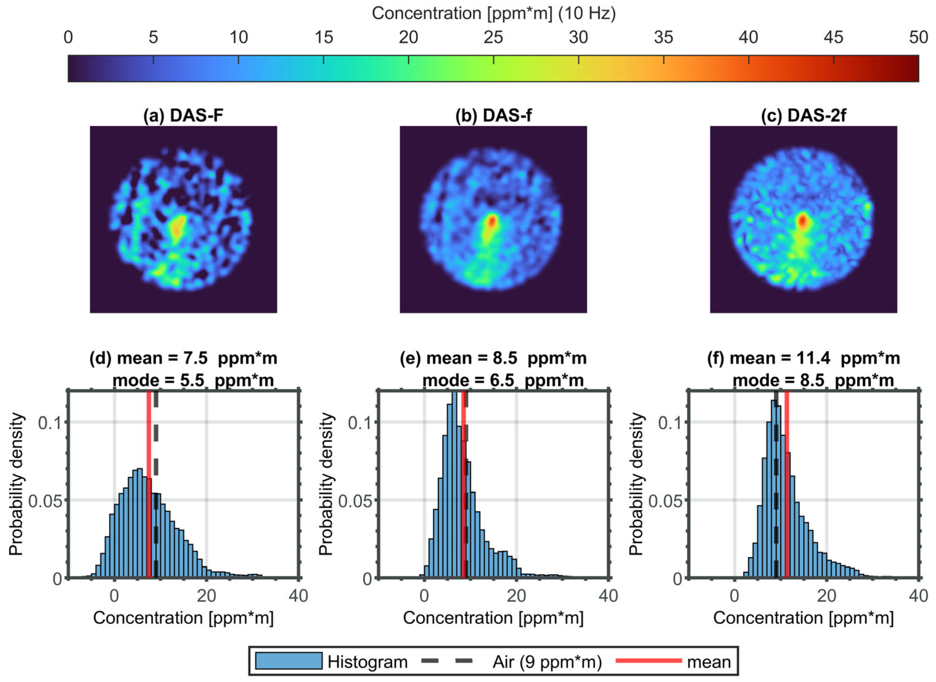
The resulting concentration images from the three methods are depicted in Figure 5a–c, with the color scale restricted to the physically meaningful range of 0 to 50 ppm*m. DAS-f and DAS-2f exhibit the ability to accurately locate the leak at the center of the image and to identify a gas plume for a methane leak rate of 0.25 mL/min within a single concentration image at a rate of 10 Hz. Conversely, the simple integral method (DAS-F) is only partially suitable for qualitatively and quantitatively investigating such small methane leaks. The key criteria in this context are the absolute accuracy (e.g., ambient air concentration of methane at approximately 9 ppm*m) and the methane contrast at the source of the methane leak, including the methane plume extending towards the bottom of the image. For an area within a few millimeters of radius around the leak source in the image center, DAS-f and DAS-2f yield methane concentrations slightly below 50 ppm*m, while the gas plume exhibits concentration values around 20 ppm*m. These values are significantly higher than both the background methane concentration in the ambient air and the spatial variation in the concentration values within the image. The latter can be estimated to be approximately 2 ppm*m for DAS-f and DAS-2f.
Based on these criteria, it is evident that DAS-F exhibits an underestimation of the ambient air concentration in significant parts of the image, including negative concentration values, as well as a larger dispersion of concentration values. Consequently, identifying the leak position and leak plume becomes much more challenging with DAS-F. The corresponding histograms representing these methane images can provide additional information, as shown in Figure 5d–f.
Considering that the methane leak rate of 0.25 mL/min is quickly diluted in the ambient air, most of the concentration image is dominated by ambient methane at approximately 9 ppm*m. Therefore, the most frequent value in the image or histogram (referred to as the mode value) is expected to be around 9 ppm*m. From this perspective, the baseline-free implementation of DAS-2f demonstrates remarkable absolute accuracy, as the mode value aligns with the ambient air concentration of methane. The ability to measure ambient methane with a sufficient spatial signal-to-noise ratio documents the performance capability of DAS-f and, particularly, DAS-2f. A methane anomaly can only be detected at the leak origin or the associated gas plume, which constitute a relatively small portion of the image. The higher methane concentrations around the leak origin and corresponding gas plume align with a positive skew in the distribution of concentration values in the histograms.

3.2. Spatiotemporal Concentration Image Sequence of the 0.25 mL/min Methane Leak Scenario

The spatiotemporal sequence of methane concentration images captures the 0.25 mL/min methane leak scenario over a duration of 6 s or 60 images at a frame rate of 10 Hz. These sequences, obtained using the three different methods, are presented in Figure 6 for DAS-2f and in Appendix C for DAS-F and -f, respectively. The methane contrast between the leakage gas and ambient methane, relative to the spatial noise in the image, is in line with the previously described advantages of DAS-f and DAS-2f over DAS-F. The localization of the leak at the center of the image, even for methane leak rates as low as 0.25 mL/min, can be observed in every concentration image of the sequences. By analyzing the concentration sequences, additional details can be extracted. Particularly, DAS-f and DAS-2f reveal the spatiotemporal dynamics of the gas plume around the leak. It becomes apparent that the gas cloud originating from the leak is not stationary, even in controlled laboratory conditions without significant atmospheric disturbances such as wind. These results highlight the practical detection of small leak rates, which requires high spatial resolution, as well as low spatiotemporal concentration noise. This necessity arises from the immediate dilution of methane from the leak into the surrounding ambient air.
According to Figure 7, the noise equivalent concentration (NEC) image is given by the pixelwise standard deviation of the last second of the concentration sequence (10 images, average time span of 1 s or bandwidth 1 Hz) of the 6 s sequence, as presented in Figure 6 and Figure A3 and Figure A4 in Appendix C. The mode values of the three NEC images for the different methods are also documented in Figure 7 and show that DAS-f and DAS-2f are up to twice as powerful as DAS-F. The temporal variations in concentration values or NEC within this last second of the image sequence can be approximated to 2 ppm*m or even lower for DAS-f and DAS-2f.

4. Discussion

In the following, the findings of this study will be discussed from different perspectives, specifically focusing on method engineering and its implications for gas leak detection or TLS in general.
It is worth mentioning that the absence of uniform methodologies for the quantitative assessment of gas leaks makes it challenging to directly compare our results with the results presented in the literature.
It is important to highlight that all three methods introduced in this study (DAS-F, DAS-f and DAS-2f) have demonstrated sufficient capability to accurately locate a methane leak with a leak rate as low as 0.25 mL/min within a single methane concentration image at a frequency of 10 Hz. This exceptional outcome very likely extends the capabilities of other state-of-the-art technologies or concepts, including conventional or passive OGI (optical gas imaging), extractive sniffing techniques for gas detection and established non-camera-based and camera-based remote detection concepts. The implementation of the DAS method for OGI (e.g., DAS-OGI) by an ICL and a fast MIR camera effectively combines sensitivity and spatiotemporal resolution. These features result in a superior performance that is essential for the detection of small gas leaks (e.g., 0.25 mL/min). In practice, this means that methane concentrations at the leak and around the leak are immediately and strongly diluted by ambient air, and only concentrations below 50 ppm*m can be detected in the vicinity of a few millimeters around the leak origin. A drawback of this active DAS-OGI approach is that a sufficient amount of light needs to be backscattered. In this study, the use of a rough aluminum plate offers almost optimal conditions in the sense of a non-cooperative target, which makes this study a kind of “best-case scenario”. The concentration image sequences show an additional information potential for leak detection and further quantification, such as a mass flow quantification approach that has been recently demonstrated [16]. These concentration images motivate a more sophisticated analysis using the spatiotemporal concentration information for more sensitive and reliable leak detection, localization and further quantification.
The active OGI concept in this work shows a remarkable potential for standoff gas leak detection for very small leaks. It can be transferred to other gases with IR activity in the wavelength range between 3 and 6 µm. This can be realized by changing the gas-specific laser without changing the camera. In this way, the focus tailored to the application can be transferred to other gases where small leak rates may be of greater importance than for methane, such as stronger greenhouse gases or toxic gases. Another application could be a leak test scenario during production to ensure the functionality and/or legal requirements of products.
To the best of the authors’ knowledge, an absorption line scan has never been investigated or demonstrated in the context of camera-based TLS. The pixel-by-pixel application of conventional DAS signal processing based on a BLC of the laser in combination with a non-linear line fit of the absorption line is not practically feasible or appropriate for the spectroscopic imaging in this work. For instance, the amount of data (e.g., 200 × 200 pixels per image times approx. 30 spectral sampling points within 10 Hz), as well as the signal-to-noise ratio for methane leak rates around 0.25 mL/min, required modified DAS signal processing for this TLS-OGI concept. Consequently, a BLC based on a single or global BLC for the image sequence was derived by means of reference image and normalization to avoid about 200 × 200 BLCs. This concept provides a better starting position with respect to a real-time realization that is planned soon. The analysis of the absorbance sequence after the BLC area analysis (unsmoothed DAS-F, see Figure 3) without further signal processing can be considered very critical in connection with a robust, accurate and sensitive concentration image estimation. Often-cited etalon noise (optical 1/f noise or low-frequency etalon) will also play a central role here [18,20,21]. Remote detection is highly prone to etalon phenomena, since the backscattered laser light quickly exhibits optical path differences in the range of a few millimeters and interferes at the detector (camera pixel). As a result, DAS-f and DAS-2f perform better than DAS-F in terms of concentration accuracy (variation or residuals) from a spatiotemporal point of view. This observation could be a result of the spectral filtering (e.g., Gaussian smoothing along the spectral dimension and spectral low-pass filtering) of DAS-f and DAS-2f, which, together with the BLC or the second derivative (spectral high-pass filtering), have a significantly different (spectral) bandpass filter effect than DAS-F. This hypothesis needs to be investigated in a more quantitative manner in the future. In particular, the baseline-free variant of DAS-2f also has the advantage of being used in situations where practically no BLC can be applied in an appropriate manner (e.g., spectral overlap of gas lines).
Furthermore, WMS is often preferred to the conventional DAS method due to its higher sensitivity (or robustness against so-called “1/f noise”). Practical studies have shown a significantly smaller standard deviation in the concentration determination for WMS-2f compared to conventional DAS signal processing [18,22]. The DAS-f and DAS-2f concepts introduced in this work might have the potential to reduce this gap between conventional DAS and WMS. However, this is beyond the scope of this work and needs to be addressed in the future. Finally, the DAS-2f approximation or principle also applies to other typical lasers sources in TLS, such as vertical cavity surface emitting lasers (VCSEL), diode lasers (DL) in the near-infrared or quantum cascade lasers (QCL), as the typical tuning behavior (mW/cm−1) in relation to the typical laser output power of the individual laser types is very similar.

5. Conclusions

This study successfully utilized TLS with mid-IR imaging, introducing a classical DAS modulation concept for the first time to enhance gas leak detection capabilities. A tunable ICL was modulated at 10 Hz across a methane absorption line at 3271 nm. The active DAS-OGI method requires backscattered laser light, so, in this study, a rough aluminum plate was used to simulate a nearly ideal non-cooperative target of 10 × 10 cm2. The detection was performed from a 2-m distance using a fast thermal camera in the mid-IR spectrum, capturing image stacks at 470 Hz for offline processing with application tailored DAS algorithms based on the Lambert–Beer law and HITRAN database.
The innovative and application-tailored algorithms named DAS-F, DAS-f and DAS-2f are intended to efficiently analyze different features of a gas absorption line, such as the area (F), peak (f) or the second derivative of the absorbance (2f). These algorithms enabled a calibration-free methane concentration determination in ppm*m and leak localization for rates as low as 0.25 mL/min, offering superior sensitivity and spatiotemporal resolution compared to conventional leak detection techniques.
DAS-f and DAS-2f displayed the leak scenario in a single concentration image with a pixel-by-pixel sensitivity of approximately 1 ppm*m, making it possible to distinguish between leak gas and ambient methane. Furthermore, concentration image sequences show the spatiotemporal dynamics of the gas plume at a high contrast-to-noise ratio. This active OGI concept can be transferred to other gases with IR activity in the wavelength range between 3 and 6 µm by changing the gas-specific laser without changing the camera. These findings could pave the path for future applications, including a real-time testing for leaks during production processes to ensure the functionality and/or legal requirements of products.
Furthermore, the introduced baseline-free and calibration-free DAS-2f concept within this work demonstrates promising characteristics and potential, including accuracy, precision, 1/f noise rejection, simplicity and computational efficiency. In general, DAS-2f could expand the application scope of convention DAS.

Author Contributions

Conceptualization, T.S., M.B. and J.H.; methodology, T.S., M.B. and J.H.; software, T.S., E.M. and S.R.; validation, T.S., E.M. and S.R.; formal analysis, T.S., M.B. and J.H.; investigation, T.S., E.M. and J.H.; resources, J.H. and J.W.; writing—original draft preparation, T.S.; writing—review and editing, M.B., J.W. and K.S.; visualization, T.S.; supervision, J.H., J.W. and K.S.; project administration, J.W. and K.S.; funding acquisition, J.W. and K.S. All authors have read and agreed to the published version of the manuscript.

Funding

This research was funded by the DFG SBF 1537/1 Ecosense and Bundesministerium für Bildung und Forschung (BMBF), 03HY202G TransHyDE.

Data Availability Statement

The data underlying the results presented in this paper are not publicly available at this time but may be obtained from the authors upon reasonable request.

Conflicts of Interest

Author Max Bergau was employed by the company Endress+ Hauser Process Solutions (DE) GmbH. The remaining authors declare that the research was conducted in the absence of any commercial or financial relationships that could be construed as a potential conflict of interest.

Appendix A

In the first step, a preprocessing of the raw images is required to apply the BLL in an appropriate manner. The raw images, including the backscattered laser intensity information, need to be background-corrected. In this case, a (homogeneous) background is estimated by an image region without laser illumination and subtracted pixelwise from the raw image. At this stage, more sophisticated background corrections are not required. In real-world scenarios with (fast-) changing backgrounds, the laser could be shortly turned off to estimate the background. Nevertheless, the background needs to be stable during a line scan. However, this background correction can be seen as a dark current correction in such a way that it holds I   I I d a r k with I = I x , y , λ .
Regarding the backscattered laser light, it is assumed the spatial intensity variations are based on the spatial reflectivity characteristics R x , y of the target by the surface roughness, such that the BLL (e.g., I =   I 0   e x p ( A )) can be rewritten as
I x , y , λ = R x , y   I 0 λ   e x p ( A x , y , λ ) .
Furthermore, there is a very high correlation between the images, so that it can be assumed that the reflectivity is wavelength-independent. Consequently, the reflectivity can be approximated by the reference image R x , y   I r e f / < I r e f > , such that I x , y , λ I x , y , λ I r e f , and Equation (A1) can be rewritten as
I x , y , λ =   I 0 λ   e x p ( A x , y , λ ) .
The corresponding image sequence is illustrated in Figure A1a and is the basis for further image processing.
Figure A1. The reference image (e.g., frame 45) and an image stack of 30 frames as a subset of the 47 background-corrected raw images per ramp of the 0.25 mL/min methane leak scenario are shown in (a) and (c), respectively, in terms of a normalized intensity according to the color bar between 0 and 2. A distribution of the intensity values within x , y     Ω of the reference image (a) is shown in the form of a histogram (b). Due to normalization, the mean value µ of the histogram is the solid red line, and the corresponding standard deviation σ is highlighted by the vertical red lines.
Applsci 14 05988 g0a1

Appendix B

Figure 4a,c show the spectra as image sequences of DAS-f and DAS-2f, respectively. From these sequences, a feature image for the corresponding method is estimated that can be directly transferred into a concentration image. This procedure for DAS-f and DAS-2f is described in the following.
  • DAS-f method:
    As illustrated in Figure 4b, the one-dimensional smoothed absorbance (DAS-f) is in line with the simulation based on the spectroscopic parameters provided by HITRAN. Furthermore, the maximum absorbance (see Figure 4b) is a suitable measure for determining the concentration that can be simply and efficiently transferred to the image sequence. The pixelwise smoothed peak absorbance can be estimated by finding the maximum image along the wavenumber dimension of the DAS-f sequence in Figure 4a. This maximum image is shown in Figure A2a. The transfer function of the smoothed peak absorbance as the DAS-f feature (e.g., y p f ) into a concentration value in ppm*m can be easily derived by further simulation, as described above. The transfer function or smoothed peak absorbance as a function of the concentration shows a linear behavior in the investigated concentration range between 0 and 50 ppm*m, as illustrated in Figure A2b. By means of this function, the feature image of DAS-f can be straightforwardly transferred into the corresponding concentration image.
  • DAS-2f method:
    The DAS-2f concentration is derived in a similar fashion as described for DAS-f.
    In this case, the peak–dip–peak measure is a suitable measure for determining the concentration that can be simply and efficiently transferred to the image sequence (cf. Figure 4c,d). The peak–dip–peak feature or image will be calculated by a maximum image (first row), minimum image (second row) and maximum image (third row) of the DAS-2f sequence (e.g., y p d p 2 f = 1 / 2   ( y m a x , r o w 1 2 f 2   y m i n , r o w 2 2 f + y m a x , r o w 3 2 f ) ). This DAS-2f feature image is shown in Figure A2c. The corresponding transfer function based on the peak–dip–peak feature ( y p d p 2 f ) of the smoothed second derivative of the absorbance as a function of the concentration is documented in Figure A2d. This DAS-2f feature also demonstrates a good linearity in the concentration between 0 and 50 ppm*m, such that a methane concentration image can be calculated straightforwardly.
Figure A2. (a,c) Image of the peak and peak–dip–peak measure as the feature extraction of the DAS-f and DAS-2f sequences. (b,d) The smoothed peak absorbance for DAS-f ( y p f ) and peak–dip–peak measure for DAS-2f ( y p d p 2 f ) as a function of the methane concentrations between 0 and 50 ppm*m.
Applsci 14 05988 g0a2

Appendix C

The spatiotemporal sequence of the methane concentration images captures the 0.25 mL/min methane leak scenario over a duration of 6 s or 60 images at a frame rate of 10 Hz for DAS-F and -f, respectively (Figure A3 and Figure A4).
Figure A3. The spatiotemporal sequence of the methane concentration images for DAS-F displays the 0.25 mL/min methane leak scenario within 6 s (or 60 images) at a frame rate of 10 Hz.
Applsci 14 05988 g0a3
Figure A4. The spatiotemporal sequence of the methane concentration images for DAS-f displays the 0.25 mL/min methane leak scenario within 6 s (or 60 images) at a frame rate of 10 Hz.
Applsci 14 05988 g0a4

References

  1. Curl, R.F.; Tittel, F.K. 7 Tunable infrared laser spectroscopy. Annu. Rep. Prog. Chem. Sect. C Phys. Chem. 2002, 98, 219–272. [Google Scholar] [CrossRef]
  2. Hodgkinson, J.; Tatam, R.P. Optical gas sensing: A review. Meas. Sci. Technol. 2013, 24, 12004. [Google Scholar] [CrossRef]
  3. Li, J.; Yu, Z.; Du, Z.; Ji, Y.; Liu, C. Standoff Chemical Detection Using Laser Absorption Spectroscopy: A Review. Remote Sens. 2020, 12, 2771. [Google Scholar] [CrossRef]
  4. Kwaśny, M.; Bombalska, A. Optical Methods of Methane Detection. Sensors 2023, 23, 2834. [Google Scholar] [CrossRef] [PubMed]
  5. Rothman, L.S. History of the HITRAN Database. Nat. Rev. Phys. 2021, 3, 302–304. [Google Scholar] [CrossRef]
  6. Gordon, I.E.; Rothman, L.S.; Hargreaves, R.J.; Hashemi, R.; Karlovets, E.V.; Skinner, F.M.; Conway, E.K.; Hill, C.; Kochanov, R.V.; Tan, Y.; et al. The HITRAN2020 molecular spectroscopic database. J. Quant. Spectrosc. Radiat. Transf. 2022, 277, 107949. [Google Scholar] [CrossRef]
  7. Zeng, Y.; Morris, J. Detection limits of optical gas imagers as a function of temperature differential and distance. J. Air Waste Manag. Assoc. 2019, 69, 351–361. [Google Scholar] [CrossRef] [PubMed]
  8. Alvarez, R.A.; Zavala-Araiza, D.; Lyon, D.R.; Allen, D.T.; Barkley, Z.R.; Brandt, A.R.; Davis, K.J.; Herndon, S.C.; Jacob, D.J.; Karion, A.; et al. Assessment of methane emissions from the U.S. oil and gas supply chain. Science 2018, 361, 186–188. [Google Scholar] [CrossRef]
  9. Ravikumar, A.P.; Wang, J.; Brandt, A.R. Are Optical Gas Imaging Technologies Effective For Methane Leak Detection? Environ. Sci. Technol. 2017, 51, 718–724. [Google Scholar] [CrossRef]
  10. Kemp, C.E.; Ravikumar, A.P.; Brandt, A.R. Comparing Natural Gas Leakage Detection Technologies Using an Open-Source “Virtual Gas Field” Simulator. Environ. Sci. Technol. 2016, 50, 4546–4553. [Google Scholar] [CrossRef]
  11. Zimmerle, D.; Vaughn, T.; Bell, C.; Bennett, K.; Deshmukh, P.; Thoma, E. Detection Limits of Optical Gas Imaging for Natural Gas Leak Detection in Realistic Controlled Conditions. Environ. Sci. Technol. 2020, 54, 11506–11514. [Google Scholar] [CrossRef]
  12. Nutt, K.J.; Hempler, N.; Maker, G.T.; Malcolm, G.P.A.; Padgett, M.J.; Gibson, G.M. Developing a portable gas imaging camera using highly tunable active-illumination and computer vision. Opt. Express 2020, 28, 18566–18576. [Google Scholar] [CrossRef]
  13. Voumard, T.; Wildi, T.; Brasch, V.; Álvarez, R.G.; Ogando, G.V.; Herr, T. AI-enabled real-time dual-comb molecular fingerprint imaging. Opt. Lett. 2020, 45, 6583–6586. [Google Scholar] [CrossRef] [PubMed]
  14. Strahl, T.; Herbst, J.; Lambrecht, A.; Maier, E.; Steinebrunner, J.; Wöllenstein, J. Methane leak detection by tunable laser spectroscopy and mid-infrared imaging. Appl. Opt. 2021, 60, C68–C75. [Google Scholar] [CrossRef] [PubMed]
  15. Bergau, M.; Strahl, T.; Scherer, B.; Wöllenstein, J. Real-time active-gas imaging of small gas leaks. J. Sens. Sens. Syst. 2023, 12, 61–68. [Google Scholar] [CrossRef]
  16. Bergau, M.; Strahl, T.; Ludlum, K.; Scherer, B.; Wöllenstein, J. Flow rate quantification of small methane leaks using laser spectroscopy and deep learning. Process Saf. Environ. Prot. 2024, 182, 752–759. [Google Scholar] [CrossRef]
  17. Ullah Khan, F.; Guarnizo, G.; Martín-Mateos, P. Direct hyperspectral dual-comb gas imaging in the mid-infrared. Opt. Lett. 2020, 45, 5335–5338. [Google Scholar] [CrossRef]
  18. Klein, A.; Witzel, O.; Ebert, V. Rapid, time-division multiplexed, direct absorption- and wavelength modulation-spectroscopy. Sensors 2014, 14, 21497–21513. [Google Scholar] [CrossRef]
  19. Lins, B.; Zinn, P.; Engelbrecht, R.; Schmauss, B. Simulation-based comparison of noise effects in wavelength modulation spectroscopy and direct absorption TDLAS. Appl. Phys. B 2010, 100, 367–376. [Google Scholar] [CrossRef]
  20. Masiyano, D.; Hodgkinson, J.; Schilt, S.; Tatam, R.P. Self-mixing interference effects in tunable diode laser absorption spectroscopy. Appl. Phys. B 2009, 96, 863–874. [Google Scholar] [CrossRef]
  21. Werle, P. Accuracy and precision of laser spectrometers for trace gas sensing in the presence of optical fringes and atmospheric turbulence. Appl. Phys. B 2011, 102, 313–329. [Google Scholar] [CrossRef]
  22. Yan, G.; Zhang, L.; Zheng, C.; Zhang, M.; Zheng, K.; Song, F.; Ye, W.; Zhang, Y.; Wang, Y.; Tittel, F.K. Mobile Vehicle Measurement of Urban Atmospheric CH4/C2H6 Using a Midinfrared Dual-Gas Sensor System Based on Interband Cascade Laser Absorption Spectroscopy. IEEE Trans. Instrum. Meas. 2022, 71, 9509411. [Google Scholar] [CrossRef]
Figure 1. The artificial leak scenario consists of a shot-blasted aluminum target with a central hole as the “leak”. This non-cooperative target ensures sufficient diffuse reflection of the incident laser radiation (interband cascade laser, ICL, by Nanoplus) that can be detected by the mid-IR camera (Infratec) and recorded as an image sequence. Pure methane as the leakage gas is released through the hole at a flow rate of 0.25 mL/min.
Applsci 14 05988 g001
Figure 2. (a) Reflectivity image sequence (contrast-corrected), including a local absorption signature of the 0.25 mL/min methane leak in the middle row of the sequence and a global intensity modulation of the laser during the line scan. (b) The mean intensity value of the inner part of the illuminated area ( x , y     Ω ) for each image is shown as a function of the wavenumber, including a simulation of the Beer–Lambert law (BLL) for a methane concentration of 9 ppm*m. (c) Baseline-corrected methane absorption for 9 ppm*m as a function of the wavenumber in comparison with the corresponding HITRAN simulation.
Applsci 14 05988 g002
Figure 3. (a) Unsmoothed absorbance sequence of methane within a 0.25 mL/min leakage scenario. (b) Concentration image in ppm*m based on the unsmoothed DAS-F method, including the corresponding histogram (c) of the concentration image. The solid and dashed red lines indicate the mean and standard deviation, respectively, of this concentration distribution, whereas the dashed black line highlights the expected ambient air concentration (9 ppm*m).
Applsci 14 05988 g003
Figure 4. (a,c) Spatially and spectrally smoothed absorbance sequence (DAS-f) and second derivative of the methane absorbance sequence (DAS-2f) of methane within a 0.25 mL/min leakage scenario. (b,d) Mean absorbance value (blue circle, averaged pixels x , y     Ω ) as the inner part of the illuminated area for each frame of the DAS-f and DAS-2f sequences shown as a function of the wavenumber in comparison with the corresponding smoothed HITRAN simulations for a methane concentration of 9 ppm*m.
Applsci 14 05988 g004
Figure 5. (ac) Methane concentration images at the 10 Hz frame rate using the DAS-F, -f and -2f methods within a 0.25 mL/min methane leakage. The color scale is restricted to the physically meaningful range between 0 and 50 ppm*m. (df) The corresponding histograms represent the methane concentration distribution of the upper concentration images of DAS-F, -f and -2f. The dashed black line highlights the expected ambient air concentration of 9 ppm*m, and the solid red line highlights the mean value.
Applsci 14 05988 g005
Figure 6. The spatiotemporal sequence of methane concentration images for DAS-2f displays the 0.25 mL/min methane leak scenario within 6 s (60 images) at a frame rate of 10 Hz.
Applsci 14 05988 g006
Figure 7. The noise equivalent concentration (NEC) image shows the pixelwise standard deviation for an average time span of 1 s (or bandwidth 1 Hz) for each method (DAS-F, -f and -2f). The mode values of the three NEC images are documented for the different methods.
Applsci 14 05988 g007
Disclaimer/Publisher’s Note: The statements, opinions and data contained in all publications are solely those of the individual author(s) and contributor(s) and not of MDPI and/or the editor(s). MDPI and/or the editor(s) disclaim responsibility for any injury to people or property resulting from any ideas, methods, instructions or products referred to in the content.