Article

Aflatoxin B1 Detoxification and Antioxidant Effect of Selected Omani Medicinal Plants against Aflatoxin B1-Induced Oxidative Stress Pathogenesis in the Mouse Liver

by
Rethinasamy Velazhahan
1,*,
Abdullah Mohammed Al-Sadi
1,
Mostafa I. Waly
2,*,
Sathish Babu Soundra Pandian
3,
Jamal Al-Sabahi
4 and
Khalid Al-Farsi
5
1
Department of Plant Sciences, College of Agricultural and Marine Sciences, Sultan Qaboos University, Al-Khoud, Muscat 123, Oman
2
Department of Food Science and Nutrition, College of Agricultural and Marine Sciences, Sultan Qaboos University, Al-Khoud, Muscat 123, Oman
3
Central Analytical and Applied Research Unit, College of Science, Sultan Qaboos University, Al-Khoud, Muscat 123, Oman
4
Central Instrumentation Laboratory, College of Agricultural and Marine Sciences, Sultan Qaboos University, Al-Khoud, Muscat 123, Oman
5
Oman Botanic Garden, Muscat 123, Oman
*
Authors to whom correspondence should be addressed.
Appl. Sci. 2024, 14(13), 5378; https://doi.org/10.3390/app14135378
Submission received: 4 May 2024 / Revised: 10 June 2024 / Accepted: 11 June 2024 / Published: 21 June 2024

Abstract

:
This study investigated the ability of aqueous leaf extracts of Heliotropium bacciferum (HE), Ocimum dhofarense (OE), and Zataria multiflora (ZE) to detoxify aflatoxin B1 (AFB1) under in vitro and in vivo conditions. The results showed that HE, OE, and ZE degraded 95%, 93%, and 92% of AFB1, respectively, after 72 h incubation at 37 °C. The degradation of AFB1 was validated by liquid chromatography–mass spectrometry analysis. A molecular ion peak at m/z 313 specific to AFB1 (C17H12O6) was observed in the mass spectrum of untreated AFB1 (control). However, the level of AFB1 was decreased to untraceable levels in response to treatment with these plant extracts. HE, OE, and ZE effectively detoxified AFB1 in a concentration-dependent manner, resulting in mortality rates of 65, 70, and 75% of brine shrimp, respectively, in contrast to 90% in the untreated AFB1 (control). The hepatoprotective effect of HE, OE, and ZE against AFB1-induced oxidative stress pathogenesis was investigated using mice as an experimental model. Glutathione depletion, impairment of total antioxidant capacity, and increase in DNA oxidative damage were observed in liver tissues of mice treated with AFB1. However, HE, OE, and ZE extract supplementation suppressed the oxidative damage associated with AFB1 treatment. Our findings indicated that HE, OE, and ZE were highly effective in the detoxification of AFB1. In addition, HE, OE, and ZE act as potent antioxidants and combat the AFB1-associated oxidative stress and liver pathogenesis, suggesting that these plants might be valuable for the development of functional foods aimed at minimizing the toxic effects of AFB1.

1. Introduction

Illness in humans due to foodborne microbial infection and the presence of mycotoxins has grown to be a severe public health issue over the past few years worldwide. Human foodborne illnesses are caused by different types of parasites, viruses, and bacteria [1]. Ingestion of mycotoxin-contaminated foods often results in sickness [2,3]. The filamentous fungi Aspergillus, Alternaria, Fusarium, and Penicillium are the major producers of foodborne mycotoxins, and aflatoxins, fumonisins, ochratoxin, patulin, deoxynivalenol, and zearalenone are the most important toxins [4]. Aflatoxin is considered the most toxic mycotoxin. Aflatoxigenic molds attack agricultural commodities during the pre- and/or post-harvest phases and produce aflatoxins [5]. Ingestion of food contaminated with aflatoxin causes a disease called “aflatoxicosis” in humans [6].
So far, 21 varieties of aflatoxins have been detected [7]. Aflatoxin G1 (AFG1; MW 328.0578), aflatoxin G2 (AFG2; MW 330.2889), aflatoxin B1 (AFB1; MW 312.2736), and aflatoxin B2 (AFB2; MW 314.2895) are common contaminants in agricultural products [8]. Aflatoxin M1 (AFM1; MW 328.0577), the principal metabolite of AFB1, is often present in the milk of lactating animals that have ingested AFB1-contaminated feed [5]. “AFB1 (C17H12O6)” is recognized as the most serious carcinogenic mycotoxin to humans and animals [9,10]. Several nations have specified maximum tolerance limits for aflatoxins in food for human consumption to safeguard the population from the risk of aflatoxin [11]. The Food and Drug Administration in the USA and the European Union have set regulatory limits of 20 ppb and 4 ppb for foods, respectively [12].
Aflatoxins cannot be completely removed or degraded from contaminated agricultural commodities due to their stability. Currently, a few biological, physical, and chemical approaches are used to detoxify food items containing aflatoxins [13,14,15,16]. However, each process has downsides, because the treated foods must be safe for human consumption and their nutritional value must not be changed. Detoxification of aflatoxins using plant products is an effective, biologically safe, and practical approach to reduce their toxic effects on humans. Extracts of Trachyspermum ammi [17], Ocimum tenuiflorum [18], Ocimum basilicum [19], zimmu (Allium sativum × A. cepa) [20], Barleria lupulina [21], Adhatoda vasica [22], Centella asiatica, Eclipta prostrata, and Hybanthus enneaspermus [23] have been used for the detoxification of aflatoxins. In the Sultanate of Oman, the existence of more than 250 species of medicinal plants has been reported. These traditional medicinal plants may be potential tools for the biological detoxification of aflatoxins. The current study’s objectives were to (i) analyze the potential of Omani medicinal plants to detoxify AFB1 under in vitro conditions; (ii) analyze the structural changes in AFB1 molecules upon treatment with the selected medicinal plant extracts; (iii) test the toxicity of the degraded products of AFB1 using brine shrimp lethality assay, and (iv) evaluate the role of selected Omani plant extracts in prevention of aflatoxin-induced toxicity during exposure to AFB1 using mice as an experimental model.

2. Materials and Methods

2.1. Aflatoxin

AFB1 (Sigma-Aldrich, St. Louis, MO, USA) standard solution (1000 μg/mL) was prepared in 100% methanol and subsequently stored in amber-colored glass vials at 4 °C.

2.2. Plant Materials

Fresh leaves or stems of 57 Omani medicinal plants belonging to 27 families, viz., Amaranthaceae (1), Anacardiaceae (1), Apocynaceae (5), Asphodelaceae (3), Asteraceae (3), Bignoniaceae (1), Boraginaceae (1), Burseraceae (3), Capparaceae (3), Ephedraceae (1), Euphorbiaceae (6), Fabaceae (4), Lamiaceae (9), Leguminosae (1), Lythraceae (1), Malvaceae (2), Malpighiaceae (1), Moringaceae (1), Myrtaceae (1), Oleaceae (1), Plantaginaceae (1), Primulaceae (1), Resedaceae (1), Rhamnaceae (1), Solanaceae (1), Sterculiaceae (1), and Zygophylaceae (2), were collected from Oman Botanic Garden, Muscat, and kept at 4 °C and processed in less than 48 h.
Aqueous extracts of the medicinal plants were prepared according to the method described by Velazhahan et al. [17] with some modifications. Briefly, leaves or stems (5 g) were homogenized in 25 mL of sterile distilled water and filtered through a sterile muslin cloth. Subsequently, the filtrate was centrifuged at 14,000× g for 15 min at 5 °C. The collected supernatant was stored at 4 °C for subsequent use.

2.3. Test for Detoxification of AFB1 Using Medicinal Plant Extracts

An aliquot (250 μL) of each plant extract was mixed with 50 μL of AFB1 (50 μg/L) in a microcentrifuge tube and incubated in a water bath at 37 °C for 72 h to degrade the toxins. Following incubation, 250 µL of chloroform was used to extract the mixture’s remaining AFB1. The chloroform fraction was collected and evaporated at 60 °C using a water bath, and the residue was dissolved in 50 μL of 70% methanol. A RIDASCREEN Aflatoxin B1 kit was applied for AFB1 examination. For control, sterile distilled water (250 μL) was mixed with 50 μL of AFB1 (50 μg/L) and processed similarly to the test samples.
For each plant extract, two replicates were used. The leaf extracts of Heliotropium bacciferum Forssk. (Boraginaceae; Accession No. 201600290), Ocimum dhofarense (Sebald) A.J.Paton (Lamiaceae; Accession No. 202000071), and Zataria multiflora Boiss. (Lamiaceae; Accession No. 201100114), which showed the highest AFB1 degradation activity (above 90%), were selected for further studies.

2.4. Analysis of Degraded AFB1 Products

Analysis of aflatoxin B1 was carried out using an Agilent 1290 infinity liquid chromatography unit coupled to an Agilent 6460 triple quadrupole mass spectrometer equipped with an electrospray ionization (ESI) source (Agilent Technologies, Basel, Switzerland). The separation of AFB1 was accomplished using a reverse-phase Symmetry C8 5 µm, 3 mm × 150 mm column (Waters, Milford, MA, USA), and a 6460 Triple Quad MS detector was used to analyze the degraded products of AFB1 following treatment with the plant extracts as per the LC-MS conditions specified by Al-Owaisi et al. [23]. A thermostated column oven (G1316C) was maintained at 45 °C during the analysis. Mobile phase A was 0.1% formic acid in acetonitrile and mobile phase B was 0.1% formic acid in HPLC-grade water with a solvent flow rate of 0.500 mL/min using a quaternary pump (G4204A). A high-performance autosampler (G4226A) was used to inject 10.0 µL of each sample. Mass spectrometry was operated in positive ion mode, the source temperature was set as 300 °C, and the ion spray voltage was set as 4000. The software used for data acquisition was MassHunter workstation Qualitative analysis ver 6.0.633.0.

2.5. Brine Shrimp Lethality Assay

Meyer et al. [24] described a brine shrimp mortality experiment for determining the toxicity of AFB1 degradation products. Briefly, 0.3 g of brine shrimp (Artemia salina) eggs (INVE Aquaculture Inc., Salt Lake City, UT, USA) were hatched in a container including artificially sterilized seawater (Tetra GmbH, Melle, Germany) in 1 L of distilled water. For 48 h, the vessel was kept at room temperature with continuous aeration and fluorescent light. After incubation, the newly hatched nauplii were collected in a small beaker containing freshly prepared and well-aerated seawater. Brine shrimp toxicity was tested in the presence of aqueous leaf extracts (100 µg/mL) containing nine different AFB1 concentrations for each plant extract exposure (5, 10, 20, 30, 45, 60, 75, 90, and 100 µg/mL). A total of 10 mL of each AFB1 concentration was added into a small Petri dish and 10 shrimp were transferred into each dish and incubated at room temperature (25 ± 2 °C). After 24 h, all shrimp (living and dead) were counted, and the percentage survival of brine shrimp was calculated for each concentration of the AFB1 with plant extract present. The control was the same procedure without a plant extract.

2.6. Preparation of Plant Extracts

The selected plant leaves (HE, OE, and ZE) were dried for a whole day at 45 °C in an oven and ground into a powder form. A 15 mL polypropylene centrifuge tube containing one gram of the plant powder and 5 ml of sterile distilled water was left to stand at 4 °C overnight. The mixture was then centrifuged at 12,000× g at 4 °C for 15 min and the supernatant was taken and its volume was adjusted to 5 mL with sterile distilled water.

2.7. Antioxidant Potential of Plant Extracts

The antioxidant ability of plant extracts was detected at concentrations from 10 to 100 µg/mL via 1,1-diphenyl-2-picrylhydrazyl (DPPH) assay using the spectrophotometric method [25].

2.8. Mouse Assay Experiments

The protocol used in this study followed the guidelines established by the Sultan Qaboos University Animal Ethics Committee (Ethical approval No. SQU/ECAU/2020-21/4). Forty CD-1 mice, weighing 50 ± 5 g, were obtained from the animal house facility, at Sultan Qaboos University. The animals were distributed into 8 groups (n = 5) and housed individually in polypropylene cages under standard growing conditions (22 ± 2 °C, 60% RH, 12 h light–dark cycle). The animals were fed a standard diet and given tap water ad libitum. On the first day of the experiment, the AFB1-treated group was given 1 mL single intraperitoneal injection of AFB1 (1.5 mg/kg body weight) per mouse, while the control group received 1 mL single intraperitoneal injection of 0.9% physiological saline. The other six groups received an intragastric intubation of plant extracts in the presence or absence of AFB1 injection (Figure 1). The effective dose of plant extracts was determined based on the results of the DPPH assay. The duration of the experiment was 30 days and food consumption was recorded daily. The body weight of the animals was recorded weekly.

2.9. Mouse Sacrifice

All mice were anesthetized at the end of the experiment using a lethal dose of a solution of 1 mg ketamine, 5 mg xylazine, and 0.2 mg acepromazine. Livers were dissected and homogenized in phosphate-buffered saline solution (PBS; pH~7.4). The homogenate was centrifuged at 6000× g for 60 min at 4 °C, and the supernatant was used for biochemical investigations.

2.10. Biochemical Tests

Protein concentration was detected using bovine serum albumin (BSA) as the standard, as established by Lowry et al. [26]. A glutathione (GSH) fluorometric assay kit (BioVision Inc., Milpitas, CA, USA) was used to determine GSH content. Total antioxidant capacity (TAC) was assayed colorimetrically using a Randox assay kit (Randox Laboratories Ltd., Crumlin, UK). DNA oxidative damage was measured using the Abcam DNA Damage Assay Kit (Abcam, Waltham, MA, USA).

2.11. Histopathological Studies

Liver tissues were kept at room temperature in 10% formalin. They were then dehydrated in graded ethanol using a Microm STP120 instrument for 12 h. Subsequently, they were cleared in xylene and embedded in paraffin using a HistoStar embedding unit. Samples were sectioned into 10 µm pieces using a rotary microtome and stained with hematoxylin and eosin. Changes in the liver tissues were examined with an Olympus BX51 microscope with an Olympus camera DP70 (Olympus Corporation, Hachioji, Tokyo, Japan) at 200× magnification.

2.12. Statistical Analysis

The results from the animal studies are expressed as means ± standard deviation (SDs). Data were analyzed using GraphPad Prism 5.03 (GraphPad Software, Boston, MA, USA). The data were analyzed using a general-linear-model ANOVA and the differences (p < 0.05) between treatment means were determined by Tukey’s test.

3. Results

3.1. Screening of Medicinal Plants for AFB1 Detoxification

Aqueous extracts derived from 57 Omani medicinal plants were assessed for their potential to detoxify AFB1. Among them, the leaf extracts of H. bacciferum, O. dhofarense, and Z. multiflora degraded 95%, 93% and 92% of AFB1, respectively, after incubation for 72 h at 37 °C (Table 1). The leaf extracts of Lavandula dhofarensis subsp. ayunensis A.G. Mill. (Lamiaceae), Lavandula subnuda Benth. (Lamiaceae), Salvia hillcoatiae Hedge (Lamiaceae), Tecomella undulata (Sm.) Seem. (Bignoniaceae), Ephedra foliata Fisch. & C.A.Mey. (Ephedraceae), and Kleinia odora (Forssk.) DC. (Asteraceae) degraded more than 80% of AFB1. Fourteen plant species’ aqueous extracts failed to detoxify AFB1, while the remaining extracts demonstrated intermediate levels of potential detoxification. The leaf extracts of H. bacciferum (HE), O. dhofarense (OE), and Z. multiflora (ZE) were selected to be used in further studies.

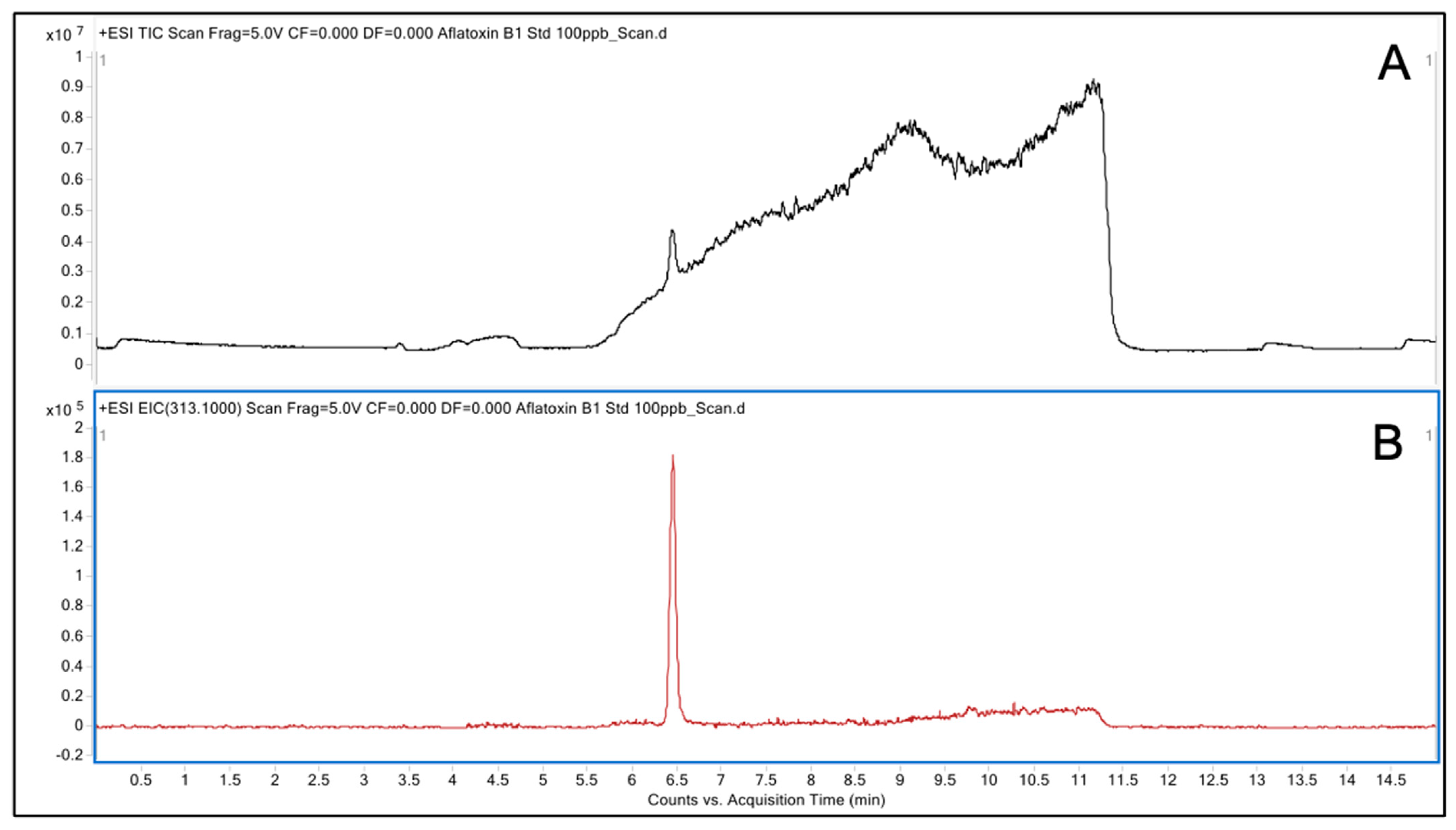
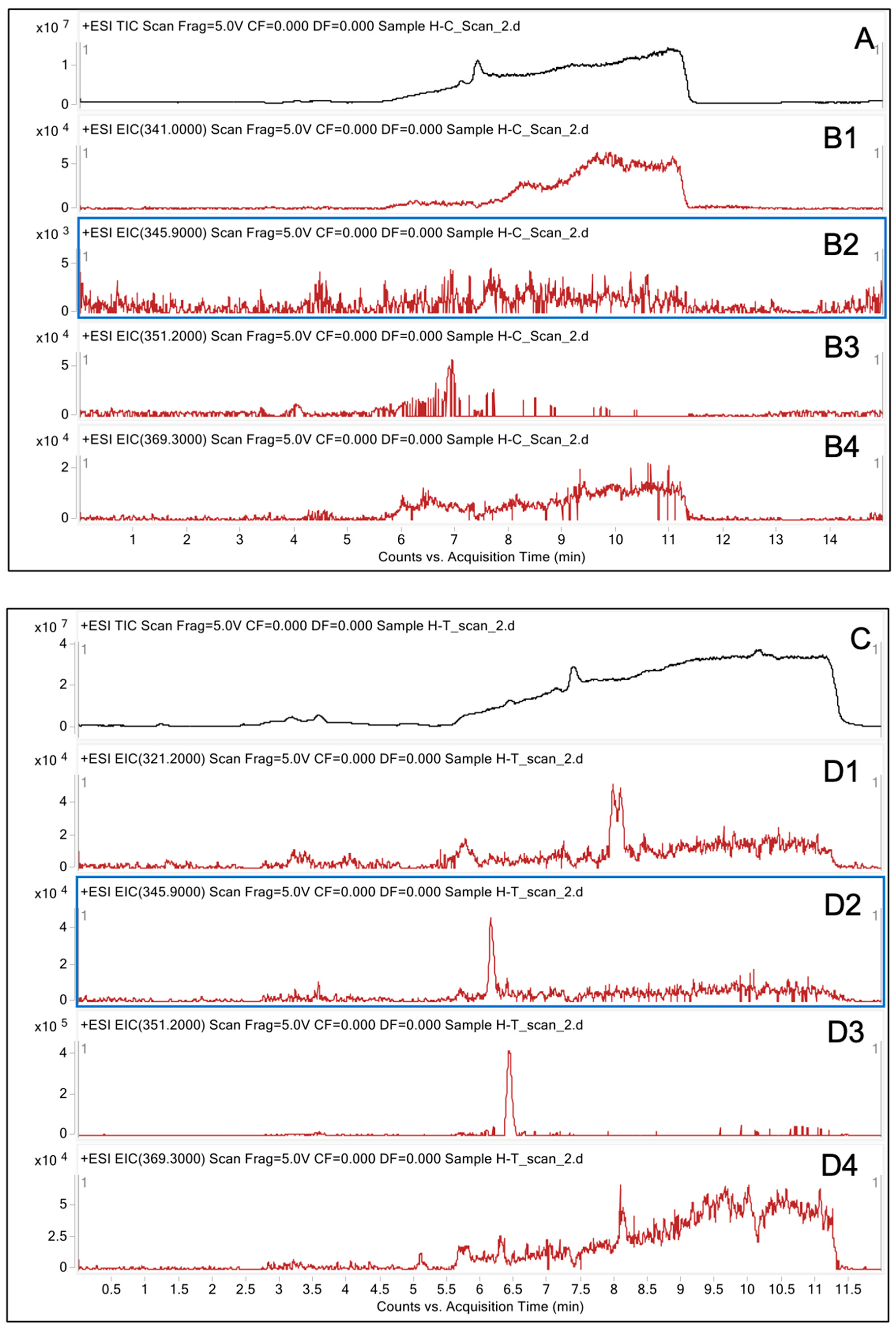
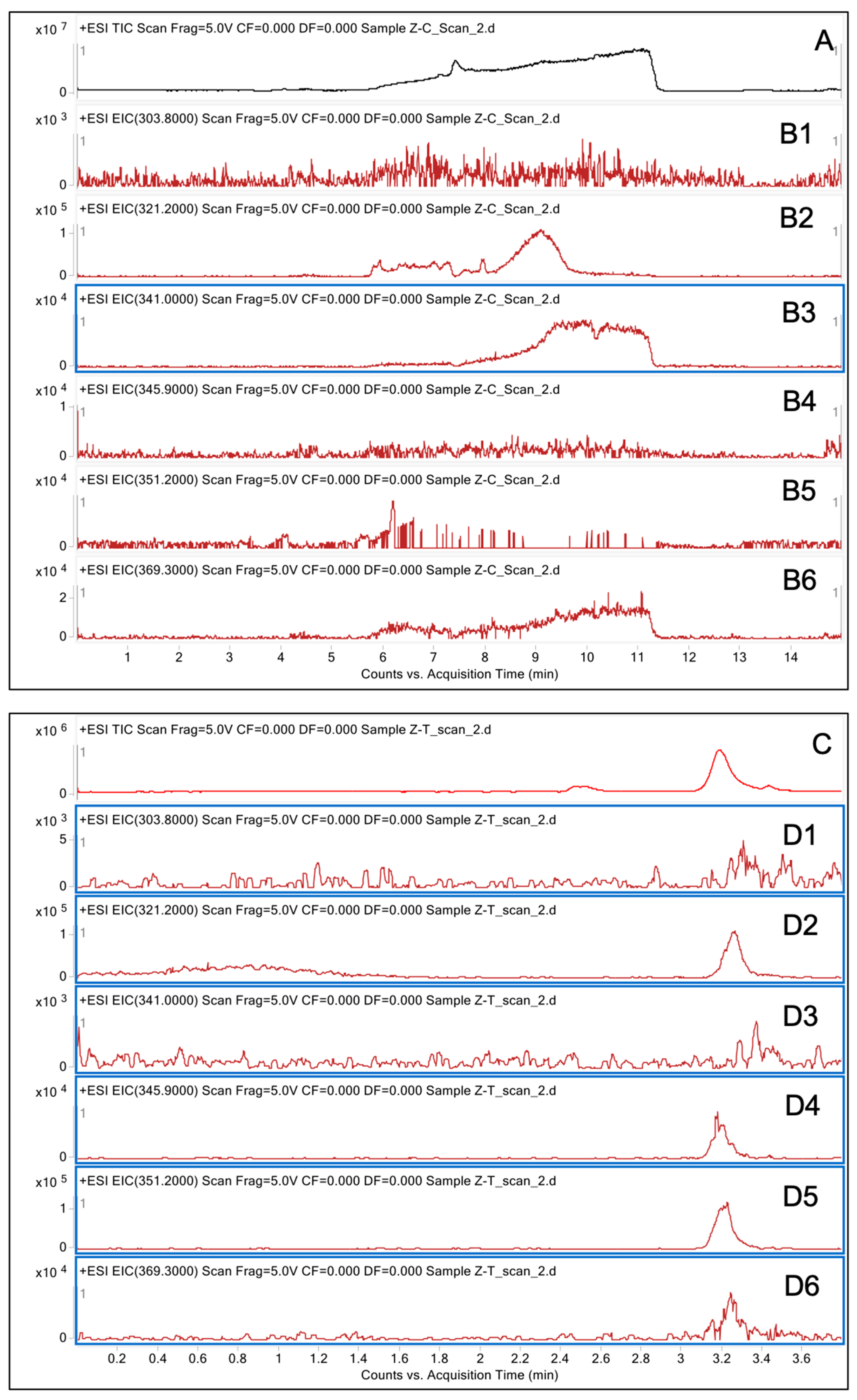
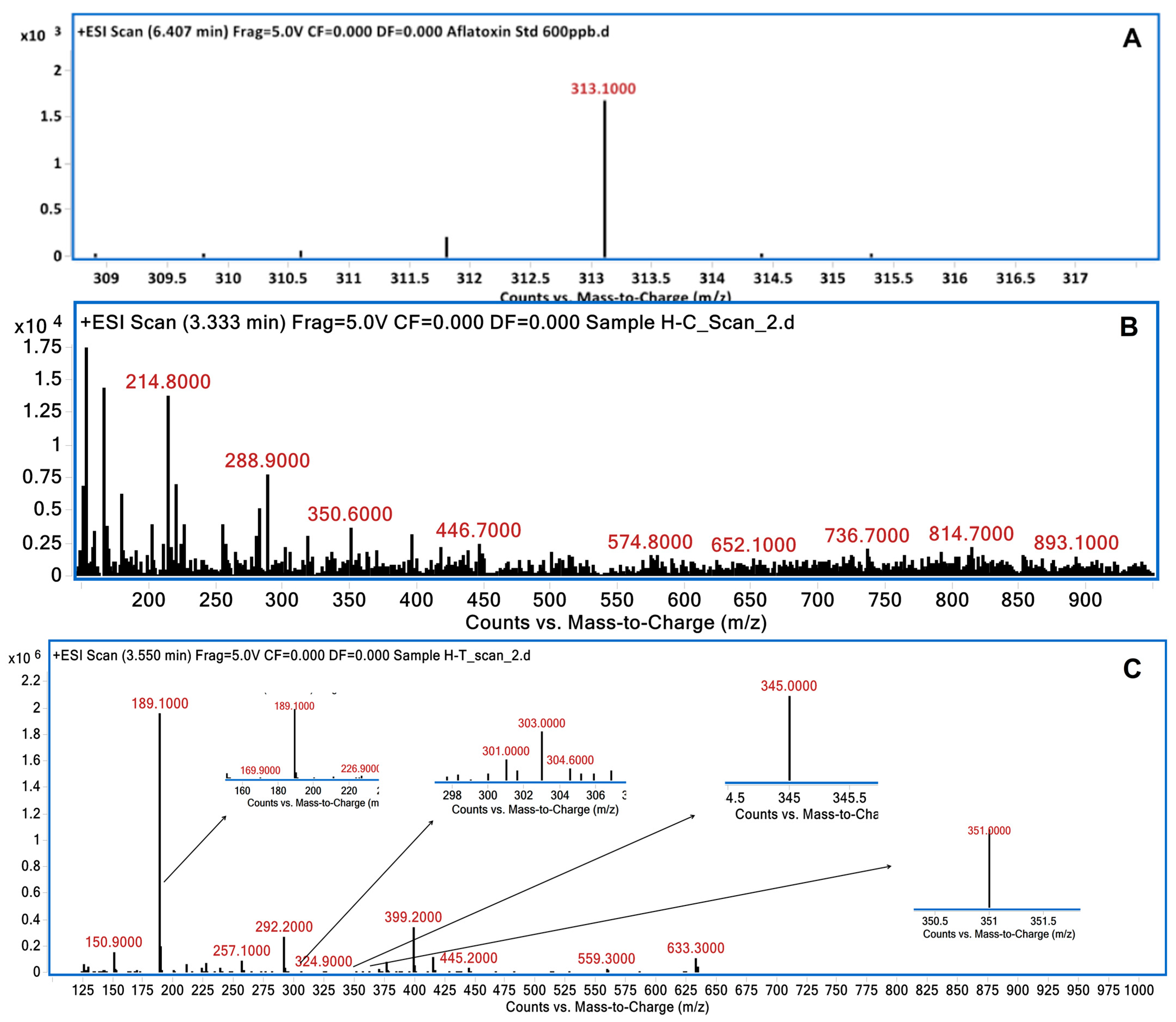
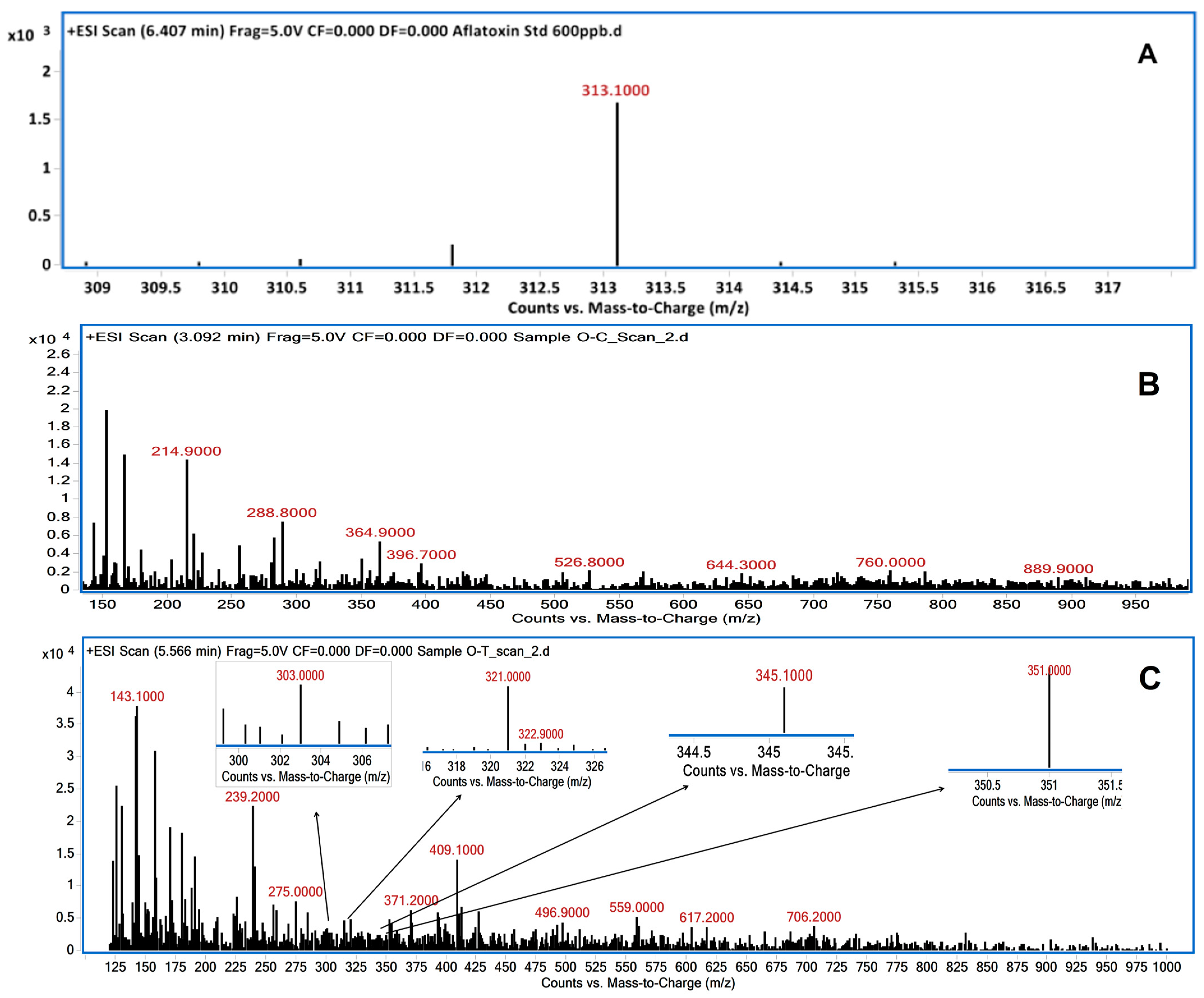
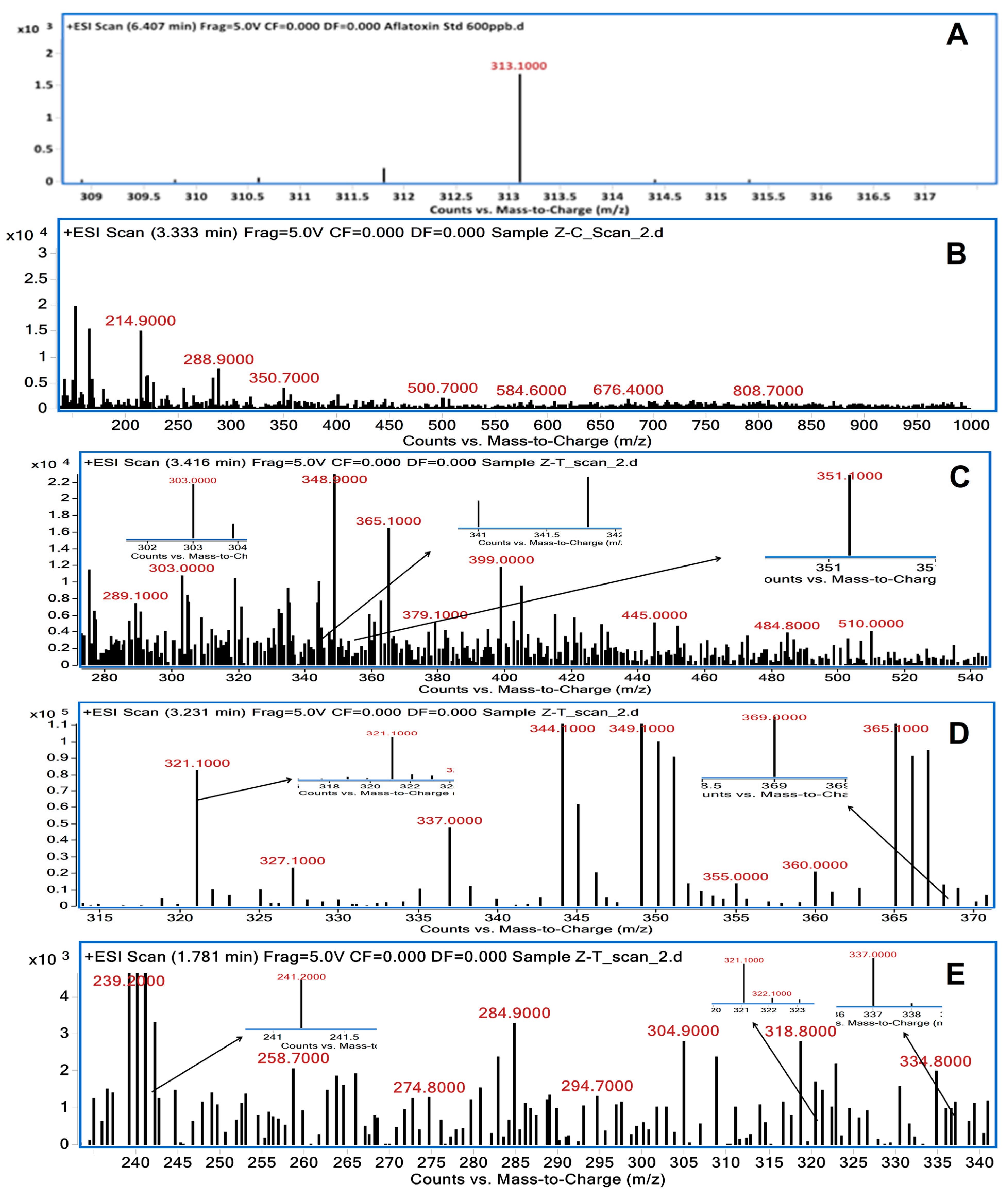
3.2. LC/MS Analysis of Degraded AFB1 Products

The degradation of AFB1 following treatment with medicinal plant extracts was confirmed by LC/MS analysis. The samples were analyzed using ESI positive scan mode, and the chromatogram displayed is the total-ion chromatogram (TIC). AFB1 fragments were extracted from the TIC and are presented as peaks in the extracted-ion chromatogram (EIC). Fragments with too low intensity to show up as peaks in the TIC were confirmed by examining the mass spectrum. LC/MS chromatograms of AFB1 treated with these extracts revealed the presence of additional peaks in comparison to untreated AFB1, confirming the degradation of AFB1 (Figure 2, Figure 3, Figure 4 and Figure 5). Untreated AFB1 (control) showed a molecular ion peak at m/z 313 in the LC/MS analysis (Figure 6A, Figure 7A, and Figure 8A). This molecular ion m/z 313 was not detected in the plant extracts (Figure 6B, Figure 7B, and Figure 8B). However, the level of AFB1 (m/z 313) after treatment with the plant extracts (HE, OE, and ZE) was reduced to undetectable levels. Fragmented AFB1 products, viz., m/z 345.9, 369.3, 303.8, 351.2, 341.0, and 321.2, were detected after treatment with the plant extracts (Figure 6C, Figure 7C, and Figure 8C–E).

3.3. Brine Shrimp Lethality Assay

Brine shrimp lethality bioassay was used to ascertain whether or not AFB1 was detoxified after being treated with the herbal extracts. At a concentration of 100 μg/mL, the three plant extracts (HE, OE, and ZE) indicated a concentration-dependent detoxification effect on AFB1, with brine shrimp survival rates of 35, 30, and 25%, respectively, compared to 10% in the untreated AFB1 (Figure 9).

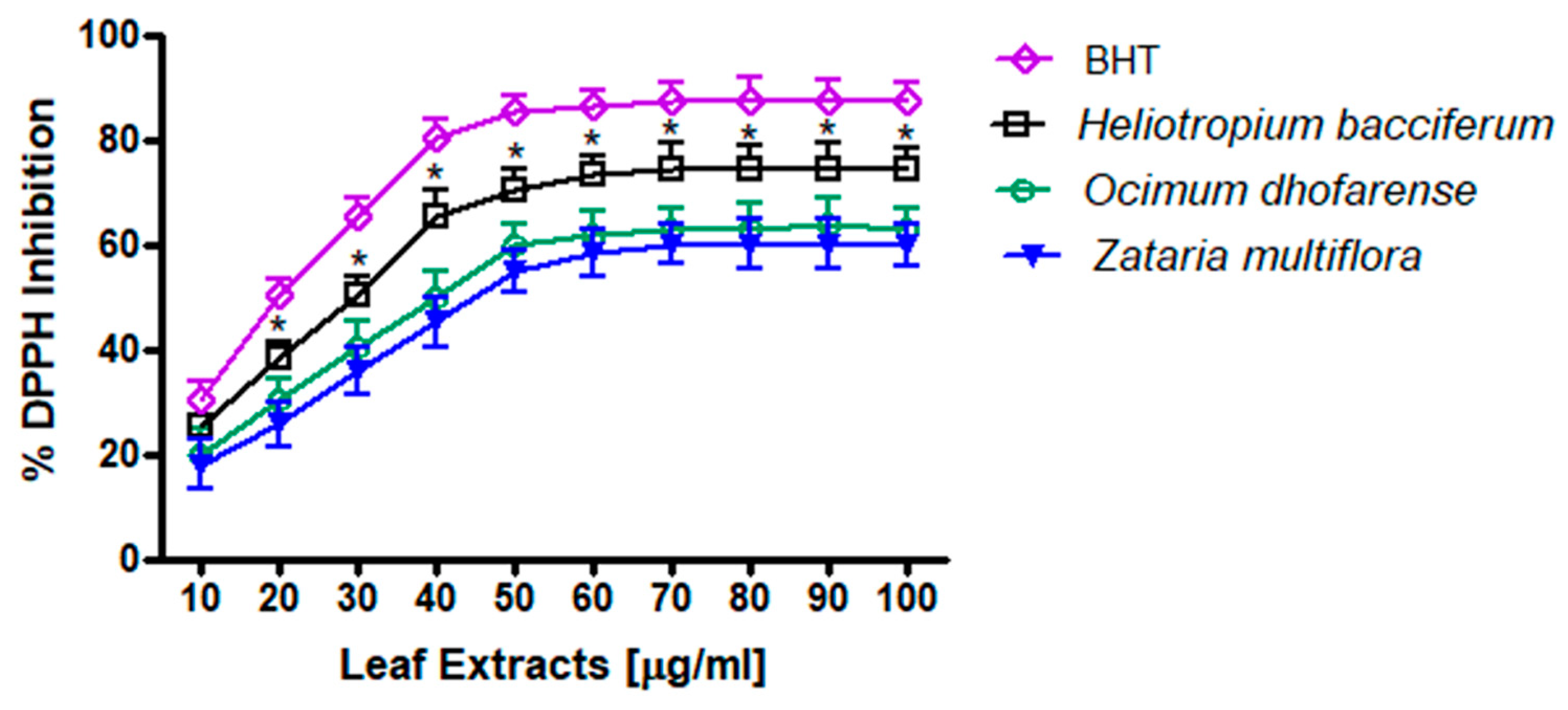
3.4. DPPH Measurements

HE, OE, and ZE demonstrated a dose-dependent inhibition of DPPH formation. HE exhibited a significantly higher inhibition rate compared to OE and ZE (p < 0.05). The effective dose, 100 µg/mL, was selected for subsequent in vivo animal studies (Figure 10).

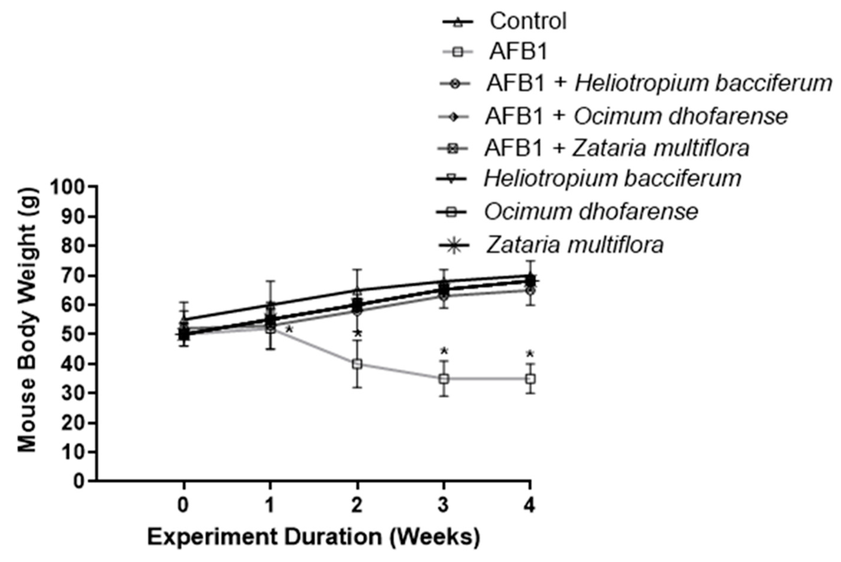
3.5. Body Weight of Mice

A steady body weight gain was found in all the mouse groups and no mortality was noticed. However, AFB1-injected mice showed a consistent decline (p < 0.05) in body weight between week 2 and week 4 compared to the control groups. The daily intragastric intubation of plant extracts prevented weight loss due to AFB1 treatment (Figure 11).

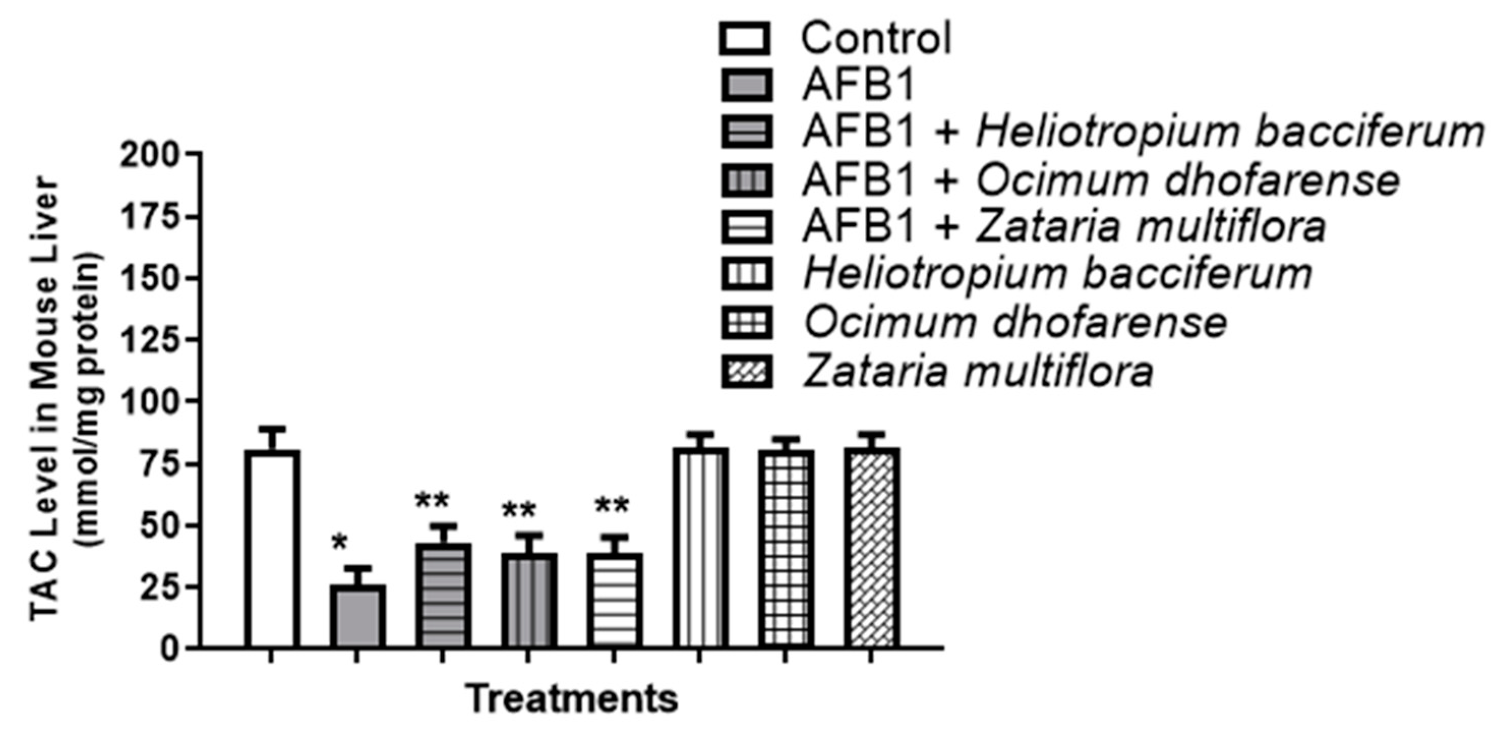
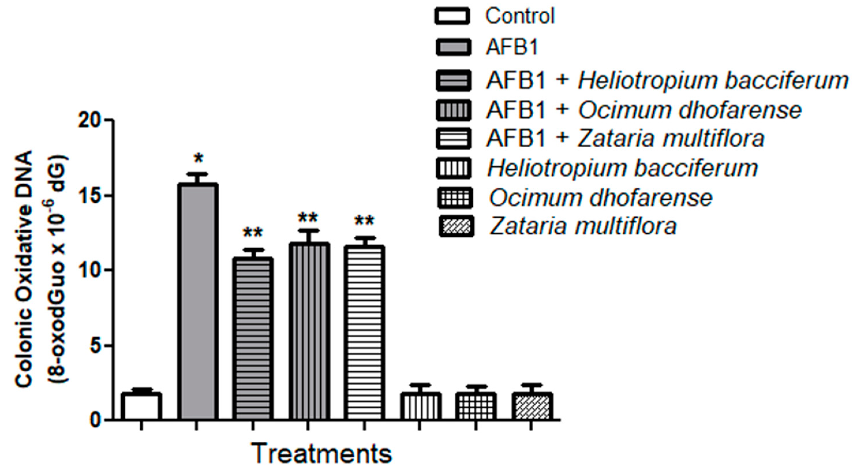
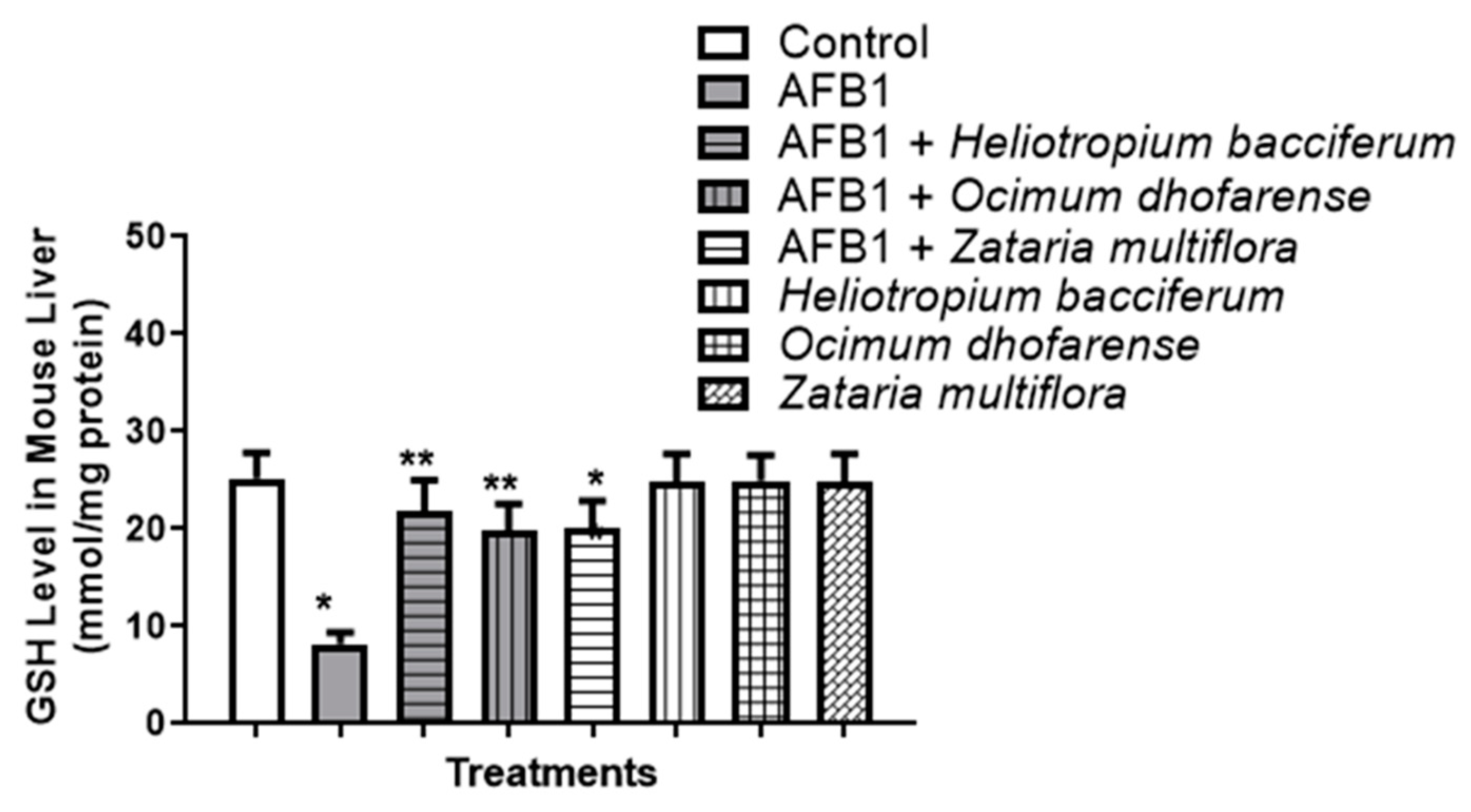
3.6. AFB1-Induced Oxidative Stress

HE, OE, and ZE supplementation counteracted AFB1-induced oxidative stress in the AFB1-injected groups by reinstating the depleted GSH level to a level comparable with that of the control group (p > 0.05) (Figure 12). The same pattern was observed for the protective effects of the three plant extracts on mitigating the AFB1-induced effect on TAC (Figure 13) and reducing DNA damage (Figure 14).

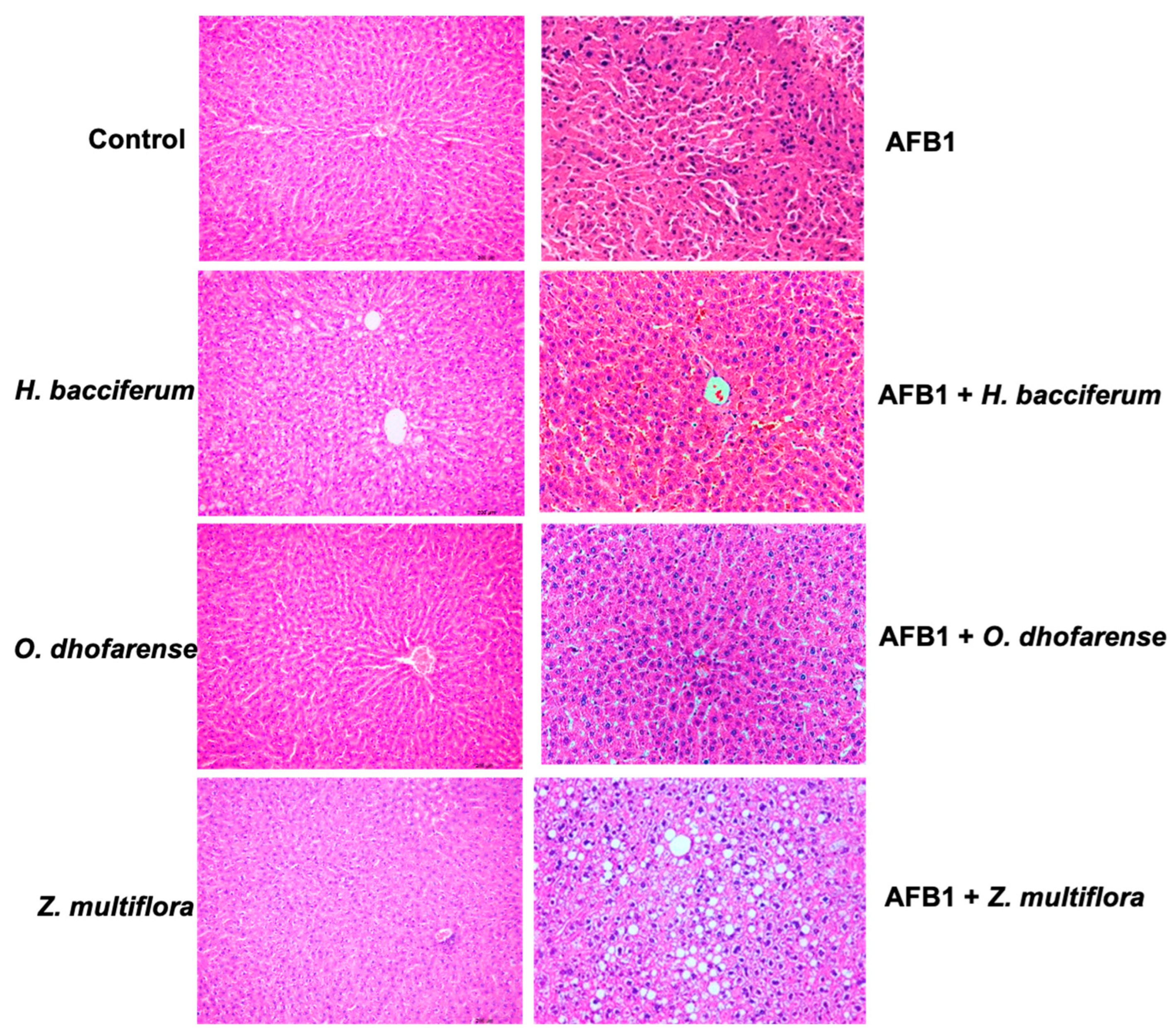
3.7. Histopathological Studies

Hepatocytes in the control group and in groups treated with plant extracts revealed normal liver lobular architecture and cell structure (Figure 15). In the AFB1-treated group, histopathological changes were noticed, including extensive areas of necrosis, loss of hepatocyte architecture around the blood vessels, changes in cytoplasmic acid in hepatic cells, and partial necrosis of hepatic cells with mild inflammation. HE, OE, and ZE plant extracts, on the contrary, demonstrated a decrease in liver damage linked to AFB1 treatment.

4. Discussion

It is evident from the results that the aqueous extracts of O. dhofarense, H. bacciferum, and Z. multiflora effectively degraded AFB1 (above 90%) in vitro following a 72 h incubation at 37 °C. Additionally, the aqueous extracts of Lavandula dhofarensis subsp. ayunensis, Lavandula subnuda, Salvia hillcoatiae, Tecomella undulata, Ephedra foliata, and Kleinia odora demonstrated the capability to degrade over 80% of AFB1. The differences in the AFB1 detoxification efficacy of plant extracts may be attributed to variations in their chemical composition. Several researchers have demonstrated the aflatoxin detoxification abilities of medicinal plants under in vitro conditions [17,18,19,22,23,27,28,29]. Hajare et al. [30] found that seed extracts of Trachyspermum ammi could degrade 80% of aflatoxin. Velazhahan et al. [17] demonstrated that T. ammi seed extract resulted in more than 90% degradation of aflatoxin G1. Vijayanandraj et al. [22] reported that Adhatoda vasica leaf extract could detoxify AFB1 up to 98%. Kannan and Velazhahan [21] found that Barleria lupulina leaf extract could degrade aflatoxins and the percentages of degradation of AFB1, AFB2, AFG1, and AFG2 were 61.1%, 71.4%, 94.4%, and 58.8%, respectively. Iram et al. [19] found that aqueous leaf extracts of Ocimum basilicum could degrade up to 90.4% of AFB1 and up to 88.6% of AFB2.
The leaves of H. bacciferum have traditionally been used to treat skin disorders [31]. The antimicrobial and antioxidant activities of H. bacciferum have been reported [32]. Many pyrrolizidine alkaloids, such as heliotrine, europine, heleurine, and supinine have been characterized in H. bacciferum [33,34,35]. Z. multiflora is an aromatic perennial shrub and is often used as a flavoring component in a wide variety of dishes. Several studies have reported its antioxidant, antimicrobial, and immunomodulatory properties [36,37,38]. The antifungal properties of Z. multiflora essential oil and its effects on the growth of A. flavus and aflatoxin production have been documented [39]. Ocimum sp. is a well-known medicinal herb and has antidiabetic and antioxidant properties [40,41]. The leaves of Ocimum sp. contain triterpenoids, flavonoids, tannins, and saponins [42]. O. sanctum essential oil was reported to suppress the growth of A. flavus and the production of AFB1 [43,44]. Furthermore, the aflatoxin detoxification properties of O. basilicum [19] and O. tenuiflorum [18] extracts have been reported. However, little is known about the aflatoxin detoxification potential of aqueous extract from O. dhofarense. Al-Harrasi et al. [45] showed the detoxification of AFB1 by O. dhofarense, H. bacciferum, and Z. multiflora essential oils. This is the first report of AFB1 detoxification by the aqueous extracts of these three medicinal plants in Oman.
The degradation of AFB1 upon treatment with HE, OE, and ZE was confirmed by LC/MS analysis. The mass spectrum of control (untreated) AFB1 showed a molecular ion peak at m/z 313, which is unique to AFB1 [46], whereas the level of AFB1 (m/z 313) was greatly reduced and fragmented AFB1 products, viz., m/z 345.9, 369.3, 303.8, 351.2, 341.0, and 321.2, appeared after treatment with the plant extracts. The product ions observed in the mass spectra indicate structural changes in AFB1 molecules during the degradation process. For example, the addition of a hydroxyl group to the furan ring results in a product at m/z 345 (C17H12O8), while the addition of H8O3 to AFB1 forms m/z 369, and the elimination of CH2 from AFB1 forms m/z 303. Furthermore, the addition of two hydroxyl groups to AFB1 forms m/z 351 (C17H18O8), which further degrades to m/z 321. The ion m/z 341 is formed by the addition of two oxygen and the elimination of four hydrogen [27]. The removal of the double bond in the furan ring structure of AFB1 is evident in the product ions m/z 303, m/z 341, and m/z 351, while the modification of the lactone group and the elimination of the double bond in the furan ring were observed in AFB1 products m/z 369 and m/z 321. The fragment ion m/z 189, which was absent in the control plant extract, appeared only after treatment of AFB1 with plant extracts.
Several studies have described the detoxification of aflatoxins by herbal extracts and the molecular structures of aflatoxin degradation products [17,19,22,27]. Modifications to the AFG1 lactone ring structure have been suggested as the detoxification mechanism of T. ammi seed extract [17]. Vijayanandraj et al. [22], while studying the AFB1 detoxification effect of Adhatoda vasica leaf extract, also reported the appearance of a product ion at m/z 189.59 and a loss of a molecular base ion at m/z 313 after incubation with A. vasica leaf extract. Structural modifications, such as changes in the lactone group and the elimination of the double bond in the furan ring structure, have been observed in the products of AFB1 and AFB2 degradation after treatment with T. ammi seed extract [27]. The continuous loss of carbon monoxide and methyl and methanol losses on the methoxy group present on the side chain of benzene were observed in the products of AFB1 degraded using O. basilicum extract [19]. Al-Owaisi et al. [23] reported AFB1 detoxification using leaf extracts of Centella asiatica, Eclipta prostrata, and Hybanthus enneaspermus and the formation of a fragment ion at m/z 189 due to two sequential losses of carbon monoxide. The furofuran ring and lactone ring of AFB1 are responsible for its cytotoxicity and mutagenic activity [47]. The disappearance of the ion peak at m/z 313 (unique to AFB1) and the generation of new ion peaks following treatment of AFB1 with HE, OE, and ZE in this study suggest the degradation of AFB1.
The results of the brine shrimp lethality bioassay indicated that HE, OE, and ZE effectively detoxified AFB1 in a concentration-dependent manner, registering 35, 30, and 25% survival rates of brine shrimp at a concentration of 100 μg/mL, respectively, compared to 10% in the untreated AFB1 group. These findings suggest a reduction in the toxicity of AFB1 upon treatment with these plant extracts.
AFB1-induced toxicity is associated with the accumulation of reactive oxygen species, resulting in oxidative stress [7,48]. Oxidative stress leads to membrane lipid peroxidation and oxidative DNA damage [49]. Several biologically active compounds from plants including Allium sativum [50], Ocimum sanctum [51], rosemary [52], Curcuma longa [53], Thonningia sanguinea [54], Adhatoda vasica [55], grape seed proanthocyanidin extract [56], Thymus vulgaris oil [57], curcumin [7,58], and curcumin plus black tea [59] have been documented to decrease AFB1-induced liver damage in animal models. The present study elucidated the potential antioxidant role of HE, OE, and ZE in alleviating AFB1-induced oxidative stress in the mouse liver. It was noticed that mice treated with AFB1 developed oxidative stress in their liver tissues, as evidenced by GSH depletion, TAC reduction, and augmented DNA oxidative damage. These findings align with those of Unsal and Kurutas [60]. They reported that AFB1 injection in mice led to a decrease in the liver redox “cellular status”. However, plant extract supplementation effectively suppressed the oxidative damage associated with AFB1 injection. These findings suggest that HE, OE, and ZE had a hepatoprotective potential against AFB1-induced oxidative stress and its associated pathogenesis. These results align with the well-established role of natural plant products in treating and preventing chronic diseases, including liver disorders [61,62,63]. In particular, Z. multiflora, O. dhofarense, and H. bacciferum have a wide medicinal application [64]. These medicinal plants are rich in phytonutrients and prevent the development of cellular oxidative stress, thus offering a novel therapeutic approach to preventing oxidative stress-induced liver pathogenesis [65].

5. Conclusions

Our findings indicated that the aqueous extracts of O. dhofarense, H. bacciferum, and Z. multiflora were highly effective in the detoxification of AFB1. The confirmation of degradation of AFB1 by these botanical extracts through liquid chromatography–mass spectrometry analysis emphasizes their effectiveness. AFB1 administration in mice resulted in oxidative stress in liver tissues. O. dhofarense, H. bacciferum, and Z. multiflora act as potent antioxidants and combat AFB1-associated oxidative stress and liver pathogenesis, suggesting that these plant extracts might be valuable for the development of functional foods aimed at minimizing the toxic effects of AFB1.

Author Contributions

R.V., A.M.A.-S., M.I.W. and K.A.-F. planned the study. R.V., M.I.W., S.B.S.P. and J.A.-S. conducted the experiments. R.V., M.I.W., A.M.A.-S., S.B.S.P., J.A.-S. and K.A.-F. wrote the manuscript. All authors have read and agreed to the published version of the manuscript.

Funding

The authors acknowledge financial support from the Ministry of Higher Education, Research, and Innovation, Sultanate of Oman (RC/RG-AGR/CROP/19/02) and Sultan Qaboos University (IG/AGR/CROP/24/03).

Institutional Review Board Statement

The experimental protocol used in this study followed the guidelines established by the Sultan Qaboos University Animal Ethics Committee (ethical approval No. SQU/ECAU/2020-21/4).

Informed Consent Statement

Not applicable.

Data Availability Statement

All data generated or analyzed during this study are included in this article, further inquiries can be directed to the corresponding authors.

Acknowledgments

The authors thank Rhonda Janke, Sultan Qaboos University, for critically reading the manuscript.

Conflicts of Interest

The authors declare that they have no conflicts of interest.

References

  1. Bintsis, T. Foodborne pathogens. AIMS Microbiol. 2017, 3, 529. [Google Scholar] [CrossRef] [PubMed]
  2. Adeyeye, S.A. Fungal mycotoxins in foods: A review. Cogent Food Agric. 2016, 2, 1213127. [Google Scholar] [CrossRef]
  3. Sarma, U.P.; Bhetaria, P.J.; Devi, P.; Varma, A. Aflatoxins: Implications on health. Indian J. Clin. Biochem. 2017, 32, 124–133. [Google Scholar] [CrossRef]
  4. Wu, F.; Groopman, J.D.; Pestka, J.J. Public health impacts of foodborne mycotoxins. Annu. Rev. Food Sci. Technol. 2014, 5, 351–372. [Google Scholar] [CrossRef] [PubMed]
  5. Kumar, P.; Mahato, D.K.; Kamle, M.; Mohanta, T.K.; Kang, S.G. Aflatoxins: A global concern for food safety, human health and their management. Front. Microbiol. 2017, 7, 2170. [Google Scholar] [CrossRef]
  6. Williams, J.H.; Phillips, T.D.; Jolly, P.E.; Stiles, J.K.; Jolly, C.M.; Aggarwal, D. Human aflatoxicosis in developing countries: A review of toxicology, exposure, potential health consequences, and interventions. Am. J. Clin. Nutr. 2004, 80, 1106–1122. [Google Scholar] [CrossRef] [PubMed]
  7. Dai, C.; Tian, E.; Hao, Z.; Tang, S.; Wang, Z.; Sharma, G.; Jiang, H.; Shen, J. Aflatoxin B1 toxicity and protective effects of curcumin: Molecular mechanisms and clinical implications. Antioxidants 2022, 11, 2031. [Google Scholar] [CrossRef] [PubMed]
  8. Jallow, A.; Xie, H.; Tang, X.; Qi, Z.; Li, P. Worldwide aflatoxin contamination of agricultural products and foods: From occurrence to control. Compr. Rev. Food Sci. Food Saf. 2021, 20, 2332–2381. [Google Scholar] [CrossRef]
  9. Roze, L.V.; Hong, S.Y.; Linz, J.E. Aflatoxin biosynthesis: Current frontiers. Annu. Rev. Food Sci. Technol. 2013, 4, 293–311. [Google Scholar] [CrossRef]
  10. Adam, M.A.A.; Tabana, Y.M.; Musa, K.B.; Sandai, D.A. Effects of different mycotoxins on humans, cell genome and their involvement in cancer. Oncol. Rep. 2017, 37, 1321–1336. [Google Scholar] [CrossRef]
  11. van Egmond, H.P.; Schothorst, R.C.; Jonker, M.A. Regulations relating to mycotoxins in food. Anal. Bioanal. Chem. 2007, 389, 147–157. [Google Scholar] [CrossRef] [PubMed]
  12. Kaale, L.D.; Kimanya, M.E.; Macha, I.J.; Mlalila, N. Aflatoxin contamination and recommendations to improve its control: A review. World Mycotoxin J. 2021, 14, 27–40. [Google Scholar] [CrossRef]
  13. Aiko, V.; Mehta, A. Occurrence, detection and detoxification of mycotoxins. J. Biosci. 2015, 40, 943–954. [Google Scholar] [CrossRef] [PubMed]
  14. Velazhahan, R. Bioprospecting of medicinal plants for detoxification of aflatoxins. Int. J. Nutr. Pharmacol. Neurol. Dis. 2017, 7, 60–63. [Google Scholar] [CrossRef]
  15. Ismail, A.; Gonçalves, B.L.; de Neeff, D.V.; Ponzilacqua, B.; Coppa, C.F.S.C.; Hintzsche, H.; Sajid, M.; Cruz, A.G.; Corassin, C.H.; Oliveira, C.A.F. Aflatoxin in foodstuffs: Occurrence and recent advances in decontamination. Food Res. Int. 2018, 113, 74–85. [Google Scholar] [CrossRef] [PubMed]
  16. Al-Mamari, A.; Al-Sadi, A.M.; Al-Harrasi, M.M.A.; Sathish Babu, S.P.; Al-Mahmooli, I.H.; Velazhahan, R. Biodegradation of aflatoxin B1 by Bacillus subtilis YGT1 isolated from yoghurt. Int. Food Res. J. 2023, 30, 142–150. [Google Scholar] [CrossRef]
  17. Velazhahan, R.; Vijayanandraj, S.; Vijayasamundeeswari, A.; Paranidharan, V.; Samiyappan, R.; Iwamoto, T.; Friebe, B.; Muthukrishnan, S. Detoxification of aflatoxins by seed extracts of the medicinal plant, Trachyspermum ammi (L.) Sprague ex Turrill-Structural analysis and biological toxicity of degradation product of aflatoxin G1. Food Control 2010, 21, 719–725. [Google Scholar] [CrossRef]
  18. Panda, P.; Mehta, A. Aflatoxin detoxification potential of Ocimum tenuiflorum. J. Food Saf. 2013, 33, 265–272. [Google Scholar] [CrossRef]
  19. Iram, W.; Anjum, T.; Iqbal, M.; Ghaffar, A.; Abbas, M.; Khan, A.M. Structural analysis and biological toxicity of aflatoxins B1 and B2 degradation products following detoxification of Ocimum basilicum and Cassia fistula aqueous extracts. Front. Microbiol. 2016, 7, 1105. [Google Scholar] [CrossRef]
  20. Sandosskumar, R.; Karthikeyan, M.; Mathiyazhagan, S.; Mohankumar, M.; Chandrasekar, G.; Velazhahan, R. Inhibition of Aspergillus flavus growth and detoxification of aflatoxin B1 by the medicinal plant zimmu (Allium sativum L. × Allium cepa L.). World J. Microbiol. Biotechnol. 2007, 23, 1007–1014. [Google Scholar] [CrossRef]
  21. Kannan, K.; Velazhahan, R. The potential of leaf extract of Barleria lupulina for detoxification of aflatoxins. Indian Phytopathol. 2014, 67, 298–302. [Google Scholar]
  22. Vijayanandraj, S.; Brinda, R.; Kannan, K.; Adhithya, R.; Vinothini, S.; Senthil, K.; Ramakoteswara Rao, C.; Paranidharan, V.; Velazhahan, R. Detoxification of aflatoxin B1 by an aqueous extract from leaves of Adhatoda vasica Nees. Microbiol. Res. 2014, 169, 294–300. [Google Scholar] [CrossRef] [PubMed]
  23. Al-Owaisi, A.; Al-Sadi, A.M.; Al-Sabahi, J.N.; Sathish Babu, S.P.; Al-Harrasi, M.M.A.; Al-Mahmooli, I.H.; Abdel-Jalil, R.; Velazhahan, R. In vitro detoxification of aflatoxin B1 by aqueous extracts of medicinal herbs. All Life 2022, 15, 314–324. [Google Scholar] [CrossRef]
  24. Meyer, B.N.; Ferrigni, N.R.; Putnam, J.E.; Jacobsen, L.B.; Nichols, D.E.; McLaughlin, J.L. Brine shrimp: A convenient general bioassay for active plant constituents. Planta Med. 1982, 45, 31–34. [Google Scholar] [CrossRef] [PubMed]
  25. Waly, M.I.; Al-Rawahi, A.S.; Al Riyami, M.; Al-Kindi, M.A.; Al-Issaei, H.K.; Farooq, S.A.; Al-Alawi, A.; Rahman, M.S. Amelioration of azoxymethane induced-carcinogenesis by reducing oxidative stress in rat colon by natural extracts. BMC Complement. Altern. Med. 2014, 14, 60. [Google Scholar] [CrossRef] [PubMed]
  26. Lowry, O.H.; Rosebrough, N.J.; Farr, A.L.; Randall, R.J. Protein measurement with the Folin phenol reagent. J. Biol. Chem. 1951, 193, 265–275. [Google Scholar] [CrossRef] [PubMed]
  27. Iram, W.; Anjum, T.; Iqbal, M.; Ghaffar, A.; Abbas, M. Structural elucidation and toxicity assessment of degraded products of aflatoxin B1 and B2 by aqueous axtracts of Trachyspermum ammi. Front. Microbiol. 2016, 7, 346. [Google Scholar]
  28. Ponzilacqua, B.; Rottinghaus, G.E.; Landers, B.R.; Oliveira, C.A.F. Effects of medicinal herb and Brazilian traditional plant extracts on in vitro mycotoxin decontamination. Food Control 2019, 100, 24–27. [Google Scholar] [CrossRef]
  29. Hamad, G.M.; Mohdaly, A.A.A.; El-Nogoumy, B.A.; Ramadan, M.F.; Hassan, S.A.; Zeitoun, A.M. Detoxification of aflatoxin B1 and ochratoxin A using Salvia farinacea and Azadirachta indica water extract and application in meat products. Appl. Biochem. Biotechnol. 2021, 193, 3098–3120. [Google Scholar] [CrossRef]
  30. Hajare, S.S.; Hajare, S.H.; Sharma, A. Aflatoxin inactivation using aqueous extract of Ajowan (Trachyspermum ammi) seeds. J. Food Sci. 2005, 70, 29–34. [Google Scholar] [CrossRef]
  31. Hammiche, V.; Maiza, K. Traditional medicine in Central Sahara: Pharmacopoeia of Tassili N’ajjer. J. Ethnopharmacol. 2006, 105, 358–367. [Google Scholar] [CrossRef]
  32. Ahmad, S.; Abdel-Salam, N.M.; Ullah, R. In vitro antimicrobial bioassays, DPPH radical scavenging activity, and FTIR spectroscopy analysis of Heliotropium bacciferum. BioMed Res. Int. 2016, 2016, 3818945. [Google Scholar] [CrossRef] [PubMed]
  33. Farrag, N.M.; Abdel-Aziz, E.M.; El-Shafae, A.M.; Ateya, A.M.; El Domiaty, M.M. Pyrrolizidine alkaloids of Heliotropium bacciferum Forssk from Egypt. Int. J. Pharmacogn. 1996, 34, 374–377. [Google Scholar] [CrossRef]
  34. Rizk, A.M.; Hammouda, F.M.; Roder, E.; Wiedenfeld, H.; Ismail, S.I.; Hassan, N.M.; Hosseiny, H.A. Constituents of plants growing in Qatar. XIII. Occurrence of pyrrolizidine alkaloids in Heliotropium bacciferum Forssk. Sci. Pharm. 1998, 56, 105–110. [Google Scholar]
  35. Aissaoui, H.; Mencherini, T.; Esposito, T.; De Tommasi, N.; Gazzerro, P.; Benayache, S.; Benayache, F.; Mekkiou, R. Heliotropium bacciferum Forssk. (Boraginaceae) extracts: Chemical constituents, antioxidant activity and cytotoxic effect in human cancer cell lines. Nat. Prod. Res. 2019, 33, 1813–1818. [Google Scholar] [CrossRef] [PubMed]
  36. Sharififar, F.; Moshafi, M.H.; Mansouri, S.H.; Khodashenas, M.; Khoshnoodi, M. In vitro evaluation of antibacterial and antioxidant activities of the essential oil and methanol extract of endemic Zataria multiflora Boiss. Food Control 2007, 18, 800–805. [Google Scholar] [CrossRef]
  37. Sajed, H.; Sahebkar, A.; Iranshahi, M. Zataria multiflora Boiss. (Shirazi thyme)- an ancient condiment with modern pharmaceutical uses. J. Ethnopharmacol. 2013, 145, 686–698. [Google Scholar] [CrossRef]
  38. Al-Balushi, Y.J.R.; Al-Sadi, A.M.; Al-Mahmooli, I.H.; Al-Harrasi, M.M.A.; Al-Sabahi, J.N.; Al-Alawi, A.K.S.; Al-Farsi, K.; Velazhahan, R. Antifungal activity of Shirazi thyme (Zataria multiflora Boiss.) essential oil against Hypomyces perniciosus, a causal agent of wet bubble disease of Agaricus bisporus. J. Agric. Mar. Sci. 2022, 27, 59–65. [Google Scholar]
  39. Gandomi, H.; Misaghi, A.; Basti, A.A.; Bokaei, S.; Khosravi, A.; Abbasifar, A.; Javan, A.J. Effect of Zataria multiflora Boiss. essential oil on growth and aflatoxin formation by Aspergillus flavus in culture media and cheese. Food Chem. Toxicol. 2009, 47, 2397–2400. [Google Scholar] [CrossRef]
  40. Gupta, S.; Mediratta, P.K.; Singh, S.; Sharma, K.K.; Shukla, R. Antidiabetic, antihypercholesterolaemic and antioxidant effect of Ocimum sanctum (Linn) seed oil. Indian J. Exp. Biol. 2006, 44, 300–304. [Google Scholar]
  41. Pattanayak, P.; Behera, P.; Das, D.; Panda, S.K. Ocimum sanctum Linn. A reservoir plant for therapeutic applications: An overview. Pharmacogn. Rev. 2010, 4, 95–105. [Google Scholar] [CrossRef] [PubMed]
  42. Jaggi, R.K.; Madaan, R.; Singh, B. Anticonvulsant potential of holy basil, Ocimum sanctum Linn. and its cultures. Indian J. Exp. Biol. 2003, 41, 1329–1333. [Google Scholar] [PubMed]
  43. Kumar, A.; Shukla, R.; Singh, P.; Dubey, N.K. Chemical composition, antifungal and antiaflatoxigenic activities of Ocimum sanctum L. essential oil and its safety assessment as plant-based antimicrobial. Food Chem. Toxicol. 2010, 48, 539–543. [Google Scholar] [CrossRef] [PubMed]
  44. Pandey, A.K.; Singh, P.; Tripathi, N.N. Chemistry and bioactivities of essential oils of some Ocimum species: An overview. Asian Pac. J. Trop. Biomed. 2014, 4, 682–694. [Google Scholar] [CrossRef]
  45. Al-Harrasi, M.M.A.; Al-Sadi, A.M.; Al-Sabahi, J.N.; Al-Farsi, K.; Waly, M.I.; Velazhahan, R. Essential oils of Heliotropium bacciferum, Ocimum dhofarense and Zataria multiflora exhibit aflatoxin B1 detoxification potential. All Life 2021, 14, 989–996. [Google Scholar] [CrossRef]
  46. Ventura, M.; Guillen, D.; Anaya, I.; Broto-Puig, F.; Lliberia, J.L.; Agut, M.; Comellas, L. Ultra-performance liquid chromatography/tandem mass spectrometry for the simultaneous analysis of aflatoxins B1, G1, B2, G2 and ochratoxin A in beer. Rapid Commun. Mass Spectrom. 2006, 20, 3199–3204. [Google Scholar] [CrossRef] [PubMed]
  47. Wang, L.; Huang, Q.; Wu, J.; Wu, W.; Jiang, J.; Yan, H.; Huang, J.; Sun, Y.; Deng, Y. The metabolism and biotransformation of AFB1: Key enzymes and pathways. Biochem. Pharmacol. 2022, 199, 115005. [Google Scholar] [CrossRef] [PubMed]
  48. Rahman, M.M.; Islam, M.B.; Biswas, M.; Khurshid Alam, A.H.M. In vitro antioxidant and free radical scavenging activity of different parts of Tabebuia pallida growing in Bangladesh. BMC Res. Notes 2015, 8, 621. [Google Scholar] [CrossRef] [PubMed]
  49. Benkerroum, N. Chronic and acute toxicities of aflatoxins: Mechanisms of action. Int. J. Environ. Res. Public Health 2020, 17, 423. [Google Scholar] [CrossRef]
  50. Abdel-Wahhab, M.A.; Aly, S.E. Antioxidants and radical scavenging properties of vegetable extracts in rats fed aflatoxin-contaminated diet. J. Agric. Food Chem. 2003, 51, 2409–2414. [Google Scholar] [CrossRef]
  51. Rastogi, S.; Shukla, Y.; Paul, B.N.; Chowdhuri, D.K.; Khanna, S.K.; Das, M. Protective effect of Ocimum sanctum on 3-methylcholanthrene, 7, 12-dimethylbenz (a) anthracene and aflatoxin B1 induced skin tumorigenesis in mice. Toxicol. Appl. Pharmacol. 2007, 224, 228–240. [Google Scholar] [CrossRef]
  52. Naiel, M.A.E.; Ismael, N.E.M.; Shehata, S.A. Ameliorative effect of diets supplemented with rosemary (Rosmarinus officinalis) on aflatoxin B1 toxicity in terms of the performance, liver histopathology, immunity and antioxidant activity of Nile Tilapia (Oreochromis niloticus). Aquaculture 2019, 511, 734264. [Google Scholar] [CrossRef]
  53. Yarru, L.P.; Settivari, R.S.; Gowda, N.K.S.; Antoniou, E.; Ledoux, D.R.; Rottinghaus, G.E. Effects of turmeric (Curcuma longa) on the expression of hepatic genes associated with biotransformation, antioxidant, and immune systems in broiler chicks fed aflatoxin. Poult. Sci. 2009, 88, 2620–2627. [Google Scholar] [CrossRef] [PubMed]
  54. Gyamfi, M.A.; Aniya, Y. Medicinal herb, Thonningia sanguinea protects against aflatoxin B1 acute hepatotoxicity in Fischer 344 rats. Hum. Exp. Toxicol. 1998, 17, 418–423. [Google Scholar] [CrossRef] [PubMed]
  55. Brinda, R.; Vijayanandraj, S.; Uma, D.; Malathi, D.; Paranidharan, V.; Velazhahan, R. Role of Adhatoda vasica (L.) Nees leaf extract in the prevention of aflatoxin-induced toxicity in Wistar rats. J. Sci. Food. Agric. 2013, 93, 2743–2748. [Google Scholar] [CrossRef] [PubMed]
  56. Rajput, S.A.; Sun, L.; Zhang, N.Y.; Khalil, M.M.; Ling, Z.; Chong, L.; Wang, S.; Rajput, I.R.; Bloch, D.M.; Khan, F.A.; et al. Grape seed proanthocyanidin extract alleviates aflatoxinB1-induced immunotoxicity and oxidative stress via modulation of NF-κB and Nrf2 signaling pathways in broilers. Toxins 2019, 11, 23. [Google Scholar] [CrossRef] [PubMed]
  57. El-Nekeety, A.A.; Mohamed, S.R.; Hathout, A.S.; Hassan, N.S.; Aly, S.E.; Abdel-Wahhab, M.A. Antioxidant properties of Thymus vulgaris oil against aflatoxin-induce oxidative stress in male rats. Toxicon 2011, 57, 984–991. [Google Scholar] [CrossRef] [PubMed]
  58. Zhang, N.Y.; Qi, M.; Zhao, L.; Zhu, M.K.; Guo, J.; Liu, J.; Gu, C.Q.; Rajput, S.A.; Krumm, C.S.; Qi, D.S.; et al. Curcumin prevents aflatoxin B1 hepatoxicity by inhibition of cytochrome P450 isozymes in chick liver. Toxins 2016, 8, 327. [Google Scholar] [CrossRef]
  59. El-Mekkawy, H.I.; Al-Kahtani, M.A.; Shati, A.A.; Alshehri, M.A.; Al-Doaiss, A.A.; Elmansi, A.A.; Ahmed, A.E. Black tea and curcumin synergistically mitigate the hepatotoxicity and nephropathic changes induced by chronic exposure to aflatoxin-B1 in Sprague–Dawley rats. J. Food Biochem. 2020, 44, e13346. [Google Scholar] [CrossRef]
  60. Unsal, V.; Kurutas, E.B. Experimental hepatic carcinogenesis: Oxidative stress and natural antioxidants. Open Access Maced. J. Med. Sci. 2017, 5, 686–691. [Google Scholar] [CrossRef]
  61. Fathalipour-Rayeni, H.; Forootanfar, H.; Khazaeli, P.; Mehrabani, M.; Rahimi, H.R.; Shakibaie, M.; Jafari, E.; Doostmohammadi, M.; Bami, M.S.; Shaghooei, P.M.; et al. Evaluation of antioxidant potential of Heliotropium bacciferum Forssk extract and wound healing activity of its topical formulation in rat. Ann. Pharm. Françaises 2022, 80, 280–290. [Google Scholar] [CrossRef] [PubMed]
  62. Zhang, L.; Zhang, J.; Zang, H.; Yin, Z.; Guan, P.; Yu, C.; Shan, A.; Feng, X. Dietary pterostilbene exerts potential protective effects by regulating lipid metabolism and enhancing antioxidant capacity on liver in broilers. J. Anim. Physiol. Anim. Nutr. 2024, 1–13. [Google Scholar] [CrossRef] [PubMed]
  63. Guan, P.; Yu, H.; Wang, S.; Sun, J.; Chai, X.; Sun, X.; Qi, X.; Zhang, R.; Jiao, Y.; Li, Z.; et al. Dietary rutin alleviated the damage by cold stress on inflammation reaction, tight junction protein and intestinal microbial flora in the mice intestine. J. Nutr. Biochem. 2024, 130, 109658. [Google Scholar] [CrossRef] [PubMed]
  64. Al-Subhi, L.; Ibrahim Waly, M. Two Cultivars of Ocimum basilicum leaves extracts attenuate streptozotocin-mediated oxidative stress in diabetic rats. Pak. J. Biol. Sci. 2020, 23, 1010–1017. [Google Scholar] [CrossRef]
  65. Shokrzadeh, M.; Chabra, A.; Ahmadi, A.; Naghshvar, F.; Habibi, E.; Salehi, F.; Assadpour, S. Hepatoprotective effects of Zataria multiflora ethanolic extract on liver toxicity induced by cyclophosphamide in mice. Drug Res. 2015, 65, 169–175. [Google Scholar] [CrossRef]
Figure 1. Experimental design.
Applsci 14 05378 g001
Figure 2. Total-ion chromatogram (A) and extracted-ion chromatogram (B) of untreated AFB1 (control).
Applsci 14 05378 g002
Figure 3. Total-ion chromatogram (TIC) and extracted-ion chromatogram (EIC) of AFB1 treated with aqueous extract of Heliotropium bacciferum. (A) TIC of H. bacciferum extract; (B1B4) EIC of H. bacciferum extract; (C) TIC of AFB1 treated with H. bacciferum extract; (D1D4) EIC of AFB1 treated with H. bacciferum extract.
Applsci 14 05378 g003
Figure 4. Total-ion chromatogram (TIC) and extracted-ion chromatogram (EIC) of AFB1 treated with aqueous extract of Ocimum dhofarense. (A) TIC of O. dhofarense extract; (B1B5) EIC of O. dhofarense extract; (C) TIC of AFB1 treated with O. dhofarense extract; (D1D4) EIC of AFB1 treated with O. dhofarense extract.
Applsci 14 05378 g004
Figure 5. Total-ion chromatogram (TIC) and extracted-ion chromatogram (EIC) of AFB1 treated with aqueous extract of Zataria multiflora. (A) TIC of Z. multiflora extract; (B1B6) EIC of Z. multiflora extract; (C) TIC of AFB1 treated with Z. multiflora extract; (D1D6) EIC of AFB1 treated with Z. multiflora extract.
Applsci 14 05378 g005
Figure 6. Mass spectra of AFB1 treated with aqueous extract of Heliotropium bacciferum. (A) Untreated AFB1 (control); (B) H. bacciferum extract; (C) AFB1 after treatment with H. bacciferum extract.
Applsci 14 05378 g006
Figure 7. Mass spectra of AFB1 treated with aqueous extract of Ocimum dhofarense. (A) Untreated AFB1 (control); (B) O. dhofarense extract; (C) AFB1 after treatment with O. dhofarense extract.
Applsci 14 05378 g007
Figure 8. Mass spectra of AFB1 treated with aqueous extract of Zataria multiflora. (A) Untreated AFB1 (control); (B) Z. multiflora extract; (CE) AFB1 after treatment with Z. multiflora extract.
Applsci 14 05378 g008
Figure 9. Effect of Heliotropium bacciferum (HE), Ocimum dhofarense (OE), and Zataria multiflora (ZE) extracts on AFB1-induced toxicity to brine shrimp.
Applsci 14 05378 g009
Figure 10. Scavenging effect of Heliotropium bacciferum, Ocimum dhofarense, and Zataria multiflora extracts and 2,6-di-tert-butyl-4-hydroxytoluene (BHT) against 1,1-diphenyl-2-picrylhydrazyl (DPPH) free radical formation. * Significantly higher as compared to O. dhofarense and Z. multiflora leaf extracts, p < 0.05.
Applsci 14 05378 g010
Figure 11. Effect of Heliotropium bacciferum, Ocimum dhofarense, and Zataria multiflora extracts and AFB1 on body weight of mice. Mice in the eight groups were examined for changes in their body weight every week for 4 weeks. * Significantly lower as compared to control group, p < 0.05. Values without superscript are not significantly different as compared to control group.
Applsci 14 05378 g011
Figure 12. Glutathione (GSH) levels in liver tissue homogenates of mice fed with Heliotropium bacciferum, Ocimum dhofarense, and Zataria multiflora extracts in the presence or absence of AFB1. * Significantly lower as compared to control group, p < 0.05. ** Significantly higher than AFB1-injected group, p < 0.05. Values without superscript are not significantly different as compared to control group, p > 0.05.
Applsci 14 05378 g012
Figure 13. Total antioxidant capacity (TAC) levels in liver tissue homogenates of mice fed with Heliotropium bacciferum, Ocimum dhofarense, and Zataria multiflora extracts in the presence or absence of AFB1. * Significantly lower as compared to control group, p < 0.05. ** Significantly higher than AFB1-injected group, p < 0.05. Values without superscript are not significantly different as compared to control group, p > 0.05.
Applsci 14 05378 g013
Figure 14. DNA oxidative damage (8-hydroxydeoxyguanosine, 8-OHdG) in liver tissue homogenates of mice fed with Heliotropium bacciferum, Ocimum dhofarense, and Zataria multiflora extracts in the presence or absence of AFB1. * Significantly higher as compared to control group, p < 0.05. ** Significantly lower than AFB1-injected group, p < 0.05. Values without superscript are not significantly different as compared to control group, p > 0.05.
Applsci 14 05378 g014
Figure 15. Histological changes in hepatic tissue of mice treated with AFB1 and Heliotropium bacciferum, Ocimum dhofarense, and Zataria multiflora extracts.
Applsci 14 05378 g015
Table 1. Detoxification of aflatoxin B1 by Omani medicinal plants.
Name of the PlantFamilyAccession NumberAFB1 Detoxification (%)
Aerva javanica (Burm.f.) Juss. ex Schult.Amaranthaceae20180050633
Searsia aucheri (Boiss.) MoffettAnacardiaceae2018007770
Desmidorchis adenensis (Deflers) Meve & LiedeApocynaceae2006044366
Desmidorchis arabica (N.E.Br.) Meve & LiedeApocynaceae2018003030
Desmidorchis flava (N.E.Br.) Meve & LiedeApocynaceae20130030832
Desmidorchis penicillata (Deflers) PlowesApocynaceae2018005960
Monolluma quadrangular (Forssk.) PlowesApocynaceae20180001155
Aloe dhufarensis LavranosAsphodelaceae20100038632
Aloe praetermissa T.A.McCoy & LavranosAsphodelaceae2010002500
Aloe vera (L.) Burm.f.Asphodelaceae20180027010
Kleinia odora (Forssk.) DC.Asteraceae20100007785
Pluchea arabica (Boiss.) Qaiser & LackAsteraceae20100038858
Pulicaria glutinosa (Boiss.) Jaub. & SpachAsteraceae20120041840
Tecomella undulata (Sm.) Seem.Bignoniaceae20081749285
Heliotropium bacciferum Forssk.Boraginaceae20160029095
Boswellia sacra Flück.Burseraceae20130006452
Commiphora gileadensis (L.) C. Christ.Burseraceae20100069518
Commiphora wightii (Arnott) BhandariBurseraceae20061680816
Capparis cartilaginea Decne.Capparaceae20100141542
Capparis spinosa L.Capparaceae20180053622
Maerua crassifolia Forssk.Capparaceae20060273224
Ephedra ciliata Fisch. & C.A.Mey.Ephedraceae20160018988
Euphorbia aff. Uzmuk S.Carter & J.R.I.WoodEuphorbiaceae20060470921
Euphorbia larica Boiss.Euphorbiaceae20180001534
Euphorbia balsamifera subsp. adenensis (Deflers) P.R.O.BallyEuphorbiaceae2007173817
Euphorbia cactus Ehrenb. ex Boiss.Euphorbiaceae2016001430
Euphorbia smithii S.CarterEuphorbiaceae20120070432
Jatropha dhofarica Radcl.-Sm.Euphorbiaceae20100040010
Vachellia flava (Forssk.) Kyal. & Boatwr.Fabaceae20190081350
Prosopis cineraria (L.) DruceFabaceae2009182358
Tephrosia purpurea subsp. apollinea (Delile) Hosni & El-KaremyFabaceae2019000650
Tephrosia nubica (Bioss.) BakerFabaceae2019000670
Lavandula dhofarensis subsp. ayunensis A.G. Mill.Lamiaceae20180001086
Lavandula subnuda Benth.Lamiaceae20190096481
Ocimum dhofarense (Sebald) A.J.PatonLamiaceae20200007193
Coleus barbatus (Andrews) Benth. ex G.DonLamiaceae20160046670
Coleus cylindraceus (Hochst. ex Benth.) A.J.PatonLamiaceae2018005549
Salvia hillcoatiae HedgeLamiaceae20190081884
Teucrium nummularifolium BakerLamiaceae20180014662
Teucrium stocksianum subsp. stenophyllum R.A.KingLamiaceae20190099722
Zataria multiflora Boiss.Lamiaceae20110011492
Indigofera articulata GouanLeguminosae20100076560
Lawsonia inermis L.Lythraceae20091799735
Abutilon fruticosum Guill. & Perr.Malvaceae2019011820
Senra incana Cav.Malvaceae2010002110
Acridocarpus orientalis A.Juss.Malpighiaceae2015004630
Moringa peregrina (Forssk.) FioriMoringaceae2006097828
Myrtus communis L.Myrtaceae2015003110
Olea europaea subsp. cuspidate (Wall. & G.Don) Cif.Oleaceae2007174120
Schweinfurthia spinosa A.G.Mill., M.Short & D.A.SuttonPlantaginaceae20180069834
Dionysia mira (Jaub. & Spach.) WendelboPrimulaceae20180066333
Ochradenus arabicus Chaudhary, Hillc. & A.G. Mill.Resedaceae20190072635
Ziziphus spina-christi (L.) Desf.Rhamnaceae20081748471
Hyoscyamus gallagheri A.G.Mill. & BiagiSolanaceae20190040735
Hermannia paniculata Franch.Sterculiaceae20190055215
Zygophyllum luntii (Baker) Christenh. & ByngZygophylaceae2019010770
Zygophyllum paulayanum (J.Wagner & Vierth.) Christenh. & ByngZygophylaceae2018004750
Disclaimer/Publisher’s Note: The statements, opinions and data contained in all publications are solely those of the individual author(s) and contributor(s) and not of MDPI and/or the editor(s). MDPI and/or the editor(s) disclaim responsibility for any injury to people or property resulting from any ideas, methods, instructions or products referred to in the content.

