Communication

Guided Access through Ceramic Crowns with Fiberglass Post Removal in Lower Molars: An In Vitro Study

by
Gustavo Freitas Fachin
1,*,
Thiago Revillion Dinato
2,
Frederico Ballvé Prates
3,
Thomas Connert
4,
Rina Andrea Pelegrine
1 and
Carlos Eduardo da Silveira Bueno
1
1
Department of Endodontics, São Leopoldo Mandic Dental Research Center, Campinas 13045-755, SP, Brazil
2
Department of Implantology, Brazilian Dental Association, Porto Alegre 90470-130, RS, Brazil
3
Specialist in Dental Radiology and Imaging through São Leopoldo Mandic (Sobracursos), Porto Alegre 90050-240, RS, Brazil
4
Department of Periodontology, Endodontology and Cariology, University Center for Dental Medicine UZB, University of Basel, 4058 Basel, Switzerland
*
Author to whom correspondence should be addressed.
Appl. Sci. 2023, 13(9), 5516; https://doi.org/10.3390/app13095516
Submission received: 1 March 2023 / Revised: 18 April 2023 / Accepted: 20 April 2023 / Published: 28 April 2023
(This article belongs to the Section Applied Dentistry and Oral Sciences)

Abstract

:

Featured Application

Fast and precise endodontic access through ceramic crowns.

Abstract

This in vitro study evaluated the accuracy of guided endodontics for the removal of intraradicular fiberglass posts from posterior mandibular teeth and the influence of the operator’s experience in this procedure. Forty root-filled mandibular first molars with an intraradicular fiberglass post were mounted onto 20 mandibular models. Guides for access were made following surface scanning and cone-beam computed tomography (CBCT) using CoDiagnostix software. The models were randomly divided between two operators (n = 20). One was an inexperienced dental surgeon (IN), and the other was a dentist experienced in the guided technique (EX). A postoperative CBCT scan was superimposed on the initial planning, and the means were calculated for the angle and for 3D deviation. The 95% confidence interval (CI) was calculated, and differences between groups were assessed by a t-test. The mean deviation angle was 2.54° (0–5.85°) for IN and 1.55° (0–2.85°) for EX. The mean 3D deviation was 0.44 mm (0.14–0.73 mm) for IN and 0.33 mm (0.11–0.76 mm) for EX. The means of the angle and 3D deviation were significantly different (p = 0.008 and p = 0.049). Guided endodontics was influenced by the operator and allowed access for removing intraradicular fiberglass posts with minimal deviation and greater accuracy for an experienced operator compared with an inexperienced one.

1. Introduction

Endodontic treatment aims primarily to preserve fully functional natural teeth, providing patients with long-term oral health and comfort. However, literature shows lower success rates in cases of retreatment [1]. In these cases, in addition to microbiological complications, technical mistakes from the previous treatment may be found, such as obstructions, morphological alterations, apical transportation, and stripping. Given the various problems that may arise, dental implants are often preferred as a plausible and simplified alternative to retreatment [2]. Therefore, we have witnessed an increase in the number of implants as a replacement for teeth with apical periodontitis—without even considering more conservative alternatives [2].
Teeth with compromised coronal structures that require endodontic retreatment may need intraradicular posts to support coronal restorations. Although intraradicular posts are commonly used, catastrophic failures requiring tooth extraction have been reported [3]. Root fractures are a recognized risk of endodontic therapy, with studies identifying them as the most common cause of tooth extraction following endodontic retreatment with the removal of intraradicular posts [4]. It is important to note that endodontic retreatment can further weaken the tooth structure and increase the risk of root fractures [4]. Over time, the types of posts used in dentistry have evolved. Initially, intraradicular posts were realized in stainless steel or carbon fiber, but nowadays fiberglass posts are the most widely used [5]. However, studies show no significant difference in the incidence of root fractures between metal and fiber posts [6,7].
Endodontically treated teeth may lose resistance due to the high loss of dental hard tissue [3]. In cases of retreatment, the tooth may be further weakened by the inevitable additional preparations. Thus, root fractures are considered a risk of endodontic therapy, and they are singled out as the most prevalent causes of extraction of teeth that underwent endodontic retreatment with the removal of intraradicular posts [4].
In spite of current technological advances, such as operating microscopes, ultrasonic instruments, and cone-beam computed tomography (CBCT) imaging, there is still a risk for technical failures in endodontic retreatment during access cavity preparation, especially during the removal of intraradicular posts. Complications such as perforations, deviations, or excessive removal of tooth structure will account for a poorer prognosis [8], and one should bear in mind that state-of-the-art technologies do require experienced or trained professionals [9].
The presence of a prosthetic crown is yet another complicating factor for endodontic retreatment. The way in which the operator guides access to the root canal is hindered when the crown’s anatomy does not follow the root’s inclination exactly, something that may require a wider access design to allow for good illumination and visualization of the operating field. Such a wider access cavity will have a significant adverse effect on the resistance of the dental crown. [10,11]. Although restoration of the access cavity has been shown to repair the prosthetic crowns [12], these crowns will usually need replacement after endodontic treatment [13].
Guided endodontics is a recent addition to the field of endodontics that has shown promising results in improving the treatment of teeth with difficult access to root canals [14]. This approach combines the use of computerized tomography and intraoral scanning to gather detailed information about the tooth’s anatomy, including the precise location and orientation of the root canals. This information is used to create a virtual 3D model, which is then used to design a customized guide. The guide is fabricated using 3D printing technology and is used to navigate through the canal system, providing accurate and precise access to the root canals. This technique has been demonstrated and reported to be safe, accurate, and predictable [15,16,17,18,19]. Additionally, guided endodontics promotes access to teeth with intraradicular fiberglass posts while minimizing risks, optimizing operating time, and reducing to a minimum the removal of dental structure—by producing the design of a small and accurate access cavity [20].
This in vitro study aims at evaluating (i) the accuracy of guided endodontics in the removal of intraradicular fiberglass posts from lower molars with all-ceramic crowns and (ii) the influence of the operator’s experience in this procedure. The null hypotheses that prompted this study were the following: (a) guided endodontics facilitates removal of intraradicular fiberglass posts with low deviation and high accuracy, and (b) there is no difference in accuracy between experienced and inexperienced dentists in the guided technique.

2. Materials and Methods

2.1. Sample Calculation

In order to calculate the required minimum sample size, a possible degree of variation was defined based on the literature in this field of study. The study carried out by Connert et al. [15] was used as a reference to determine the acceptable deviation value between operators.
The sample calculation was carried out using the SAS 9.4 software (SAS Institute, Cary, NC, USA), with the result that a minimum sample size of 13 teeth would be needed to detect differences in accuracy between the two study groups (power of 80%, alpha level of 0.05). Therefore, 20 teeth assigned to each group were sufficient to ensure a sample with statistical reliability and prevent possible model losses due to breakage during the procedure.

2.2. Selection and Preparation of Teeth

Forty mandibular first molars of similar size were selected, all with a fully formed apex and a single distal canal, and extracted for reasons not relevant to the present study. Teeth with anomalies, previous endodontic treatment, calcifications, or sharp curvatures were excluded from this study. Teeth were kept in a solution of 0.1% thymol before further processing [21].
Root canals were instrumented to working length using reciprocating files R25 for the mesial and R40 (VDW GmbH, Munich, Germany) for the distal canals. The canals were irrigated with 2 mL of 2.5% sodium hypochlorite solution (Asfer Industria Quimica Ltda., São Caetano do Sul, Brazil) at each 3 mm penetration. After that, irrigation was also performed with 17% EDTA solution (Kdent, Joinville, Brazil) for 3 min. The canals were then dried and obturated with AH Plus (Dentsply Sirona, York, PA, USA) and gutta-percha (Tanariman Industrial Ltda., Manacapuru, Brazil). Preparation for the post was standardized at 10 mm from the canal entrance. A fiberglass post (#1 Reforpost; Angelus, São Paulo, Brazil) was cemented with RelyX U200 (3M ESPE, St. Paul, MN, USA). The remaining access cavity was filled with composite resin Z250 (3M ESPE, St. Paul, MN, USA).

2.3. Model Preparation

A fully dentate human mandibular arch was randomly selected and scanned—to be used as a model—by AutoScan-DS-Ex Pro (Shinning 3D Tech Co., Hangzhou, China). The mandibular first molars of this dental arch were virtually removed, leaving a cylindrical space in their place. Twenty of these models were 3D-printed in resin (P Pro Master Model Grey; Straumann, Basel, Switzerland) using a high-resolution 3D printer (Straumann® CARES® P series; Straumann, Basel, Switzerland). The previously prepared teeth were then fixed to the model in their corresponding positions with self-curing acrylic resin (Jet, Artigos Odontológicos Clássico Ltda., São Paulo, Brazil). After being fixed, the teeth received prosthetic preparation to accommodate complete crowns of similar height dimensions. Finally, lithium disilicate crowns Emax Ceram® (Ivoclar Vivadent AG, Schaan, Liechtenstein) were cemented with resin cement ReLyX™ U200 (3M ESPE, St. Paul, MN, USA) (Figure 1).

2.4. Virtual Planning

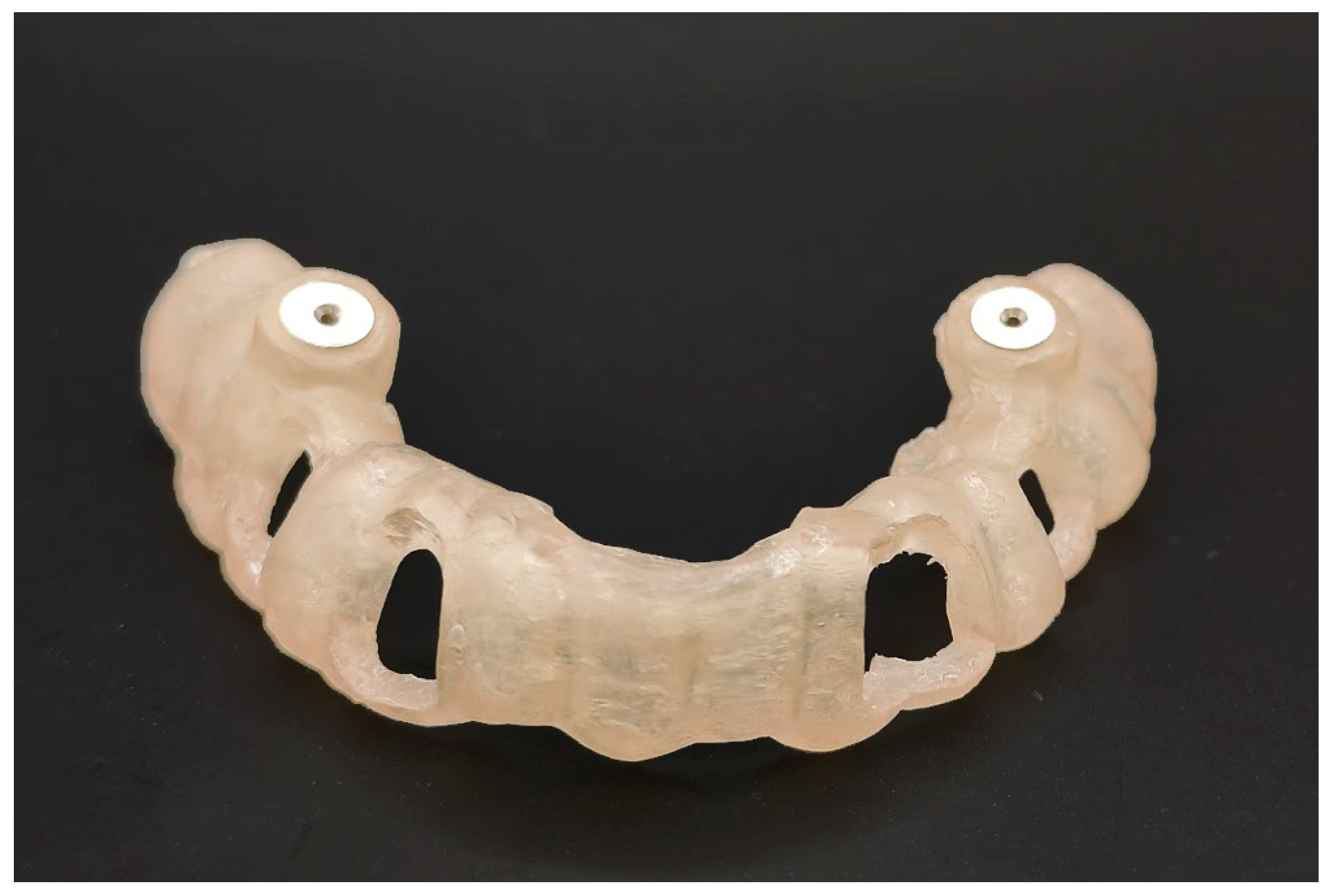
Both a CBCT (Prexion, San Mateo, CA, USA) and a surface scan with iTero (Align Technology, San Jose, CA, USA) were carried out for each model. The generated images were merged using CoDiagnostiX™ planning software (Version 9.14, Dental Wings GmbH, Chemnitz, Germany). A 1 mm diameter drilling trajectory was drawn following the exact position and depth of the fiberglass post. A virtual sleeve with a 1 mm internal diameter and 5 mm length (steco-system-technik GmbH & Co. KG, Hamburg, Germany) was put into place to guide the corresponding drill. The neighboring teeth were included in the design of the template to provide stability. The guide was then 3D-printed (Straumann® CARES® P series; Straumann, Basel, Switzerland), and the metal sleeves were subsequently implemented (Figure 2).

2.5. Procedure

The 20 manufactured models were randomly divided between two operators, ten models each. The inexperienced operator (IN) was a dental surgeon with no experience in the guided technique. The experienced operator (EX) was an endodontist with experience in the use of guides.
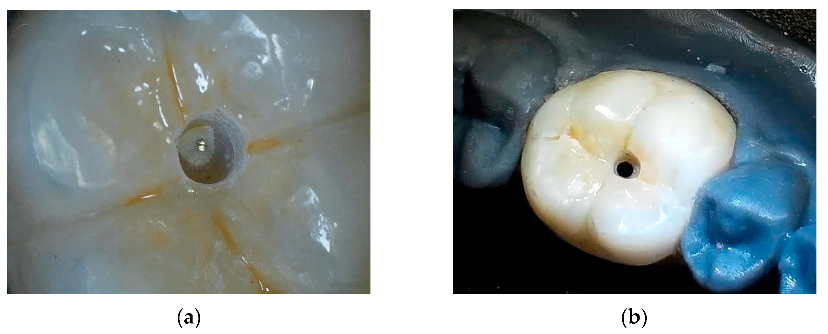
Both operators received instructions on how to stabilize the guide in position and how to carry out the guided technique in order to perform the preparation. Initially, the ceramic crown was marked using a pencil, aided by the guide indicating the place of initial penetration. Using a spherical diamond tip with high rotation (No. 1012; KG Sorensen®, São Paulo, Brazil), the ceramic crown was prepared at the marked location until it reached the resin core. Subsequently, the guide was repositioned, and penetration was performed using a bur designed for Guided Endodontics with a diameter of 1 mm and a 21 mm length (Atec Dental, Ebringen, Germany). An NSK Surgic Pro engine (NSK, Tochigi, Japan) was used, operating at 40,000 rpm with a 20:1 contra-angle handpiece (SG20; NSK, Tochigi, Japan). The access cavity was prepared under irrigation with cold saline. The drill was directed to the planned depth, finishing its course when touching gutta-percha (Figure 3). To ensure optimal performance, it is recommended by the manufacturer to use each drill to carry out ten preparations.

2.6. Evaluation of Accuracy

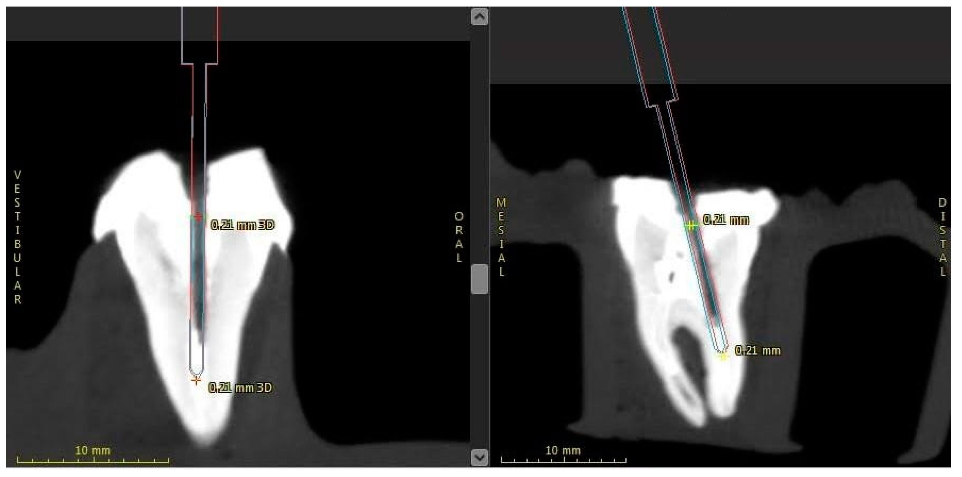
For the evaluation of the accuracy of the preparation, a postoperative CBCT scan was performed and imported into the CoDiagnostiX program. The postoperative scan was then superimposed on the initial CBCT scan to calculate the difference in the angle and the 3D deviation attained at the base and at the end of the drill path by an in-built tool of the software (Figure 4).

2.7. Statistical Analysis

Two calibrated radiologists carried out the evaluation of accuracy using the CoDiagnostiX treatment evaluation tool. The interclass correlation was calculated using a one-way random effect model and found to be 0.96 (95% CI 0.94–0.99), showing excellent agreement between the evaluators. The 95% confidence interval (CI) was calculated for each variable measured. A t-test for independent samples was used to compare the means of both the angle variable and the 3D deviation variable between the two groups. A significance level of 0.05 was adopted. The software used for the statistical analysis was SPSS (Version 25; IBM Corporation, Armonk, NY, USA).

3. Results

The mean deviation angle was 2.54° (2.7° median; 95% CI: 0–5.85°) for IN and 1.55° (1.82° median; 95% CI: 0–2.85°) for EX. The mean 3D deviation was 0.44 mm (0.45 mm median; 95% CI: 0.14–0.73 mm) for IN and 0.33 mm (0.31 mm median; 95% CI: 0.11–0.76 mm) for EX (Table 1). The fiberglass posts were removed from all samples with the exception of one, in the IN group, due to perforation.
The means for both angle and 3D deviation variables were significantly different (p = 0.008 and p = 0.049, respectively).

4. Discussion

The present study showed that the guided endodontics technique allowed the complete removal of fiberglass posts. The results indicate that the mean deviation in guided accesses is in accordance with previous studies [17,22]. Zehnder et al. [14], in an in vitro study, reported a mean deviation of 1.81° with the guided technique, and this was corroborated by our findings, with a mean deviation of 1.55° for the EX group. Therefore, the first null hypothesis of the present study was verified, confirming that guided endodontics promotes minimal deviations from planned trajectories and complete removal of intraradicular fiberglass posts.
Teeth were kept in a thymol solution (0.1%) to preserve their microhardness until further processing [21]. Although using 3D-printed teeth could be more practical in terms of sample standardization, as they were used in a study by Connert et al. [9], dental hardness is bound to influence the trajectory of the drill since the dental material resistance could lead the operator to use more or less strength to remove the dental structure. This could explain the difference in results presented by Connert et al. [9] and by the present study, as there is a significant difference between experienced and inexperienced operators. This leads to the rejection of the second null hypothesis. Despite the implementation of natural teeth and individualized ceramic crowns, the samples generated in this study demonstrated a significant level of homogeneity. This was achieved through the use of reproducible printed study models, which ensured consistency between the samples. It is important to note that the in vitro nature of this study allowed for greater stability in the study models compared to those found in the oral cavity, and clinicians must be aware of this difference when interpreting the results.
In the guided endodontics technique, the sleeve keeps the angulation of the drill precisely on the planned axis. Possible inaccuracies are partially related to the adjustment between the drill and sleeve—necessary to prevent heat from being generated or the drill bit from getting stuck during the access cavity preparation. The tolerance gap, as well as the operator’s inexperience, can lead to a deviation in angulation, which would explain the differences found between the two sample groups tested. As shown in the study by Laederach et al. [23], eccentric forces exerted by the operator in a guided system will impact the accuracy of the procedure. Despite the statistically significant differences between the two operators, their difference in deviation was only 1°, and both removed the posts successfully. However, it seems that operators need training in the guided technique in order to achieve higher accuracy.
In a guided in vivo approach, Schwindling et al. [20] demonstrated the complete removal of a fiberglass post from an upper central incisor. Perez et al. [24] have also demonstrated the feasibility of the technique in the posterior segment, where the volume of the guide and limitation of mouth opening would seem to be disadvantages. Our findings confirm that the guided technique is highly effective in removing fiberglass posts from both anterior and posterior teeth and can be replicated reliably.
Due to its accuracy, the guided endodontic technique for the removal of fiberglass posts is less prone to failure and reduces the removal of crown material to a minimum. Wide crown openings may be necessary when one uses the conventional endodontic technique, and they always have a significant impact on the prosthetic work [25]. In the study by Gerogianni et al. [26], it was shown that ceramic crowns restored after endodontic access significantly decreased their resistance under load, with the access size being the main reason. Nondestructive methods have not been found in our literature review, but guided endodontics has been shown to be a conservative alternative, for it makes possible minimally invasive access and preserves prosthetic work.
The present study, which was conducted in vitro, has certain limitations that need to be highlighted. As this study does not fully replicate the complex biological environment of a clinical setting, it is important to exercise caution when interpreting the results. One area where guided endodontic procedures can present several challenges is due to factors such as limited access and visibility, the number and location of teeth providing guide support, as well as the potential for movement or shifting of the guide during the procedure [27,28]. Although guided procedures have demonstrated efficacy in many cases, it is essential to carefully consider the specific challenges and limitations of each case to determine the suitability and potential risks associated with the procedure. The incomplete seating of the surgical template can cause deviations generated by the guide to accumulate and affect the drilling accuracy [29]. Additionally, the lack of comparison with other techniques, such as conventional endodontic treatment, restricts the extent to which the results from this study can be applied. While the accuracy of the procedure is crucial, its clinical significance ultimately depends on its ability to improve the prognosis of the tooth and the success of the restoration.
It is important to note that while guided endodontic access can be a minimally invasive and effective technique for accessing the root canal, it should not replace a proper inspection of the pulp chamber. The risk of missing important anatomy cannot be overlooked, and a thorough examination is necessary to ensure the best possible outcome for the treatment [30]. Clinicians should exercise caution when utilizing guided endodontic access and prioritize a comprehensive evaluation of the internal anatomy to avoid any potential complications. Although guided endodontics offers high precision, our study highlights the importance of the operator’s influence on the outcome of the procedure. As demonstrated in our results, inexperienced operators were more likely to encounter complications such as perforation. Therefore, it is essential for clinicians to be aware of the potential risks associated with guided endodontics and to ensure adequate training and experience before implementing the technique in practice. While our study provides valuable insights into the operator’s influence on guided endodontics, it is necessary to conduct further research that systematically investigates the various factors that impact the success of this technique. In addition to exploring potential confounding variables, such as operator experience, tooth location, and guide stabilization, comparative studies between guided endodontics and traditional endodontic techniques in clinical situations can be conducted to evaluate efficacy. This can enhance our understanding of the complexities of guided endodontics, help identify areas for improvement, and lead to the development of evidence-based strategies to minimize complications and improve patient outcomes.

5. Conclusions

With the inherent limitations of an in vitro study, it was evidenced that guided endodontics may be a viable alternative in the management of endodontic retreatments in posterior teeth with intraradicular fiberglass posts while providing minimally invasive access through ceramic crowns. Experienced operators removed fiberglass posts with higher accuracy than inexperienced operators.

Author Contributions

Conceptualization, G.F.F. and C.E.d.S.B.; methodology, G.F.F.; software, T.R.D. and F.B.P.; validation, G.F.F., T.R.D. and F.B.P.; formal analysis, C.E.d.S.B.; resources, G.F.F.; writing—original draft preparation, G.F.F.; writing—review and editing, T.C.; supervision, R.A.P. All authors have read and agreed to the published version of the manuscript.

Funding

This research received no external funding.

Institutional Review Board Statement

The research protocol of the present study was approved by the Ethics Committee (Technical Opinion #3,498,971) of Faculdade São Leopoldo Mandic.

Informed Consent Statement

Informed consent was obtained from all subjects involved in the study.

Data Availability Statement

The data that support the findings of this study are available from the corresponding author, G.F. Fachin, upon request.

Conflicts of Interest

The authors declare no conflict of interest.

References

  1. Ng, Y.L.; Mann, V.; Gulabivala, K. Outcome of secondary root canal treatment: A systematic review of the literature. Int. Endod. J. 2008, 41, 1026–1046. [Google Scholar] [CrossRef] [PubMed]
  2. Ruskin, J.D.; Morton, D.; Karayazgan, B.; Amir, J. Failed root canals: The case for extraction and immediate implant placement. J. Oral Maxillofac. Surg. 2005, 63, 829–831. [Google Scholar] [CrossRef]
  3. Garcia-Guerrero, C.; Parra-Junco, C.; Quijano-Guauque, S.; Molano, N.; Pineda, G.A.; Marin-Zuluaga, D.J. Vertical root fractures in endodontically-treated teeth: A retrospective analysis of possible risk factors. J. Investig. Clin. Dent. 2018, 9, e12273. [Google Scholar] [CrossRef]
  4. Riis, A.; Taschieri, S.; Del Fabbro, M.; Kvist, T. Tooth Survival after Surgical or Nonsurgical Endodontic Retreatment: Long-term Follow-up of a Randomized Clinical Trial. J. Endod. 2018, 44, 1480–1486. [Google Scholar] [CrossRef] [PubMed]
  5. Scribante, A.; Vallittu, P.K.; Ozcan, M. Fiber-Reinforced Composites for Dental Applications. BioMed Res. Int. 2018, 2018, 4734986. [Google Scholar] [CrossRef] [PubMed]
  6. Sarkis-Onofre, R.; Amaral Pinheiro, H.; Poletto-Neto, V.; Bergoli, C.D.; Cenci, M.S.; Pereira-Cenci, T. Randomized controlled trial comparing glass fiber posts and cast metal posts. J. Dent. 2020, 96, 103334. [Google Scholar] [CrossRef]
  7. Figueiredo, F.E.; Martins-Filho, P.R.; Faria, E.S.A.L. Do metal post-retained restorations result in more root fractures than fiber post-retained restorations? A systematic review and meta-analysis. J. Endod. 2015, 41, 309–316. [Google Scholar] [CrossRef]
  8. Lang, H.; Korkmaz, Y.; Schneider, K.; Raab, W.H. Impact of endodontic treatments on the rigidity of the root. J. Dent. Res. 2006, 85, 364–368. [Google Scholar] [CrossRef]
  9. Connert, T.; Krug, R.; Eggmann, F.; Emsermann, I.; ElAyouti, A.; Weiger, R.; Kuhl, S.; Krastl, G. Guided Endodontics versus Conventional Access Cavity Preparation: A Comparative Study on Substance Loss Using 3-dimensional-printed Teeth. J. Endod. 2019, 45, 327–331. [Google Scholar] [CrossRef]
  10. Wood, K.C.; Berzins, D.W.; Luo, Q.; Thompson, G.A.; Toth, J.M.; Nagy, W.W. Resistance to fracture of two all-ceramic crown materials following endodontic access. J. Prosthet. Dent. 2006, 95, 33–41. [Google Scholar] [CrossRef]
  11. Lund, C.; Guevara, P. The effect of endodontic access on the failure load of lithium disilicate and resin nanoceramic CAD/CAM crowns. Gen. Dent. 2018, 66, 54–59. [Google Scholar]
  12. Wiegand, A.; Kanzow, P. Effect of Repairing Endodontic Access Cavities on Survival of Single Crowns and Retainer Restorations. J. Endod. 2020, 46, 376–382. [Google Scholar] [CrossRef] [PubMed]
  13. Lynch, C.D.; Hale, R.; Chestnutt, I.G.; Wilson, N.H.F. Reasons for placement and replacement of crowns in general dental practice. Br. Dent. J. 2018, 225, 229–234. [Google Scholar] [CrossRef]
  14. Zehnder, M.S.; Connert, T.; Weiger, R.; Krastl, G.; Kuhl, S. Guided endodontics: Accuracy of a novel method for guided access cavity preparation and root canal location. Int. Endod. J. 2016, 49, 966–972. [Google Scholar] [CrossRef] [PubMed]
  15. Krastl, G.; Zehnder, M.S.; Connert, T.; Weiger, R.; Kuhl, S. Guided Endodontics: A novel treatment approach for teeth with pulp canal calcification and apical pathology. Dent. Traumatol. 2016, 32, 240–246. [Google Scholar] [CrossRef]
  16. Van Der Meer, W.J.; Vissink, A.; Ng, Y.L.; Gulabivala, K. 3D Computer aided treatment planning in endodontics. J. Dent. 2016, 45, 67–72. [Google Scholar] [CrossRef] [PubMed]
  17. Buchgreitz, J.; Buchgreitz, M.; Mortensen, D.; Bjorndal, L. Guided access cavity preparation using cone-beam computed tomography and optical surface scans-An ex vivo study. Int. Endod. J. 2016, 49, 790–795. [Google Scholar] [CrossRef] [PubMed]
  18. Connert, T.; Zehnder, M.S.; Weiger, R.; Kuhl, S.; Krastl, G. Microguided Endodontics: Accuracy of a Miniaturized Technique for Apically Extended Access Cavity Preparation in Anterior Teeth. J. Endod. 2017, 43, 787–790. [Google Scholar] [CrossRef]
  19. Lara-Mendes, S.T.O.; Barbosa, C.F.M.; Machado, V.C.; Santa-Rosa, C.C. A New Approach for Minimally Invasive Access to Severely Calcified Anterior Teeth Using the Guided Endodontics Technique. J. Endod. 2018, 44, 1578–1582. [Google Scholar] [CrossRef]
  20. Schwindling, F.S.; Tasaka, A.; Hilgenfeld, T.; Rammelsberg, P.; Zenthofer, A. Three-dimensional-guided removal and preparation of dental root posts-concept and feasibility. J. Prosthodont. Res. 2020, 64, 104–108. [Google Scholar] [CrossRef]
  21. Aydin, B.; Pamir, T.; Baltaci, A.; Orman, M.N.; Turk, T. Effect of storage solutions on microhardness of crown enamel and dentin. Eur. J. Dent. 2015, 9, 262–266. [Google Scholar] [CrossRef] [PubMed]
  22. Buchgreitz, J.; Buchgreitz, M.; Bjorndal, L. Guided root canal preparation using cone beam computed tomography and optical surface scans-An observational study of pulp space obliteration and drill path depth in 50 patients. Int. Endod. J. 2019, 52, 559–568. [Google Scholar] [CrossRef] [PubMed]
  23. Laederach, V.; Mukaddam, K.; Payer, M.; Filippi, A.; Kuhl, S. Deviations of different systems for guided implant surgery. Clin. Oral Implant. Res. 2017, 28, 1147–1151. [Google Scholar] [CrossRef]
  24. Perez, C.; Finelle, G.; Couvrechel, C. Optimisation of a guided endodontics protocol for removal of fibre-reinforced posts. Aust. Endod. J. 2019, 46, 107–114. [Google Scholar] [CrossRef]
  25. Gorman, C.M.; Ray, N.J.; Burke, F.M. The effect of endodontic access on all-ceramic crowns: A systematic review of in vitro studies. J. Dent. 2016, 53, 22–29. [Google Scholar] [CrossRef]
  26. Gerogianni, P.; Lien, W.; Bompolaki, D.; Verrett, R.; Haney, S.; Mattie, P.; Johnson, R. Fracture Resistance of Pressed and Milled Lithium Disilicate Anterior Complete Coverage Restorations Following Endodontic Access Preparation. J. Prosthodont. 2019, 28, 163–170. [Google Scholar] [CrossRef] [PubMed]
  27. El Kholy, K.; Lazarin, R.; Janner, S.F.M.; Faerber, K.; Buser, R.; Buser, D. Influence of surgical guide support and implant site location on accuracy of static Computer-Assisted Implant Surgery. Clin. Oral Implants Res. 2019, 30, 1067–1075. [Google Scholar] [CrossRef]
  28. Tahmaseb, A.; Wismeijer, D.; Coucke, W.; Derksen, W. Computer technology applications in surgical implant dentistry: A systematic review. Int. J. Oral Maxillofac. Implants 2014, 29 (Suppl.), 25–42. [Google Scholar] [CrossRef]
  29. Pessoa, R.; Siqueira, R.; Li, J.; Saleh, I.; Meneghetti, P.; Bezerra, F.; Wang, H.L.; Mendonca, G. The Impact of Surgical Guide Fixation and Implant Location on Accuracy of Static Computer-Assisted Implant Surgery. J. Prosthodont. 2022, 31, 155–164. [Google Scholar] [CrossRef] [PubMed]
  30. Mendes, E.B.; Soares, A.J.; Martins, J.N.R.; Silva, E.; Frozoni, M.R. Influence of access cavity design and use of operating microscope and ultrasonic troughing to detect middle mesial canals in extracted mandibular first molars. Int. Endod. J. 2020, 53, 1430–1437. [Google Scholar] [CrossRef]
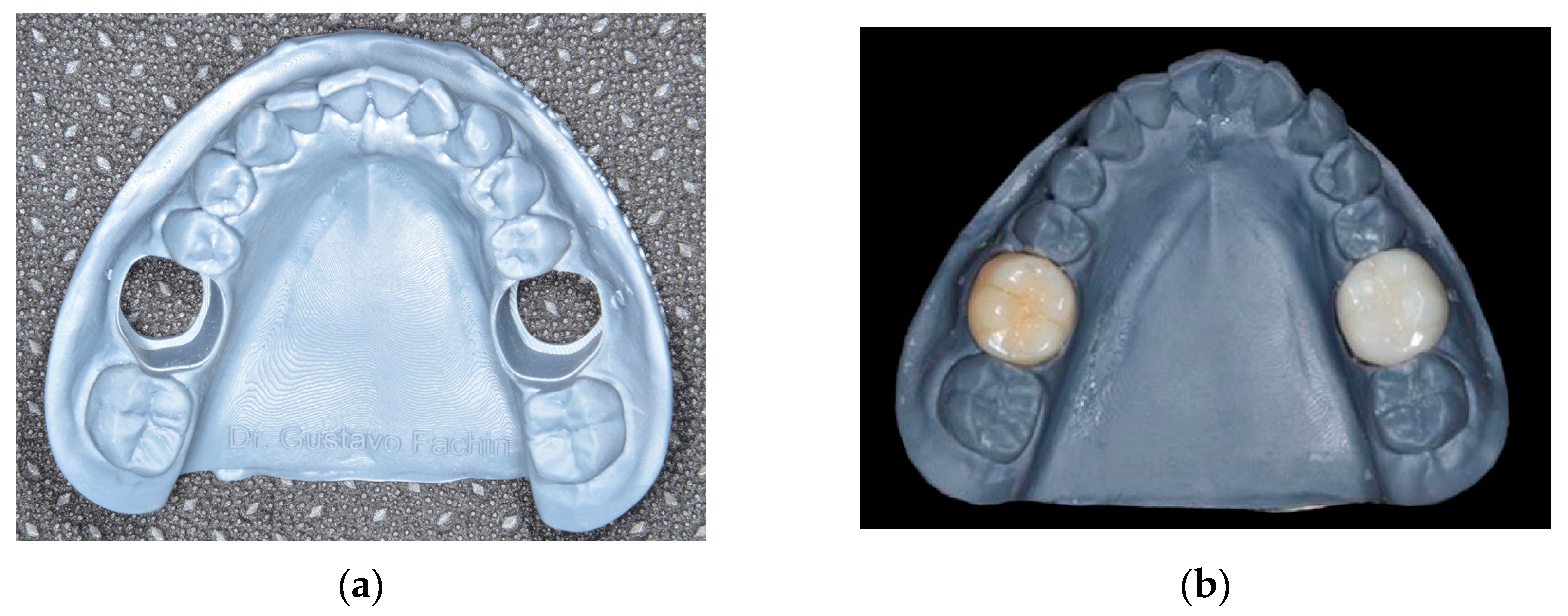
Figure 1. The manufactured model (a), and the model with cemented Emax crowns (b).
Applsci 13 05516 g001
Figure 2. Printed guide with sleeves.
Applsci 13 05516 g002
Figure 3. Procedural stages: location of the fiberglass post after initial drilling (a) and final image of the procedure (b), showing little removal of dental structure.
Applsci 13 05516 g003
Figure 4. The figure displays the analysis of the samples, showcasing both the virtual planning (blue marker) and the executed access cavity (red marker) for the procedure. The precision of the procedure is reflected in the near superimposition of the blue and red lines.
Applsci 13 05516 g004
Table 1. Comparison of the means of the variables studied.
Inexperienced Operator (IN)Experienced Operator (Ex)Differencep *
Mean (SD)
Median [min–max]
Mean (SD)
Median [min–max]
Mean [CI 95%]
Angle2.54° (1.371)
2.7° [0–5.85]
1.55° (0.791)
1.82 [0–2.85]
0.998
[0.281–1.714]
0.008
3D deviation0.44 mm (0.174)
0.45 mm [0.14–0.73]
0.33 mm (0.178)
0.31 mm [0.11–0.76]
0.113
[0.001–0.226]
0.049
SD = standard deviation; CI = confidence interval; * t-test for independent samples.
Disclaimer/Publisher’s Note: The statements, opinions and data contained in all publications are solely those of the individual author(s) and contributor(s) and not of MDPI and/or the editor(s). MDPI and/or the editor(s) disclaim responsibility for any injury to people or property resulting from any ideas, methods, instructions or products referred to in the content.