Article

Effects of the Germinated Glycine max and Angelica gigas Nakai Mixture on Hepatic Lipid Metabolism and Bone Turnover Balance in Ovariectomized Rats

1
Department of Laboratory Animal Medicine, College of Veterinary Medicine, Jeonbuk National University, Gobong-ro 79, Iksan 54596, Republic of Korea
2
Gbio Co., Ltd., Hyoyeol-ro 111, 3F, Buk-gu, Busan 46509, Republic of Korea
*
Author to whom correspondence should be addressed.
These authors contributed equally to this work.
Appl. Sci. 2023, 13(8), 5084; https://doi.org/10.3390/app13085084
Submission received: 15 March 2023 / Revised: 12 April 2023 / Accepted: 18 April 2023 / Published: 19 April 2023
(This article belongs to the Section Food Science and Technology)

Abstract

:
Women going through menopause experience changes in their hormone levels, such as decreased estrogen secretion. Those changes can lead to weight gain and steatosis (fatty liver) due to abnormal lipid metabolism and bone turnover. Natural herbal medicines have been developed to treat and prevent menopausal symptoms. The aim of this study was to evaluate how a mixture of germinated Glycine max (GG) and Angelica gigas Nakai (AG) affected serum lipid profile levels, hepatic damage, inflammation, and bone turnover in ovariectomized (OVX) rats. The animals were randomly allocated into six groups: Sham control group (Sham), OVX control group (OVX), OVX + 50 mg/kg b.w. of GG (GG50) group, and OVX + 25, 50, 100 mg/kg b.w. of GG and AG mixture (GAM; GAM25, GAM50, GAM100) groups. After four weeks of treatment, the GAM groups exhibited decreases in serum lipid profile levels (TC, TG, and Low Density Lipoprotein (LDL)) and increases in High Density Lipoprotein (HDL). Proinflammatory mediators (IL-1β, IL-6, TNF-α, and iNOS) were reduced after the administration of GAM, and the degree of liver damage (ALT, AST) also decreased. Bone resorption (CTX1, NTX1, osteoclasts in H&E staining) decreased in the GAM-treated groups, and bone morphometric markers (BMD, BV/TV, Tb.N) improved compared with the OVX group. Additionally, increased bone formation (ALP, mineralization) and decreased bone resorption (TRACP) were confirmed in in vitro experiments. These results suggest that GAM has anti-obesity and anti-inflammatory effects by preventing dyslipidemia and that it alleviates bone mass loss in OVX rats by inhibiting osteoclastogenesis.

1. Introduction

Menopause occurs as the ovaries age, causing their functioning to decrease. When the ovaries stop functioning, the consequent reduction in estradiol and estrogen production affects many organ systems [1]. As a result, menopause can cause symptoms such as osteoporosis, increased body weight, memory loss, and depression. Osteoporosis is a common disease related to postmenopausal syndrome that has a chronic effect on health [2]. Osteoporosis or osteopenia is reported to occur in half of menopausal women. Osteoporosis develops from an imbalance between bone formation and bone resorption, resulting in low bone mass and microarchitectural deterioration of bone, which increases susceptibility to bone fractures due to low bone mineral density (BMD) [3].
Osteoclasts are terminally differentiated multinucleate cells whose main function is to dissolve bone matrix and minerals in the resorption phase of bone remodeling [4]. The effects of estrogen on osteoclasts are thought to be mediated indirectly through non-osteoclastic cells. For instance, estrogen deficiency caused by menopause or ovariectomy correlates with increased secretion of tumor necrosis factor-α (TNF-α), interleukin (IL)-1, and IL-6 from osteoblasts, bone marrow stromal cells, and peripheral blood monocytes [5,6,7,8,9]. Ovariectomized (OVX) rats have symptoms of physiological menopause disorder that are similar to those found in human menopause and thus are often used as models when studying human menopause. Removing the ovaries accelerates bone absorption and significantly reduces bone density and the amount of collagen, which forms connective tissues such as cartilage or bone, and increases body lipids, especially abdominal fat accumulation, due to estrogen deficiency [10]. Bisphosphonate and estrogen, which are currently used to treat osteoporosis, were proven effective through experiments on OVX rats [11].
Hormone replacement therapy at the onset of menopause is reported to decrease the symptoms of osteoporosis by about 50% and have long-lasting effects [12]. However, several studies have reported that hormone replacement therapy can increase the risk of side effects such as cardiovascular disease, breast tenderness, thromboembolic disease, breast cancer, endometrial cancer, and stroke [13]. Studies are currently being conducted to minimize the symptoms of menopause without incurring the side effects caused by hormone replacement therapy. One alternative for treating and improving menopausal symptoms is phytoestrogen derived from natural products, which is known to relieve menopause symptoms by activating estrogen receptors based on its structural similarity. Among the candidate treatments, soybean is getting attention as a complementary and alternative medicine that might be used for the long-term treatment of menopause-related disease [14,15,16,17].
Soybean (Glycine max) is widely used in healthy foods and is nutritionally supported for the prevention of menopausal symptoms, prostate cancer, and osteoporosis. Soybean contains high protein, bioactive compounds such as isoflavone, and vitamins [18]. Several studies have shown that soybean compounds are associated with improvement in cardiovascular disease and glucose tolerance and have anti-osteoporosis effects and antioxidant activity. The germination of soybean has been found to increase its nutritional value and bioactive compound’s amounts. Soybean has long been an ingredient in traditional medicine and functional foods. Germinated soybean (Glycine max, GG) is reported to have higher levels of bioactive compounds and phytoestrogen, such as genistein, than non-germinated soybean [19]. However, in people who are sensitive to estrogen, the phytoestrogen in soybeans can activate estrogen receptors, affecting the occurrence and progression of diseases common in middle age, such as uterine myoma and breast cancer. Angelica gigas Nakai (AG) is used as an herbal medicine in East Asia [20]. Traditionally, AG has been used to prevent various diseases [21,22], and it is also known to be effective for wound treatment, body fat reduction, and anti-inflammation. Decursin is the major active component of the AG root and has been shown to have beneficial effects against disease models for cancer, oxidative stress, and inflammation [23].
GG has been reported to have good effects in various fields, but studies on inflammatory mechanisms related to lipid metabolism are insufficient. For these reasons, a mixture of GG and AG (GAM) might have anti-osteoporosis effects and mitigate body fat increase, which could make it useful as a functional food source. Therefore, we investigated the effects of GAM on hepatic lipid metabolism and bone turnover balance in an OVX rat model. By evaluating the effects of this dietary intervention, we hope to improve understanding about the potential benefits of GAM for improving menopausal health.

2. Materials and Methods

2.1. Sample Preparation

Soybean embryos were purchased from KNbio Co., Ltd. (Hamyang, Republic of Korea), thoroughly washed, digested in purified water at 20 °C for 24 h, and pretreated in a germinated state. Thereafter, the GG was dried at 70 °C for 24 h using a hot air dryer. Then, 10 L of 70% ethanol fermented liquor was added to 1 kg of dried germinated soybean embryo, and the extract was obtained after extracting the quench at 65 °C for 5 h. AG was purchased from Jinbu GAP Danggwi Agricultural Cooperative Corporation (Pyeongchang, Korea). It was washed clean and dried in a hot air dryer at 80 °C until it had a moisture content of 10% or less. Then, 10 L of 70% ethanol fermented liquor was added to 1 kg of the dry product to obtain an extract after extracting the quench at 65 °C for 8 h. After filtration, the concentrate was freeze dried to a powder.

2.2. Cell Culture and Treatment

2.2.1. Osteoblasts

MC3T3-E1 murine osteoblast cells were purchased from the Korean Cell Line Bank (KCLB, Seoul, Republic of Korea) and maintained in alpha minimum essential medium (α-MEM) with 10% fetal bovine serum (FBS, Welgene, Daegu, Republic of Korea), 100 unit/mL penicillin (Welgene, Republic of Korea), and 100 μg/mL streptomycin (Welgene, Republic of Korea). The cells were seeded in 96-well plates at 1 × 10⁴ cells/well. After 24 h, the cells were differentiated by replacing fresh α-MEM medium with 10 mM β-glycerol phosphate, 50 mg/mL ascorbic acid, and GG or GAM at the indicated doses.

2.2.2. Osteoclasts

RAW264.7 murine macrophage cells were purchased from KCLB (Republic of Korea) and maintained in Dulbecco’s modified Eagle medium (DMEM, Welgene, Republic of Korea) with 10% inactivated FBS (Welgene, Republic of Korea), 100 unit/mL penicillin (Welgene, Republic of Korea), and 100 μg/mL streptomycin (Welgene, Republic of Korea). The cells were seeded in 96-well plates at 1 × 10⁴ cells/well. After 3 days, the cells were differentiated by replacing the medium with DMEM containing 2% FBS, RANKL (1 ng/mL), and GG or GAM at the indicated doses every 48 h for 7 days.

2.3. Cell Viability Assay

Cytotoxicity was measured using a 3-(4,5-dimethylthiazol-2-yl)-2,5-diphenyltetrazolium bromide (MTT) assay kit (Sigma-Aldrich, St. Louis, MO, USA). MC3T3-E1 cells were seeded in 96-well plates at 1 × 10⁴ cells/well in triplicate. When the confluences reached 70–80%, the cells were treated with the indicated concentrations of GG or GAM and incubated for 24 h at 37 °C, 5% CO2. Next, 10 μL of 5 mg/mL MTT solution was added to each well and incubated at 37 °C and 5% CO2 for 2 h. Then, we removed the medium from each well, added 100 μL of dimethyl sulfoxide (DMSO), and measured the optical density at 490 nm using a microplate reader (BioTek Instruments, Winooski, VT, USA).

2.4. Alanine Phosphate Activity and Tartrate-Resistant Acid Phosphatase Activity

Alanine phosphatase (ALP) activity and tartrate-resistant acid phosphatase (TRACP) activity were measured using a TRACP&ALP assay kit (MK301, TaKaRa, Kusatsu, Singa, Japan). Osteoclast cells differentiated from RAW264.7 cells were washed once with Dulbeco’s phosphate-buffered saline (DPBS). Then, 50 μL of extraction solution and substrate solution were added to each well, and they were incubated for 60 min at 37 °C. We added 50 μL of stop solution and measured the absorbance at 405 nm using a microplate reader (BioTek Instrument, Winooski, VT, USA) after color formation.

2.5. Alizarin Red Staining

Osteoblast cells differentiated from MC3T3-E1 cells were washed twice with DPBS buffer and fixed with 4% paraformaldehyde for 15 min. The cells were then washed again with deionized H2O (dH2O), stained with alizarin red, and incubated for 30 min at room temperature. The cells were washed with dH2O and visualized using a light microscope (ZEISS, Jena, Germany) and high-power fields (×200).

2.6. Animals and Surgical Procedure

Female Sprague-Dawley (SD) rats (6-weeks old) were purchased from Damool Science (Daejeon, Republic of Korea). All rats were housed in a conventional system (room temperature: 22–24 °C, humidity: 50–60%, 12 h/12 h light and dark cycles) and allowed 1 week of adaptation before the beginning of the experiment. We randomly divided 42 rats into six groups (seven rats per group): Sham control group (Sham), OVX control group (OVX), OVX + GG 50 mg/kg (GG 50) group, and OVX + GAM 25, 50, or 100 mg/kg (GAM25, GAM50, GAM100) groups. Before the surgical operation, each rat was anesthetized using isoflurane (Hana Pharm. Co., Ltd., Hwaseong, Republic of Korea). One week after the surgical operation, the experimental groups were treated with GG or GAM once a day by oral gavage for 4 weeks. The Sham and OVX groups were treated with an equal volume of physiological saline by oral gavage. After 4 weeks of administration, blood was collected from the abdominal aorta vein. After 30 min, the blood was centrifuged at 40,000× g for 15 min (Union 32R Plus, Hanil Science Industrial Co., Kimpo, Korea), and the resulting supernatant was stored at −70 °C for further assays. The whole femur, liver, and uterus were collected from each animal after they were euthanized. The animal experiment was approved by the Institutional Animal Ethics Committee of the Jeonbuk National University Animal Care and Use Committee (Approval Number: JBNU 2022-096).

2.7. Micro-Computed Tomography Analysis

The femurs from all the animals were examined using microcomputed tomography (µ-CT) at the Center for University-wide Research Facilities (CURF) at Jeonbuk National University (Jeonju, Republic of Korea) on a SkyScan1076 (SKYSCAN, Aartselaar, Belgium). Those examinations were used to determine changes in the trabecular bone. The bone volume and density of the femurs were analyzed using the CTAn program (SKYSCAN, Aartselaar, Belgium). Additionally, BMD was calculated using a phantom and Hounsfield units (HU). The bone threshold value was determined between the low phantom (0.25) 1067.09838 HU and high phantom in μ-CT HU (0.75) 3249.32035 HU.

2.8. Hematoxylin and Eosin Staining

Rat femurs were isolated and fixed in 10% formalin for 3 days. After fixation, they were decalcified in a 10% HCl solution for 3 h. All selected femurs were embedded in paraffin and cut into 5-μm sections using a microtome. The tissue section samples were deparaffinized using xylene and a graduated alcohol series with dH2O and stained with hematoxylin and eosin (H&E). The stained femurs were imaged using a light microscope (ZEISS, Jena, Germany) and high-power fields (×400).

2.9. ELISA of Serum in Rats

The serum osteocalcin (OC) concentration was analyzed using an OC ELISA kit (MBS728975, MyBioSource, San Diego, CA, USA). The total ALP concentration was analyzed using an ALP assay kit (ab83369, Abcam, London, UK). The cross-linked C terminal telopeptide of type 1 collagen (CTX1) concentration was analyzed using a rat CTX1 ELISA kit (MBS728629, MyBioSource, San Diego, CA, USA). The cross-linked N terminal telopeptide of type 1 collagen (NTX1) concentration was analyzed using a rat NTX1 ELISA kit (MBS8800850, MyBioSource, San Diego, CA, USA). The serum triglyceride (TG) concentration was analyzed using a PicoSensTM TG assay kit (BM-TGR-100, Biomax, Guri, Republic of Korea). The serum total cholesterol (TC) concentration was analyzed using a PicoSensTM TC assay kit (BM-CHO-100, Biomax, Guri, Republic of Korea). The serum low density lipoprotein (LDL) concentration was analyzed using a rat LDL ELISA kit (MBS702165, MyBioSource, CA, USA). The serum high density lipoprotein (HDL) concentration was analyzed using an HDL, LDL/VLDL assay kit (BM-CDL-100, Biomax, Guri, KR). The serum aspartate transaminase (AST) concentration was analyzed using a rat AST ELISA kit (ab263883, Abcam, London, UK). The serum alanine aminotransferase (ALT) concentration was analyzed using a rat ALT ELISA kit (ab285264, Abcam, London, UK). All assays were conducted according to the manufacturer instructions.

2.10. Western Blot Analyses

The collected livers were washed with ice-cold phosphate-buffered saline (PBS) and lysed in lysis buffer with protease inhibitors and phosphatase inhibitors (04082219, GenDEPOT, HU, USA). We centrifuged the samples at 15,000× g for 20 min and then quantified the proteins in the supernatants using a bicinchoninic acid (BCA) reagent. The proteins were denatured, separated by 12% sodium dodecyl sulphate–polyacrylamide gel electrophoresis, and transferred to a polyvinylidene difluoride membrane (18289A 10, Bio-Rad Laboratories, Hercules, CA, USA). After blocking them in 5% bovine serum albumin (BSA) in PBS-T for 2 h, we incubated them overnight at 4 °C with a primary antibody (iNOS, IL-1β, IL-6, TNF-α, or β-actin) diluted 1: 1000 in 5% BSA in PBS-T. Then, the membranes were incubated with a secondary antibody diluted 1:2000 at room temperature for 1 h. The membranes were developed using SuperSignal West Femto maximum sensitivity substrate (Thermo Fisher Scientific, Waltham, MA, USA) and visualized on a WSE-6200 luminograph II (ATTO, Tokyo, Japan). Band density was normalized to that of β-actin as a loading control.

2.11. Statistical Analysis

The statistical analyses were conducted in GraphPad Prism version 5.0 (GraphPad Software, San Diego, CA, USA). Data are presented as the mean ± SEM. The significance of differences between groups was determined by one-way ANOVA and considered significant when p < 0.05 was obtained by Tukey’s multiple range test. All the sets of in vitro experiments were carried out in triplicate at a minimum [24].

3. Results

3.1. GAM Prevents Weight Gain in the OVX-Induced Rat Model

OVX animals are a well-known model for postmenopausal conditions and are characterized by an increase in body weight [25]. We therefore evaluated the effect of GAM on body weight in our OVX rat model. The reduction in uterine weight in the OVX control group confirmed that the modeling proceeded properly. The OVX rats showed an increase in body weight compared with the Sham group (Table 1). However, the GAM-treated groups showed reduced body weight compared with the OVX group. Therefore, GAM significantly prevented weight gain in the OVX-induced rat model.

3.2. GAM Inhibits Lipid Profile Changes in the OVX-Induced Rat Model

The goal was to evaluate the effects of GAM on the lipid metabolism of OVX-induced rats. It is well known that estrogen plays an important role in lipid metabolism in humans [26]. Recently, several studies in humans reported that a deficiency of estrogen promoted weight gain by causing lipid accumulation [27,28]. Serum TC increased by 41% in the OVX group compared with the Sham group (Figure 1a). Interestingly, the serum TC concentration decreased by 25.9% in the GAM50 group compared with the OVX group. The serum TG concentration increased by 35% in the OVX group compared with the Sham group (Figure 1b) and decreased by 40.9% in the GAM50 group compared with the OVX group. The serum LDL concentration increased by 87.8% in the OVX group compared with the Sham group (Figure 1c) and decreased by 77.6% and 65.7% in the GAM25 and GAM50 groups, respectively, compared with the OVX group. The serum HDL concentration decreased by 2.0% in the OVX group compared with the Sham group (Figure 1d) and increased by 15.3% and 39.2% in the GAM50 and GAM100 groups, respectively, compared with the OVX group. These results suggest that GAM improved the body lipid profiles of the OVX-induced model rats.

3.3. GAM Prevents Liver Damage in the OVX-Induced Rat Model

The severity of fatty liver disease is directly related to classic components of metabolic syndrome, such as damage to the liver and central obesity [29]. Therefore, the effects of GAM on liver damage were explored in our OVX-induced rat model using ELISA kits for AST and ALT. Damage to hepatocytes causes abnormally high AST levels in blood serum. The serum AST and ALT concentrations increased by 62.3% and 10.4%, respectively, in the OVX group compared with the Sham group (Figure 2) and significantly decreased in the GAM groups compared with the OVX group. These results show that GAM prevented liver damage in our OVX-induced rat model.

3.4. GAM Decreases Inflammatory Mediators in the OVX-Induced Rat Model

Proinflammatory substances such as IL-6 and TNF-α are overproduced in fatty livers [30]. To evaluate the effects of GAM on inflammation in our OVX-induced rat model, the levels of pro-inflammatory factors in liver tissue were measured using Western blotting. TNF-α, IL-1β, IL-6, and iNOS expression all increased the livers of the OVX group compared with the Sham group (Figure 3). The expressions of TNF-α, IL-1β, IL-6, and iNOS decreased significantly, by 72.7%, 26.9%, 62.9, and 93.6%, respectively, in the GAM100 group. Therefore, GAM inhibited inflammation in our OVX-induced rat model.

3.5. GAM Prevents Estrogen Deficiency-Induced Bone Loss in the OVX-Induced Rat Model

During menopause, estrogen levels significantly decrease, which increases the risk of osteoporotic fractures. To evaluate the anti-osteoporosis effects of GAM, we investigated BMD, percent bone volume ratio, and trabecular bone number. As shown in Figure 4a, μ-CT indicated that trabecular bone in the femur was decreased by OVX, and GAM treatment dose-dependently protected against OVX-induced bone loss. As shown in Figure 4b, BMD, percent bone volume ratio, trabecular bone number, trabecular bone volume, and trabecular bone thickness were also dose-dependently improved by GAM. Therefore, GAM prevented OVX-induced bone loss in our OVX-induced rat model.

3.6. GAM Stimulated Bone Formation in the OVX-Induced Rat Model

To evaluate the effects of GAM on bone formation in our OVX-induced rat model, Serum ALP, which is a biomarker routinely examined in clinical laboratory tests, and serum OC were analyzed using ELISA kits. The total ALP concentration increased in the OVX group (38.1%) compared with the Sham group. However, after GAM treatment for 4 weeks, the total ALP concentration was decreased by 22.7% and 29.5% in the GAM50 and GAM100 groups, respectively, compared with the OVX group (Figure 5a). The serum OC concentration increased in the OVX group (10.1%) compared with the Sham group (Figure 5b) and significantly decreased in the GAM50 and GAM100 groups (16.4% and 24.7%, respectively) compared with the OVX group. Therefore, GAM prevented bone loss in our OVX-induced rat model by stimulating bone formation.

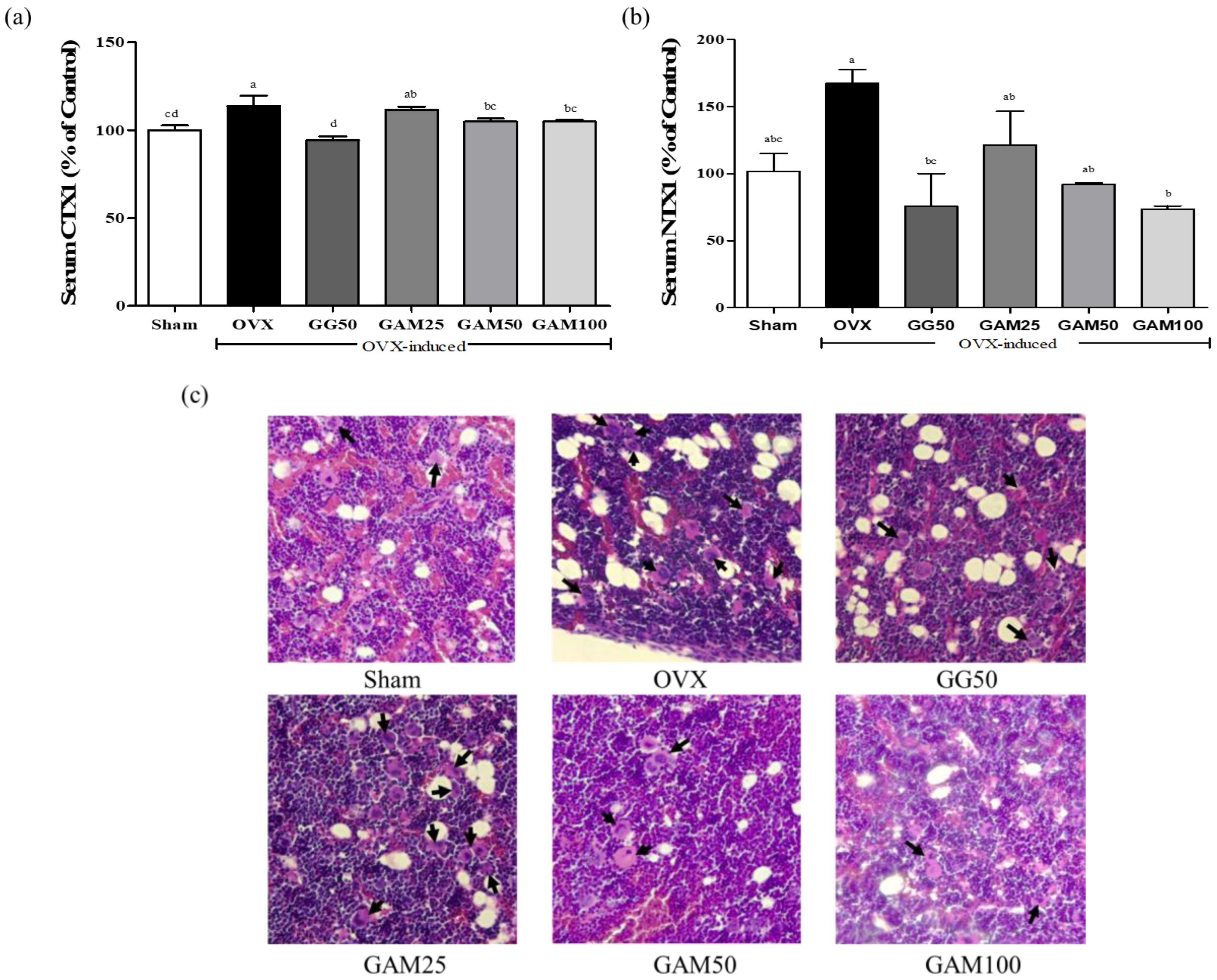
3.7. GAM Inhibits Bone Absorption in the OVX-Induced Rat Model

Many pathological conditions, such as postmenopausal osteoporosis and rheumatoid arthritis, are characterized by progressive and excessive bone resorption due to osteoclast differentiation activity [31]. CTX1 and NTX1 are products of type 1 collagen degradation, and serum levels of CTX1 and NTX1 are positively correlated with bone resorption. To confirm the effects of GAM on OVX-induced bone absorption, CTX1 and NTX1 were measured using ELISA kits. As shown in Figure 6a, the serum CTX1 level increased by 13.6% in the OVX group compared with the Sham group, and 4 weeks of GAM administration decreased the serum CTX1 concentration in the GAM50 group by 8.9% compared with the OVX group. As shown in Figure 6b, the serum NTX1 level increased by 67.3% in the OVX group compared with the Sham group, and GAM decreased that concentration. Histological assessments revealed that the number of osteoclasts, which resorb bone in trabecular bone, was decreased by GAM treatment (Figure 6c). Therefore, GAM prohibited bone absorption in our OVX-induced rat model by inhibiting bone resorption.

3.8. GAM Stimulated Osteoblast Differentiation in MC3T3-E1 Cells

We treated osteoblastic cells with various ratios of GG and AG (7:3, 5:5, 3:7 and 1:9) and analyzed their bone formation-enhancing effects and osteoblast differentiation via ALP activity. GAM did not have cytotoxic effects on MC3T3-E1 cells (Figure 7b). After the induction of osteoblast differentiation, the highest ALP activity was observed following treatment with 7:3 GG + AG (Figure 7a). To evaluate the effects of GAM on osteoblasts differentiated from MC3T3-E1 cells, we incubated the cells with various concentrations (10, 50, 100 μg/mL) of GG or GAM. As shown in Figure 7c, GAM increased ALP activity in osteoblasts differentiated from MC3T3-E1 cells for 7 days and 14 days. Alizarin red staining indicates osteogenic mineralization. GAM treatment increased osteogenic mineralization in the osteoblasts differentiated from MC3T3-E1 cells (Figure 7d,e). Therefore, GAM stimulated osteoblast differentiation.

3.9. GAM Inhibits TANKL-Induced Osteoclast Differentiation in RAW264.7 Cells

The receptor activator of nuclear factor-kappa B ligand (RANKL) is a cytokine essential to osteoclastogenesis [32]. To evaluate the effects of GAM on RANKL-induced osteoclastogenesis, RAW264.7 cells were incubated with various concentrations (10, 50, 100 μg/mL) of GG or GAM. After the induction of osteoclast differentiation, we measured TRACP using an ELISA kit. TRACP is an enzyme expressed in high amounts by bone-resorbing osteoclasts and inflammatory macrophages. As shown in Figure 8, GAM inhibited osteoclast differentiation in RANKL-induced RAW264.7 cells. Therefore, GAM dose-dependently inhibited RANKL-induced osteoclast differentiation.

4. Discussion

Soybean is a major source of protein and provides nutritional benefits to most of the world’s population [18]. However, when extracting the soybean protein from soybean flour, the glycoside form of isoflavone can be converted into an aglycon of different proportions, depending on the leaching method used [33]. Germination processes have been developed to overcome related disadvantages. Many studies have shown the benefits of germinated soybean, including improvements in nutritional quality by preventing lipid oxidation, increases in nutrients such as riboflavin and ascorbic acid, hydrolysis of raffinose and stachyose that can cause flatulence, and decreases in trypsin inhibitors [34,35,36,37]. In addition, the total isoflavone content increases rapidly during the early stage of germination. Isoflavones can prevent osteoporosis through their phytoestrogen effects [19,38]. ALP activity was measured in MC3T3-E1 cells to determine the best ratio of GG and AG. At a GG: AG ratio of 7:3, ALP activity peaked (Figure 7a). In our study, the results suggested that the GG: AG ratio of 7:3 could effectively treat postmenopausal osteoporosis.
GAM treatment decreased body weight (Table 1). It was previously reported that low estrogen levels after menopause can cause fat to gradually accumulate around internal abdominal organs, causing central obesity. Abdominal fat accumulation correlates with many metabolic changes after menopause, such as decreasing HDL-cholesterol and increasing LDL-cholesterol [39,40]. The levels of LDL, TC, TG, and lipoproteins peak during the menopausal transition and early postmenopausal stage [41]. In this study, SD rats subjected to OVX had higher concentrations of LDL, TC, and TG than the Sham group rats (Figure 1a–c), and HDL expression was lower than in the Sham group (Figure 1d). The OVX rat model thus shows that estrogen deficiency results in lipid accumulation by increasing the LDL level. Treatment with GAM for 4 weeks decreased the concentrations of LDL, TC, and TG and increased the HDL level compared with the OVX group. These results showed that GAM prevented lipid accumulation in our OVX-induced rat model. This is consistent with recent reports suggesting that GAM has anti-obesity effects in OVX model rats by improving lipid metabolism.
The severity of fatty liver disease is directly related to classic components of metabolic syndrome such as liver damage, central obesity, nonalcoholic fatty liver diseases, insulin resistance, and hyperinsulinemia [29]. Excessive fat accumulation in the liver associated with menopause plays an important role in the development of insulin resistance [42]. Hepatocyte and adipocyte accumulation is well-known in animal models of menopause. AST and ALT are found in almost all body tissues, but they are absent from bones. AST determines the reversible exchange between glutamate and aspartate, and ALT catalyzes the conversion of alanine and α-ketoglutarate into pyruvate and glutamate, thereby contributing to cellular nitrogen metabolism and liver gluconeogenesis. Therefore, damage to any organ causes a rapid increase in the levels of AST and ALT in the blood stream [43]. We assessed the anti-inflammatory and liver-protective effects of GAM and found that the AST and ALT concentrations in the OVX group were higher than those in the Sham group. Treatment with GAM for 4 weeks decreased the concentrations of AST and ALT compared with OVX group (Figure 2). These results showed that GAM protects the liver from damage in the OVX-induced rat model. GAM could have protective effects from damage to the liver by lipid accumulation.
Whether the cause is liver fat accumulation or portal circulation inflammation, inflammatory cytokines such as IL-6 and TNF-α are overproduced in a damaged liver. Furthermore, produced inflammatory substances such as IL-6, TNF-α, and IL-1β can enhance osteoclastogenesis [44]. Our results suggested that the expression of IL-6, TNF-α, IL-1β, and iNOS increased in the OVX group, indicating liver damage (Figure 3), and GAM decreased the expression of these inflammatory biomarkers compared with the OVX group (Figure 3). These results showed that GAM had anti-inflammatory effects that protected the liver from lipid accumulation and alleviated osteoporosis.
Bone remodeling requires a balance between bone formation (osteoblasts) and bone resorption (osteoclasts). BMD and trabecular bone volume were assessed using a μ-CT system to evaluate the extent of osteoporosis. BMD indicates the amount of calcium and other types of minerals in analyzed areas of bone. A deficiency of bone replacement during menopause can cause an increase in bone mass loss because bone resorption outpaces bone formation, leading to a decrease in BMD [45]. The μ-CT system is highly accurate for evaluating the trabecular bone volume. We investigated bone quality in our osteoporosis model by measuring BMD, percent bone volume ratio, and trabecular bone number, and we confirmed OVX-induced bone loss in the μ-CT images of the femur. Our histological visualization of the μ-CT results shows that GAM treatment filled the trabecular bone. Additionally, our finding that the bone morphometric markers (BMD, BV/TV, Tb.N) increased (Figure 4b) indicates that GAM treatment for 4 weeks prevented trabecular bone loss in estrogen deficiency-induced osteoporosis (Figure 4b). Therefore, GAM could have anti-osteoporosis effects in postmenopausal osteoporosis.
ALP activity is an important biochemical marker for bone formation during osteoblast differentiation [46]. After menopause, bone resorption markers increase due to the activation of bone resorption by estrogen deficiency, so bone formation must also increase to fill the higher number of resorption cavities, causing an increase in the serum levels of both ALP and OC [43,44]. That is why high levels of ALP in elderly people suggest the presence of osteoporosis [44]. We investigated bone formation activity by measuring ALP and OC levels in our OVX-induced model rats. We found that the serum ALP and OC levels were higher in the OVX group than in the Sham group (Figure 5), suggesting that OVX increases bone formation markers to maintain the balance of bone turnover. The serum concentrations of both ALP and OC decreased following GAM treatment for 4 weeks, which suggests that GAM prevents OVX-induced bone loss by decreasing bone resorption. Furthermore, in our in vitro experiment, we found that increasing ALP levels indicate increasing bone mineralization. Because ALP activity can indicate the degree of differentiation of osteoblasts, it is used to evaluate the mineralization ability of osteoblasts [47]. We measured ALP using alizarin red staining in osteoblasts differentiated from MC3T3-E1 cells (Figure 7) and found that GAM treatment increased ALP levels compared with the control group (Figure 7b). The histological visualization of alizarin red staining showed that GAM treatment increased mineralization in osteoblasts differentiated from MC3T3-E1 cells (Figure 7d). Increased ALP activity and alizarin red staining both reflect osteogenic mineralization. Therefore, GAM could prevent postmenopausal osteoporosis by regulating the ALP concentration.
The inhibition of osteoclastogenic signals could be an effective method for treating bone loss-related diseases. CTX1 and NTX1 are released by osteoclasts and are therefore markers of bone resorption [48]. Bone resorption activity was investigated by measuring CTX1 and NTX1 in our OVX-induced rat model. We found that the levels of CTX1 and NTX1 and the number of osteoclasts seen with H&E staining were higher in the OVX group than in the Sham group (Figure 6). Therefore, OVX causes excessive bone resorption by stimulating osteoclastogenesis. We also investigated osteoclast activity by measuring TRACP activity in RANKL-induced osteoclasts differentiated from RAW264.7 cells (Figure 8) and found that GAM treatment decreased CTX1 and NTX1 levels and TRACP activity (Figure 6 and Figure 8). Our histological visualization of H&E staining shows that GAM treatment inhibited the differentiation of osteoclasts in our OVX-induced rat model (Figure 6c). Therefore, GAM could decrease estrogen deficiency-induced bone resorption by inhibiting osteoclastogenesis in postmenopausal osteoporosis.
The results of this study suggest that GAM decreases postmenopausal inflammation, fat accumulation, liver damage, and bone absorption. We have here demonstrated the potential of GAM for development as a beneficial ingredient for food to improve menopausal symptoms.

5. Conclusions

This study has demonstrated that combining GG and AG at a 7:3 ratio had anti-obesity, anti-inflammation, and anti-osteoporotic effects in an OVX-induced rat model. Moreover, we confirmed the anti-osteoporotic effects in pre-osteoblast MC3T3-E1 cells by showing that GAM stimulated mineralization and in RANKL-induced osteoclast differentiation from RAW264.7 cells by showing that GAM suppressed osteoclastogenesis. Therefore, GAM could be a potential candidate for the treatment and prevention of postmenopausal symptoms. Comprehensive human studies are required to investigate the role of GAM as a therapeutic functional food against osteoporosis and associated disorders.

Author Contributions

Conceptualization, J.K.; methodology, S.L., Y.C., J.C. and H.C.; performing animal experiments, S.L., J.C. and H.C.; performing cells experiments, Y.C.; data curation, S.L. and J.C.; writing—original draft preparation, S.L. and J.C.; writing—review and editing, J.C. and J.K.; project administration, T.S.J. and J.K. All authors have read and agreed to the published version of the manuscript.

Funding

This work was supported by project for Collabo R&D between Industry, Academy, and Research Institute funded Korea Ministry of SMEs and Startups in 2021(Project No. S3104795).

Institutional Review Board Statement

The study was conducted according to the guidelines of the Declaration of Helsinki, and approved by the Ethics Committee of Jeonbuk National University Institutional Animal Care and Use Committee (protocol code: JBNU 2022-096).

Informed Consent Statement

Not applicable.

Data Availability Statement

Not applicable.

Acknowledgments

Authors thank Yu Jeong Chang, Center for University-wide Research Racilities (CURF) at Jeonbuk National University, for using Micro-Computed Tomagraphy.

Conflicts of Interest

The authors declare no conflict of interest.

References

  1. World Health Organization. Research on the Menopause in the 1990s: Report of a WHO Scientific Group; WHO: Geneva, Switzerland, 1996.
  2. Monteleone, P.; Mascagni, G.; Genazzani, A.; Simoncini, T. Symptoms of menopause-global prevalence, physiology and implication. Nat. Rev. Endocrinol. 2018, 14, 199–215. [Google Scholar] [CrossRef]
  3. Tian, L.; Baek, S.H.; Jang, J.; Kim, S.Y. Imbalanced bone turnover markers and low bone mineral density in patients with osteonecrosis of the femoral head. Int. Orthop. 2018, 42, 1545–1549. [Google Scholar] [CrossRef]
  4. Ono, T.; Nakashima, T. Recent advances in osteoclast biology. Histochem. Cell Biol. 2018, 149, 325–341. [Google Scholar] [CrossRef]
  5. Wang, T.; He, C. TNF-α and IL-6: The link between immune and bone system. Curr. Drug Targets 2020, 21, 213–227. [Google Scholar] [CrossRef] [PubMed]
  6. Song, X.X.; Jin, L.Y.; Li, X.F.; Yu, B.W. Substance P mediates estrogen modulation proinflammatory cytokines release in intervertebral disc. Inflammation 2021, 44, 506–517. [Google Scholar] [CrossRef] [PubMed]
  7. Wu, B.; Song, Q.; Zhang, Y.; Wang, C.; Yang, M.; Zhang, J.; Han, W.; Jiang, P. Antidepressant activity of ω-3 polyunsaturated fatty acids in ovariectomized rats: Role of neuroinflammation and microglial polarization. Lipids Health Dis. 2020, 19, 4. [Google Scholar] [CrossRef] [PubMed]
  8. Pfeilschifter, J.; Chenu, C.; Bird, A.; Mundy, G.R.; Roodman, G.D. Interleukin-1 and tumor necrosis factor stimulate the formation of human osteoclastlike cells in vitro. J. Bone Miner. Res. 1989, 4, 113–118. [Google Scholar] [CrossRef] [PubMed]
  9. Bertolini, D.R.; Nedwin, G.E.; Bringman, T.S.; Smith, D.D.; Mundy, G.R. Stimulation of bone resorption and inhibition of bone formation in vitro by human tumour necrosis factors. Nature 1986, 319, 516–518. [Google Scholar] [CrossRef]
  10. Brennan, O.; Kennedy, O.D.; Lee, T.C.; Rackard, S.M.; O’brien, F.J. Effects of estrgen deficienct and bisphosphonate therapy on osteocyte viability and microdamage accumulation in an ovine model of osteoporosis. J. Orthop. Res. 2011, 29, 419–424. [Google Scholar] [CrossRef]
  11. Lelovas, P.P.; Xanthos, T.T.; Thoma, S.E.; Lyritis, G.P.; Dontas, I.A. The laboratory rat as an animal model for osteoporosis research. Comp Med. 2008, 58, 424–430. [Google Scholar]
  12. Cagnacci, A.; Venier, M. The controversial history of hormone replacement therapy. Medicina 2019, 55, 602. [Google Scholar] [CrossRef] [PubMed]
  13. Tit, D.M.; Bungau, S.; Lovan, C.; Cseppento, D.C.N.; Endres, L.; Sava, C.; Sabau, A.M.; Furau, G.; Furau, C. Effects of the hormone replacement therapy and of soy isoflavones on bone resorption in postmenopause. J. Clin. Med. 2018, 7, 297. [Google Scholar] [CrossRef] [PubMed]
  14. Gorzkiewicz, J.; Bartosz, G.; Sadowska-Bartosz, I. The potential effects of phytoestrogens: The role in neuroprotection. Molecules 2021, 26, 2954. [Google Scholar] [CrossRef] [PubMed]
  15. Abdi, F.; Rahnemaei, F.A.; Roozbeh, N.; Pakzad, R. Impact of phytoestrogens on treatment of urogenital menopause symptoms: A systematic review of randomized clinical trials. Eur. J. Obstet. Gynecol. Reprod. Biol. 2021, 261, 222–235. [Google Scholar] [CrossRef]
  16. Pan, M.; Li, Z.; Yeung, V.; Xu, R.j. Dietary supplementation of soy germ phytoestrogens or estradiol improves spatial memory performance and increases gene expression of BDNF, TrkB receptor and synaptic factors in ovariectomized rats. Nutr. Metab. 2010, 7, 75. [Google Scholar] [CrossRef]
  17. Nahas, E.A.P.; Nahas-Neto, J.; Orsatti, F.L.; Carvalho, E.P.; Oliveira, M.L.C.S.; Dias, R. Efficacy and safety of a soy isoflavone extract in postmenopausal women: A randomized, double-blind, and placebo-controlled study. Maturitas 2007, 58, 249–258. [Google Scholar] [CrossRef]
  18. Chen, K.I.; Erh, M.H.; Su, N.W.; Liu, W.H.; Chou, C.C.; Cheng, K.C. Soyfoods and soybean products: From traditional use to modern applications. Appl. Microbiol. Biotechnol. 2012, 96, 9–22. [Google Scholar] [CrossRef]
  19. Zhu, D.; Hettiarachchy, N.S.; Horax, R.; Chen, P. Isoflavone contents in germinated soybean seeds. Plant Foods Hum. Nutr. 2005, 60, 147–151. [Google Scholar] [CrossRef]
  20. Kang, J.W.; Cho, H.E.; Choi, H.M.; Lee, I.C. Anti-wrinkle properties of Angelica gigas Nakai Root extracts using mineral-rich water. J. Cosmet. Dermatol. 2023, 22, 328–334. [Google Scholar] [CrossRef]
  21. Lee, Y.; Kim, K.M.; Kang, J.S. Preventive effect of Angelica gigas Nakai extract oral administration on dry eye syndrome. Asian Pac. J. Trop Med. 2021, 11, 37–43. [Google Scholar] [CrossRef]
  22. Ok, S.; Oh, S.R.; Jung, T.S.; Jeon, S.O.; Jung, S.O.; Ryu, D.S. Effects of Angelica gigas Nakai as an Anti-Inflammatory Agent in In Vitro and In Vivo Atopic Dermatitis Models. Evid. Based Complement. Alternat. Med. 2018, 2018, 2450712. [Google Scholar] [CrossRef] [PubMed]
  23. Kim, J.H.; Jeong, S.J.; Kwon, H.Y.; Park, S.Y.; Lee, H.J.; Lieske, J.C.; Kim, S.H. Decursin prevents cisplatin-induced apoptosis via the enhancement of antioxidant enzymes in human renal epithelial cells. Biol. Pharm. Bull. 2010, 33, 1279–1284. [Google Scholar] [CrossRef]
  24. Kouhestani, S.; Jafari, A.; Babaei, P. Kaempferol attenuates cognitive deficit via regulating oxidative stress and neuroinflammation in an ovariectomized rat model of sporadic dementia. Neural Regen. Res. 2018, 10, 1827–1832. [Google Scholar] [CrossRef]
  25. Lee, G.H.; Hoang, T.H.; Lee, H.Y.; Lim, Y.J.; Kim, J.H.; Jung, S.J.; Chae, S.W.; Rashid, M.M.U.; Chae, H.J.; Yoon, S.J. Ramie leaf Extract Alleviates Bone Loss in Ovariectomized Rats—The Involvement of ROS and Its Associated Signalings. Nutrients 2023, 15, 745. [Google Scholar] [CrossRef] [PubMed]
  26. Ko, S.H.; Kim, H.S. Menopause-associated lipid metabolic disorders and foods beneficial for postmenopausal women. Nutrients 2020, 12, 202. [Google Scholar] [CrossRef]
  27. Wu, X.; Kim, M.J.; Yang, H.J.; Park, S. Chitosan alleviated menopausal symptoms and modulated the gut microbiota in estrogen-deficient rats. Eur. J. Nutr. 2021, 60, 1907–1919. [Google Scholar] [CrossRef]
  28. Nanashima, N.; Horie, K.; Yamanouchi, K.; Tomisawa, T.; Kitajima, M.; Oey, I.; Maeda, H. Blackcurrant (Ribes nigrum) extract prevents dyslipidemia and hepatic steatosis in ovariectomized rats. Nutrients 2020, 12, 1541. [Google Scholar] [CrossRef]
  29. Targher, G.; Arcaro, G. Non-alcoholic fatty liver disease and increased risk of cardiovascular disease. Atherosclerosis 2007, 191, 235–240. [Google Scholar] [CrossRef]
  30. Jao, H.Y.; Hsu, J.D.; Lee, Y.R.; Lo, C.S.; Lee, H.J. Mulberry water extract regulates the osteoblast/osteoclast balance in an ovariectomic rat model. Food Funct. 2016, 7, 4753–4763. [Google Scholar] [CrossRef]
  31. Sapir-Koren, R.; Livshits, G. Postmenopausal osteoporosis in rheumatoid arthritis: The estrogen deficiency-immune mechanisms link. Bone 2017, 103, 102–115. [Google Scholar] [CrossRef]
  32. Udagawa, N.; Koide, M.; Nakamura, M.; Nakamichi, Y.; Yamashita, T.; Uehara, S.; Kobayashi, Y.; Guruya, Y.; Yasuda, H.; Fukada, C.; et al. Osteoclast differentiation by RANKL and OPG signaling pathways. J. Bone Miner. Metab. 2021, 39, 19–26. [Google Scholar] [CrossRef] [PubMed]
  33. Pandijaitan, N.; Hettiarachchy, N.; Ju, Z.Y.; Crandall, P.; Sneller, C.; Dombek, D. Evaluation of genistin and genistein contents in soybean varieties and soy protein concentrate prepared with 3 basic methods. J. Food Sci. 2000, 65, 399–402. [Google Scholar] [CrossRef]
  34. McGrain, A.K.; Chen, J.C.; Wilson, K.A.; Tan-Wilson, A.L. Degradation of trypsin inhibitors during soybean germination. Phytochemistry 1989, 28, 1013–1017. [Google Scholar] [CrossRef]
  35. Sattar, A.; Neelofar; Akhtar, M.A. Irradiation and germination effects on phytate, protein and amino acids of soybean. Plant Food Hum. Nutr. 1990, 40, 185–194. [Google Scholar] [CrossRef]
  36. Rhy, S.B.; Zheng, L.; Wang, X. Changes in phospholipase D experessiopn in soybeans during seed development and germination. J. Am. Oil Chem. Soc. 1996, 73, 1171–1176. [Google Scholar]
  37. Ohanenye, I.C.; Tsopmo, A.; Ejike, C.E.C.C.; Udenigwe, C.C. Germination as bioprocess for enhancing the quality and nutritional prospects of legume proteins. Trands Food Sci. Technol. 2022, 101, 213–222. [Google Scholar] [CrossRef]
  38. Choi, C.W.; Choi, S.W.; Kim, H.J.; Lee, K.S.; Kim, S.H.; Kim, S.L.; Do, S.H.; Seo, W.D. Germinated soy germ with increased soyasaponin Ab improves BMP-2-induced bone formation and protects against in vivo bone loss in osteoporosis. Sci. Rep. 2018, 8, 12970. [Google Scholar] [CrossRef]
  39. Feresin, R.G.; Johnson, S.A.; Elam, M.L.; Jo, E.; Arjmandi, B.H.; Hakkak, R. Effects of obesity on bone mass and quality in ovariectomized female Zucker rats. J. Obes. 2014, 2014, 690123. [Google Scholar] [CrossRef]
  40. Dalvand, S.; Gheshlagh, R.G.; Kurdi, A. Prevalence of poststroke depression in Iranian patients: A systematic review and meta-analysis. Neuropsychiatr. Dis. Treat. 2018, 14, 3073–3080. [Google Scholar] [CrossRef] [PubMed]
  41. Orekhov, A.N.; Myasoedova, V.A. Low density lipoprotein-induced lipid accumulation is a key phenomenon of atherogenesis at the arterial cell level. Vessel Plus 2019, 3, 3. [Google Scholar] [CrossRef]
  42. Kang, S.J.; Choi, B.R.; Kim, S.H.; Yi, H.Y.; Park, H.R.; Song, C.H.; Ku, S.K.; Lee, Y.J. Anti-climacterium effects of pomegranate concentrated solutions in ovariectomized ddY mice. Exp. Ther. Med. 2017, 13, 1249–1266. [Google Scholar] [CrossRef]
  43. Yadav, S.; Jangra, R.; Sharma, B.R.; Sharma, M. Current Advancement in Biosensing techniques for determination of Alanine aminotransferase and Aspartate aminotransferase-a Mini Review. Process. Biochem. 2022, 114, 71–76. [Google Scholar] [CrossRef]
  44. Kameda, T.; Mano, H.; Yuasa, T.; Mori, Y.; Miyazawa, K.; Shiokawa, M.; Nakamaru, Y.; Hiroi, E.; Hiura, K.; Kameda, A.; et al. Estrogen Inhibits Bone Resorption by Directly Inducing Apoptosis of the Bone-resorbing Osteoclasts. J. Exp. Med. 1997, 186, 489–495. [Google Scholar] [CrossRef] [PubMed]
  45. Park, E.; Lim, E.; Yeo, S.; Yong, Y.; Yang, J.; Jeong, S.Y. Anti-Menopausal effects of Cornus officinalis and Ribes fasciculatum extract in vitro and in vivo. Nutrients 2020, 12, 369. [Google Scholar] [CrossRef] [PubMed]
  46. Liu, X.W.; Ma, B.; Zi, Y.; Xiang, L.B.; Han, T.Y. Effects of rutin on osteoblast MC3T3-E1 differentiation, ALP activity and Runx2 protein expression. Eur. J. Histochem. 2021, 65, 3195. [Google Scholar] [CrossRef]
  47. Mukaiyama, K.; Kamimura, M.; Uchiyama, S.; Ikegami, S.; Nakamura, Y.; Kato, H. Elevation of serum alkaline phosphatase (ALP) level in postmenopausal women is caused by high bone turnover. Aging Clin. Exp. Res. 2015, 27, 413–418. [Google Scholar] [CrossRef]
  48. Lee, H.H.; Jang, J.W.; Lee, J.K.; Park, C.K. Rutin improves bone histomorphometric values by reduction of osteoclastic activity in osteoporosis mouse model induced by bilateral ovariectomy. J. Korean Neurosurg. Soc. 2020, 63, 433–443. [Google Scholar] [CrossRef]
Figure 1. Effects of GAM on body lipids on OVX-induced rat models. The effects of GAM on (a) TC, (b) TG, (c) LDL, and (d) HDL were evaluated using ELISA assay kit. a–d Mean values with different letters differed significantly (p < 0.05) among groups, as shown by Tukey’s test.
Applsci 13 05084 g001
Figure 2. Effects of GAM on liver functions in OVX-induced rat models. The effects of GAM on (a) AST and (b) ALT were evaluated using ELISA assay kits. a–c Mean values with different letters differed significantly (p < 0.05) among groups, as shown by Tukey’s test.
Applsci 13 05084 g002
Figure 3. Effects of GAM on inflammation markers in OVX-induced rat models. The effects of GAM on (a) the protein expression of inflammatory mediators were evaluated using Western blot analysis and the levels of (b) IL-1β, (c) IL-6, (d) TNF-α, and (e) iNOS. a–e Mean values with different letters differed significantly (p < 0.05) among groups, as shown by Tukey’s test.
Applsci 13 05084 g003
Figure 4. Effects of GAM on bone density in OVX-induced rat models. The effects of GAM on bone density were evaluated using micro-computed tomography analyses: (a) transverse μ-CT images of the experimental groups, and (b) bone mineral density, percent bone volume ration, and trabecular bone number measurements from a 3D image analyzer. a–c Mean values with different letters differed significantly (p < 0.05) among groups, as shown by Tukey’s test.
Applsci 13 05084 g004
Figure 5. Effects of GAM on bone formation biomarkers in OVX-induced rat models. The effects of GAM on (a) ALP and (b) OC were evaluated using ELISA kits. a–c Mean values with different letters differed significantly (p < 0.05) among groups, as shown by Tukey’s test.
Applsci 13 05084 g005
Figure 6. Effects of GAM on bone resorption biomarkers in OVX-induced rat models. The effects of GAM on (a) CTX1 and (b) NTX1 were evaluated using ELISA assay kits. (c) Rats were euthanized after 4 weeks of GAM treatment, and their femurs were dissected, fixed, decalcified, and sectioned. The sections were stained with H&E and visualized using a light microscope (×400). Osteoclasts are tagged with “Black arrow”. a–d Mean values with different letters differed significantly (p < 0.05) among groups, as shown by Tukey’s test.
Applsci 13 05084 g006
Figure 7. Effects of GAM on ALP activity in MC3T3-E1 cells. (a) Effects of GAM on ALP activity at different GG:AG ratios. (b) The effects of GAM on MC3T3-E1 cell viability were evaluated using the MTT assay. (c) The effects of GAM on ALP activity were evaluated using an ALP activity assay kit. a–c Mean values with different letters differed significantly (p < 0.05) among groups, as shown by Tukey’s test. (d) The effects of GAM on mineralization in MC3T3-E1 cells. (e) The effects of GAM on osteoblast differentiation in MC3T3-E1 cells were visualized. The data are the mean ± SD of at least three experiments.
Applsci 13 05084 g007
Figure 8. Effects of GAM on osteoclast differentiation in RAW264.7 cells. The effects of GAM on TRACP activity in RANKL-induced osteoclasts in RAW264.7 cells were evaluated using a TRACP activity assay kit. a,b Mean values with different letters differed significantly (p < 0.05) among groups, as shown by Tukey’s test. Data are representative of at least three experiments.
Applsci 13 05084 g008
Table 1. Effects of GAM mixtures on body and organ weights of the OVX-induced rat model.
ShamOVXGG50GAM25GAM50GAM100
Initial body weight (g)174.5 ± 9.0 NS170.7 ± 8.8173.5 ± 8.3171.6 ± 8.1171.9 ± 8.2171.1 ± 8.9
Final body weight (g)241.4 ± 5.7 b274.0 ± 8.7 a260.6 ± 8.6 ab266.4 ± 5.4 ab265.8 ± 3.9 ab249.7 ± 6.3 ab
Organs (% of body weight)
Liver (%)3.5 ± 0.4 NS3.5 ± 0.43.7 ± 0.43.7 ± 0.43.6 ± 0.23.9 ± 0.3
Uterus (%)0.26 ± 0.03 a0.08 ± 0.07 b0.1 ± 0.09 b0.05 ± 0.01 b0.06 ± 0.06 b0.08 ± 0.05 b
a,b Mean values with different letters differed significantly (p < 0.05) among groups, as shown by Tukey’s test. NS Not significantly different among groups.
Disclaimer/Publisher’s Note: The statements, opinions and data contained in all publications are solely those of the individual author(s) and contributor(s) and not of MDPI and/or the editor(s). MDPI and/or the editor(s) disclaim responsibility for any injury to people or property resulting from any ideas, methods, instructions or products referred to in the content.

