Article

The Effect of Different Archwires on Initial Orthodontic Pain Perception: A Prospective Controlled Cohort Study

by
Maria Lavinia Bartolucci
1,*,
Serena Incerti Parenti
1,
Livia Solidoro
1,
Ingrid Tonni
2,
Francesco Bortolotti
1,
Corrado Paganelli
2 and
Giulio Alessandri-Bonetti
1
1
Section of Orthodontics and Dental Sleep Medicine, Department of Biomedical and Neuromotor Sciences, University of Bologna, via san Vitale 59, 40125 Bologna, Italy
2
Department of Medical and Surgical Specialties, Radiological Sciences and Public Health, Dental School, University of Brescia, 25121 Brescia, Italy
*
Author to whom correspondence should be addressed.
Appl. Sci. 2023, 13(8), 4929; https://doi.org/10.3390/app13084929
Submission received: 27 March 2023 / Revised: 10 April 2023 / Accepted: 13 April 2023 / Published: 14 April 2023
(This article belongs to the Special Issue Advanced Dental Materials and Appliances)

Abstract

:
The early stages of orthodontic treatment are frequently associated with pain that can vary in intensity and duration, representing one of the main reasons for treatment discontinuation. Whilst the use of drugs is recognised as being effective to control orthodontic pain, there are no reliable data indicating the best first archwire for efficacy and minimum discomfort. A prospective controlled cohort study was conducted to compare the intensity and the characteristics of orthodontic pain during the first 15 days of treatment with 2 archwires. Fifty subjects were enrolled and divided into two groups: one received 0.012 inch stainless steel (SS) as the first archwire; the other, a 0.014 inch super-elastic nickel–titanium (Ni-Ti) archwire. Patients compiled a visual analogue scale to measure pain intensity over 15 days, a questionnaire for pain characteristics, the Somatosensory Amplification Scale and the State-Trait Anxiety Inventory to control the psychosocial component of pain. Dental casts were digitally analysed to evaluate the initial arch length discrepancy. In the first 3 days of treatment, the mean VAS values of the SS group were significantly lower than those of the Ni-Ti group (p < 0.05). No significant differences emerged between the groups concerning pain characteristics. The 0.012 inch SS archwire could be used at the beginning of orthodontic treatment to minimise pain perception and improve compliance.

1. Introduction

Orthodontic treatment aims to reach a full alignment of teeth and the optimal coordination of upper and lower maxillary bones in order to obtain an aesthetic harmony of a smile and face within the anatomical and functional limits of the patient. Clinical effective alignment should aim to aid the optimal speed of tooth movement, inducing the minimum level of discomfort for the patient [1].
The International Association for the Study of Pain reported an exhaustive definition of pain that was described as “an unpleasant sensory and emotional experience associated with, or resembling that associated with, actual or potential tissue damage” [2]. Therefore, pain sensation is the result of sensory, affective and cognitive components, involving several psychological processes. Pain stimulates both behavioural and emotional reactions that can in turn influence its perception to a variable extent, depending on character traits. In this regard, the literature reports that the presence of somatosensory amplification and trait anxiety entail increased pain perception, both quantitative and qualitative [3,4,5]. A recent study investigating the effect of clinical, demographic, psychological and genetic factors on pain levels experienced during fixed orthodontic treatment reported that pain in teeth is mainly related to the introduction of a fixed appliance rather than to the force applied, and patients with high catastrophising scores report higher values of discomfort [6]. Pain represents a common side effect during fixed orthodontic treatment, conditioning the treatment acceptance more than the social discomfort caused by the braces [7]; furthermore, treatment discontinuation and poor compliance have been attributed to pain experienced in the early stages of orthodontic therapy [8]. Consequently, pain prevention and reduction should be the main concerns of an orthodontist. According to the literature, fixed appliances cause higher pain intensity and teeth sensitivity than removable appliances [9]; the discomfort starts 2 h after the application of the forces [10], raises over the next 24–48 h and tends to disappear within 6–7 days [11]. Despite non-steroidal anti-inflammatory drugs (NSAIDs) being widely used to reduce pain perception during orthodontic treatment [12], it has been shown that this class of drugs can reduce the rate of tooth movement [13,14] through the inhibition of prostaglandins. Considering also systemic side effects such as gastrointestinal, renal and hepatic toxicity [15,16] brought about by NSAIDs, their use in orthodontics should be reconsidered. As far as archwires are concerned, several studies investigated which archwire was more effective in terms of alignment, pain provoked and consequent root resorption, achieving contrasting results [17,18,19,20]. A few studies have made comparisons between different archwires viz. super-elastic nickel–titanium (Ni-Ti) and multi-stranded stainless steel (SS) archwires [19,21,22,23] and conventional Ni-Ti and super-elastic Ni-Ti archwires [17,24], but no conclusions can be drawn concerning the differences in the rate of alignment or pain [25]. No data are available on the possible differences between single-strand SS and different types of Ni-Ti archwires. There is a lack of well-designed and conducted investigations to determine if the performance of the initial arch material makes a clinically relevant difference to the alignment of teeth and pain in the initial stage of orthodontic treatment [25,26]. Moreover, clinical observations have suggested the existence of a direct relationship between the degree of crowding and the intensity of pain perception after the application of orthodontic forces [27], but other studies have reported no statistically significant correlation among these parameters [18]. The aim of this prospective controlled cohort study was to compare the perception of pain experienced using a 0.014 inch super-elastic Ni-Ti and a 0.012 inch SS wire during the first 15 days of orthodontic treatment.

2. Materials and Methods

A prospective controlled cohort study was conducted on patients recruited at the Department of Medical and Surgical Specialties, section of Orthodontics, University of Brescia. The study protocol was approved by the Ethics Committee of the University of Brescia, with the number NP4160.
The inclusion criterion was healthy patients with more than 10 years [28] in permanent dentition (with the exception of the second and third molars) who needed to undergo fixed orthodontic appliance therapy. The exclusion criteria were the presence of acute dental conditions (pulpitis, gingival or periodontal inflammation), orofacial pain and systemic diseases that could modify pain sensitivity (i.e., diabetes and peripheral neuropathy) and not self-reliant subjects, necessitating physical and psychological support for the difficulties to understand the instructions and/or to report pain perception. Conditions that could alter nociception were excluded, such as the concurrent use of anti-inflammatory and analgesic drugs, antidepressants, anticonvulsants and oral contraceptives [29].
The fulfilment of the inclusion and exclusion criteria was assessed by an expert operator with a clinical examination and the administration of an anamnestic questionnaire.
In order to control the psychosocial component of pain and to verify the homogeneous distribution of subjects into groups, which represented their compatibility, the Somatosensory Amplification Scale (SSAS) [4] and the State-Trait Anxiety Inventory (STAI) [30] were administered to all the subjects included. The first questionnaire (SSAS) provides an assessment of the propensity to detect and focus on normal physiological states or uncomfortable bodily sensations and automatically interpret them as signs of a serious disease [31]. This scale is a 10-item self-report questionnaire and the respondents rate the degree to which each statement is “characteristic of you in general” on an ordinal scale of 1 to 5; SSAS scores over 30 may reflect a highly somatising condition (score range from 10 to 50) [4]. The STAI is a self-assessment questionnaire made up of 40 items, 20 on state anxiety and 20 on trait anxiety. The subjects report on a 1- to 4-point scale how much each sentence describes their own personality, providing a measurement of how much anxiety affects their character. The score can range from 20 to 80, and the higher the value, the more anxious the individual, with a cut-off point of 40 suggesting possible clinically significant symptoms [30]. The present study focused on trait anxiety, with the aim of identifying stable conditions of anxiety regardless of a particular situation, viz. anxiety expressed as habitual way of responding to external stimuli rather than as a contingent symptom. All the participants signed an informed consent.
Dental impressions were taken using alginate Hydrogum 5 (Zhermack S.p.a., Rovigo, Italy) and dental casts were made of extra-hard dental stone type 4. MBT pre-informed brackets with a 0.22 slot (Mini Diagonal©, Leone Spa, Sesto Fiorentino, Italy) were bonded on upper or lower teeth and the patients were divided into two groups: one received a 0.012 inch SS wire (AJ Wilcock Australian, PTY Ltd., Whittlesea, VIC, Australia) as a first archwire whilst the other group received a 0.014 inch Ni-Ti super-elastic wire (Memoria©, Leone Spa, Sesto Fiorentino, Italy). The archwires were tied with elastic orthodontic ligatures (Ormco, West Collins Orange, CA, USA). The patients were blind to the type of archwire received and the same orthodontist treated all the included patients.
After the placement of orthodontic appliances, an operator who was blinded to the allocation group gave the same verbal information to all the included patients. Each subject was asked to avoid analgesic drug assumption during the investigation and was instructed to report the characteristics of the pain perceived during the following 15 days by means of a questionnaire. The patients were instructed to compile a 100 mm visual analogue scale (VAS) twice a day (about 8 AM and 8 PM), putting a mark at the level that best represented their pain at the time of assessment, where 0 represented no pain and 100 represented “the worst pain imaginable”, as previously described [32]. Every evening the patients were also asked to report the quality of pain and the activities exacerbating discomfort, answering yes or no to the following questions: “is pain compressive?”; “do you feel pain keeping teeth in contact but not during active mastication?”; “do you feel pain while eating?”; and “is the pain spontaneous?”. This questionnaire was previously administered [33,34] to evaluate the quality and the intensity of orthodontic pain over time [34]. Dental casts of each subject were photographed and analysed with the Delta-Dent (Outside Format, Milan, Italy) digital program to evaluate the initial degree of arch length discrepancy (ALD) (Figure 1): it refers to an imbalance between the available and required spaces in a dental arch to accommodate all the teeth when perfectly aligned [35]. This measurement allowed the evaluation of the comparability of groups concerning the degree of crowding so that we could exclude this variable during the pain perception assessment.

2.1. Sample Size

Considering as a primary outcome the comparison of VAS values between the groups at each time point and the results of a pilot study defining an α error = 0.05 and a β error = 0.20, the effect size was 0.916 and the sample size required 21 patients for each group.

2.2. Statistical Methods

The Shapiro–Wilk test demonstrated that the VAS and the ALD values of each group at different times were deviated from normality; therefore, data were described using median values and an interquartile range, and a non-parametric statistical analysis was performed. Comparisons of the ALD and VAS values between the groups at each time point were performed using the Mann–Whitney test. The Friedman test was performed in order to compare the VAS values at different time points in each study group. The SSAS and TAI questionnaire scores were compared between the groups using the t-test for independent samples. A multiple linear regression was performed in order to investigate if the type of archwire, the amount of crowding and the SSAS score influenced the VAS values in the first three days of treatment.

3. Results

Starting from a group of 78 patients, 50 patients (20 males and 30 females, mean age 19.32 ± 8.96 years) were included in the study after the application of the inclusion and exclusion criteria. Of these, 25 patients were assigned to the 0.012 SS group and 25 to the 0.014 Ni-Ti group. The sample is described in Table 1. No patients withdrew from the study.
The SSAS and STAI scores showed no statistically significant differences between the groups (Table 1). The results obtained from the STAI scale revealed that subjects of both groups reached scores higher than the cut-off point of 40, which indicated that all subjects enrolled in this study reported a high level of anxiety. No statistically significant differences between the groups were found concerning ALD (Table 1). These results confirmed the comparability between the groups in terms of ALD, sensory amplification and anxiety.
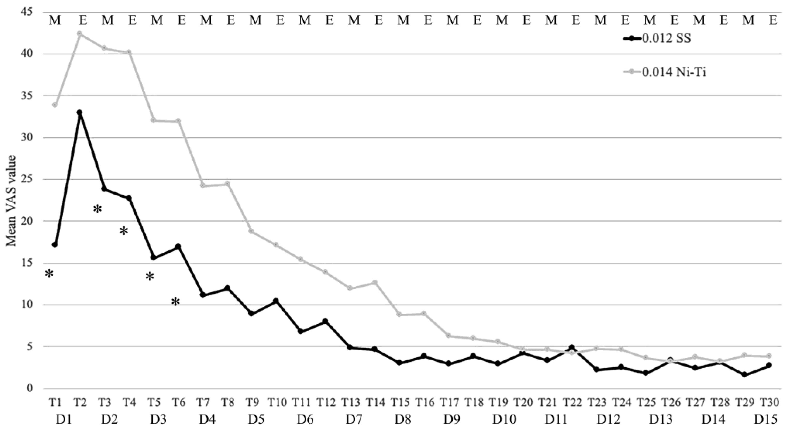
There were statistically significant differences between the groups at T1, T3, T4, T5 and T6 (p < 0.05); the mean VAS values of the 0.012 SS group were significantly lower at each time point when compared with those of the 0.014 Ni-Ti group, especially during the most painful days (Figure 2 and Table 2). The linear regression analysis showed that neither the archwire type nor the amount of crowding nor the SSAS score influenced the VAS values in the first 3 days of treatment (p > 0.05).
From the analysis of the questionnaire, it emerged that in most subjects, pain occurred immediately after the activation of orthodontic appliances (96% in the 0.012 SS group and 88% in the 0.014 Ni-Ti group, p = 0.555); only one subject in the 0.014 Ni-Ti group did not feel pain during the examination period. No statistically significant difference was found between the groups regarding the most painful day (p = 0.294); the most painful day was the first day for 58% of all the subjects, the second day for 20% and the third day for 12%. Moreover, no significant difference was found concerning the day of pain disappearance; in 18% of all subjects, the pain never disappeared during the examination time points (12% in the 0.012 SS group and 24% in the 0.014 Ni-Ti group, p = 0.495). The pain characteristics are reported in Table 3. Pain during chewing was the most reported by the subjects of both groups (84%), followed by compressive pain (78%), pain during occlusion (48%) and spontaneous pain (40%). No statistically significant differences were found between the groups.

4. Discussion

The aim of the present clinical study was to compare the perception of pain caused by the activation of two different types of archwires during the initial orthodontic alignment phase. No differences between the two archwires were found concerning the quality of pain. The mean VAS values of the 0.012 SS group were lower than those of the 0.014 Ni-Ti group at each time point, with a statistically significant difference in the first three days of treatment (T1, T3, T4, T5 and T6), which were characterised by the higher level of pain perceived. This outcome disagreed with the common opinion that light, continuous forces exerted by super-elastic Ni-Ti wires induce low pain perception.
Concerning the occurrence of pain, the present results showed that it began immediately after the activation of arches in both groups (96% of the 0.012 SS group and 88% of the 0.014 Ni-Ti group), and that in 58% of all subjects, a higher value of pain intensity was reached on the first day of treatment, with no significant difference between the groups. These data were in accordance with other authors who reported that pain started within 12 h and raised in the following 24–48 h [8,36].
In 52% of subjects, the pain disappeared within 7 days of the appliance activation. Although no significant difference was found between the groups, 48% of subjects reported pain beyond 7 days and 9 subjects beyond 15 days after fixed appliance activation. Most of the studies that have evaluated the characteristics of pain during an initial dental alignment with a fixed orthodontic appliance performed a standard follow-up of 7 days [1,11,18,33,37,38,39]; however, the results obtained in the present investigation justified a longer follow-up period and suggested its application in further clinical trials.
The Australian archwire is a high-tensile SS wire that is heat-treated to yield a higher resilience than normal SS and produces high forces that dissipate over short periods. The Ni-Ti archwires exert continuous forces and have a low modulus of elasticity, a high springback and a low deactivation force.
Therefore, we could suppose that the heavier orthodontic forces expressed by the 0.012 SS archwire, characterised by a faster decay of intensity after their activation, could cause a less traumatic biological response at the tooth level and, consequently, lower pain perception and faster pain reduction compared with the lighter but continuous forces expressed by the 0.014 Ni-Ti archwire.
One the most frequent methods used by orthodontists to reduce pain perception is the administration of NSAIDs such as acetylsalicylic acid and ibuprofen. However, their use should be discouraged because several studies have demonstrated that NSAIDs reduce the rate of tooth movement [9,10,13,14] in addition to causing a number of other side effects such as kidney and gastrointestinal toxicity and an increased cardiovascular risk [40,41,42]. The literature suggests several alternative methods to control orthodontic pain during the initial alignment phase such as laser therapy [34,43], written information [44] and supplemental vibrational forces [45], with contrasting results. Furthermore, in the early stages of orthodontic therapy, the experience of pain reduces patients’ compliance [8]; thus, the choice of the first archwire appears to be of crucial importance. The present study suggested that a 0.012 inch SS archwire caused lower pain perception than a 0.014 Ni-Ti archwire at the beginning of the orthodontic treatment. A large number of variables related to the appliance or to a single subject can affect the reliability of pain reporting. The type and material of the appliance with the age of the subjects and their metabolic profile should be controlled [25].
In order to control these variables as much as possible, in this study all subjects received the same orthodontic appliance and the mean age was comparable between the groups. Subjects affected by systemic diseases or in therapy with drugs that could alter the metabolic system or pain threshold were excluded. Moreover, pain is a biopsychosocial experience that goes far from mere nociception; orthodontic discomfort seems to be related not only to the magnitude of the force exerted on the teeth, but also relies on the psychological wellbeing of the individual [8]. The control of the psychosocial component of pain by means of SSAS and STAI scales represented a strength of the present investigation. These questionnaires allowed us to verify the comparability of the two groups that resulted in homogeneity. Despite the statistical comparability of the two groups, the slightly higher values of crowding in the Ni-Ti group could have influenced pain perception.
Furthermore, although no correlation has been found between initial dental crowding and the intensity of orthodontic pain [18], the ALD of each enrolled patient was also evaluated to assess the baseline comparability between the groups. Data obtained from the analysis demonstrated no statistically significant differences, so homogeneity between the groups was also confirmed for this parameter. A limitation of the present study was that the perception of pain during the initial alignment was not evaluated considering upper and lower arch separately, and this was a confounding factor. In this regard, there is no consensus in the literature concerning the possibility of different pain perception between the maxillary or mandibular dental arch. Fernandes [17] reported a higher perception of pain at the lower arch 11 h after the application of an orthodontic force; these data were in accordance with another study in which orthodontic pain was induced by elastic separators positioned at the level of the first molars [46]. However, many studies have not found a significant difference in pain perception between upper and lower jaws [33,34,39]. Due to the inter-individual variation related to pain perception, the ideal study design to control this variable should be a crossover in which each patient represents the control for themself. However, a “wash-out” period of a few months is mandatory to perform this type of study in order to avoid bias related to pain memory [47]. This is not easily applicable to patients who need to undergo orthodontic therapy with fixed braces. Methodologically sound randomised controlled trials are needed to obtain standardised protocols that could simplify the clinical practice, indicating the best archwire sequence to control orthodontic pain. This aspect could be crucial to increase the cooperation of the patients.
Pain is a complex, multi-factorial experience that is influenced by gender, sex, systemic conditions, social and psychological status. The sample of the present study was controlled for the variables that could cause alterations in pain perception, and the results showed that a 0.012 SS archwire induced lower pain than a 0.014 super-elastic Ni-Ti archwire during the first days of an orthodontic treatment with a fixed multi-bracket appliance. These results suggested that a 0.012 SS should be used as first archwire in order to cause less discomfort to the patients, allowing a better acceptance of the treatment and a higher compliance.

Author Contributions

Conceptualisation, F.B. and G.A.-B.; methodology, M.L.B. and S.I.P.; formal analysis, I.T.; investigation, L.S.; data curation, C.P.; writing M.L.B.; supervision, G.A.-B. and C.P. All authors have read and agreed to the published version of the manuscript.

Funding

This research received no external funding.

Institutional Review Board Statement

The study was conducted in accordance with the Declaration of Helsinki and approved on 11 September 2020 by the Ethics Committee of the University of Brescia, Brescia, Italy, with the number NP4160.

Informed Consent Statement

Informed consent was obtained from all subjects involved in the study.

Data Availability Statement

The data presented in this study are available on request from the corresponding author.

Conflicts of Interest

The authors declare no conflict of interest.

References

  1. Scott, P.; Sherriff, M.; Dibiase, A.T.; Cobourne, M.T. Perception of Discomfort during Initial Orthodontic Tooth Alignment Using a Self-Ligating or Conventional Bracket System: A Randomized Clinical Trial. Eur. J. Orthod. 2008, 30, 227–232. [Google Scholar] [CrossRef] [PubMed] [Green Version]
  2. Raja, S.N.; Carr, D.B.; Cohen, M.; Finnerup, N.B.; Flor, H.; Gibson, S.; Keefe, F.J.; Mogil, J.S.; Ringkamp, M.; Sluka, K.A.; et al. The Revised International Association for the Study of Pain Definition of Pain: Concepts, Challenges, and Compromises. Pain 2020, 161, 1976–1982. [Google Scholar] [CrossRef] [PubMed]
  3. Barsky, A.J.; Silbersweig, D.A. The Amplification of Symptoms in the Medically III. J. Gen. Intern. Med. 2023, 38, 195–202. [Google Scholar] [CrossRef] [PubMed]
  4. Nakao, M.; Barsky, A.J. Clinical Application of Somatosensory Amplification in Psychosomatic Medicine. Biopsychosoc. Med. 2007, 1, 17. [Google Scholar] [CrossRef] [Green Version]
  5. Ploghaus, A.; Narain, C.; Beckmann, C.F.; Clare, S.; Bantick, S.; Wise, R.; Matthews, P.M.; Rawlins, J.N.; Tracey, I. Exacerbation of Pain by Anxiety Is Associated with Activity in a Hippocampal Network. J. Neurosci. 2001, 21, 9896–9903. [Google Scholar] [CrossRef] [Green Version]
  6. Lin, W.; Farella, M.; Antoun, J.S.; Topless, R.K.; Merriman, T.R.; Michelotti, A. Factors Associated with Orthodontic Pain. J. Oral Rehabil. 2021, 48, 1135–1143. [Google Scholar] [CrossRef]
  7. Sergl, H.G.; Klages, U.; Zentner, A. Functional and Social Discomfort during Orthodontic Treatment—Effects on Compliance and Prediction of Patients’ Adaptation by Personality Variables. Eur. J. Orthod. 2000, 22, 307–315. [Google Scholar] [CrossRef]
  8. Monk, A.B.; Harrison, J.E.; Worthington, H.V.; Teague, A. Pharmacological Interventions for Pain Relief during Orthodontic Treatment. Cochrane Database Syst. Rev. 2017, 11, CD003976. [Google Scholar] [CrossRef] [Green Version]
  9. Krishnan, V. Orthodontic Pain: From Causes to Management—A Review. Eur. J. Orthod. 2007, 29, 170–179. [Google Scholar] [CrossRef] [Green Version]
  10. Long, H.; Wang, Y.; Jian, F.; Liao, L.N.; Yang, X.; Lai, W.L. Current Advances in Orthodontic Pain. Int. J. Oral Sci. 2016, 8, 67–75. [Google Scholar] [CrossRef]
  11. Erdinç, A.M.E.; Dinçer, B. Perception of Pain during Orthodontic Treatment with Fixed Appliances. Eur. J. Orthod. 2004, 26, 79–85. [Google Scholar] [CrossRef] [PubMed]
  12. Patel, S.; McGorray, S.P.; Yezierski, R.; Fillingim, R.; Logan, H.; Wheeler, T.T. Effects of Analgesics on Orthodontic Pain. Am. J. Orthod. Dentofac. Orthop. 2011, 139, e53-8. [Google Scholar] [CrossRef] [PubMed]
  13. Walker, J.B.; Buring, S.M. NSAID Impairment of Orthodontic Tooth Movement. Ann. Pharmacother. 2001, 35, 113–115. [Google Scholar] [CrossRef] [PubMed]
  14. Bartzela, T.; Türp, J.C.; Motschall, E.; Maltha, J.C. Medication Effects on the Rate of Orthodontic Tooth Movement: A Systematic Literature Review. Am. J. Orthod. Dentofac. Orthop. 2009, 135, 16–26. [Google Scholar] [CrossRef]
  15. Süleyman, H.; Demircan, B.; Karagöz, Y. Anti-Inflammatory and Side Effects of Cyclooxygenase Inhibitors. Pharmacol. Rep. 2007, 59, 247–258. [Google Scholar] [PubMed]
  16. Farkouh, M.E.; Greenberg, B.P. An Evidence-Based Review of the Cardiovascular Risks of Nonsteroidal Anti-Inflammatory Drugs. Am. J. Cardiol. 2009, 103, 1227–1237. [Google Scholar] [CrossRef] [PubMed]
  17. Fernandes, L.M.; Ogaard, B.; Skoglund, L. Pain and Discomfort Experienced after Placement of a Conventional or a Superelastic NiTi Aligning Archwire. A Randomized Clinical Trial. J. Orofac. Orthop. 1998, 59, 331–339. [Google Scholar] [CrossRef]
  18. Abdelrahman, R.S.; Al-Nimri, K.S.; Al Maaitah, E.F. Pain Experience during Initial Alignment with Three Types of Nickel-Titanium Archwires: A Prospective Clinical Trial. Angle Orthod. 2015, 85, 1021–1026. [Google Scholar] [CrossRef]
  19. Sandhu, S.S.; Sandhu, J. A Randomized Clinical Trial Investigating Pain Associated with Superelastic Nickel-Titanium and Multistranded Stainless Steel Archwires during the Initial Leveling and Aligning Phase of Orthodontic Treatment. J. Orthod. 2013, 40, 276–285. [Google Scholar] [CrossRef] [Green Version]
  20. Cioffi, I.; Piccolo, A.; Tagliaferri, R.; Paduano, S.; Galeotti, A.; Martina, R. Pain Perception Following First Orthodontic Archwire Placement--Thermoelastic vs Superelastic Alloys: A Randomized Controlled Trial. Quintessence Int. 2012, 43, 61–69. [Google Scholar]
  21. Jones, M.; Chan, C. The Pain and Discomfort Experienced during Orthodontic Treatment: A Randomized Controlled Clinical Trial of Two Initial Aligning Arch Wires. Am. J. Orthod. Dentofac. Orthop. 1992, 102, 373–381. [Google Scholar] [CrossRef] [PubMed]
  22. West, A.E.; Jones, M.L.; Newcombe, R.G. Multiflex versus Superelastic: A Randomized Clinical Trial of the Tooth Alignment Ability of Initial Arch Wires. Am. J. Orthod. Dentofac. Orthop. 1995, 108, 464–471. [Google Scholar] [CrossRef] [PubMed]
  23. Evans, T.J.; Jones, M.L.; Newcombe, R.G. Clinical Comparison and Performance Perspective of Three Aligning Arch Wires. Am. J. Orthod. Dentofac. Orthop. 1998, 114, 32–39. [Google Scholar] [CrossRef]
  24. O’brien, K.; Lewis, D.; Shaw, W.; Combe, E. A Clinical Trial of Aligning Archwires. Eur. J. Orthod. 1990, 12, 380–384. [Google Scholar] [CrossRef] [PubMed]
  25. Jian, F.; Lai, W.; Furness, S.; McIntyre, G.T.; Millett, D.T.; Hickman, J.; Wang, Y. Initial Arch Wires for Tooth Alignment during Orthodontic Treatment with Fixed Appliances. Cochrane Database Syst. Rev. 2013, 4, CD007859. [Google Scholar] [CrossRef] [PubMed]
  26. Wang, Y.; Liu, C.; Jian, F.; Mcintyre, G.T.; Millett, D.T.; Hickman, J.; Lai, W. Initial Arch Wires Used in Orthodontic Treatment with Fixed Appliances. Cochrane Database Syst. Rev. 2018, 2018, CD007859. [Google Scholar] [CrossRef] [PubMed] [Green Version]
  27. Bergius, M.; Kiliaridis, S.; Berggren, U. Pain in Orthodontics. A Review and Discussion of the Literature. J. Orofac. Orthop. 2000, 61, 125–137. [Google Scholar] [CrossRef] [PubMed]
  28. Beltramini, A.; Milojevic, K.; Pateron, D. Pain Assessment in Newborns, Infants, and Children. Pediatr. Ann. 2017, 46, e387–e395. [Google Scholar] [CrossRef] [Green Version]
  29. Fillingim, R.B. Individual Differences in Pain: Understanding the Mosaic That Makes Pain Personal. Pain 2017, 158, S11. [Google Scholar] [CrossRef] [Green Version]
  30. Spielberger, C.D.; Gorsuch, R.L.; Lushene, R.; Vagg, P.R.; Jacobs, G. Manual for the State-Trait Anxiety Inventory; Consulting Psychologists Press: Palo Alto, CA, USA, 1983. [Google Scholar]
  31. Barsky, A.J.; Goodson, J.D.; Lane, R.S.; Cleary, P.D. The Amplification of Somatic Symptoms. Psychosom. Med. 1988, 50, 510–519. [Google Scholar] [CrossRef]
  32. Chiarotto, A.; Maxwell, L.J.; Ostelo, R.W.; Boers, M.; Tugwell, P.; Terwee, C.B. Measurement Properties of Visual Analogue Scale, Numeric Rating Scale, and Pain Severity Subscale of the Brief Pain Inventory in Patients With Low Back Pain: A Systematic Review. J. Pain 2019, 20, 245–263. [Google Scholar] [CrossRef] [PubMed]
  33. Tortamano, A.; Lenzi, D.C.; Haddad, A.C.S.S.; Bottino, M.C.; Dominguez, G.C.; Vigorito, J.W. Low-Level Laser Therapy for Pain Caused by Placement of the First Orthodontic Archwire: A Randomized Clinical Trial. Am. J. Orthod. Dentofac. Orthop. 2009, 136, 662–667. [Google Scholar] [CrossRef] [PubMed]
  34. Marini, I.; Bartolucci, M.L.; Bortolotti, F.; Innocenti, G.; Gatto, M.R.; Alessandri Bonetti, G. The Effect of Diode Superpulsed Low-Level Laser Therapy on Experimental Orthodontic Pain Caused by Elastomeric Separators: A Randomized Controlled Clinical Trial. Lasers Med. Sci. 2015, 30, 35–41. [Google Scholar] [CrossRef] [PubMed]
  35. Bernabé, E.; Flores-Mir, C. Estimating Arch Length Discrepancy through Little’s Irregularity Index for Epidemiological Use. Eur. J. Orthod. 2006, 28, 269–273. [Google Scholar] [CrossRef] [PubMed] [Green Version]
  36. Bergius, M.; Berggren, U.; Kiliaridis, S. Experience of Pain during an Orthodontic Procedure. Eur. J. Oral Sci. 2002, 110, 92–98. [Google Scholar] [CrossRef] [PubMed]
  37. Fleming, P.S.; Dibiase, A.T.; Sarri, G.; Lee, R.T. Pain Experience during Initial Alignment with a Self-Ligating and a Conventional Fixed Orthodontic Appliance System. A Randomized Controlled Clinical Trial. Angle Orthod. 2009, 79, 46–50. [Google Scholar] [CrossRef]
  38. Polat, O.; Karaman, A.I. Pain Control during Fixed Orthodontic Appliance Therapy. Angle Orthod. 2005, 75, 214–219. [Google Scholar]
  39. Turhani, D.; Scheriau, M.; Kapral, D.; Benesch, T.; Jonke, E.; Bantleon, H.P. Pain Relief by Single Low-Level Laser Irradiation in Orthodontic Patients Undergoing Fixed Appliance Therapy. Am. J. Orthod. Dentofac. Orthop. 2006, 130, 371–377. [Google Scholar] [CrossRef]
  40. Zeilhofer, H.U.; Brune, K. Analgesic Strategies beyond the Inhibition of Cyclooxygenases. Trends Pharmacol. Sci. 2006, 27, 467–474. [Google Scholar] [CrossRef]
  41. Schjerning, A.M.; McGettigan, P.; Gislason, G. Cardiovascular Effects and Safety of (Non-Aspirin) NSAIDs. Nat. Rev. Cardiol. 2020, 17, 574–584. [Google Scholar] [CrossRef]
  42. Bindu, S.; Mazumder, S.; Bandyopadhyay, U. Non-Steroidal Anti-Inflammatory Drugs (NSAIDs) and Organ Damage: A Current Perspective. Biochem. Pharmacol. 2020, 180, 114147. [Google Scholar] [CrossRef] [PubMed]
  43. Alsayed Hasan, M.M.A.; Sultan, K.; Ajaj, M.; Voborná, I.; Hamadah, O. Low-Level Laser Therapy Effectiveness in Reducing Initial Orthodontic Archwire Placement Pain in Premolars Extraction Cases: A Single-Blind, Placebo-Controlled, Randomized Clinical Trial. BMC Oral Health 2020, 20, 209. [Google Scholar] [CrossRef]
  44. Montebugnoli, F.; Parenti, S.I.; D’Antò, V.; Alessandri-Bonetti, G.; Michelotti, A. Effect of Verbal and Written Information on Pain Perception in Patients Undergoing Fixed Orthodontic Treatment: A Randomized Controlled Trial. Eur. J. Orthod. 2020, 42, 494–499. [Google Scholar] [CrossRef] [PubMed]
  45. Bakdach, W.M.M.; Hadad, R. Effectiveness of Supplemental Vibrational Force in Reducing Pain Associated with Orthodontic Treatment: A Systematic Review. Quintessence Int. 2020, 51, 742–752. [Google Scholar] [CrossRef] [PubMed]
  46. Eslamian, L.; Borzabadi-Farahani, A.; Hassanzadeh-Azhiri, A.; Badiee, M.R.; Fekrazad, R. The Effect of 810-Nm Low-Level Laser Therapy on Pain Caused by Orthodontic Elastomeric Separators. Lasers Med. Sci. 2014, 29, 559–564. [Google Scholar] [CrossRef] [PubMed]
  47. Mills, E.J.; Chan, A.-W.; Wu, P.; Vail, A.; Guyatt, G.H.; Altman, D.G. Design, Analysis, and Presentation of Crossover Trials. Trials 2009, 10, 27. [Google Scholar] [CrossRef] [PubMed] [Green Version]
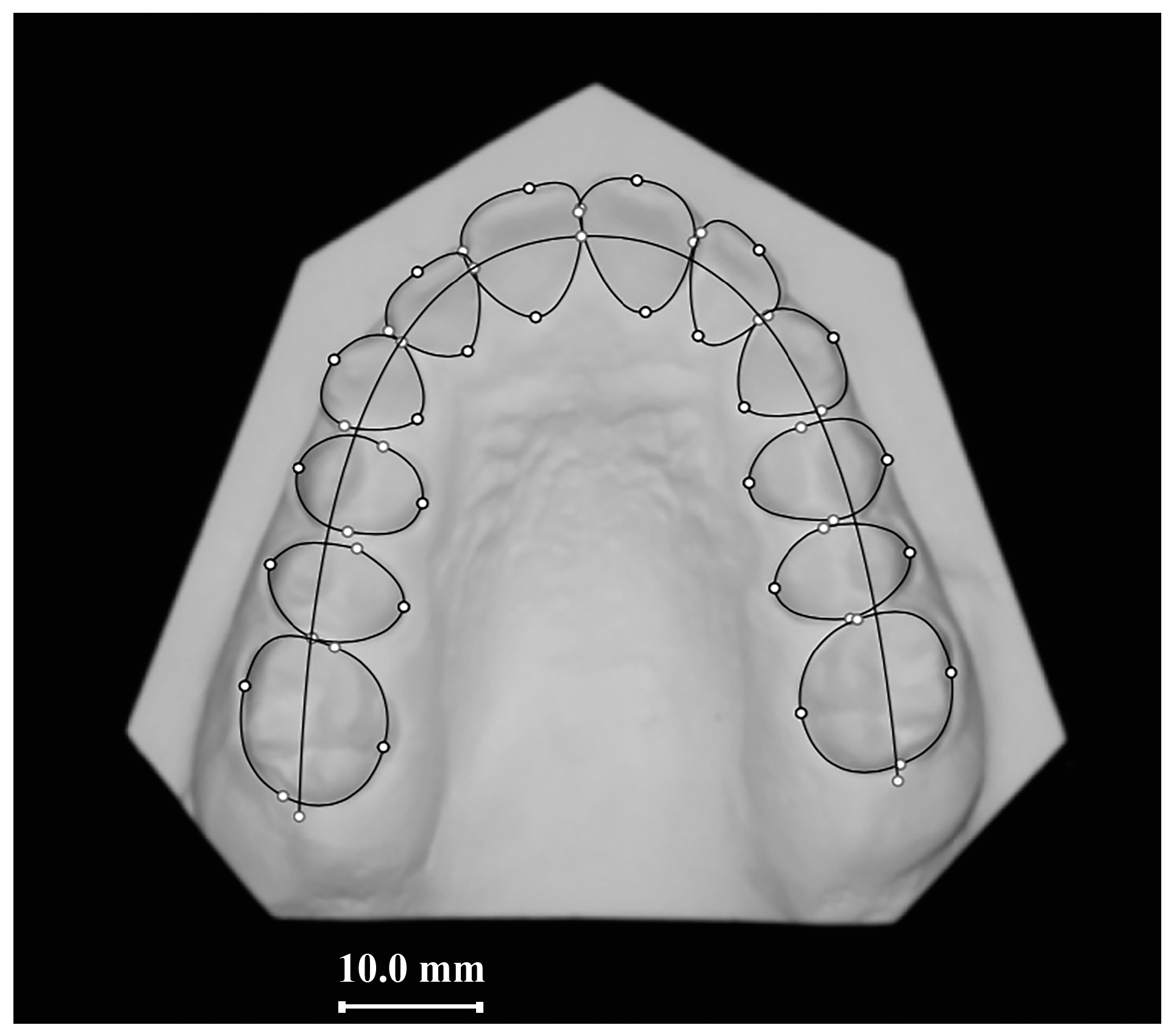
Figure 1. ALD evaluation with Delta-Dent software v1.0.
Applsci 13 04929 g001
Figure 2. Mean VAS values in 0.012 SS and 0.014 Ni-Ti groups during the first 15 days of treatment. * Statistically significant difference at each time point between groups (p < 0.05); M = morning; E = evening.
Applsci 13 04929 g002
Table 1. Sample description. The p-value refers to the comparison between groups (t-test for independent samples or Mann–Whitney test).
GroupGenderAge
(Mean ± SD)
Arch Length Discrepancy
(mm, Median and Interquartile Range)
SSAS
(Mean ± SD)
TAI
(Mean ± SD)
0.012 SS (n = 25)(9M; 16F)19.92 ± 9.4−0.43 (−3.95–2.76)21.88 ± 6.1942.20 ± 6.01
0.014 Ni-Ti (n = 25)(11M; 14F)18.32 ± 8.31−1.7 (−3.70–4.40)24.36 ± 5.4143.36 ± 3.92
p-Value0.5640.5280.7640.1380.423
Table 2. VAS values in 0.012 SS and 0.014 Ni-Ti groups at each time point. Data are reported as mean and interquartile ranges. The p-value refers to the comparison between groups at each time point (Mann–Whitney test). * p < 0.05.
0.012 SS0.014 Ni-Tip-Value
T117.0 (4.0–26.0)34.0 (8.5–58.0)0.027 *
T226.0 (11.5–47.0)41.0 (15.0–70.0)0.268
T321.0 (4.0–33.0)38.0 (20.0–65.5)0.017 *
T413.0 (6.0–34.0)33.0 (16.5–66.5)0.046 *
T58.0 (2.0–24.0)24.0 (11.0–57.5)0.018 *
T69.0 (2.0–31.0)32.0 (10.5–51.5)0.039 *
T710.0 (1.0–17.0)15.0 (2.0–46.5)0.147
T86.0 (1.5–18.0)15.0 (3.0–45.0)0.117
T94.0 (0.0–16.0)9.0 (1.5–37.0)0.199
T103.0 (0.0–16.0)6.0 (0.5–33.5)0.250
T111.0 (0.0–11.5)0.0 (0.0–35.5)0.686
T120.0 (0.0–12.5)0.0 (0.0–28.0)0.833
T130.0 (0.0–3.0)0.0 (0.0–30.5)0.456
T140.0 (0.0–2.0)0.0 (0.0–24.5)0.241
T150.0 (0.0–2.0)0.0 (0.0–15.0)0.170
T160.0 (0.0–0.0)0.0 (0.0–14.0)0.135
T170.0 (0.0–3.5)0.0 (0.0–12.5)0.206
T180.0 (0.0–3.5)0.0 (0.0–10.5)0.256
T190.0 (0.0–1.0)0.0 (0.0–6.5)0.462
T200.0 (0.0–0.5)0.0 (0.0–6.0)0.615
T210.0 (0.0–0.5)0.0 (0.0–5.5)0.697
T220.0 (0.0–1.0)0.0 (0.0–5.5)0.840
T230.0 (0.0–0.0)0.0 (0.0–1.0)0.659
T240.0 (0.0–0.0)0.0 (0.0–1.0)0.639
T250.0 (0.0–0.0)0.0 (0.0–1.0)0.445
T260.0 (0.0–0.0)0.0 (0.0–1.0)0.532
T270.0 (0.0–0.0)0.0 (0.0–1.0)0.496
T280.0 (0.0–0.0)0.0 (0.0–1.0)0.505
T290.0 (0.0–0.0)0.0 (0.0–1.0)0.259
T300.0 (0.0–0.0)0.0 (0.0–1.0)0.290
Table 3. Pain characteristics. Frequency reported in number of subjects. The p-value refers to the comparison between groups (χ2 test).
Compressive PainPain during OcclusionPain during ChewingSpontaneous Pain
0.012 SS (N = 25)20 (80%)14 (56%)20 (80%)9 (36%)
0.014 Ni-Ti (N = 25)19 (76%)10 (40%)22 (88%)11 (44%)
p-Value0.7330.2580.4400.564
Disclaimer/Publisher’s Note: The statements, opinions and data contained in all publications are solely those of the individual author(s) and contributor(s) and not of MDPI and/or the editor(s). MDPI and/or the editor(s) disclaim responsibility for any injury to people or property resulting from any ideas, methods, instructions or products referred to in the content.