Article

Automatic Tumor Identification from Scans of Histopathological Tissues

Department of Information Technologies, Vilnius Gediminas Technical University, Sauletekio Al. 11, LT-10223 Vilnius, Lithuania
*
Author to whom correspondence should be addressed.
Appl. Sci. 2023, 13(7), 4333; https://doi.org/10.3390/app13074333
Submission received: 6 March 2023 / Revised: 27 March 2023 / Accepted: 28 March 2023 / Published: 29 March 2023
(This article belongs to the Special Issue AI Technology in Medical Image Analysis)

Abstract

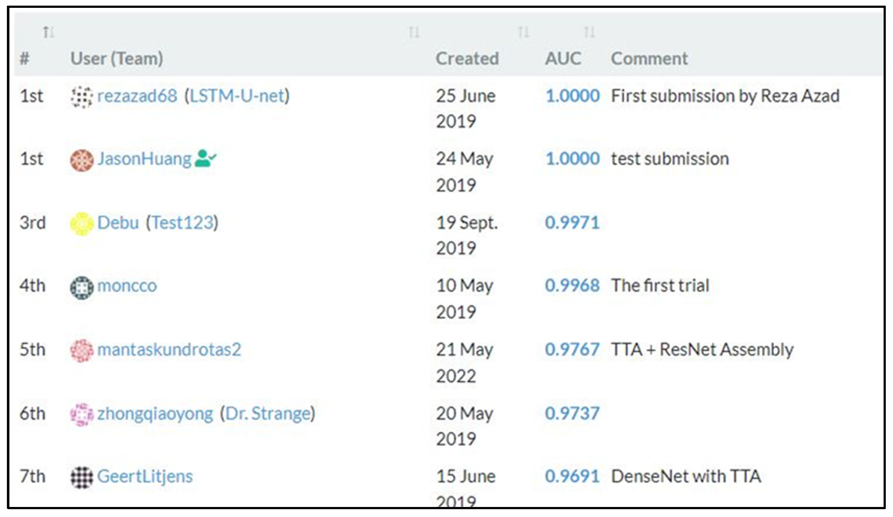
Latest progress in development of artificial intelligence (AI), especially machine learning (ML), allows to develop automated technologies that can eliminate or at least reduce human errors in analyzing health data. Due to the ethics of usage of AI in pathology and laboratory medicine, to the present day, pathologists analyze slides of histopathologic tissues that are stained with hematoxylin and eosin under the microscope; by law it cannot be substituted and must go under visual observation, as pathologists are fully accountable for the result. However, a profuse number of automated systems could solve complex problems that require an extremely fast response, accuracy, or take place on tasks that require both a fast and accurate response at the same time. Such systems that are based on ML algorithms can be adapted to work with medical imaging data, for instance whole slide images (WSIs) that allow clinicians to review a much larger number of health cases in a shorter time and give the ability to identify the preliminary stages of cancer or other diseases improving health monitoring strategies. Moreover, the increased opportunity to forecast and take control of the spread of global diseases could help to create a preliminary analysis and viable solutions. Accurate identification of a tumor, especially at an early stage, requires extensive expert knowledge, so often the cancerous tissue is identified only after experiencing its side effects. The main goal of our study was to expand the ability to find more accurate ML methods and techniques that can lead to detecting tumor damaged tissues in histopathological WSIs. According to the experiments that we conducted, there was a 1% AUC difference between the training and test datasets. Over several training iterations, the U-Net model was able to reduce the model size by almost twice while also improving accuracy from 0.95491 to 0.95515 AUC. Convolutional models worked well on groups of different sizes when properly trained. With the TTA (test time augmentation) method the result improved to 0.96870, and with the addition of the multi-model ensemble, it improved to 0.96977. We found out that flaws in the models can be found and fixed by using specialized analysis techniques. A correction of the image processing parameters was sufficient to raise the AUC by almost 0.3%. The result of the individual model increased to 0.96664 AUC (a more than 1% better result than the previous best model) after additional training data preparation. This is an arduous task due to certain factors: using such systems’ applications globally needs to achieve maximum accuracy and improvement in the ethics of Al usage in medicine; furthermore if hospitals could give scientific inquiry validation, while retaining patient data anonymity with clinical information that could be systemically analyzed and improved by scientists, thereby proving Al benefits.
Keywords: machine learning; automatization; histopathology; medical imaging machine learning; automatization; histopathology; medical imaging