Article

The Effect of Callus and Corns Removal Treatments on Foot Geometry Parameters, Foot Pressure, and Foot Pain Reduction in Women

by
Paweł Piotr Antończak
1,2,
Magdalena Hartman-Petrycka
3,
Agnieszka Garncarczyk
1,
Katarzyna Adamczyk
1,
Dominika Wcisło-Dziadecka
1,* and
Barbara Błońska-Fajfrowska
3
1
Department of Cosmetology, School of Pharmaceutical Science in Sosnowiec, Medical University of Silesia, Kasztanowa 3 Str., 41-200 Sosnowiec, Poland
2
Beauty Fit—Health and Beauty Center, Jana Kilińskiego 8 Str., 41-200 Sosnowiec, Poland
3
Department of Basic Biomedical Science, School of Pharmaceutical Science in Sosnowiec, Medical University of Silesia, Kasztanowa 3 Str., 41-200 Sosnowiec, Poland
*
Author to whom correspondence should be addressed.
Appl. Sci. 2023, 13(7), 4319; https://doi.org/10.3390/app13074319
Submission received: 21 February 2023 / Revised: 15 March 2023 / Accepted: 23 March 2023 / Published: 29 March 2023
(This article belongs to the Section Applied Biosciences and Bioengineering)

Abstract

:
Foot pain and deformities are significant problems that increase with age and are significantly more prevalent in women. Calluses and corns are both common and inconvenient, and the accompanying pain complaints significantly reduce the quality of life. Professional foot care is one of the key elements in the prevention of foot disorders and improves the comfort of people with such problems. The aim of the present research was to assess the impact of podiatry treatments on the foot conditions, including parameters related to foot geometry, the forefoot pressure on the ground, and the occurrence of pain in women over 50. In each volunteer, history and physical examination were performed and an objective assessment with the use of the digital foot scanner and the baropodometric measurements was performed. The examinations were conducted before the beginning of the procedures and up to a week after each of the three podiatry treatments performed at monthly intervals. Specialist foot care treatments influenced the values of the hallux valgus angle, the varus angle of the fifth toe, and the longitudinal arch. After each treatment, the percentage of feet with normal longitudinal arch was significantly greater than before the treatments. The treatments influenced the value of pressure in the forefoot. The percentage of feet with pain decreased significantly after each treatment. Performed podiatry treatments, consisting of the removal of changes related to keratosis, not only normalized the parameters related to foot geometry and the forefoot pressure on the ground but also reduced foot pain.

1. Introduction

The aging process progresses throughout the body. Many changes also occur in the skin and nails of the feet. They are common problems that cause foot pain and reduce quality of life. Proper specialist foot care is one of the key elements in the prevention of foot disorders and in improvement of the comfort and quality of life of people [1,2].
These hyperkeratotic lesions lead to the loss of proper body stability in the elderly. Elderly and obese people, especially with limited mobility, have problems with self-care. They are exposed to hygienic negligence and changes resulting from incorrect pressure on specific areas of the feet. Such patients should receive specialist foot care treatments [2,3,4].
Podiatry care includes the prevention of foot diseases and procedures related to the improvement of the appearance and condition of the feet. Specialist foot care treatments are performed in podiatry clinics. The treatments involve the shortening and correcting of nails and removing calluses and corns [4,5].
The main problem, which increases with age and is more common in women, is the presence of hyperkeratosis on the areas of maximum pressure of the foot. Frequent skin micro-injuries, caused by friction and overloading of the feet, induce the secretion of inflammatory mediators and growth factors. These factors stimulate epidermal cells to produce keratin with an excessive accumulation of a cohesive hard layer of stratum corneum [2,6,7].
When the stratum corneum does not exfoliate properly but increases its thickness and cohesiveness, calluses and corns are formed. Cohesive thickenings of the epidermis often cause significant pain. Calluses and corns are one of the most frequently reported problems in podiatry clinics and can be removed using podiatry equipment [6,8,9].
Each specialist foot care treatment should be preceded by an interview and physical examination. Depending on the technique of examining the feet and lower limbs, there are various methods: orthopedic, anthropometric (measurements of the lower limbs and computer podoscopy), tensometric (baropodometric), radiographic, goniometric, functional, and stabilographic (posturographic) [10].
The human foot shows a great diversity of shape, mainly in terms of the axis of the calcaneus, the arch of the longitudinal medial edge of the foot, the position of the forefoot in relation to the rear foot, and the transverse arch of the forefoot. These differences show specific features and occur singly or—more often—in combinations, called foot developmental variants. Pathologies are diagnosed in the case of exceeding commonly established norms in terms of developmental variants. The alignment of the calcaneus axis determines the correct, valgus, or varus foot. In a normal foot, the longitudinal arch of the medial edge of the foot is maintained when the foot is loaded. In avalgus foot, the incidence of toe zone deformities—hallux valgus—is higher than in normal and varus feet [11].
Flat foot (pes planus) is diagnosed when the skin is in contact with the ground above the navicular bone. An excessive longitudinal arch with a high instep is called a hollow foot (pes excavatus, pes cavus). The variants of the foot are also differentiated by the transverse arch. The load distribution in a healthy foot is concentrated on the ball of the big toe and the little toe. The lowered position of the medial heads of the metatarsal bones is characteristic of the transversally flat foot (pes transversus, pes transverso-planus). The position of the forefoot to the rearfoot is differentiated by the position of the first metatarsal bone in relation to the axis of the talus. In a normal foot, the vertical projection of the first metatarsal shows a slight deviation from the axis of the talus. The deviation of the entire forefoot, medially, is called forefoot adduction (pes adductus), and, laterally, it is called forefoot abduction (pes abductus). A severe medial deviation at the 1st sphenoid joint is called varus of the 1st metatarsal (metatarsus primus varus). This abnormality is often accompanied by hallux valgus [12,13,14].
Muscle failure is the most common cause of foot joint deformities in adults. Most deformities within the tarsus and metatarsals have their origin in imbalances in the action of the shin and foot muscles, resulting from damage to the musculoskeletal elements or nerve paralysis. These deformations also apply to the toes. The most common deformities in the toe zone include hallux valgus, claw toe, hammer toe, hammer toe, and rigid toe [11,14].
Pain in foot deformities and skin diseases (calluses and corns) are strongly correlated with age and are more common in women. The percentage of foot deformities and skin changes increases with age. The progressive degenerative changes of the musculoskeletal system elements in various developmental variants of the feet, as well as characteristic changes in the skin of the feet, are the cause of chronic pain. Foot pain significantly affects every day functioning and reduces the of life [15,16,17].
Foot diseases are common among mature and elderly people. The prevalence of certain disorders, such as flat foot, flat valgus, varus, hollow, or deformities of the toe zone, reaches up to 65% in the elderly population. Diseases of the musculoskeletal system and related symptoms increase the frequency of falls and may cause significant limitations in everyday activities.
The subject of interest of this study is the assessment of the effectiveness of routinely performed podiatry procedures, including the mechanical removal of calluses and corns.
The aim of the study was to assess the impact of podological procedures on the condition of feet, including parameters related to the geometry of the foot and the pressure of the forefoot exerted on the ground, as well as the occurrence of pain during standing and walking in women over 50.

2. Materials and Methods

2.1. Characteristics of Volunteers and Groups

After obtaining the approval of the Bioethical Committee of the Medical University of Silesia in Katowice, 200 feet of 100 women over 50 (68 ± 6.5) were examined. The research started in 2015. The project is still being continued and also includes the evaluation of other procedures.
The research was conducted after obtaining the written consent of the volunteers. They were informed about the purpose of the research and its course. The criteria for inclusion in the study were: voluntary participation, reporting discomfort or pain in the feet while standing, and prophylaxis of lower limb diseases. The criteria for exclusion from the study included: the occurrence of injuries, unhealed wounds and post-operative conditions in the feet, the presence of infectious lesions, pain resulting from rheumatic diseases, untreated Morton’s neuralgia or unhealed heel spurs, and the lack/withdrawal of consent to participate in the study. On the basis of an interview about the state of health, those people who stood and moved independently without the need to support themselves were qualified for the study.
Then, in order to assess the physiological effect of podological treatments, the following groups were distinguished in the AV group (all volunteers): group T (treatment)—76 feet of 38 women who had undergone callus and/or corns removal procedures; group C (control)—80 feet of 40 women with keratosis but without calluses and corns, thus they had no treatments performed; group NT (no treatment)—44 feet of 22 women who, despite the presence of calluses and/or corns, did not agree to treatment, thus they were excluded from further examinations (Table 1).
In groups T and C subgroups were distinguished according to the following criteria:
(1)
based on foot geometry parameters—transverse flat foot according to the Wejsflog index (W):
  • transverse flat foot (TF),
  • normal transverse arch (TNA).
(2)
based on the force of pressure on the ground (P) in the forefoot area:
  • insufficient pressure in the forefoot (TIP),
  • normal pressure in the forefoot (TNP),
  • excessive pressure in the front area (TEP).
Each volunteer has undergone a study involving: a podiatry interview, visual assessment and palpation of the feet, as well as objective measurement methods.

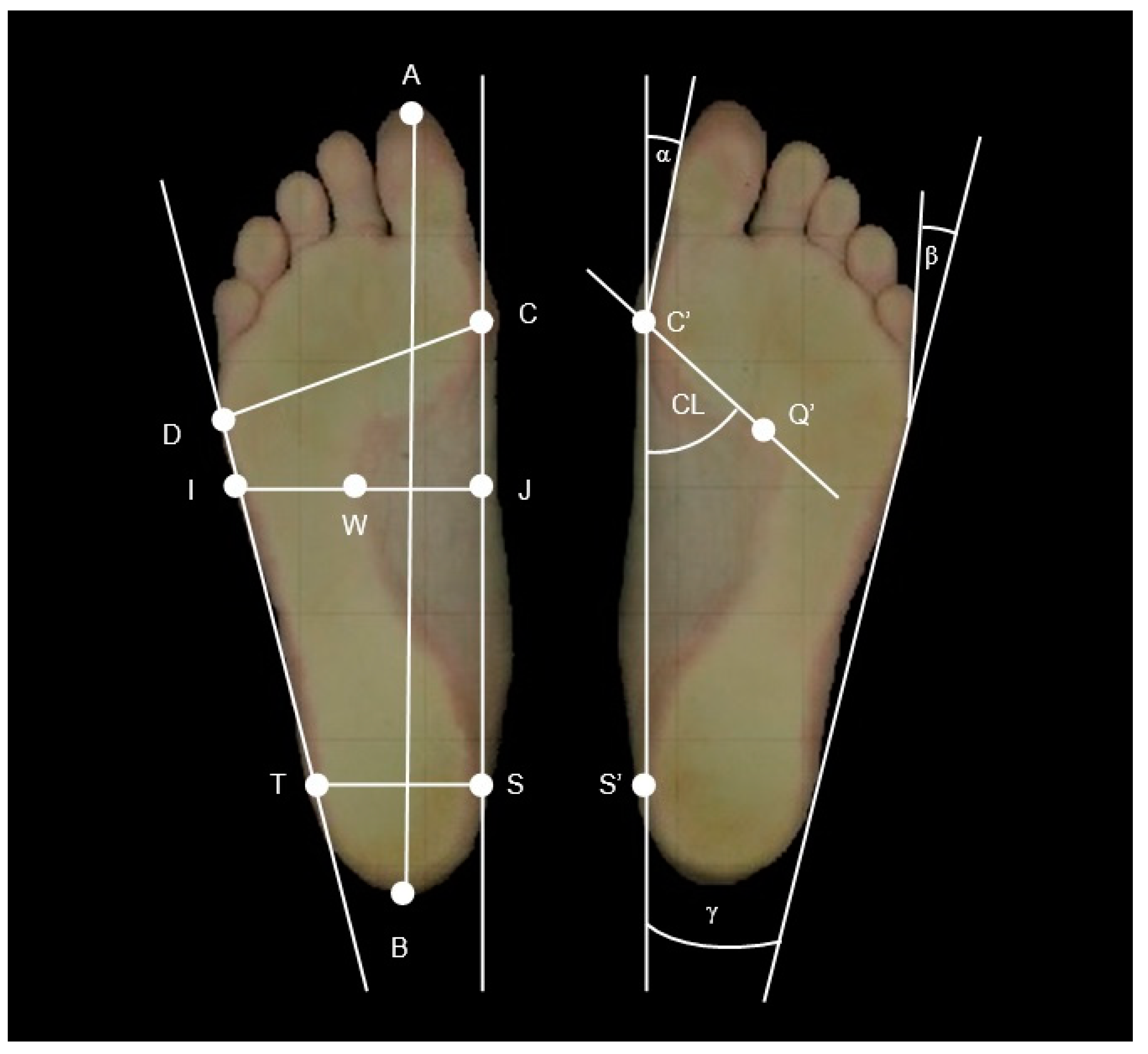
2.2. Podoscope Examination

A 2D digital podoscope examination was performed, which consisted of scanning the feet and obtaining a digital image. Using Podoscan 2D (Sensor Medica, Guidonia, Italy), precise footprints of the subjects were obtained. With the use of the FreeStep Professional software v.1.3.01, the parameters for both feet were then determined on the resulting image. The 2D digital podoscope is an advanced analysis tool for footprints, foot geometry, and foot loading.
After scanning, the length and width of the foot and the length of the medial longitudinal arch (cm), as well as the heel angle γ (o), were obtained and further analyzed.
The Clarke index (CL) and Sztriter–Godunow index (KY) were used to assess the longitudinal arch, while the Wejsflog index (W) and heel angle (γ) were used for the transverse arch. Moreover, the hallux valgus (angle α) and varus of the fifth toe (angle β) were measured (Figure 1).
The Clarke angle (CL) is determined by the line C′–S′ (the tangent to the medial edge of the foot) and the line passing through the point Q′ (the point of the deepest depression) and C′ (the point where the medial tangent meets the edge of the foot). (Figure 1: in the classification of feet according to the CL index, a flat foot was diagnosed in the range of x—30°, a foot with a reduced arch 31–41°, a normal foot 42–54°, and a hollow foot (with an increased arch) in the range of 55°–x.)
The Sztriter–Godunow index (KY) determines the ratio of the length of the segment running in the center of the arch of the longitudinal arch (W–I) to the length of the segment drawn by the adjacent and non-adjacent part of the scanned plantar surface of the foot (J–I), perpendicular to the medial tangent (Figure 1). Determining the KY index allows to establish the diagnosis depending on the value obtained, respectively: KY = 0.00–0.25—hollow foot; KY = 0.26–0.45—normal foot; KY = 0.46–0.490—reduced foot I°; KY = 0.50–0.75—reduced foot II°; and KY = 0.76–1.00—flat foot.
The Wejsflog index (W) is the ratio of the length (AB) to the width of the foot (CD) and determines the degree of transverse arching of the foot (Figure 1). The length to width ratio of the foot should be 3:1; however, in practice, the values of the Wejsflog index are in the range of 2 < W < 3. Values closer to 2 indicate transverse flat feet, while values closer to 3 prove the correct transverse arch of the foot. Based on the data from the literature, the borderline value of a transversely flat foot was assumed to be 2.55.
The calcaneal angle (γ) is determined by two tangents to the medial and lateral edges of the foot (Figure 1). The lines forming the angle γ intersect beyond the heel. The norm for the γ angle is 15–18° and it is assumed that values above 18°, similarly to lower values of the W index, indicate transverse flat feet.
The hallux valgus angle (α) is between the tangent to the medial edge of the foot and the tangent from the point at the widest point of the forefoot (the area of the metatarsophalangeal joint of the first toe) to the most medial point on the edge of the big toe (the area of the interphalangeal joint) (Figure 1).
The varus angle of the toe V (β) is contained between the tangent to the lateral edge of the foot and the tangent from the point at the widest point of the forefoot on the outside (near the metatarsophalangeal joint of the toe V) to the most lateral point on the edge of the toe V (area of the interphalangeal joint proximal finger V) (Figure 1).

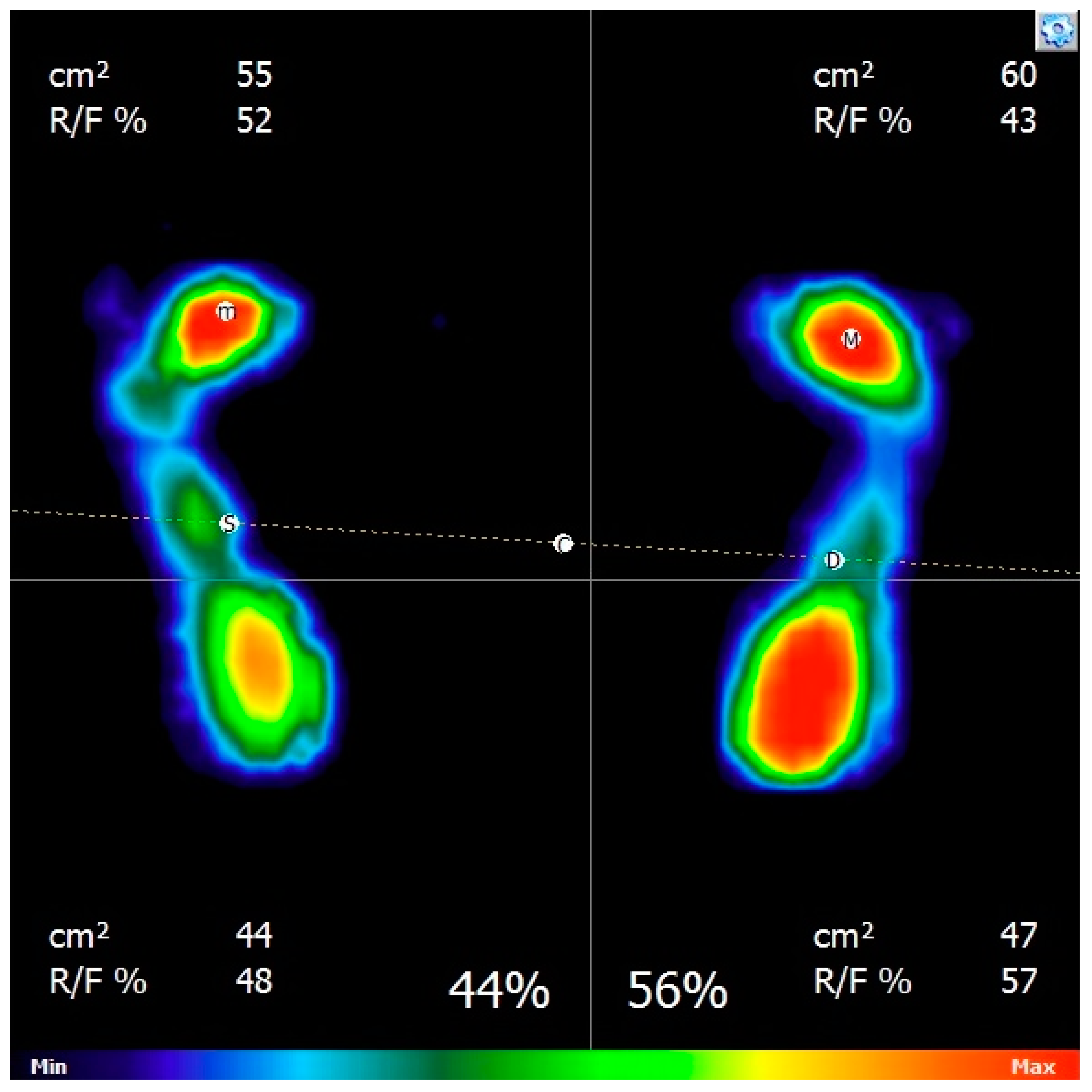
2.3. Baropodometric Examination

The next phases of the study included the evaluation of the foot loading rate in statics using the FreeMED force platform (Sensor Medica, Guidonia, Italy). The baropodometric study of feet with the use of the ground force reaction platform consisted of measuring foot pressure distribution on the ground while standing.
According to the software, normal values are: the ratio of the pressure on the left foot to the right foot in the amount of 50:50 ± 5%. According to the manufacturer and distributor’s data, the load on the front foot area is in the range of 40–50%, and the rear foot area is in the range of 50–60%.
The examination was carried out with body weight only and statically—in a standing position. The following parameters obtained from the pressure on the left and right foot were analyzed: total foot area directly adjacent to the ground (cm2), pressure area of the front foot (cm2), and the percentage of pressure on the ground for the front and rear foot areas (%).
Additionally, the software used allows for the graphic visualization of the obtained results (Figure 2).

2.4. Podiatric Procedures

The treatments that were performed on volunteers in a podiatry office involved in the mechanical removal of keratosis, calluses, and corns.
In each volunteer from group T, treatments were performed 3 times every month, and check-up and tests were conducted between the 3rd and the 7th day from when the procedure was performed. A podiatry interview preceded each examination.

2.5. Statistical Analysis

Individual test results were collected in a Microsoft Excel 2007 spreadsheet, and, after data import, statistical analysis was performed in STATISTICA 10. Data on an interval scale with a normal distribution are presented as average ± standard deviation. Qualitative data are presented in the form of number and percentage values (%). The Shapiro–Wilk test and the quantile plot (Q-Q) were used to assess the normality of the distribution of the obtained results.
Homogeneity of variance was assessed by Levene’s test. The chi2 test was used to test the significance of changes in nonparametric data: the characteristic changes of the longitudinal and transverse arch of the feet based on the indexes obtained in podoscopic examination in groups; percentage of feet with a specific pressure category in groups—differences in the assignment to the appropriate category; and assessment of the presence of pain. The ANOVA test was used to test the significance of changes in podoscopic and baropodometric parameters after subsequent treatments in group T, and Dunnett’s test was used in the post hoc analysis. Values for which the significance level p < 0.05 were considered statistically significant. The statistical analysis was performed by an independent statistician, who was then included as a co-author of this paper.

3. Results

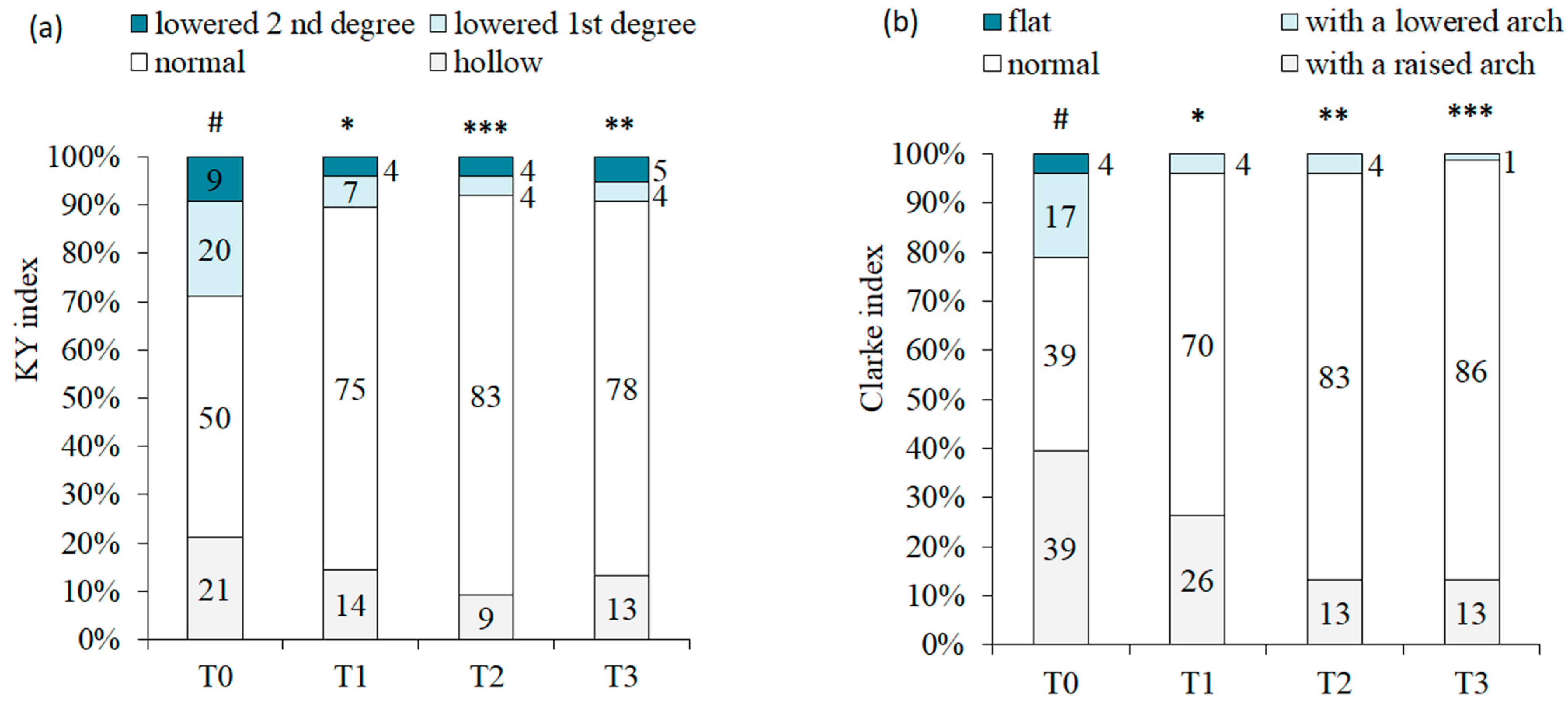
3.1. Evaluation of Changes in Podoscopic Parameters

Studies with the use of a podoscope showed significant differences between groups T and C in terms of the average width of the foot and the average value of the alpha angle, while significant differences were shown both between groups T and C and between groups T and NT in the range of the average value of the β angle (Table 2).
The research showed significant differences in the percentages of feet with different longitudinal arches according to the Sztriter–Godunow index. Differences were found between groups T and C as well as C and NT (Table 2).
There were significant intergroup differences in the average gamma heel angle between each of the studied groups (Table 2).
The treatments affected the foot geometry (Table 3). The analysis of the average foot length showed significant differences in the subgroup of transversely flat feet. The length of the foot slightly increased after each treatment and after the third treatment it was the longest.
There were also significant differences in the average foot width in the whole T group and TF subgroup after the procedures compared to the width before the removal of calluses and corns. The width slightly increased, and it was the largest after the 3rd treatment. Podiatry treatments did not significantly affect the average length of the medial longitudinal arch; however, there was a tendency to increase the length of the medial longitudinal arch with each procedure in the subgroup of transverse flat feet (pbefore_after = 0.06). Specialist foot care treatments influenced the values of the hallux valgus angle α in the T group as a whole due to changes in the TF subgroup. It was found that angle α decreased after each procedure and was the lowest after the third procedure. Podiatry treatments had a significant effect on the values of the varus angle β of the fifth toe in group T and in both analyzed subgroups. Varus angle β decreased after the first treatment, there was further improvement after the second treatment, and the third treatment strengthened the former positive change (Table 3).
Podiatry treatments influenced the longitudinal arch according to Sztriter–Godunow (KY) and Clarke index (CL). After each treatment, the percentage of feet with a normal longitudinal arch was significantly greater than before the treatments (Figure 3).

3.2. Evaluation of Changes in Baropodometric Parameters

There were no significant differences in the average value of the pressure (%) of the forefoot area on the ground in all groups (Table 4).
In the subgroup with excessive pressure in the front area (EP), performing three treatments significantly influenced the value of the pressure in the forefoot. After the third treatment, it was the lowest (Table 5).

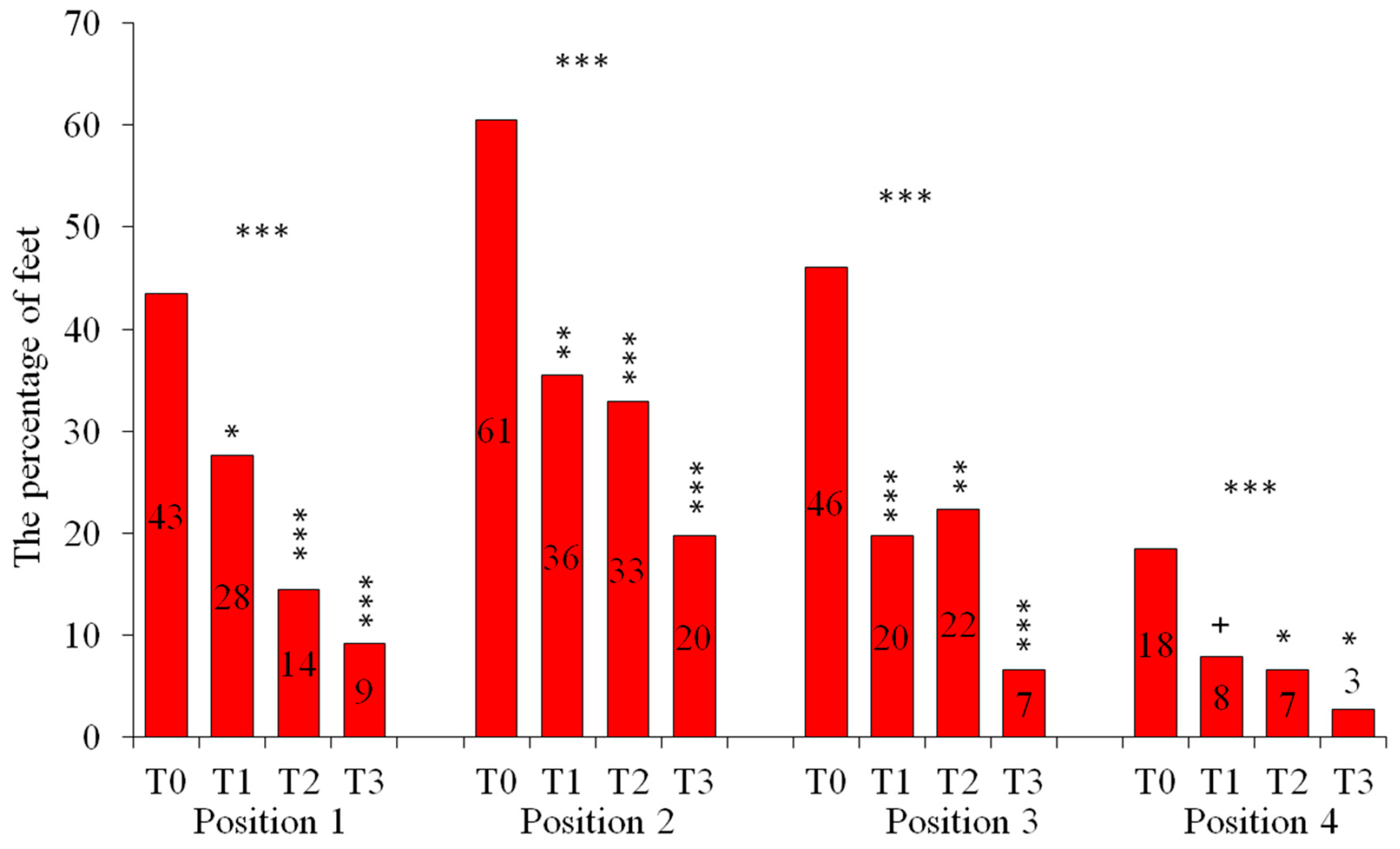
3.3. Assessment of the Presence of Pain

The podiatry interview regarding foot pain allowed for a statistical analysis of the percentage of feet affected by specific ailments in the studied groups. The highest percentage regarded pain when loading the forefoot (Table 6).
More than half of the feet in group T were affected by increased pain in the forefoot when weight was transferred to this area. The lowest percentage of affected feet was found in the control group. A significant difference in symptoms was found between these groups.
There were highly significant differences between groups T and C and between groups T and NT regarding the occurrence of stabbing pain in the foot while walking. Almost half of the examined feet in group T experienced pain.
The percentage of feet with heel pain was low and statistical analysis showed no intergroup differences.
Pain assessment showed that the percentage of feet with pain when standing, as well as during the transfer of body weight to the forefoot, significantly decreased in group T after 1, 2, and 3 procedures (Figure 4). The percentage of feet with pain decreased significantly after each treatment compared to T0 (before treatment).
Assessment of stabbing pain in the forefoot and during loading the hindfoot also showed a significantly lower percentage of feet with stabbing pain when walking compared to the initial condition and a significantly lower percentage of feet with pain in the hindfoot area when transferring the body weight to the heels compared to the initial condition. A significant reduction in the percentage of feet with stabbing pain was found after the first treatment and its radical reduction after the third treatment. Although the initial percentage of feet with hindfoot pain was low, in this case, it was also significantly reduced after the 2nd and 3rd treatments.

4. Discussion

Foot problems in late adulthood (over 50), especially in the elderly (over 65), are among the most frequently cited problems of this developmental period in published studies. Foot dysfunctions increase with age and are significantly more common in women [18,19].
Between 20 and 45% of women aged over 65 years of age will develop one or more foot problems, which, although not specific to older women. These include structural defects of the toes including hallux valgus, hammer, mallet or claw toes, skin disorders, fungal infection of the toes and toe nails, thickened toe nails, ulcers, fissures or cracks between the toes, corns and calluses, and flat or high-arched feet. Foot pain is common and may be associated with other co-morbid diseases of the foot [20]. Conducted research showed that 60% of women over 50 had at least one callus or corn.
This study showed that calluses and corns with the predominant location on the forefoot were common changes associated with hyperkeratosis and in many women over 50, causing pain when standing and at the time of weight transfer to the forefoot of the affected limb.
Pain prevention and treatment are particularly important in the elderly due to the increased risk of falls due to these ailments. Awale et al. [21] showed that both foot pain and the condition of the foot arch may play an important role in increasing the risk of falls.
The presence of lesions and pain was the basis for an assessment of anthropometric parameters related to the state of the foot arch and baropodometric parameters related to possible overloading or underloading of the feet in individual groups, as well as the evaluation of the effects of removing these lesions in group T.
Plantar callosities under lesser metatarsals are often accompanied by the hallux valgus, and the cause of callosity is thought to be associated with the foot deformity, such as the metatarsal length discrepancy, the abnormal metatarsal head height, cavus, flat foot, and rheumatoid conditions [22]. Moreover, hypermobility of the first ray, which is caused by an instability of the first metatarsocuneiform joint, is one of the factors that induces hallux valgus [23]. However, it is unclear which variable is most involved in the cause of callosity in hallux valgus deformity [22].
Therefore, the wider angle α in group T may suggest a connection to the presence of calluses in the forefoot. These calluses and related ailments were the reason why women sought specialist foot care. The results of our research are consistent with those of Murahashi et al. [22], which showed a correlation between the occurrence of hyperkeratosis in the forefoot and hallux valgus.
The conducted research showed that the average value of the hallux valgus angle in transversely flat feet decreased significantly after three procedures. The decrease in the average value of angle α was demonstrated after each treatment, regardless of the arching state; however, this change was not significant in the case of feet with normal arches.
It was noted that there was a significant decrease in the value of angle β after removing the hyperkeratosis from the forefoot.
Removal of the lesions had a significant impact on the condition of the longitudinal arch, which was previously suggested by the tendency to elongate the medial longitudinal arch. It was shown that the percentage of normal feet by both Clarke’s and KY indexes was higher after each treatment compared to the percentage of normal feet before removal of lesions. After each treatment, the percentage of flat and hollow feet was also lower.
The presence of calluses on the forefoot seems to be related to the disturbance of foot geometry, manifested by an increased angle α and/or β, and callus removal reduces these angles which promotes changes towards the restoration of normal longitudinal foot arch.
There was no correlation between the presence of calluses and corns on the forefoot with the presence of longitudinally flat feet, lowered feet, or with increased arches according to Clarke index; however, it was observed that hyperkeratosis occurred more often in women with a hollow foot according to the KY index. People with a hollow foot burden the forefoot and heel area the most [24]; hence, this is probably the cause of hyperkeratosis in the overloaded area. The highest percentage of normal feet according to the Clarke index was recorded in women from group C and those with calluses and corns who did not undergo treatment due to the lack of pain.
Formation of calluses and corns on the forefoot is related to the foot arch condition—a normal longitudinal arch and high transverse arch (slender foot) reduce the likelihood of the occurrence of hyperkeratosis.
It was shown that both the length and width of the foot increased in women with a transverse flat foot according to the W index after the third treatment. The average width of the foot increased significantly after each treatment. Women who experienced discomfort and pain in the feet probably adopted an analgesic posture before the procedures, which could lead to contracture of the flexor muscles. After removing the hard lesions, the muscle tone was probably lowered, and thus the foot was lengthened and wider.
Calluses mask transverse flatfoot and their removal lengthens and widens the foot, thus revealing the real arch of the foot.
Therefore, further analysis was carried out on the baropodometric parameters. Biomechanical changes involving overloading increase the risk of blisters, fissures, calluses, and corns [25].
Conflicting scientific reports on the characteristics of the foot pressure distribution pattern for foot deformity make it difficult to establish, in the framework of clinical assessment, whether the observed abnormalities in the foot loading may be typical of the deformity. The literature confirms the changes in the pressures on the plantar surface of the foot associated with hallux valgus or flatfoot; however, the results are inconclusive.
Due to the large spectrum of parameters of the pressure sensors and transducers in the measuring devices used, the lack of standardization of the units used in the measurements obtained on various measuring devices, as well as the lack of specific standards and differences in the calibration of the apparatus, comparing the results of various authors of the works is particularly difficult [26]. However, the use of the baropodometric method increases the value of the clinical diagnostic examination of the patient’s foot abnormalities, extending the indirect method of anthropometric examination performed with any kind of podoscope [27].
Schulze et al. [28] showed that differences in loading the feet significantly influence the parameters of baropodometry. Increasing body weight with an additional external mass significantly increases the pressure value and the contact surface of the feet with the ground. Researchers hypothesized that long-term foot loading may cause flattening of the longitudinal and transverse arches in the feet and may exacerbate the symptoms in people suffering from flat feet. It is likely that the lack of significant differences in the average surface area values in our study is connected with the fact that women in all groups had a similar average BMI index.
A beneficial effect of the procedures, visible in the baropodometric examination, is a significant reduction in the average value of the pressure of the forefoot in the group of women whose forefoot was overloaded. Before the procedures, the average pressure in this group was 56.6%, and it decreased after each procedure until it reached the average of 47.8% after the third procedure. It is worth noting that the correct upper limit of this pressure is considered to be 50%, thus this parameter was within the normal range after the third treatment.
The study showed that removal of calluses and corns from the forefoot reduces the pressure in this area of the foot on the ground in women overloading this area. Treatment of calluses and corns usually involves a mechanical debridment of lesions [29].
In Europe, hyperkeratosis is removed with a scalpel by specialists or it is treated with pharmaceuticals that dissolve the callous layer at home [29,30].
The mechanical and pharmacological removal of these changes may reduce the subjective symptoms associated with discomfort and pain. Mechanical removal with a scalpel has been shown to provide immediate pain reduction compared to the use of keratolytics. Removal of lesions also positively influences the structure and parameters of skin hydration [30,31].
The purpose of Biz’s et al. prospective study was to evaluate the safety and effectiveness of Minimally Invasive Distal Metatarsal Metaphyseal Osteotomy (DMMO) in treating central metatarsalgia. Maestro parameters, relative morphotypes, and bone callus formations were assessed. Ninety-three patients (93 feet) with a mean age of 62.4 (31–87) years were evaluated. They showed that DMMO is a safe and effective method for the treatment of metatarsalgia. Although Maestro criteria were useful to calculate the metatarsal bones to be shortened, a significant clinical improvement of all scores was achieved, and the ideal harmonious morphotype was restored only in a few feet [32].
The authors’ own research showed that the percentage of feet affected by pain decreased after each treatment compared to the initial condition after removing the lesions. The lowest percentage of the feet affected by pain were: when standing, when transferring the body weight to the forefoot, stabbing while walking and pain when transferring the body weight to the hindfoot area—in all cases, these were found after the third treatment. This result indicates a justified need to repeat treatments in order to relieve ailments caused by hyperkeratosis, which has not been considered previously in other published research papers.
The limitation of the presented research is the inability to assess the pressure of individual small selected areas of the foot. The results present the assessment of pressure changes in the division into front and rear feet. Values expressed in percentages and area were used, while other research laboratories use platforms that enable even more accurate assessment of pressure in specific indicated areas of interest to us. Due to the differences in the parameters of the sensors and transducers in the measuring devices, the lack of standardization of the units, as well as the lack of specific standards and differences in the calibration of the devices, comparing the results with other studies is very difficult. In addition, the study was designed due to the higher frequency of women reporting to the podiatry office with foot problems. It would be worthwhile to conduct a similar study in men in the future. The obtained results of the impact of the treatments on the physiology of the feet could then be compared with each other.

5. Conclusions

Diagnostic tests in the field of podiatry provide epidemiological data on the occurrence of foot diseases, skin and nail diseases, their dependencies on deformities, the impact of foot deformities and skin revelers on the load on the feet, gait and balance disorders, and the assessment of the impact of routine podiatric procedures.
Podiatry treatments consisting in the removal of changes related to keratosis not only normalizes the parameters related to foot geometry and the forefoot pressure on the ground but also reduce foot pain, thus improving the well-being and quality of life of people who regularly undergo such procedures to maintain good foot conditions.
The results of the foot examinations carried out in people who start using podiatry procedures may allow to raise the rank of routinely performed procedures, especially in elderly people, in whom abnormalities and dysfunctions within the feet are common. The topic of the work dealt with the practical aspects of the impact of routinely performed procedures that can help many patients with pain, for whom invasive treatment was not possible.

Author Contributions

Conceptualization, P.P.A. and B.B.-F.; Methodology, P.P.A. and B.B.-F.; Validation, P.P.A., B.B.-F. and M.H.-P.; Formal analysis, P.P.A., B.B.-F., M.H.-P., D.W.-D., A.G. and K.A.; Writing—original draft preparation, P.P.A.; Investigation, P.P.A., B.B.-F., and M.H.-P.; Writing—review and editing, P.P.A., D.W.-D., A.G., K.A., B.B.-F. and M.H.-P.; Visualization, P.P.A. and M.H.-P.; Supervision, B.B.-F. All authors have read and agreed to the published version of the manuscript.

Funding

This research was financed by Medical University of Silesia in Katowice/Poland on the basis of decision no. KNW-2-021/N/7/N and KNW-1-001/K/7/O.

Institutional Review Board Statement

The authors confirm that the ethical policies of the journal, as noted on the journal’s author guidelines page, have been adhered to, and the appropriate ethical review committee approval has been received. The Bioethical Committee of the Medical University of Silesia in Katowice guidelines were followed.

Informed Consent Statement

Informed consent was obtained from all subjects involved in the study.

Data Availability Statement

Not applicable.

Acknowledgments

As the first author, I would like to thank Magdalena Hartman-Petrycka for her invaluable help in the statistical analysis of the results, as well as Barbara Błońska-Fajfrowska for supervising the conducted research and providing the foot and body posture diagnostics laboratory.

Conflicts of Interest

The authors declare no conflict of interest.

References

  1. Chan, H.Y.; Lee, D.T.; Leung, E.M.; Man, C.-W.; Lai, K.-M.; Leung, M.-W.; Wong, I.K. The effects of a foot and toenail care protocol for older adults. Geriatr. Nurs. 2012, 33, 446–453. [Google Scholar] [CrossRef] [PubMed]
  2. Holloway, S.; Mahoney, K. Periwound skin care considerations for older adults. Br. J. Community Nurs. 2021, 26, 26–33. [Google Scholar] [CrossRef] [PubMed]
  3. Ratajczak, P.; Mutka, K.; Michalak, M.; Kopciuch, D.; Paczkowska, A.; Zaprutko, T.; Kus, K. Patients’ awareness of the prevention and treatment of the selected podiatry diseases. J. Cosmet. Dermatol. 2022, 21, 2889–2896. [Google Scholar] [CrossRef] [PubMed]
  4. Garcia, C.A.; Soler, F.C. Effect of debridement of plantar hyperkeratoses on gait in older people—An exploratory trial. Arch. Gerontol. Geriatr. 2018, 78, 7–13. [Google Scholar] [CrossRef]
  5. Stolt, M.; Suhonen, R.; Kielo, E.; Katajisto, J.; Leino-Kilpi, H. Foot health of nurses. A cross-sectional study. Int. J. Nurs. Pract. 2017, 23, 256. [Google Scholar] [CrossRef]
  6. Bailey, J. Nail and Foot Procedures. Prim. Care 2022, 49, 63–83. [Google Scholar] [CrossRef]
  7. Elwell, R. The management and treatment of hyperkeratosis. Br. J. Nurs. 2017, 27, 468–470. [Google Scholar] [CrossRef]
  8. Farndon, L.; Concannon, M.; Stephenson, J. A survey to investigate the association of pain, foot disability and quality of life with corns. J. Foot Ankle Res. 2015, 8, 70. [Google Scholar] [CrossRef] [Green Version]
  9. Akdemir, O.; Bilkay, U.; Tiftikcioglu, Y.O.; Ozek, C.; Yan, H.; Zhang, F.; Akin, Y. New alternative in treatment of callus. J. Dermatol. 2011, 38, 146–150. [Google Scholar] [CrossRef]
  10. Papaliodis, D.N.; Vanushkina, M.A.; Richardson, N.G.; DiPreta, J.A. The foot and ankle examination. Med. Clin. N. Am. 2014, 98, 181–182. [Google Scholar] [CrossRef]
  11. Hagedorn, T.J.; Dufour, A.B.; Riskowski, J.L.; DiPreta, J.A. Foot disorders, foot posture, and foot function: The Framingham foot study. PLoS ONE 2013, 8, e74364. [Google Scholar] [CrossRef]
  12. Toullec, E. Adult flatfoot. Orthop. Traumatol. Surg. Res. 2015, 101, 11–17. [Google Scholar] [CrossRef] [Green Version]
  13. Wicart, P. Cavus foot, from neonates to adolescents. Orthop. Traumatol. Surg. Res. 2012, 98, 813–828. [Google Scholar] [CrossRef] [Green Version]
  14. Menz, H.B.; Dufour, A.B.; Riskowski, J.L.; Hillstrom, H.J.; Hannan, M.T. Foot posture, foot function and low back pain: The Framingham Foot Study. Rheumatology 2013, 52, 2275–2282. [Google Scholar] [CrossRef] [PubMed] [Green Version]
  15. Thomas, M.J.; Roddy, E.; Zhang, W.; Menz, H.; Hannan, M.; Peat, G.M. The population prevalence of foot and ankle pain in middle and old age: A systematic review. Pain 2011, 152, 2870–2880. [Google Scholar] [CrossRef] [PubMed]
  16. Menz, H.B. Chronic foot pain in older people. Maturitas 2016, 91, 110–114. [Google Scholar] [CrossRef] [Green Version]
  17. Rao, S.; Riskowski, J.L.; Hannan, M.T. Musculoskeletal conditions of the foot and ankle: Assessments and treatment options. Best Pract. Res. Clin. Rheumatol. 2012, 26, 345–368. [Google Scholar] [CrossRef] [PubMed] [Green Version]
  18. Drzał-Grabiec, J.; Podgórska, J.; Rykała, J.; Walicka-Cupryś, K.; Truszczyńska, A. Zmiany w ukształtowaniu stóp u osób starszych. Post Rehab. 2013, 4, 13–19. [Google Scholar]
  19. Stolt, M.; Suhonen, R.; Puukka, P.; Viitanen, M.; Voutilainen, P.; Leino-Kilpi, H. Foot health and self-care activities of older people in home care. J. Clin. Nurs. 2012, 21, 3082–3095. [Google Scholar] [CrossRef]
  20. Guidozzi, F. Foot problems in older women. Climacteric 2017, 20, 518–521. [Google Scholar] [CrossRef]
  21. Awale, A.; Hagedorn, T.J.; Dufour, A.B.; Menz, H.B.; Casey, V.A.; Hannan, M.T. Foot Function, Foot Pain, and Falls in Older Adults: The Framingham Foot Study. Gerontology 2017, 63, 318–324. [Google Scholar] [CrossRef] [Green Version]
  22. Murahashi, Y.; Iba, K.; Teramoto, A.; Takahashi, K.; Okada, Y.; Kamiya, T.; Takashima, H.; Watanabe, K.; Ohnishi, H.; Yamashita, T. Relationship Between Plantar Callosity and Foot Deformity in Hallux Valgus Using Weightbearing Computed Tomography. J. Foot Ankle Surg. 2021, 60, 1207–1211. [Google Scholar] [CrossRef] [PubMed]
  23. Biz, C.; Favero, L.; Stecco, C.; Aldegheri, R. Hypermobility of the first ray in ballet dancer. Muscles Ligaments Tendons J. 2013, 21, 282–288. [Google Scholar]
  24. Fernández- Seguín, L.M.; Diaz Mancha, J.A.; Sánchez Rodríguez, R.; Escamilla Martínez, E.; Gómez Martín, B.; Ramos Ortega, J. Comparison of plantar pressures and contact area between normal and cavus foot. Gait Posture 2014, 39, 789–792. [Google Scholar] [CrossRef] [PubMed]
  25. Skopljak, A.; Muftic, M.; Sukalo, A.; Masic, I. Pedobarography in diagnosis and clinical application. Acta Inform. Med. 2014, 22, 374–378. [Google Scholar] [CrossRef] [Green Version]
  26. Rosário, J.L. A review of the utilization of baropodometry in postural assessment. J. Bodyw. Mov. Ther. 2014, 18, 215–219. [Google Scholar] [CrossRef]
  27. Menz, H.B. Biomechanics of the Ageing Foot and Ankle: A Mini-Review. Gerontology 2015, 61, 381–388. [Google Scholar] [CrossRef] [PubMed]
  28. Schulze, C.; Lindner, T.; Woitge, S.; Finze, S.; Mittelmeier, W.; Bader, R. Effects of wearing different personal equipment on force distribution at the plantar surface of the foot. Sci. World J. 2013, 16, 827671. [Google Scholar] [CrossRef]
  29. Gijon-Nogueron, G.; Garcia-Paya, I.; Ortega-Avila, A.B.; Paez-Moguer, J.; Cervera-Marin, J.A. Changes in the parameters of gait after a mechanical debridement of a plantar callosities. J. Tissue Viability 2015, 24, 12–16. [Google Scholar] [CrossRef]
  30. Hashmi, F.; Nester, C.J.; Wright, C.R.; Lam, S. The evaluation of three treatments for plantar callus: A three-armed randomised, comparative trial using biophysical outcome measures. Trials 2016, 17, 251. [Google Scholar] [CrossRef] [Green Version]
  31. Hashmi, F.; Nester, C.; Wright, C.; Newton, V.; Lam, S. Characterising the biophysical properties of normal and hyperkeratotic foot skin. J. Foot Ankle Res. 2015, 8, 35. [Google Scholar] [CrossRef] [PubMed] [Green Version]
  32. Biz, C.; Corradin, M.; Trepin Kuete Kanah, W.; Dalmau-Pastor, M.; Zornetta, A.; Volpin, A.; Ruggieri, P. Medium-Long-Term Clinical and Radiographic Outcomes of Minimally Invasive Distal Metatarsal Metaphyseal Osteotomy (DMMO) for Central Primary Metatarsalgia: Do Maestro Criteria Have a Predictive Value in the Preoperative Planning for This Percutaneous Technique? Biomed. Res. Int. 2018, 15, 1947024. [Google Scholar]
Figure 1. Graphical visualization of the determined parameters: Clarke’s index (CL), Sztriter–Godunow index (KY), Wejsflog index (W), heel angle (γ), hallux valgus angle (α), and varus of the fifth toe (β).
Applsci 13 04319 g001
Figure 2. Image data obtained after the static test of the baropodometric examination. R—rear of the foot; F—front of the foot.
Applsci 13 04319 g002
Figure 3. Percentage of feet with different longitudinal arches determined by Sztriter–Godunow index (KY) (a) and Clarke index (CL) (b) in group T before the treatments (T0), # pbefore_after < 0.001; and after 1 (T1), 2 (T2), and 3 (T3) treatments; * p < 0.05; ** p < 0.01; *** p < 0.001.
Applsci 13 04319 g003
Figure 4. Percentage of feet with pain before treatments (T0) and after 1st (T1), 2nd (T2) and 3rd (T3) treatment when loading feet in different positions. Position 1—pain when standing on straight legs. Position 2—pain when shifting weight to the forefoot. Position 3—stabbing pain in the front of the foot when walking. Position 4—pain when transferring weight to the heel area. *** p < 0.001, ** p < 0.01, * p < 0.05, + p = 0.054.
Applsci 13 04319 g004
Table 1. Characteristics of the studied groups and subgroups.
Group/
Subgroup
No. of VolunteersNo. of FeetAge (Years)BMI (kg/m2)
Avg.SDMinMaxAvg.SDMinMax
AV10020068.06.550.085.033.15.321.846.4
T387666.57.650.083.032.15.623.546.4
TF214268.96.656.083.033.05.624.446.4
TNA173463.97.950.078.031.05.423.540.8
TIP163266.19.651.083.031.74.923.540.9
TNP163267.64.758.074.033.56.025.546.4
TEP61265.38.350.071.029.66.024.339.2
C408069.36.054.085.033.85.321.843.9
NT224468.34.855.077.033.34.625.843.8
Table 2. Anthropometric parameters determined on the scanned image of the feet and the characteristics of the longitudinal and transverse arch of the feet based on the indexes obtained in podoscopic examination in groups T—treatment (N = 76); C—control (N = 80); NT—no treatment (N = 44). Avg.—average; SD—standard deviation; p—level of significance; ns—not significant; * p < 0.05; ** p < 0.01; *** p < 0.001 (+ ANOVA, Dunnett, ++ Chi2).
ParameterTCNTp +
Avg.SDAvg.SDAvg.SDT
vs.
C
T
vs.
NT
C
vs.
NT
Foot length (cm)24.21.124.11.024.11.1nsnsns
Foot width (cm)9.70.59.50.59.60.4*nsns
Length of the medial longitudinal
arch (cm)
12.90.612.90.512.90.6nsnsns
Hallux valgus angle α (°)18.410.914.48.817.610.8*nsns
Varus angle of the fifth toe β (°)17.24.414.64.714.54.1***ns
ParameterFoot%%%p ++
Sztriter–Godunow index (KY)hollow211532*ns**
normal506045
lowered 1st degree20811
lowered 2nd degree91811
Clarke Angle index (CL)with a raised arch393027nsnsns
normal394952
with a lowered arch171920
flat430
Wejsflog index (W)properly arched414943nsnsns
flat transversely595157
Heel angle gamma (γ)slim547570******
normal422330
flat transversely430
Groups: T—volunteers undergoing podiatry procedures; C—volunteers without calluses and corns; and NT—volunteers with calluses and corns who have not undergone podological procedures.
Table 3. Foot geometry before treatments (T0) and after 1 (T1), 2 (T2), and 3 (T3) procedures in group T—all feet treated (N = 76); and in subgroups F—transverse flat feet (N = 42); and NA—feet with normal transverse arches (N = 34) according to Wejsflog Index. Avg.—average; SD—standard deviation; p—level of significance; ns—not significant; * p < 0.05; ** p < 0.01; *** p < 0.001; + p = 0.06 (ANOVA, Dunnett).
TFNA
Avg.SDpPost hocAvg.SDpPost hocAvg.SDpPost hoc
Foot length (cm)T024.151.14ns 23.701.01** 24.811.01ns
T124.151.13ns23.711.03ns24.790.96ns
T224.151.13ns23.721.03ns24.780.98ns
T324.181.11ns23.761.01*24.790.95ns
Foot width (cm)T09.730.53** 9.900.53* 9.470.43ns
T19.760.53*9.940.53ns9.510.41ns
T29.760.53*9.940.53ns9.500.42ns
T39.780.52***9.960.52**9.520.42ns
Length of the medial longitudinal arch (cm)T012.930.61ns 12.700.54+ 13.250.55ns
T112.970.74ns12.700.54ns13.370.81ns
T212.930.59ns12.710.53ns13.250.53ns
T312.940.58ns12.730.54ns13.250.50ns
Hallux valgus angle α (°)T018.3810.90*** 22.4411.20*** 12.487.26ns
T117.6111.01*21.5611.57ns11.877.03ns
T217.6110.63*21.4210.98+12.067.22ns
T316.8710.73***20.5111.34***11.587.09ns
Varus angle of the fifth toe β (°)T017.214.40*** 18.074.21*** 15.974.43***
T115.974.16***16.494.27**15.233.93ns
T215.334.54***16.164.46***14.134.46***
T315.394.44***16.244.98***14.163.21***
Table 4. Foot pressure surface parameters and percentage of feet with a specific pressure category in groups T—treatment (N = 76); C—control (N = 80); NT—no treatment (N = 44). Avg.—average; SD—standard deviation; p—level of significance; ns—not significant (+ ANOVA, Dunnett, ++ Chi2).
ParameterTCNTp +
Avg.SDAvg.SDAvg.SDT
vs.
C
T
vs.
NT
C
vs.
NT
Total feet pressure surface (cm2)113.727.6114.522.4115.525.7nsnsns
Area of pressure front zone (cm2)61.217.461.514.262.817.5nsnsns
Pressure of the front zone
of the foot (%)
40.711.140.39.340.610.9nsnsns
Foot areaPressure category%%%p ++
Front of the footinsufficient454543nsnsns
normal384436
excessive171120
Rear of the footinsufficient171120nsnsns
normal384436
excessive454543
Table 5. Foot pressure surface parameters in subgroups EP—with excessive pressure; NP—with normal pressure; and IP—with insufficient pressure before (T0) and after 1 (T1), 2 (T2), and 3 (T3) treatment. Avg.—average; SD—standard deviation; p—level of significance; ns—not significant; * p < 0.05; ** p < 0.01 (ANOVA, Dunnett).
EPNPIP
Avg.SDpPost hocAvg.SDpPost hocAvg.SDpPost hoc
Total area of the load area (cm2)T0112.320.0nsns127.230.1nsns102.723.0nsns
T1118.718.7ns123.638.6ns104.221.9ns
T2110.423.9ns122.131.0ns108.924.9ns
T3112.121.0ns122.529.3ns108.023.4ns
Surface loaded front area of the foot (cm2)T065.312.6nsns69.917.4nsns52.214.6nsns
T167.210.8ns69.419.0ns54.014.0ns
T263.914.5ns66.418.0ns56.214.9ns
T363.312.7ns66.418.6ns55.815.2ns
Load the front area of the foot (%)T056.626.25*ns44.973.32nsns31.066.56nsns
T153.696.54ns44.247.03ns33.126.28ns
T253.1510.78ns43.346.60ns33.476.36ns
T347.777.21**42.247.03ns33.687.90ns
Table 6. Percentage of feet with pain reported in group T—treatment (N = 76); C—control (N = 80); NT—no treatment (N = 44). * p < 0.05; ** p < 0.01; *** p < 0.001 (Chi2).
The Presence of PainTCNTp
%T vs. CT vs. NTC vs. NT
Pain when standing on straight legs (position 1)433023ns*ns
Increased pain when loading the forefoot (position 2)613848**nsns
Stabbing in the forefoot when walking (position 3)461414******ns
Pain when loading the hindfoot (position 4)181816nsnsns
Disclaimer/Publisher’s Note: The statements, opinions and data contained in all publications are solely those of the individual author(s) and contributor(s) and not of MDPI and/or the editor(s). MDPI and/or the editor(s) disclaim responsibility for any injury to people or property resulting from any ideas, methods, instructions or products referred to in the content.

