Article

The Application of Mixed Reality in Root Canal Treatment

by
Damian Dolega-Dolegowski
1,
Magdalena Dolega-Dolegowska
1,
Agnieszka Pregowska
2,*,
Krzysztof Malinowski
3,4 and
Klaudia Proniewska
3,4
1
Projekt LIDER XI NCBiR-HoloMed, Jagiellonian University Medical College, Kopernika 7E Str., 31-034 Krakow, Poland
2
Institute of Fundamental Technological Research, Polish Academy of Sciences, Pawinskiego 5B, 02-106 Warsaw, Poland
3
Center for Digital Medicine and Robotics, Jagiellonian University Medical College, Kopernika 7E Str., 31-034 Krakow, Poland
4
Department of Bioinformatics and Telemedicine, Jagiellonian University Medical College, Medyczna 7 Str., 30-688 Krakow, Poland
*
Author to whom correspondence should be addressed.
Appl. Sci. 2023, 13(7), 4078; https://doi.org/10.3390/app13074078
Submission received: 29 January 2023 / Revised: 10 March 2023 / Accepted: 20 March 2023 / Published: 23 March 2023
(This article belongs to the Special Issue Materials and Technologies in Oral Research 2nd Edition)

Abstract

:

Featured Application

The results obtained show the huge potential of immersive technologies in supporting dental treatment, in particular endodontic procedures. The proposed solution enables a combination of the image, in fact, a 3D hologram, which is visible in Microsoft HoloLens 2 goggles with the enlarged image obtained in a surgical microscope. The plans include connecting the microscope’s video track with HoloLens 2 goggles, which would act as a receiver. In this case, the dentist has the opportunity to perform the procedure and at the same time analyze the 3D model of the treated tooth as well as two-dimensional X-ray images or CT scans. AR/MR-based solutions may contribute to an increase in the effectiveness and shorten the time of endodontic treatment.

Abstract

The priority of modern dentistry is to keep patients’ teeth for as long as possible. Tooth extraction is a procedure performed as a last resort when conservative methods and endodontic surgery procedures have not brought the expected results. As a consequence, the number of patients in dental offices, who require first and repeated endodontic treatment, is increasing. Thus, the development of new technologies in the medical industry, including microscopy, computer tomography (CT), as well as diode and neodymium-YAG-erbium lasers, enables dentists to increase the percentage of successful treatments. Moreover, mixed reality (MR) is a very new technology, in which the 3D view can help plan or simulate various types of tasks before they will be carried out in real life. In dentistry, 3D holography can be applied to display CT data to plan endodontic treatment. The most important element in effective root canal treatment is the precise imaging of the root canal. The CT scans allow dentists to view the anatomy of the patient’s tooth with much higher precision and understanding than using 2D radiography (RTG-radiographic photo) pictures. Recently, the development of new 3D technologies allows dentists to obtain even more data from existing CT scans. In this paper, the CT scan data were applied to generate patient teeth in 3D and simulate the view of the root canal’s anatomy in MR devices, i.e., Microsoft HoloLens 2. Using DICOM RAW data from the CT exam, we generated a 3D model of the jaw with a tooth. In the next step, the crown of the tooth was removed in a similar way to how a dentist would do this using a dental handpiece. Furthermore, all root canals were cleaned of everything inside. This way we achieved empty tunnels, namely root canals. Finally, we added appropriate lighting, similar to the type of lighting that dentists use. The proposed approach enables to display of the root canals in the same way as during the endodontic procedure using a microscope. It allows for the visualization of the root canal and changing its direction, in which dimensional accuracy is crucial. It turns out that mixed reality can be considered a complementary method to the traditional approach, which reduces the amount of time for the root canal treatment procedure by up to 72.25%, depending on the complexity of the case, and increases its effectiveness. Thus, the mixed reality-based system can be considered an effective tool for planning dental treatment.

1. Introduction

The development of minimally invasive dentistry has made it possible to preserve natural teeth for many [1]. The number of teeth requiring primary or secondary endodontic treatment is growing. Secondary treatment is considered more difficult and is associated with a lower percentage of successful treatments and a higher number of complications. The development of new technologies, including new tools to improve diagnosis and root canals treatment, such as new generations of rotary files, surgical microscopes, computed tomography, and diode and neodymium-YAG-erbium lasers, enables dentists to reduce the failure rate [2]. Many factors influence the efficiency of both types of endodontic treatment (primary and secondary), including the most important one, namely, the precise imaging of the root canal system, tooth root, and surrounding tissues [3,4,5]. The commonly used technique in endodontic treatment is two-dimensional (2D) dental radiographs [6]. In turn, two-dimensional images of three-dimensional areas inevitably lead to the loss of information. More information about tooth anatomy is introduced by the application of cone beam computed tomography (CBCT) in three-dimensional (3D) endodontics [3,7,8,9,10,11,12]. As a consequence, performing CBCT can have a huge impact on decision-making during the diagnosis and planning of endodontic therapy, especially in complicated endodontic cases [13,14]. Taking into account these reports, the European Societies of Endodontics supports the use of CBCT in endodontic treatment, as long as the ALARA (as low as reasonably achievable) principle is followed [15]. The failure of previous treatment procedures, which were caused by difficulties related to anatomical abnormalities or iatrogenic actions (perforation, broken file), influenced the decrease in the treatment efficiency [14]. Using a 3D hologram based on CBCT, the dentist can visualize the real anatomy of the tooth, including hidden curves, calcifications, perforations, or other structures, such as broken tools. Thanks to such detailed diagnostics and case analyses, it is possible to reduce iatrogenic errors during procedures using hand or rotary files. If the hidden curvatures of the root canal are not properly recognized, they lead to an increased load on the instrument and may lead to its fracture.
Immersive technologies, including virtual reality (VR), augmented reality (AR), and mixed reality (MR), offer new possibilities for imaging root canals [16,17,18,19,20]. The application of VR for root canals was presented by Mladenovic et al. [21]. Eleven students used a virtual simulator, i.e., Dental Simulator v1.13 for iOS and Android in the Serbian language, for learning how to perform local anesthesia during the COVID-19 pandemic. Immersive technologies can also be applied to the planning of maxillofacial procedures and surgery training [22] or aesthetic dentistry [23]. An interesting case of the application of AR in dental care, namely, one patient with multiple missing teeth, diastemata, and impaired masticatory, as well as phonetic ability, was presented by Touati et al. in [24]. It turned out that the use of AR improved communication between the dentist and the dental technician. In turn, the combination of CBCT and AR in the area of navigation of dental implant placement was shown by Ma et al. [25]. First, the method was developed on a model and then verified experimentally on a single volunteer. In turn, Katić et al. showed an accuracy of 85% in mandibular visualization with AR technologies [26]. It turns out that the use of immersion technology saves time with no loss in navigation accuracy. The mixed reality was also proposed to create anatomical structures intraoperatively in oral and maxillofacial surgery by Dang et al. [27]. The method was validated experimentally for both a pig jaw and one patient operation, i.e., the case of a maxillary cyst next to the root of tooth 13. Moreover, AR technologies may also be helpful in anesthetic injection [28]. The paper shows this in the example of training students, specifically a group of 21 participants.
In our previous paper, we concentrated on dental education with the application of MR, namely, implementing Vertucci’s classification of dental root morphology to run in a Microsoft HoloLens 2 MR goggles-based dental course [29]. In this paper, we propose an MR-based supporting system for root canal treatment. The root canal holograms were obtained from CTs of teeth to provide an efficient supporting tool in root canal treatment. We consider the five case studies to demonstrate the potential benefits of the MR-based technology application in dental treatment.

2. Materials and Methods

This study was approved by the ethics committee of the Jagiellonian University Medical College under 1072.6120.276.2020, as well as by the ethics committee of the Liderdent Centrum 14/KBL/OIL/2022.

2.1. Root Canal Treatment

Prior to treatment as well as during the whole procedure, the analysis of computed tomography scans was performed. The opposite of a 2D X-ray, this is a procedure that requires time to carefully evaluate each plane. To obtain a precise radiological image, a pantographic curve should be drawn, as shown in Figure 1. The analysis of each of the resulting planes: transverse, longitudinal, and axial, can be carried out with an accuracy of 0.1 mm. Table 1 lists the items that are assessed pre-treatment on CBCT scans.
To diagnose the case and choose an effective treatment method, it is necessary to carefully analyze each of the planes, which are presented in Figure 2. In the upper right window, the CT scanner application software generates a three-dimensional image of the bone structure with teeth. However, root canals are not visible here. In the next step, based on CT, a three-dimensional computer model of the bone structure (i.e., tooth and alveolar bone) is prepared. In the remaining three windows, pictures of the examined tooth’s cross-sections (namely, axial, transverse, and longitudinal) are visible.
In this paper, we concentrate on root canal treatments, in particular, on five different case studies. The first case concerns the endodontic treatment of tooth 17 with carious pulp exposure. The second case is in connection with the treatment of tooth 16 due to periodic pain., The third is the prosthetic reconstruction of tooth 15 and implant placement in the position of tooth 14. Case study four reported a supragingival fracture of tooth 11, and case study five is the retreatment of tooth 46 due to incomplete canals and the presence of a broken instrument in the mesial lingual canal. The input to the proposed supporting system is a panoramic X-ray and computed tomography of the maxilla and mandible. During these activities, time was measured for each case separately, mainly in the traditional approach, as well as the proposed solution. In this study, we do not consider a comparison with the control group. In all calculations, which are included in the analysis, the same canals were compared in the transverse CT plane and on 3D models. As a comparative parameter, the time when using MR and the time when using traditional methods was recorded. Both were measured with a stopwatch.

2.2. Mixed Reality-Based Solution

In this paper, we proposed an advanced computer system to support endodontic treatments, which is designed to run using MR goggles, i.e., the Microsoft HoloLens 2. It enables visualization of root canals and active interaction with them using eye movement and gestures. The system operates on the CT scan data, which were used to create 3D models. In the next step, the developed models were displayed on a Microsoft HoloLens 2 device to allow dentists to evaluate the root canals of the teeth. In Figure 3, the scheme of the development of the 3D model is shown, while Figure 4 presents the MR- based system to support endodontic treatments. The dentists have the option to rotate, scale, and move generated tooth models using HoloLens 2 standard gestures that are similar to the ones used in a real environment, to catch something and manipulate it. Users have almost real experience in using the application without the need for advanced and time-consuming training in using applications. The 3D models were developed and implemented in Autodesk Maya and exported to Unity. Finally, the 3D holograms are displayed in the Microsoft HoloLens 2 MR goggles. It turns out that the proposed approach allowed us to view more information at the same time than when using 2D pictures or 3D pictures. The 3D models displayed using Microsoft HoloLens 2 allowed users to see more and have a much better understanding of the complicated 3D structures.

3. Results

Various types of software and hardware tools are required to develop and visualize holographic 3D models. In terms of hardware, the system is designed to run on Microsoft HoloLens 2. The structural models of teeth and root canals are reconstructed by a developed Python application and the models are exported into Unreal Engine (Epic Games, Potomac, MD, USA). To create the holographic 3D models of patients’ teeth, which are presented in Figure 5, DICOM RAW data from CT scans were used. By applying semi-automated algorithms, the CT scan is segmented. Every elementary volume component (i.e., voxels or Hounsfield units) of the CT scan is labeled manually and then processed. Here, the fill-between-slices algorithm was used. The application, which enables the creation of 3D holograms, was written in Python, see Figure 6. In the first step, a CT scan (DICOM data) was loaded. Next, each layer was reviewed to obtain the required range of minimal and maximal values for the entire CT scan. The values obtained are applied to select the appropriate range to obtain the best quality model. For example, the range of 0–50,000, where 0 denotes a black color and 50,000 white colors. The created range selection enables us to decide which pixels should be skipped in the generated model. It can be compared to printing black pictures on white paper. The printer is only placing black ink in black spots on white paper and skips everything where it should print in white color. Creating 3D models is the same situation. We decide which color values from the DICOM pictures should be used for creating the model and which are skipped as empty spaces.
In Figure 6, the model is created with values in the range of 2350–32,246, the selected pixels are marked red on an example preview of one CT layer. The values can be amended until the 3D model preview returns the best-quality model. Following that, we can run “cleaning” and smoothing algorithms. The final result is converted into OBJ format which is used to export the model to Microsoft HoloLens 2. To achieve the best quality and performance, a new HoloLens 2 project is created using the Unreal Engine application. Then, the Unreal new HoloLens 2 project obj file is configured with the created model. Next, we need to add appropriate lighting to view the model correctly and save the project at the end so it can be installed and run on a HoloLens 2 device.

3.1. Case 1—The Endodontic Treatment of Tooth 17 with Carious Pulp Exposure

Figure 7 shows the analysis of the root canals in the transverse plane. Each image was analyzed separately. The colored lines indicate the canals visible on the scan. The plane was slowly moved with mouse clicks to show the subsequent sections of the canals, which were then marked with different colors. Each of the canals required the summation of at least two values to approximate the entire length of the root canal. The most complicated turned out to be the distal palatal canal, which required summing up the individual lengths from five scans. Table 2 presents the minimum number of scans for each root canal needed to analyze its entire course and shows the percentage of the length of the canal visible on the scan.
In the first case, the patient was referred to endodontic treatment, while in tooth 17, carious pulp exposure was detected. It was a deep subgingival carious lesion. Thus, the patient considered extraction and implant placement, and as a consequence, the patient was referred for microCBCT. Upon analyzing the scans together with the patient, a decision was made to perform endodontic treatment. On the CT performed, four roots with canals of significant curvature are visible. All four canals were chemomechanically prepared and filled. Finally, the endodontic treatment was successful. The procedure was performed without any complications thanks to the high accuracy of recognition of the course of all four tooth canals. The 3D of tooth 17 with carious pulp exposure was shown in Figure 8.

3.2. Case 2—The Treatment of Tooth 16 Due to Periodic Pain

In the presented case, the patient was referred for repeated endodontic treatment due to periodic pain in tooth 16. The CT scans are presented in Figure 9. It shows underfilled canals and an inflammatory change in the periapical tissues. Treatment in rubber dam isolation under microscope magnification began with the removal of the composite cement from the tooth chamber. The mouths of three canals were located. The palatial and mesial buccal canals were cleared. Patency of the distal buccal canal was not obtained. Despite the use of a surgical microscope, due to the curvature of the canal, the cause of the obstruction was not determined. Thus, computed tomography images, which are shown in Figure 9, enabled the identification of a broken file in the apex of the root, the fragment of which extends beyond the apical foramen. There is a high probability of pushing a broken instrument into the maxillary sinus if an attempt is made to remove it. Based on these CT scans, a 3D model of the tooth with marked canals and a broken file was created. The resulting hologram could be enlarged, reduced, and rotated. Thanks to the ability to assess the curvature of the canal simultaneously in three spaces and the position of the broken instrument, it was possible to adjust the appropriate tool to bypass it. An ultrasonic tip was used on which a Miller probe was mounted. The tip of the tool was bent at an appropriate angle and inserted along a specific path after analyzing the hologram of the distal canal. This was made possible by combining real-world images (Miller’s needle) with digitally generated virtual objects, namely, 3D tooth holograms, as in Figure 10. As in the first case, Table 3 shows the percentage share of individual CT scans in the visualization of the entire path of each root canal.

3.3. Case 3—The Treatment of the Tooth 15

In this case, the patient reported for the prosthetic reconstruction of tooth 15 and implant placement in the position of tooth 14. CT was performed. Tooth 15 was treated endodontically with incomplete canals and a periapical lesion was qualified for retreatment. Two canals are visible on the CT, which merge into one near the apex, as shown in Figure 11. On the 3D hologram, the location of the point where the two canals merge and the further course to the apex were assessed, as shown in Figure 12. Under the magnification of the microscope, the filling material was removed, the canals were cleared, a rotation system was used to widen the canals and the final material was filled. Following the procedure, a control two-dimensional X-ray was taken. Table 4 shows the minimum number of scans to be evaluated and the percentage of the canal area visible on each scan.

3.4. Case 4—The Treatment of Tooth 11

The patient reported a supragingival fracture of tooth 11. The panoramic X-ray shows the filled canal and the shadow of the periapical lesion and the heterogeneous filling in the area of the root apex. It was decided to perform CBCT, which revealed the perforation, as shown in Figure 13. The created 3D model made it possible to assess the course of the track leading to a false location as well as to the location of perforations, as shown in Figure 14. It was planned to remove the material up to the point of the beginning of the deviation from the correct canal curvature. Tools were selected: Munce drills and ultrasonic tips. It was planned to close the perforation and unblock and fill the apical part of the canal. A surgery date was set. Table 5 shows the minimum number of scans to be evaluated and the percentage of the canal area visible on each scan.

3.5. Case 5—The Treatment of Tooth 46

In this case, the patient was referred for endodontic retreatment of tooth 46 due to incomplete canals and the presence of a broken instrument in the mesial lingual canal. The CT scan of the tooth, which was used for the analysis of the root canals in the transverse plane in the case of tooth 46, is shown in Figure 15. The generated 3D model made it possible to determine the exact position of the tool concerning the lingual tubercle and the number of root tissues surrounding the file, as shown in Figure 16. By determining the location of the proximal end of the broken file, access and visibility in the operating microscope were obtained. Munce burs and ultrasonic tips were used for access. Thanks to the precise location provided by the hologram, access was obtained without unnecessary loss of root tissue, which could lead to perforation or root fracture in the future. The broken file was removed using a Miller needle as an ultrasonic tip. The canals were developed and filled with permanent material. This stage of treatment ended with a traditional X-ray. As in the above cases, the visibility of all root canals in the transverse plane was analyzed on individual scans, which are presented in Table 6.
To summarize, in the presented study, we analyzed five CBCT scans, which were randomly selected from the patient database of the LiderDent medical center. On their basis, holographic 3D models were created. In the next step, a dentist with many years of experience analyzed the CT scans and a week later the same dentist analyzed the 3D models. Table 7 shows the comparison of the proposed system based on mixed reality with traditional methods (i.e., CBCT), taking into account the amount of time involved. It turns out that the use of an MR-based support system saved about 52% (the average number, taking into account all considered cases) of the dentist’s time. Each root canal was analyzed separately in the transverse plane. It is not possible to obtain the entire canal curvature in one scan. Accurate analysis is necessary by moving the plane every 0.1 mm, and in some cases, it was possible to obtain the entire approximate length of the canal in two scans. Obtaining an accurate image of the dental canal was possible thanks to the application of the holograms that provided a three-dimensional picture of the entire canal, and that were created from CT scans.
In the first case, the analysis of 3D holograms made it possible to open the distal buccal canal. In the second case, the MR-based solutions facilitated the location of four canal orifices, the selection of appropriate tools, and the path of their insertion into the canals, which allowed to avoid complications in the form of file breakage with significant canal curvatures. In the third case, the 3D holograms of tooth 15 made it possible to determine the junction of the two canals, while another 3D hologram of tooth 11 shows the extent of the perforation, its location concerning the reference point, and its location in the root wall. In the last case, the proposed approach allowed us to determine the location of the broken file and the number of surrounding tissues. Appropriate access was obtained without unnecessary loss of root tissue, which could result in perforation.

4. Discussion

The traditional methods of endodontic treatment are closely related to the dentist’s experience, while insufficient experience can easily lead to misdiagnosis or unsuccessful endodontic treatment outcomes. The visual limitations of 2D imaging cause errors and inaccuracies in the location of root canals, which is one of the main factors affecting the effectiveness of treatment [30]. Such limitations can lead to complications during treatment in the form of sudden, unexpected breakage of rotary instruments or perforation triggering an incorrect understanding of the anatomy [31]. Anatomy analysis in three dimensions is a breakthrough in the clinical approach aimed at improving safety through appropriate treatment planning and selection of tools [32].
Here, immersive technologies create a huge opportunity to increase the efficiency of endodontic treatments, while it enables the identification of all root canal orifices with high accuracy, focusing on the first stage of endodontic treatment: opening the tooth chamber [33,34,35]. The potential use of AR to avoid random errors when accessing the bottom of the tooth chamber has been evaluated in several studies [33,35]. The results of their research show that this new technology provides dentists with useful information that facilitates the procedure. Mahrous et al. concentrated on the analysis of endodontic access to cavities, in terms of accuracy, using both the application of AR-based techniques and conventional techniques [36]. It turns out that the use of AR enables the achievement of higher accuracy of endodontic access to cavities. In the paper by Aichert et al., AR was used to determine the position of the maxilla and mandible based on computed tomography [37], while in the paper by Bruellmann et al., AR was applied to the detection of the canal’s orifices [32]. In the preliminary study by Mladenovic et al., VR was applied to the endodontic curriculum concerning root canal anatomy [21]. It presented a computer-assisted learning (CAL) approach for third-year undergraduate students. A survey was conducted in which students’ opinions regarding the recognition of anatomical features of the canals using VR and the use of two-dimensional radiographs and CBCT data were examined. It turns out that both methodologies gave similar results in terms of understanding the topic and the student’s ability to use the acquired knowledge. Moreover, it shows a positive approach of dental students to the application of immersion technologies in education. Virtual reality was also applied as a complementary method to endodontics training on mannequins [38]. It turns out that it helps improve the fine motor skills of the students. Moreover, AR/VR/MR applications in dental education provide material savings and enable the practice of dental procedures on computer models [39]. The potential of VR in virtual surgical planning, especially in oral and maxillofacial reconstruction, has been shown in several studies [40,41,42]. It has the potential to reduce the shortcoming of preoperative medical imaging due to visuospatial vision. Another interesting solution is the application of virtual reality to reduce the level of stress associated with visiting the dentist, in particular in the case of children, by distracting the patient [43].
It is known that 3D visualization helps to understand complex tasks or provides a more in-depth picture of the situation. For example, to create the preclinical endodontics system, the combination of surface scanners and Dental Align software [44] is useful. Three-dimensional visualization, namely MR, was also applied in the case of mesiodens extraction [45]. The holograms of the jaw, which are based on CT data, were shared with three jaw surgeons before the operation.
Prior to starting endodontic treatment, it is necessary to conduct a thorough diagnosis to assess the anatomy of the tooth and the condition of the periapical tissues. In addition to the intraoral examination and history taking, an X-ray examination is necessary to make a proper diagnosis and to plan treatment. A two-dimensional X-ray is not always sufficient. Especially in the case of re-treatment, cone-beam computed tomography has a greater diagnostic value. Thus, this paper evaluated the application of mixed reality for root canal treatments due to its complex nature. The created 3D model, which is shown in Figure 8, is helpful at the first stage of treatment when locating canal orifices. Thanks to the visible curvature of the roots, dentists can choose tools that will be able to bend at the right angle around the root tip. In the case of hand and nickel-titanium rotary files, it is possible to bend the tip of the tool at an appropriate angle. Thanks to the possibility of the simultaneous assessment of canal curvatures in all planes, it is also possible to choose the right file insertion path for each canal. The results obtained show that the analysis of 3D holograms takes about 52% less time than the analysis of computed tomography scans. The significant advantage of the proposed approach is the fact that in one image, the entire tooth and the anatomy of its canals are visible, while the CBCT is not able to provide the entire internal structure of the tooth at the same time.
Moreover, the application of immersive technologies has one real disadvantage, which is a large financial outlay [46]. At the moment, the limitation is also caused by small datasets in the case of VR/AR/MR applications for teaching future medical staff and young doctors, especially in dentistry [47]. Other limitations may be connected with the fact that dentists must have at least basic knowledge of the VR/AR/MR tools and their software. Staying in virtual reality for a long time can also have health consequences, including loss of concentration, neck pain, and headaches or dizziness [48]. In turn, the case of virtual reality applications, as a tool to help reduce the fear of dental procedures, has limitations in behavioral management [38,49]. The immersive technology does not prevent the users from hearing sounds associated with dental treatment, which can also cause fear and anxiety [50].

5. Conclusions

To summarize, the MR-based solution proposed can be successfully used as a support system for root canals at the bottom of the pulp chamber. It provides the possibility to analyze all root canals simultaneously in three planes, and the time needed for diagnosis and treatment planning is significantly reduced. The huge advantages of the proposed approach are the ability to select files in the case of significantly curved canals. Moreover, thanks to 3D holograms that show the curvature of the canals in the mesial-distal and buccal-lingual dimensions, the path of insertion of the tool can be determined. In the case of the assessment of CT scans, it requires a precise analysis of each plane and, as it were, creating a path of the root canal in the imagination. This step can be difficult for less experienced dentists. Since the MR-based tool proposed provides a more accurate visualization of the entire canal, it increases the effectiveness of treatment by reducing errors resulting from the imprecise diagnostics of 2D images. It also contributes to more data than CT scans, and this technology can contribute to reducing the number of iatrogenic errors and complications associated with root canal treatment. Thus, immersive technologies can be an efficient alternative approach to traditional methods in dental practices.
Dental support using the HoloLens is characterized by being hands-free, and it enables the projection of clinical data. MR dentist support with superimposed holographic images enables confirmation of the 3D position and anatomical morphology of the presented teeth, which is expected to improve surgical safety and reduce postoperative complications. Dental procedures using HoloLens 2 have been reported for impacted teeth extraction. It is expected to be applied in dental offices and hospitals in the future. It is important to perform preoperative CBCT visualization to determine the 3D position of the teeth and to decide upon the approach for the procedure. However, direct, intraoperative verification in 3D with HoloLens support is the field of future expertise that is feasible to avoid mistakes during the procedures. Various intraoperative techniques for monitoring dental procedures will allow for the assessment of the anatomy of the teeth as well as cranial and peripheral nerves during the procedure. They are extremely valuable in detecting and preventing neurological injuries. Intraoperative monitoring is now becoming part of standard medical practice and is routinely used in many surgical procedures, including dental surgery. The use of a wide range of imaging is supported by 3D technology and immersion technologies, each of which has a unique application and can be used together in the same surgery, which can lead to better patient outcomes. As the benefits of monitoring become apparent, the use of various monitoring techniques during dedicated surgery has expanded. This article shows the concept of different 3D data visualizations supported by mixed reality methods that can be used for advanced intraoperative monitoring. The role of the multidisciplinary team in evaluating and improving patient care in providing high-quality perioperative care to detect and prevent possible deficiencies should be emphasized.
Moreover, the essential limitation for the mass use of this technology in dental offices, in particular private dental practices, may be connected with the financial outlay necessary to incur hardware and software.

Author Contributions

Conceptualization, A.P., D.D.-D. and K.P.; methodology, M.D.-D., D.D.-D. and A.P.; software, D.D.-D.; validation, M.D.-D.; formal analysis, A.P. and D.D.-D.; investigation, A.P., D.D.-D. and K.M.; resources, M.D.-D. and K.P.; data curation, D.D.-D. and M.D.-D.; writing—original draft preparation, A.P., D.D.-D. and M.D.-D.; writing—review and editing, A.P., D.D.-D., M.D.-D., K.M. and K.P.; visualization, D.D.-D.; supervision, A.P.; project administration, K.P.; funding acquisition, K.P. All authors have read and agreed to the published version of the manuscript.
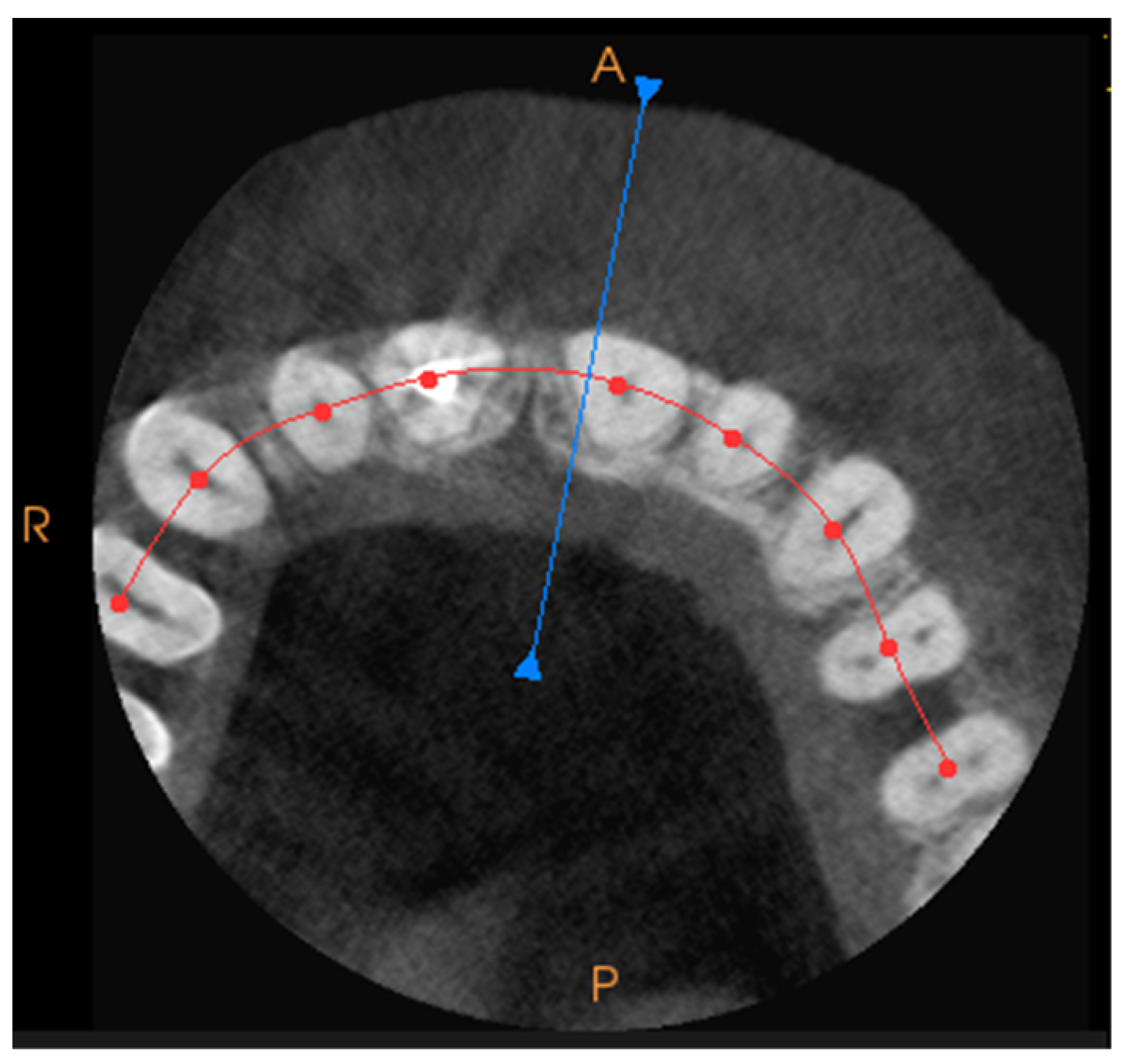
Funding

This study was partially supported and financed by the National Centre for Research and Development under Grant Lider No. LIDER/17/0064/L-11/19/NCBR/2020.

Institutional Review Board Statement

The study was conducted in accordance with the Declaration of Helsinki, and approved by the ethics committee of the Jagiellonian University Medical College under 1072.6120.276.2020, as well as by the ethics committee of the Liderdent Centrum 14/KBL/OIL/2022.

Informed Consent Statement

Not applicable.

Data Availability Statement

The data will be made available on request.

Conflicts of Interest

The authors declare no conflict of interest.

References

  1. Majumder, H.; Sharma, A.S.; Jadhav, A.K.; Deshpande, S.S. Role Of Endodontics In Forensic Odontology—A Review. J. Pharm. Negat. Results 2022, 13, 7138–7142. [Google Scholar]
  2. Costa, L.A.; Eiro, N.; Vaca, A.; Vizoso, F.J. Towards a New Concept of Regenerative Endodontics Based on Mesenchymal Stem Cell-Derived Secretomes Products. Bioengineering 2023, 10, 4. [Google Scholar] [CrossRef]
  3. Jeger, F.B.; Lussi, A.; Bornstein, M.M.; Jacobs, R.; Janner, S.F. Die digitale Volumentomographie in der Endodontologie. Eine Übersicht für den Praxisalltag [Cone beam computed tomography in endodontics: A review for daily clinical practice]. Schweiz. Mon. Zahnmed. 2013, 123, 661–668. [Google Scholar]
  4. De Mota Almeida, F.J.; Knutsson, K.; Flygare, L. The effect of cone beam CT (CBCT) on therapeutic decision-making in endodontics. Dentomaxillofac. Radiol. 2014, 43, 20130137. [Google Scholar] [CrossRef] [Green Version]
  5. Patel, S.; Durack, C.; Abella, F.; Shemesh, H.; Roig, M.; Lemberg, K. Cone beam computed tomography in Endodontics—A review. Int. Endod. J. 2015, 48, 3–15. [Google Scholar] [CrossRef]
  6. Koskela, A.; Manai, F.; Basagni, F.; Liukkonen, M.; Rosini, M.; Govoni, S.; Monte, M.D.; Smedowski, A.; Kaarniranta, K.; Amadio, M. Nature-Inspired Hybrids (NIH) Improve Proteostasis by Activating Nrf2-Mediated Protective Pathways in Retinal Pigment Epithelial Cells. Antioxidants 2022, 11, 1385. [Google Scholar] [CrossRef]
  7. Lofthag-Hansen, S.; Huumonen, S.; Gröndahl, K.; Gröndahl, H.G. Limited cone-beam CT and intraoral radiography for the diagnosis of periapical pathology. Oral Surg. Oral. Med. Oral. Pathol. Oral. Radiol. Endod. 2007, 103, 114–119. [Google Scholar] [CrossRef]
  8. Hedesiu, M.; Baciut, M.; Baciut, G.; Nackaerts, O.; Jacobs, R.; SEDENTEXCT Consortium. Comparison of cone beam CT device and field of view for the detection of simulated periapical bone lesions. Dentomaxillofac. Radiol. 2012, 41, 548–552. [Google Scholar] [CrossRef] [Green Version]
  9. Leonardi Dutra, K.; Haas, L.; Porporatti, A.L.; Flores-Mir, C.; Nascimento Santos, J.; Mezzomo, L.A.; Corrêa, M.; De Luca Canto, G. Diagnostic Accuracy of Cone-beam Computed Tomography and Conventional Radiography on Apical Periodontitis: A Systematic Review and Meta-analysis. J. Endod. 2016, 42, 356–364. [Google Scholar] [CrossRef]
  10. Stavropoulos, A.; Wenzel, A. Accuracy of cone beam dental CT, intraoral digital and conventional film radiography for the detection of periapical lesions. An ex vivo study in pig jaws. Clin. Oral Investig. 2007, 11, 101–106. [Google Scholar] [CrossRef]
  11. Mozzo, P.; Procacci, C.; Tacconi, A.; Martini, P.T.; Andreis, I.A. A new volumetric CT machine for dental imaging based on the cone-beam technique: Preliminary results. Eur. Radiol. 1998, 8, 1558–1564. [Google Scholar] [CrossRef]
  12. Scarfe, W.C.; Farman, A.G. What is cone-beam CT and how does it work? Dent. Clin. N. Am. 2008, 52, 707–730. [Google Scholar] [CrossRef]
  13. Patel, S.; Brown, J.; Semper, M.; Abella, F.; Mannocci, F. European Society of Endodontology position statement: Use of cone beam computed tomography in Endodontics: European Society of Endodontology (ESE) developed by. Int. Endod. J. 2019, 52, 1675–1678. [Google Scholar] [CrossRef] [Green Version]
  14. Mazzotti, C.; Mounssif, I.; Rendón, A.; Mele, M.; Sangiorgi, M.; Stefanini, M.; Zucchelli, G. Complications and treatment errors in root coverage procedures. Periodontol. 2000 2023. [Google Scholar] [CrossRef]
  15. Huang, T.K.; Yang, C.H.; Hsieh, Y.H.; Wang, J.C.; Hung, C.C. Augmented reality (AR) and virtual reality (VR) applied in dentistry. Kaohsiung J. Med. Sci. 2018, 34, 243–248. [Google Scholar] [CrossRef]
  16. Farronato, M.; Lucchina, A.G.; Mortellaro, C.; Fama, A.; Galbiati, G.; Farronato, G.; Maspero, C. Bilateral Hyperplasia of the Coronoid Process in Pediatric Patients: What is the Gold Standard for Treatment? J. Craniofac. Surg. 2019, 30, 1058–1063. [Google Scholar] [CrossRef]
  17. Joda, T.; Gallucci, G.O.; Wismeijer, D.; Zitzmann, N.U. Augmented and virtual reality in dental medicine: A systematic review. Comput. Biol. Med. 2019, 108, 93–100. [Google Scholar] [CrossRef]
  18. Fahim, A.; Saleem, Z.; Malik, K.A.; Atta, K.; Mahmood, R.; Alam, M.K.; Sethi, A. Exploring challenges and mitigation strategies towards practicing Teledentistry. BMC Oral Health 2022, 22, 658. [Google Scholar] [CrossRef]
  19. Reymus, M.; Fabritius, R.; Keßler, A.; Hickel, R.; Edelhoff, D.; Stawarczyk, B. Fracture load of 3D-printed fixed dental prostheses compared with milled and conventionally fabricated ones: The impact of resin material, build direction, post-curing, and artificial aging-an in vitro study. Clin. Oral Investig. 2020, 24, 701–710. [Google Scholar] [CrossRef]
  20. Fahim, S.; Maqsood, A.; Das, G.; Ahmed, N.; Saquib, S.; Lal, A.; Khan, A.A.G.; Alam, M.K. Augmented Reality and Virtual Reality in Dentistry: Highlights from the Current Research. Appl. Sci. 2022, 12, 3719. [Google Scholar] [CrossRef]
  21. Mladenovic, R.; Bukumiric, Z.; Mladenovic, K. Practice of local anesthesia applications in 3D environment during the COVID-19 pandemic. J. Dent. Educ. 2020. [Google Scholar] [CrossRef]
  22. Ayoub, A.; Pulijala, Y. The application of virtual reality and augmented reality in Oral & Maxillofacial Surgery. BMC Oral Health 2019, 19, 238. [Google Scholar]
  23. Procházka, A.; Dostálová, T.; Kašparová, M.; Vyšata, O.; Charvátová, H.; Sanei, S.; Mařík, V. Augmented Reality Implementations in Stomatology. Appl. Sci. 2019, 9, 2929. [Google Scholar] [CrossRef] [Green Version]
  24. Touati, R.; Fehmer, V.; Ducret, M.; Sailer, I.; Marchand, L. Augmented Reality in Esthetic Dentistry: A Case Report. Curr. Oral Health Rep. 2021, 8, 23–28. [Google Scholar] [CrossRef]
  25. Ma, L.; Jiang, W.; Zhang, B.; Qu, X.; Ning, G.; Zhang, X.; Liao, H. Augmented reality surgical navigation with accurate CBCT-patient registration for dental implant placement. Med. Biol. Eng. Comput. 2019, 57, 47–57. [Google Scholar] [CrossRef]
  26. Katić, D.; Sudra, G.; Speidel, S.; Castrillon-Oberndorfer, G.; Eggers, G.; Dillmann, R. Knowledge-Based Situation Interpretation for Context-Aware Augmented Reality in Dental Implant Surgery. In Medical Imaging and Augmented Reality. MIAR 2010; Liao, H., Edwards, P.J., Pan, X., Fan, Y., Yang, G.Z., Eds.; Lecture Notes in Computer Science; Springer: Berlin/Heidelberg, Germany, 2010; Volume 6326. [Google Scholar]
  27. Dang, N.P.; Chandelon, K.; Barthélémy, I.; Devoize, L.; Bartoli, A. A proof-of-concept augmented reality system in oral and maxillofacial surgery. J. Stomatol. Oral Maxillofac. Surg. 2021, 122, 338–342. [Google Scholar] [CrossRef]
  28. Mladenovic, R.; Dakovic, D.; Pereira, L.; Matvijenko, V.; Mladenovic, K. Effect of augmented reality simulation on administration of local anaesthesia in paediatric patients. Eur. J. Dent. Educ. 2020, 24, 507–512. [Google Scholar] [CrossRef]
  29. Dolega-Dolegowski, D.; Proniewska, K.; Dolega-Dolegowska, M.; Pregowska, A.; Hajto-Bryk, J.; Trojak, M.; Chmiel, J.; Walecki, P.; Fudalej, P.S. Application of holography and augmented reality based technology to visualize the internal structure of the dental root—A proof of concept. Head Face Med. 2022, 18, 12. [Google Scholar] [CrossRef]
  30. Faus-Matoses, V.; Faus-Llácer, V.; Moradian, T.; Riad Deglow, E.; Ruiz-Sánchez, C.; Hamoud-Kharrat, N.; Zubizarreta-Macho, Á.; Faus-Matoses, I. Accuracy of Endodontic Access Cavities Performed Using an Augmented Reality Appliance: An In Vitro Study. Int. J. Environ. Res. Public Health 2022, 19, 11167. [Google Scholar] [CrossRef]
  31. Zubizarreta-Macho, Á.; Ferreiroa, A.; Agustín-Panadero, R.; Rico-Romano, C.; Lobo-Galindo, A.B.; Mena-Álvarez, J. Endodontic re-treatment and restorative treatment of a dens invaginatus type II through new technologies. J. Clin. Exp. Dent. 2019, 11, e570–e576. [Google Scholar] [CrossRef]
  32. Bruellmann, D.D.; Tjaden, H.; Schwanecke, U.; Barth, P. An optimized video system for augmented reality in endodontics: A feasibility study. Clin. Oral Investig. 2013, 17, 441–448. [Google Scholar] [CrossRef] [PubMed]
  33. Zubizarreta-Macho, Á.; Castillo-Amature, C.; Montiel-Company, J.M.; Mena-Álvarez, J. Efficacy of Computer-Aided Static Navigation Technique on the Accuracy of Endodontic Microsurgery. A Systematic Review and Meta-Analysis. J. Clin. Med. 2021, 10, 313. [Google Scholar] [CrossRef] [PubMed]
  34. Song, T.; Yanh, C.; Dianat, O.; Azimi, E. Endodontic guided treatment using augmented reality on a head-mounted display system. Healthcare Technol. Lett. 2018, 5, 201–207. [Google Scholar] [CrossRef]
  35. Faus-Llácer, V.; Hamoud-Kharrat, N.; Marhuenda Ramos, M.T.; Faus-Matoses, I.; Zubizarreta-Macho, Á.; Ruiz Sánchez, C.; Faus-Matoses, V. Influence of the Geometrical Cross-Section Design on the Dynamic Cyclic Fatigue Resistance of NiTi Endodontic Rotary Files-An In Vitro Study. J. Clin. Med. 2021, 10, 4713. [Google Scholar] [CrossRef]
  36. Mahrous, A.; Elgreatly, A.; Qian, F.; Schneider, G.B. A comparison of pre-clinical instructional technologies: Natural teeth, 3D models, 3D printing, and augmented reality. J. Dent. Educ. 2021, 85, 1795–1801. [Google Scholar] [CrossRef] [PubMed]
  37. Aichert, A.; Wein, W.; Ladikos, A.; Reichl, T.; Navab, N. Image-based tracking of the teeth for orthodontic augmented reality. Med. Image Comput. Comput. Assist. Interv. 2012, 15, 601–608. [Google Scholar] [PubMed] [Green Version]
  38. Constantini Leopardi, A.; Adanero Velasco, A.; Espí Mayor, M.; Miegimolle Herrero, M. Effectiveness of Virtual Reality Goggles as Distraction for Children in Dental Care—A Narrative Review. Appl. Sci. 2023, 13, 1307. [Google Scholar] [CrossRef]
  39. Gaviria, L.; Salcido, J.P.; Guda, T.; Ong, J.L. Current trends in dental implants. J. Korean Assoc. Oral Maxillofac. Surg. 2014, 40, 50–60. [Google Scholar] [CrossRef]
  40. Ulbrich, M.; Van den Bosch, V.; Bönsch, A.; Gruber, L.J.; Ooms, M.; Melchior, C.; Motmaen, I.; Wilpert, C.; Rashad, A.; Kuhlen, T.W.; et al. Advantages of a Training Course for Surgical Planning in Virtual Reality for Oral and Maxillofacial Surgery: Crossover Study. JMIR Serious Games 2023, 11, e40541. [Google Scholar] [CrossRef]
  41. Monterubbianesi, R.; Tosco, V.; Vitiello, F.; Orilisi, G.; Fraccastoro, F.; Putignano, A.; Orsini, G. Augmented, Virtual and Mixed Reality in Dentistry: A Narrative Review on the Existing Platforms and Future Challenges. Appl. Sci. 2022, 12, 877. [Google Scholar] [CrossRef]
  42. Blanchard, J.; Koshal, S.; Morley, S.; McGurk, M. The use of mixed reality in dentistry. Br. Dent. J. 2022, 233, 261–265. [Google Scholar] [CrossRef] [PubMed]
  43. Ba-Hattab, R.; Helvacioglu-Yigit, D.; Anweigi, L.; Alhadeethi, T.; Raja, M.; Atique, S.; Daas, H.; Glanville, R.; Celikten, B.; Orhan, K.; et al. Impact of Virtual Reality Simulation in Endodontics on the Learning Experiences of Undergraduate Dental Students. Appl. Sci. 2023, 13, 981. [Google Scholar] [CrossRef]
  44. Choi, S.; Choi, J.; Peters, O.A.; Peters, C.I. Design of an interactive system for access cavity assessment: A novel feedback tool for preclinical endodontics. Eur. J. Dent. Educ. 2023. [Google Scholar] [CrossRef] [PubMed]
  45. Koyama, Y.; Sugahara, K.; Koyachi, M.; Tachizawa, K.; Iwasaki, A.; Wakita, I.; Nishiyama, A.; Matsunaga, S.; Katakura, A. Mixed reality for extraction of maxillary mesiodens. Maxillofac. Plast. Reconstr. Surg. 2023, 45, 1. [Google Scholar] [CrossRef] [PubMed]
  46. Rodrigues, P.; Nicolau, F.; Norte, M.; Norte, M.; Zorzal, E.; Botelho, J.; Machado, V.; Proença, L.; Alves, R.; Zagalo, C. Daniel Simões Lopes & José João Mendes Preclinical dental students self-assessment of an improved operative dentistry virtual reality simulator with haptic feedback. Sci. Rep. 2023, 13, 2823. [Google Scholar]
  47. Iwanaga, J.; Kamura, Y.; Nishimura, Y.; Terada, S.; Kishimoto, N.; Tanaka, T.; Tubbs, R.S. A new option for education during surgical procedures and related clinical anatomy in a virtual reality workspace. Clin. Anat. 2021, 34, 496–503. [Google Scholar] [CrossRef]
  48. Moro, C.; Štromberga, Z.; Raikos, A.; Stirling, A. The effectiveness of virtual and augmented reality in health sciences and medical anatomy. Am. Assoc. Anat. 2018, 10, 549–559. [Google Scholar] [CrossRef] [Green Version]
  49. Sorribes De Ramón, L.A.; Ferrández Martínez, A.F.; García Carricondo, A.R.; Espín Gálvez, F.; Alarcón Rodríguez, R. Effect of virtual reality and music therapy on anxiety and perioperative pain in surgical extraction of impacted third molars. J. Am. Dent. Assoc. 2023, 154, 206–214. [Google Scholar] [CrossRef]
  50. Zaidman, L.; Lusky, G.; Shmueli, A.; Halperson, E.; Moskovitz, M.; Ram, D.; Fux-Noy, A. Distraction With Virtual Reality Goggles in Paediatric Dental Treatment: A Randomised Controlled Trial. Int. Dent. J. 2023, 73, 108–113. [Google Scholar] [CrossRef]
Figure 1. The CT scan of a tooth with a pantographic curve, with the blue line marking the angle and perspective for the front view, can be generated separately. The red line marks the pantographic curve.
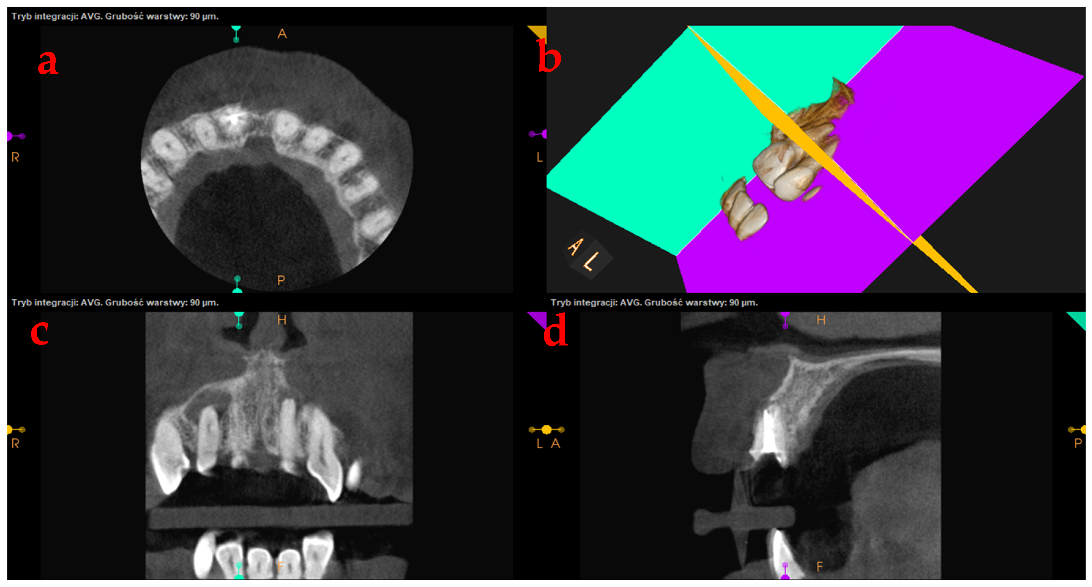
Applsci 13 04078 g001
Figure 2. The procedure leading to the creation of a three-dimensional bone model is (a) a CT scan, axial tooth’s cross-sections, (b) a three-dimensional image of the bone structure and teeth; yellow: axial plane, green: longitudinal plane, pink: transverse plane, (c) tooth’s transverse cross-sections, and (d) tooth’s longitudinal cross-sections.
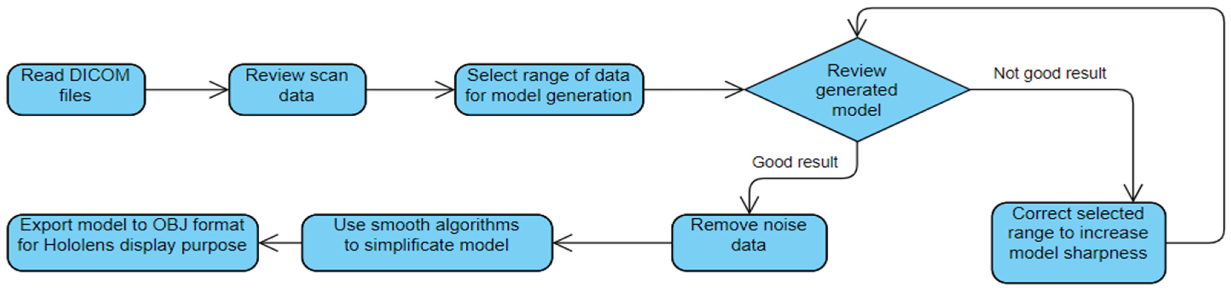
Applsci 13 04078 g002
Figure 3. The scheme of the MR-based supporting system for root canal treatment.
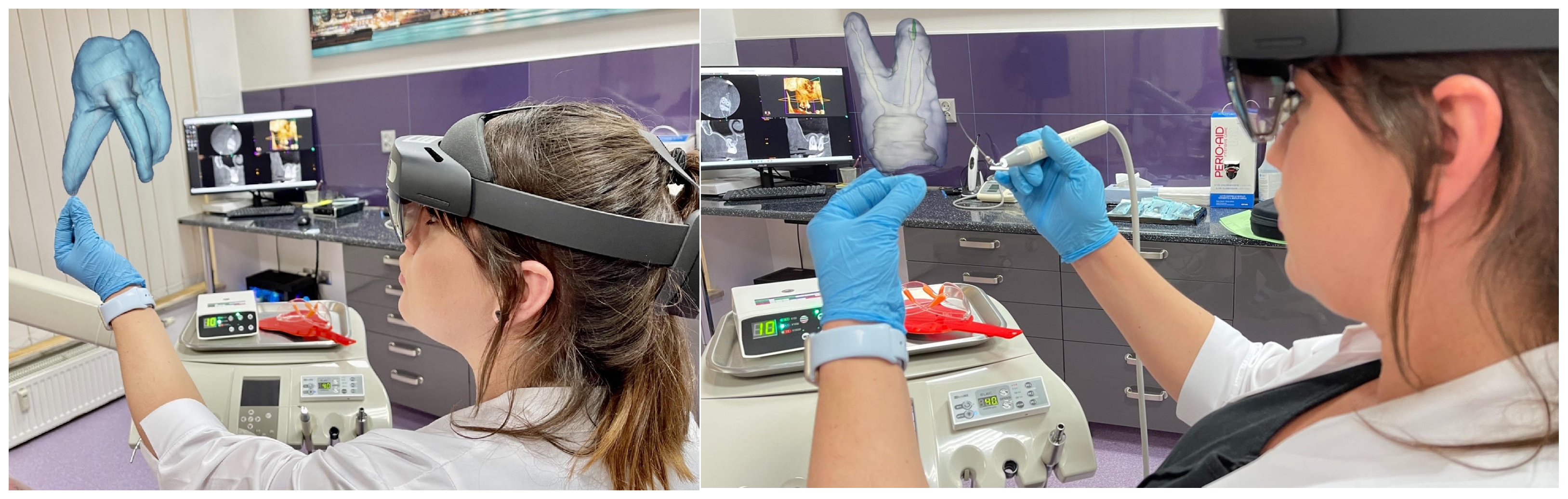
Applsci 13 04078 g003
Figure 4. The MR-based supporting system for root canal treatment.
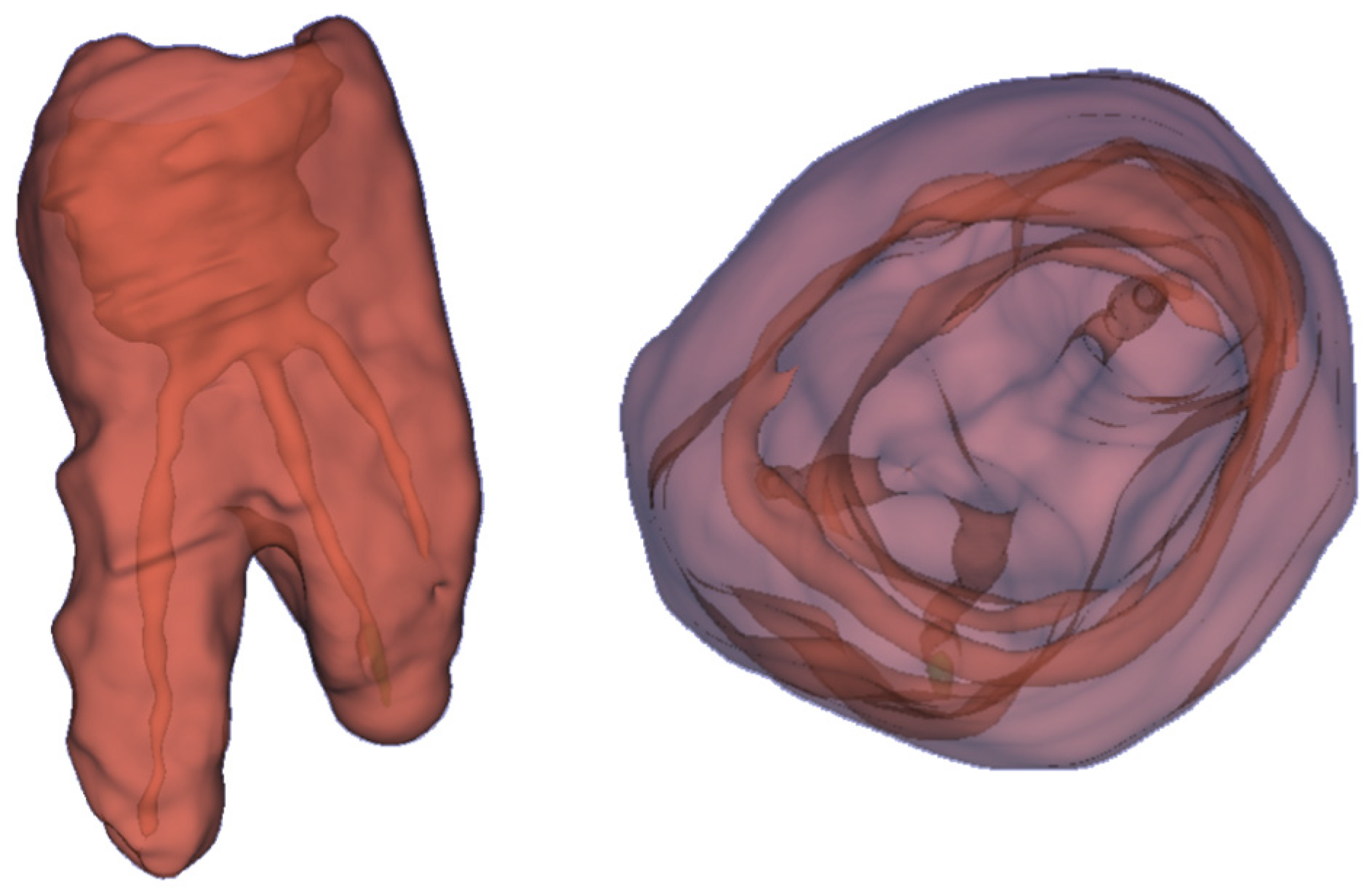
Applsci 13 04078 g004
Figure 5. The 3D holograms of the tooth, with visible root canals.
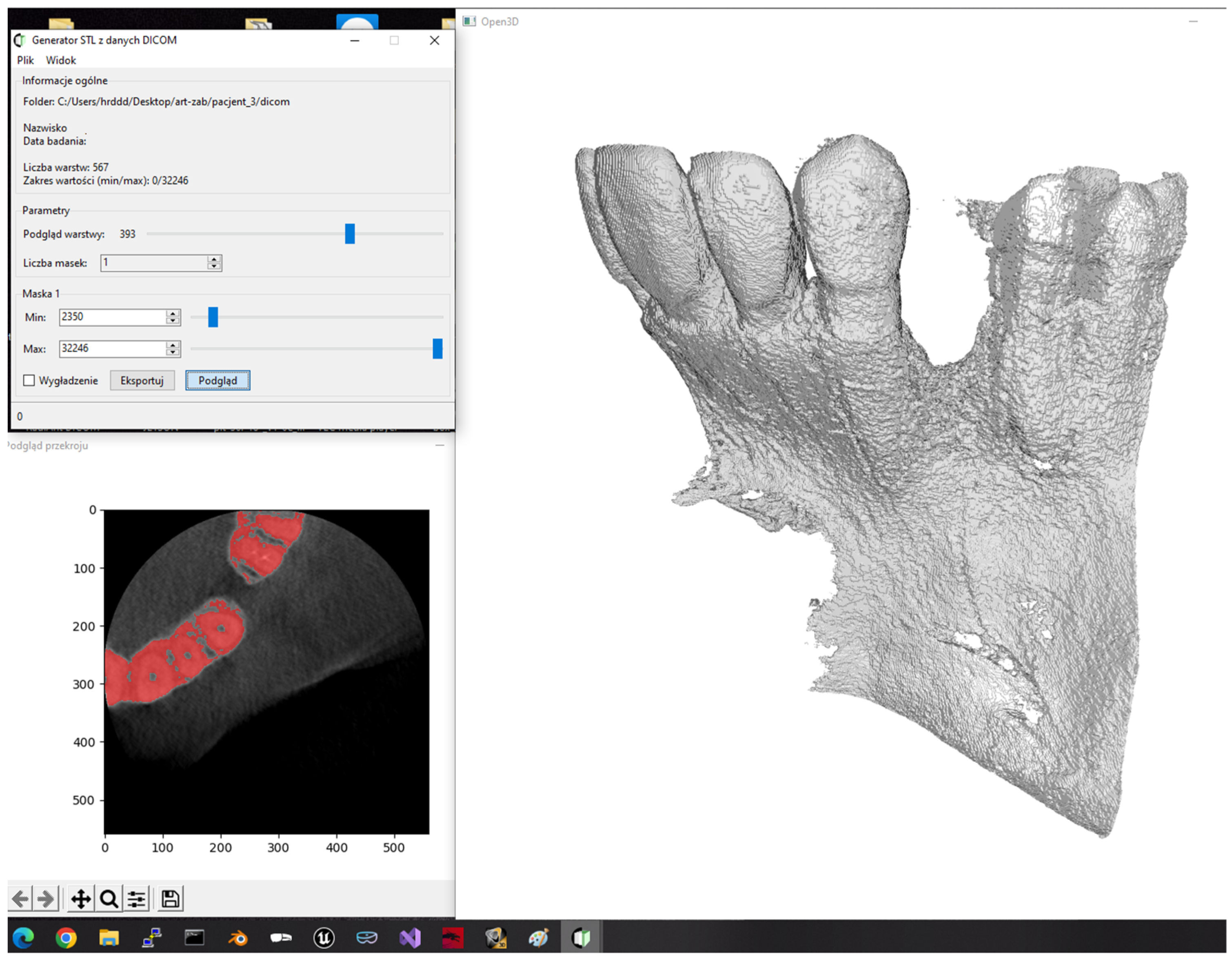
Applsci 13 04078 g005
Figure 6. The screenshot of the application enables the creation of 3D holograms based on the CT scan (DICOM data).
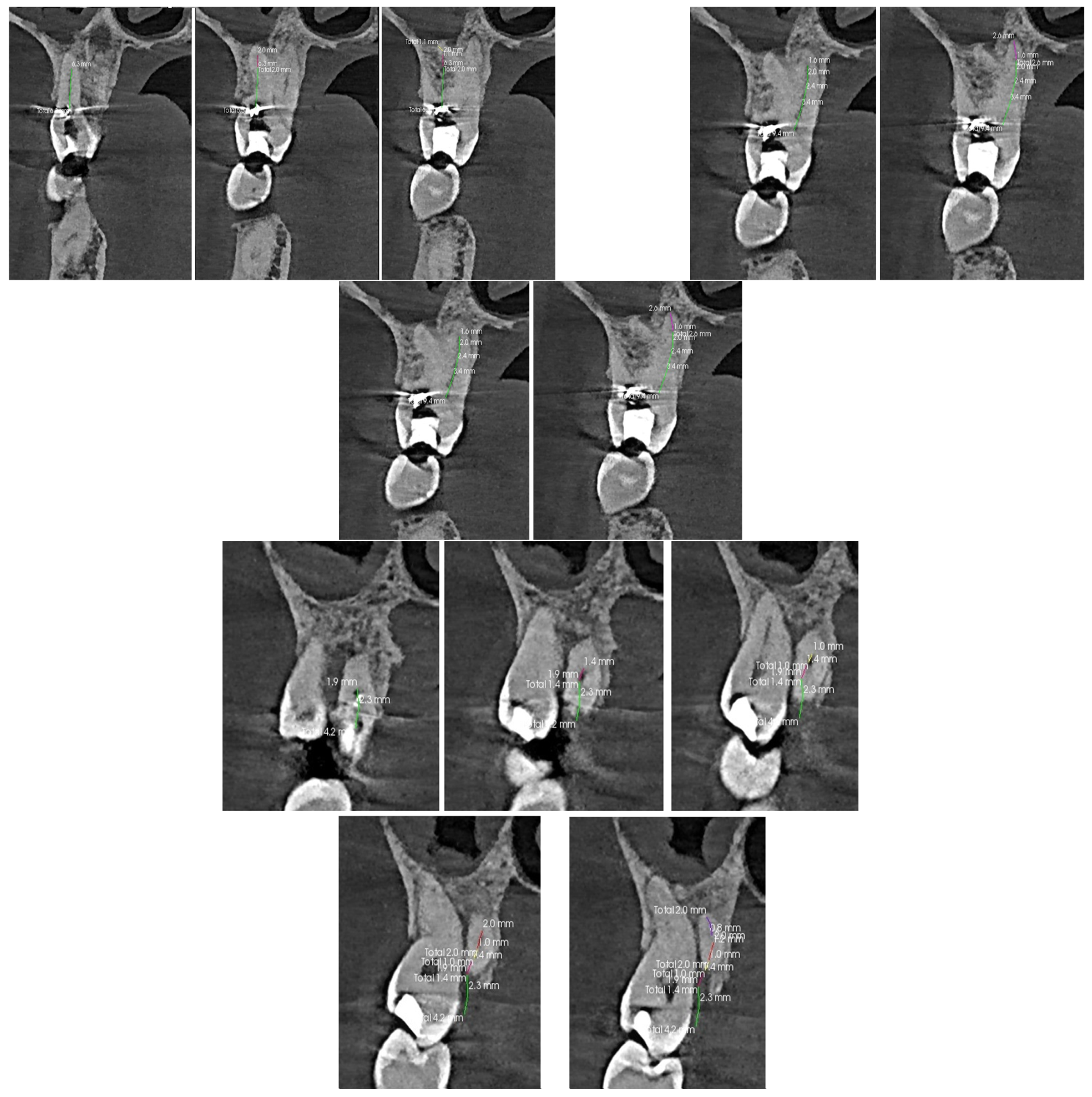
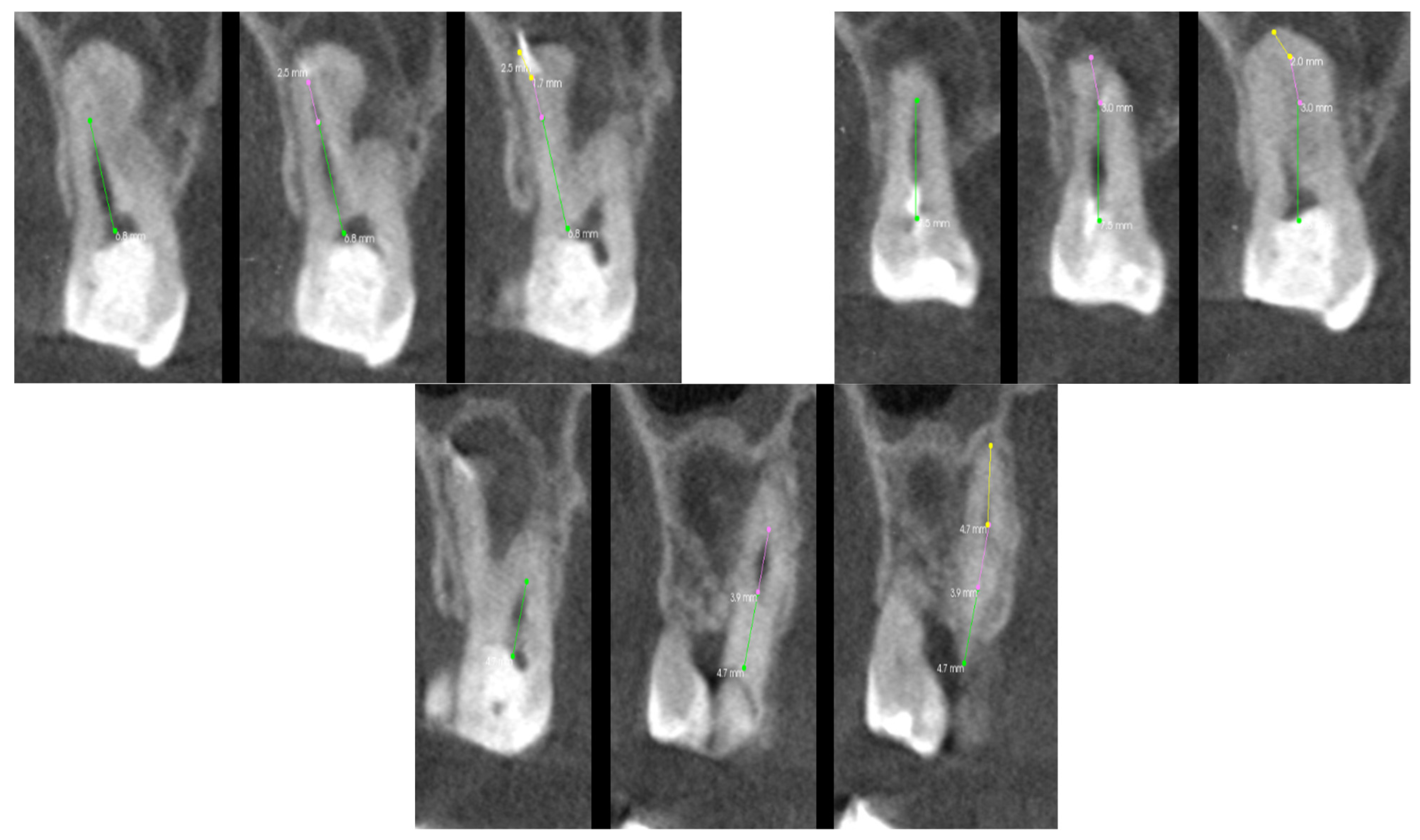
Applsci 13 04078 g006
Figure 7. The CT scan of the tooth was used for the analysis of the root canals in the transverse plane, in the case of tooth 17 with carious pulp exposure.
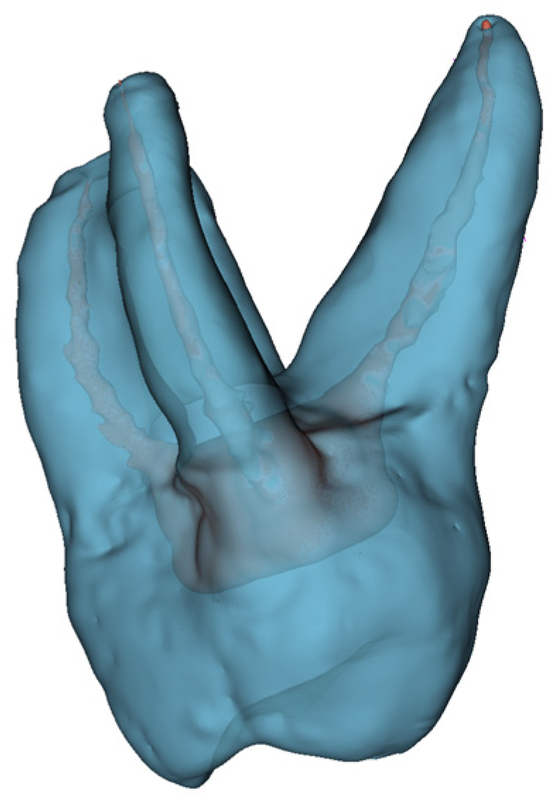
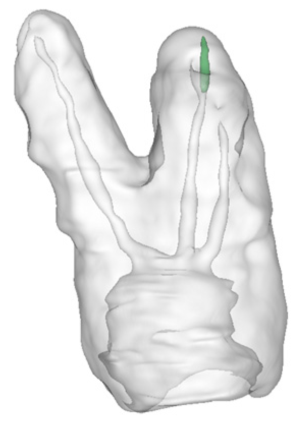
Applsci 13 04078 g007
Figure 8. The 3D of tooth 17 with carious pulp exposure.
Applsci 13 04078 g008
Figure 9. The CT scan of the tooth was used for the analysis of the root canals in the transverse plane, in the case of tooth 16, where the yellow line denotes the axial plane, and the green line denotes the longitudinal plane.
Applsci 13 04078 g009
Figure 10. The 3D hologram of tooth 16, is subjected to endodontic treatment due to periodic pain.
Applsci 13 04078 g010
Figure 11. The CT scan of the tooth was used for the analysis of the root canals in the transverse plane, in the case of tooth 15.
Applsci 13 04078 g011
Figure 12. The 3D hologram of tooth 15, subjected to endodontic treatment.
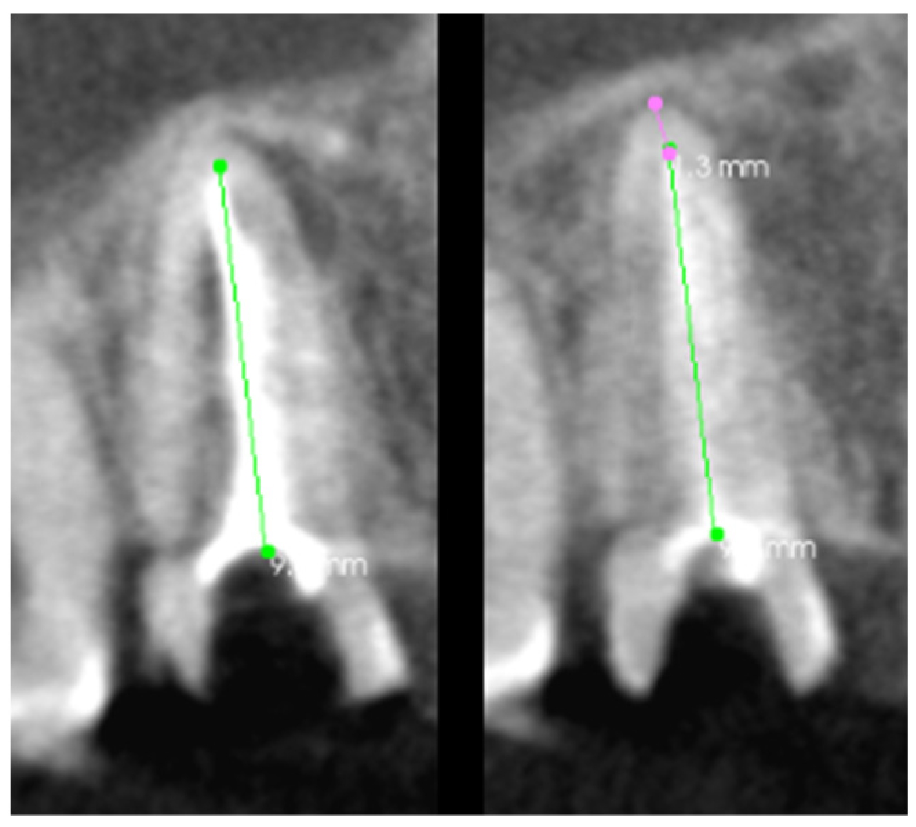
Applsci 13 04078 g012
Figure 13. The CT scan of the tooth was used for the analysis of the root canals in the transverse plane, in the case of tooth 11.
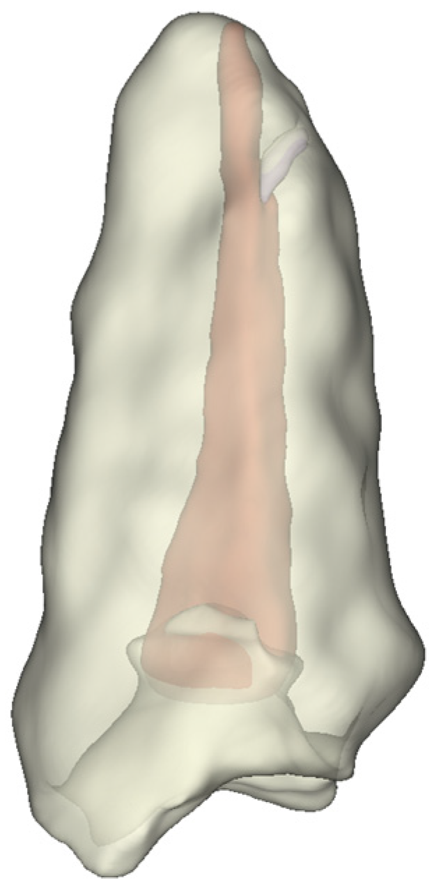
Applsci 13 04078 g013
Figure 14. The 3D hologram of tooth 11, subjected to endodontic treatment.
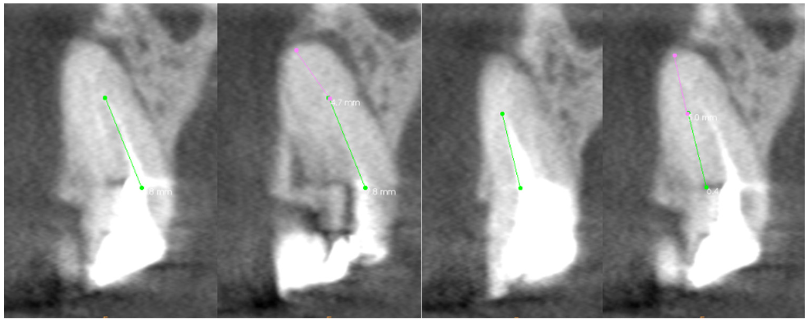
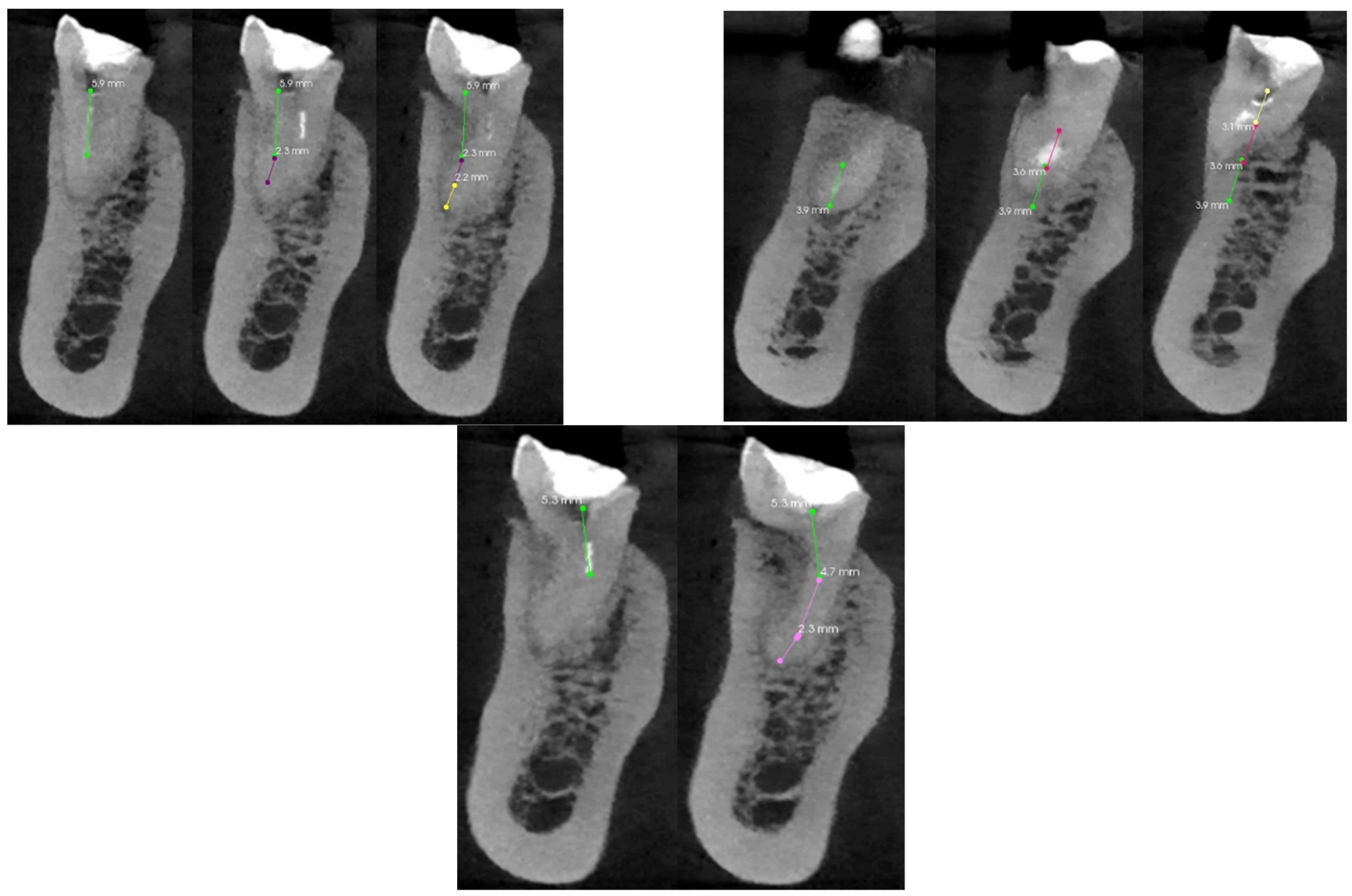
Applsci 13 04078 g014
Figure 15. The CT scan of the tooth was used for the analysis of the root canals in the transverse plane, in the case of tooth 46.
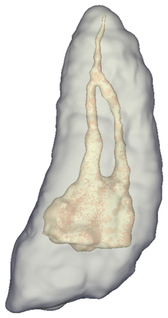
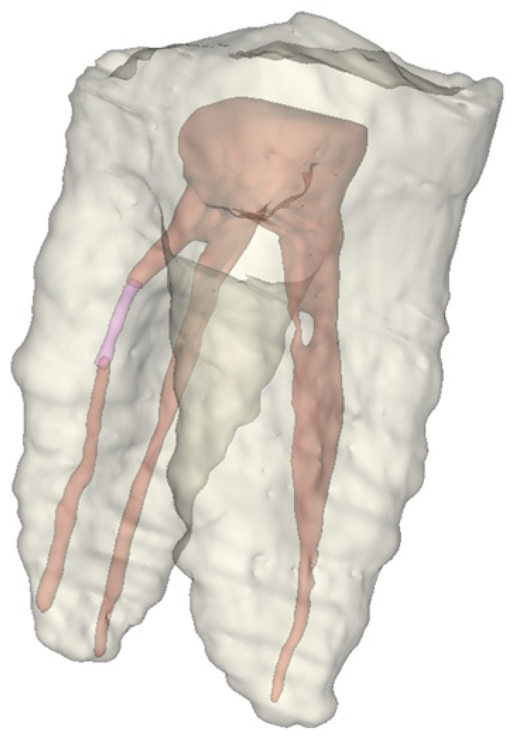
Applsci 13 04078 g015
Figure 16. The 3D hologram of tooth 46, subjected to endodontic treatment.
Applsci 13 04078 g016
Table 1. Parameters of the patient’s tooth condition are assessed based on cone beam computed tomography.
Axial PlaneTransverse PlaneLongitudinal Plane
Location of the root canal orifices at the bottom of the tooth chamberThe curvature of the root canal in the buccal-lingual/buccal-palatal dimensionThe curvature of the root canal in the mesial-distal dimension
Axial and Transverse Planes
Connection of two canals, e.g., MB1 and MB2, or buccal and lingual lower molars
Transverse and Longitudinal Planes
Location of the perforation concerning the length of the root canal
Distance of the broken file from the root tip
Axial, Transverse, and Longitudinal Planes
Uniformity of the filling material
The presence of a broken file
The presence of perforations
The presence of obliteration and calcification
Table 2. The minimum number of scans for each root canal needed to analyze its entire course and the percentage of the length of the canal visible on the scan, in the case of tooth 17 with carious pulp exposure.
Type of Root CanalThe Number of Shots Needed to Visualize/Evaluate the Entire Root Canal [pcs.]Length Visibility of r Per Picture [%]
Canal mesial buccal367.00; 21.00; 12.00
Canal distal buccal279.00; 21.00
Canal mesial palatal278.00; 22.00
Canal distal palatal 540.00; 13.00; 9.00; 19.00; 19.00
Table 3. The minimum number of scans for each root canal is needed to analyze its entire course and the percentage of the length of the canal visible on the scan, in the case of the treatment of tooth 16 due to periodic pain.
Type of Root CanalThe Number of Shots Needed to Visualize/Evaluate the Entire Root Canal [pcs.]Length Visibility of r per Picture [%]
Canal mesial buccal360.00; 24.00; 16.00
Canal distal buccal362.00; 23.00; 15.00
Canal palatal 335.00; 30.00; 35.00
Table 4. The minimum number of scans for each root canal is needed to analyze its entire course and the percentage of the length of the canal visible on the scan, in the case of the treatment of tooth 15.
Type of Root CanalThe Number of Shots Needed to Visualize/Evaluate the Entire Root Canal [pcs.]Length Visibility of r per Picture [%]
Canal buccal256.00; 44.00
Canal palatal262.00; 38.00
Table 5. The minimum number of scans for each root canal is needed to analyze its entire course and the percentage of the length of the canal visible on the scan, in the case of the treatment of tooth 11.
Type of Root CanalThe Number of Shots Needed to Visualize/Evaluate the Entire Root Canal [pcs.]Length Visibility of r per Picture [%]
Canal 288.00; 12.00
Table 6. The minimum number of scans for each root canal is needed to analyze its entire course and the percentage of the length of the canal visible on the scan, in the case of the treatment of tooth 11.
Type of Root CanalThe Number of Shots Needed to Visualize/Evaluate the Entire Root Canal [pcs.]Length Visibility of r per Picture [%]
Canal mesial buccal357.00, 22.00, 21.00
Canal mesial lingual243.00, 57.00
Canal distal337.00, 34.00, 29.00
Table 7. The comparison of the MR application with the traditional methods in root canal treatments.
Tooth NumberCBCT Analysis TimeTime to Analyze 3D ModelsTime Saved When Using MR Compared to Traditional Methods [%]
175 min 50 s3 min 28s40.57
164 min 13 s2 min 15 s46.46
153 min 40 s1 min 10 s54.83
112 min 35 s43 s72.25
464 min 55 s2 min 38 s46.44
Disclaimer/Publisher’s Note: The statements, opinions and data contained in all publications are solely those of the individual author(s) and contributor(s) and not of MDPI and/or the editor(s). MDPI and/or the editor(s) disclaim responsibility for any injury to people or property resulting from any ideas, methods, instructions or products referred to in the content.

