Article

Investigation of the Protective Effects of Urtica dioica, Capsella bursa-pastoris and Inula racemosa on Acetaminophen-Induced Nephrotoxicity in Swiss Albino Male Mice

by
Sumaira Yousuf
1,
Shabnam Shabir
1,
Mohammad Murtaza Mehdi
1,
Shailesh Srivastav
2,
Zuhair M. Mohammedsaleh
3,
Zaid Bassfar
4,
Mohammed M. Jalal
3,
Mamdoh S. Moawadh
3,
Yahya F. Jamous
5,
Sandeep Kumar Singh
6,
Emanuel Vamanu
7,* and
Mahendra P. Singh
8,9,*
1
School of Bioengineering and Biosciences, Lovely Professional University, Phagwara 144411, India
2
Department of Zoology, Shia P G College, Lucknow 226020, India
3
Department of Medical Laboratory Technology, Faculty of Applied Medical Sciences, University of Tabuk, Tabuk 71491, Saudi Arabia
4
Department of Information Technology, Faculty of Computers and Information Technology, University of Tabuk, Tabuk 71491, Saudi Arabia
5
National Center of Vaccines and Bioprocessing, King Abdulaziz City for Science and Technology (KACST), Riyadh 11442, Saudi Arabia
6
Indian Scientific Education and Technology Foundation, Lucknow 226002, India
7
Faculty of Biotechnology, University of Agricultural Sciences and Veterinary Medicine, 011464 Bucharest, Romania
8
Department of Zoology, DDU Gorakhpur University, Gorakhpur 273009, India
9
Centre of Genomics and Bioinformatics, DDU Gorakhpur University, Gorakhpur 273009, India
*
Authors to whom correspondence should be addressed.
Appl. Sci. 2023, 13(6), 3925; https://doi.org/10.3390/app13063925
Submission received: 10 February 2023 / Revised: 11 March 2023 / Accepted: 17 March 2023 / Published: 20 March 2023
(This article belongs to the Special Issue Bioactive Compounds from Natural Products - Volume II)

Abstract

:
Acetaminophen (APAP) is the most commonly used nonprescription antipyretic-analgesic drug. This medication is thought to be safe at the suggested dosage (4 g/24 h), but its overdose (up to 2.5 g/kg) can cause severe injuries to the human body, including renal injury. APAP has various toxic effects on nephrons, as it leads to an excessive free radical generation that, in turn, results in a disturbance in the redox homeostasis of cells, causing oxidative stress. To replenish this oxidative stress, there is an ultimate urge for natural therapies that can retain the cellular homeostasis of nephrons by diminishing the overdose impression of acetaminophen. The principle objective of this work is to appraise nephrotoxicity due to APAP and its amelioration through the antioxidant properties of aqueous extracts of selected medicinal plants: Urtica dioica, Capsella bursa-pastoris, and Inula racemosa (UD, CBP, and IR, respectively). The pH stability of the nutraceuticals used was examined by determining the impact of pH 4, pH 7 and pH 9 on the DPPH radical scavenging activity of aqueous plant extracts. Gas chromatography-Mass spectroscopy (GC–MS) analytical technique was performed to determine the volatile organic phytochemical profiles of all three medicinal plants. Male Swiss albino mice were used for the present investigation. The animals were distributed into five groups of (n = 6), a total of 30 mice, for in vivo analysis. Group 1 served as the control group; group 2 received a single IP dose of APAP (600 mg/kg); group 3 received APAP pretreated with UD (300 mg/kg); group 4 received APAP pretreated with CBP (300 mg/kg); and group 5 received APAP pretreated with IR (300 mg/kg). Overdose of the APAP- induced a significant (p < 0.05) alterations in the total protein concentration, weight and the nephrological architecture in renal tissue, as observed through biochemical assays and histopathological examinations. Due to nephrotoxicity, there was a substantial (p < 0.05) drop in body weight and total protein contents in the APAP alone group when compared to the treatment groups. There was remarkable protection against APAP-induced alterations in the total protein of renal homogenate in the treatment groups. Histopathological analysis (H&E staining) of the mice kidneys indicated severe deterioration in the APAP alone group, whereas the therapy groups showed considerable nephroprotection towards APAP-induced abnormalities. The biochemical findings and histopathological study of the kidneys revealed that the herbal extracts (UD, CBP, and IR) have a nephroprotective potential against APAP-induced nephropathy. The trend of efficacy was observed as UD > CBP > IR. However, extensive study is needed to determine the likely ameliorative mechanism of these nutraceuticals.

Graphical Abstract

1. Introduction

Exposure to various toxins is the main cause of nephropathy in vivo [1]. One of these causes is medications, such as acetaminophen, also known as paracetamol (N-acetyl-p-aminophenol) (APAP), which is sold under the brand names Tylenol and Panadol. This medication, along with other analgesics and antipyretics, can cause nephrotoxicity when taken in excess [2]. When taken in therapeutic doses, it has no negative effects, but prolonged usage and overdosing can have fatal consequences for the kidneys and liver [3]. Although the extrahepatic symptoms of acetaminophen poisoning are presently not very well documented in the literature, acetaminophen-induced liver necrosis has already been intensively investigated. Nearly 1–2% of patients who consume an excessive amount of acetaminophen develop renal dysfunction [4]. Although numerous underlying mechanisms have been identified, including the activity of N-deacetylase and prostaglandin synthetase enzymes, it has been hypothesized that the cytochrome P-450 mixed function oxidase isoenzymes present in the kidney are the pathophysiology of nephrotoxicity in acetaminophen intoxication [5].
Acetaminophen-induced nephropathy (renal damage) can happen in two distinct ways: primarily through acetaminophen’s detrimental consequences on the kidneys or subsequently through the medication’s liver damage. As acetaminophen is metabolized by the liver, a hazardous metabolite known as N-acetyl-p-benzoquinone imine is formed (NAPQI) [6]. NAPQI is routinely mitigated by glutathione (GSH), a common antioxidant found in hepatocytes. When the dosage of APAP is excessive, GSH is depleted, and NAPQI accumulates in the liver cells, causing hepatotoxicity. As the kidneys filter the blood and remove wastes, including all those generated by a diseased liver, liver damage can, in turn, lead to renal impairment [7].
In addition, deacetylation of APAP in the kidneys results in the production of the nephrotoxic metabolite para-aminophenol, which results in renal cortex necrosis. Conventional medications, such as N-acetyl cysteine (NAC), used for replenishing this toxicity, have a narrow therapeutic window of just 8 h [8]. Therefore, there is an urgent need for natural antioxidants that could minimize the overdose effects. The use of herbal remedies with antioxidant characteristics has been explored since oxidative stress is implicated in the progression of liver and kidney damage caused by APAP [9,10]. In the Ayurveda healthcare system, the herbs Urtica dioica (UD), Capsella bursa-pastoris (CBP), and Inula racemosa (IR) have been employed as well-known hepatoprotective and nephroprotective medicines. These three species include phytochemicals renowned for their ethnomedicinal properties, including polyphenols, quercetin, alkaloids and vitamins. UD is identified as stinging nettle or common nettle [11,12]. It is well known for its medicinal benefits. Leaves of this plant are used as vegetables in soups and salads [13]. Phenolic compounds detected in nettle plants are chlorogenic acid, 2-O-caffeoyl malic acid, P-coumaroyl malic acid, rutin, caffeic acid, isoquercetin, kaempferol 3-Orutinoside, isorhamnetin hexoside and isorhamnetic-3-O-rutinoside [14]. Some fatty acids are also present, especially cis-9,12-linoleic acid, palmitic, linolenic acids, phenolic compounds and some essential amino acids [15].
The prior investigation discovered that UD leaves are packed with antioxidants and possess a range of health benefits, such as anti-genotoxic, anticarcinogenic, and anti-inflammatory properties [16] and hepatoprotective and cardiovascular properties [17]. Studies have demonstrated that nettle leaf extract has antihyperglycemic activity [18]. UD extract ameliorated acetaminophen-induced hepatotoxicity in rats, as explained by one examination by Razak et al. (2020) [19]. UD is a potential neuroprotective, hepatoprotective and nephroprotective chemopreventive and therapeutic agent against oxidative stress induced by potassium bromate in rodents [11,20]. Capsella bursa-pastoris has an ancient legacy of conventional use all over the world. It is frequently known as the shepherd’s purse. The aerial parts possess the polypeptides acetylcholine, tyramine, histamine and choline. Due to this phytochemical composition, the plant induces various health benefits, such as anti-inflammatory, cardiovascular, antioxidant, sedative, hepatoprotective and antitumor effects [21,22]. Inula racemosa is known as pushkara in Ayurveda and mano in Hindi. The roots of this plant are considered to have medicinal value for treating several chronic disorders, such as asthma, tuberculosis, cough and liver toxicity [23,24]. The roots of this plant contain sesquiterpene lactones that activate the nuclear erythroid 2-related factor 2-antioxidant response element (Nrf2-ARE) intrinsic antioxidant pathway and maintain redox homeostasis [25].
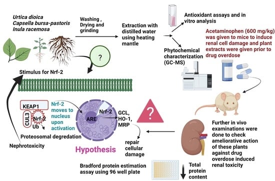
Phytochemicals are gaining popularity worldwide, with multiple studies supporting their effectiveness in the cell as well as animal experiments. Polyphenolic compounds abundant in medicinal plants act as an inducer for the Nrf-2 signaling pathway [26]. The Nrf-2 antioxidant response element signal mechanism is an essential antioxidant stress response system. Nrf-2 is a key molecule (transcription factor) that binds to a protein called Kelch-like ECH-associating protein 1 (KEAP-1), which is found in the cellular cytoplasm in an inactive state. Phytoconstituents have been found in earlier studies to promote the Nrf-2 pathway and augment the natural antioxidant and anti-inflammatory capabilities of the body by activating the cytoplasmic Nrf-2 via proteosomal degradation of Nrf2-KEAP1 complex [27].
The current work used an APAP-induced nephrotoxicity mice model to explore the nephroprotective effect of UD, CBP, and IR. GC-MS analysis was performed to characterize the volatile organic bioactive components in extracts of medicinal plants. Depending on their chemical architecture and biochemical production, plant volatile compounds are classified as phenylpropanoids, benzenoids, terpenoids, and derivatives of fatty acids. To the extent possible, no published studies have focused on the renoprotective effects of the plant extracts mentioned in this study against APAP-induced nephrotoxicity in mice. The total body weight and kidney tissue protein content were determined to assess the nephroprotective effects of UD, CBP, and IR extracts in the APAP-induced nephrotoxicity mice model. To determine ultrastructural alterations in the mice’s kidneys, histological sections of the kidney were examined.

2. Materials and Methods

2.1. Chemicals and Equipment

All the reagents and chemicals consumed during the present study were of analytical grade. 1, _1_Diphenyl-2-picrylhydrazyl (DPPH), and Bradford protein estimation kits were procured from Himedia. Acetaminophen powder (C8H9NO2) and solvents such as ethanol, methanol, chloroform, dimethyl sulfoxide (DMSO, C2H6OS), formic acid (CH2O2), sodium acetate (C2H3NaO2), acetic acid (CH3COOH), hydrochloric acid (HCl), phosphate dibasic heptahydrate (H15Na2O11P), sodium chloride (NaCl), Tris-Cl (pH 7.4), TRITON-X, sodium dodecyl-sulfate (SDS), sodium deoxycholate, phenylmethylsulfonyl fluoride (PMSF), and distilled water were obtained from Loba Chemie Ltd. (India). An electrical blender (Philips Electrical Co. Kolkata, India), heating mantle (200 watts), and Whatman filter paper were used for the preparation of extracts. A rotary evaporator (R-200) was used to evaporate extraction solvents from the herbal extracts. Gas chromatography-mass spectrophotometry (GC-MS) analyzer (Shimadzu Analytical, Columbia, MD, U.S.A). A microprocessor UV–Vis spectrophotometer single beam (Labtronics, Panchkula, HR, India) and spectrophotometer cuvettes were used. Deionized water was prepared using a Milli-Q® water purification system and ELISA reader (Bio-Rad iMark Microplate reader).

2.2. Sample Collection and Identification of Plant Materials

In the month of April 2022, young Urtica dioica leaves and entire Capsella bursa-pastoris plants were taken from the local orchards and fields of Sopore, District Baramulla, Jammu and Kashmir Province, India. The plants used in this study are therapeutic plants that thrive wild in nearly every vegetal area in North Kashmir, Jammu and Kashmir (J&K), India. These plants also grow in the Kashmir Himalayan regions of India. Inula racemosa has also been planted at Sher-e-Kashmir University of Agricultural Sciences and Technology (SKUAST). The roots of IR used in this research were obtained from the Ayurvedic Unit of SKAUST, Sopore Jammu and Kashmir, India, in the month of May 2022. For authentication and authorization of plant materials, the herbariums of the collected plants were constructed in accordance with scientific guidelines and deposited in the KASH Herbarium at the University of Kashmir, J&K, India [28]. A taxonomist at the University of Kashmir certified and authenticated plant materials such as Urtica dioica (6021-KASH), Inula racemosa (6022-KASH), and Capsella bursa-pastoris (6023-KASH).

2.3. Extraction of Plant Material

The preparation of plant extracts was performed in the specialized laboratory of the Lovely Professional University, Punjab, India, in the month of June 2022. The collected plant samples were cleaned, shade-dried for 10 days, and processed into a fine powder using a mechanical grinder. Twenty grams of powder was added to 200 mL of distilled water and allowed to undergo further extraction via a simple boiling process using a heating mantle. The extraction process was carried out for 10 min, and further, the extracts were filtered to collect the filtrate using Whatman’s No. 1 filter paper. To remove any particulate debris, the filtrates obtained were centrifuged at 2500 rpm at 4 °C for 5 min. Further, the collected extracts were concentrated using a rotary evaporator that evaporates the solvent of the filtrates, and the final crude product was weighed and further stored in a refrigerator at 4 °C for impending analysis [29].

2.4. Analysis of Antioxidant Activity and Quantification of Phytochemicals in Selected Plants

The antioxidant potency of the medicinal plant aqueous fractions (UD, CBP, ad IR) was ascertained using the DPPH antioxidant assay, as previously noted by Mensor et al. (2001) and the ABTS antioxidant assay, as initially described by Re et al. (1999) [30,31]. Quantitative measurements and characterization of phytoconstituents and the antiproliferative activity of all these plants were previously explained in our published research [32].

2.5. pH Stability Test of Herbs

To be applied as antioxidants in food systems, it is very urgent to take into consideration the optimum technological and processing factors, such as temperature and pH, affecting the stability [33]. The effect of pH on extracts and their antiradical activity was measured using solutions prepared with the required volume of extracts in acetate buffer 50 mM (pH 3.5) and phosphate buffer 50 mM (pH 8.5). The preparations were heated for 15 min, and the residual antioxidant activity was examined, as previously mentioned. Antioxidant extracts were preincubated at pH 4, 7, and pH 9, and the remaining antioxidant activity was assessed to test for pH stability. An aliquot was kept at 4 °C under refrigeration in the dark. After 48 h, the antioxidant activity of each aliquot was evaluated via the DPPH radical scavenging assay [34].

2.6. Gas Chromatography-Mass Spectrometry (GC–MS) Analysis

GC–MS analysis was executed to examine the volatile organic phytochemical profiles of the various crude preparations. To refine the samples, the solutions were divided on a 5% diphenyl/95%dimethyl polysiloxane polar column with dimensions of 30 m, 0.25 mmID, 0.25 um df with a temperature range: 320/350 °C. The oven column temperature was 70 °C for 2 min with an injection temperature of 250 °C and then elevated by 6 °C every min to 260 °C, and the pressure was maintained at 61.3 KPa for 10 min. The split mode was used to introduce the particle-free diluted sample at a ratio of 1:50 (10 mL/min split flow). Sample (1 µL) was administered while helium as a carrier gas was used at a flow rate of 1 mL/min (constant). The column flow was 1 mL/min with a total flow of 14 mL/min, a linear velocity of 36.7 cm/min and a purge flow of 3 mL/min. The total elution time was 63 min.
As a result of peak area normalization, the relative proportions of the components of the crude extract were calculated. In high-resolution mode, the mass spectral scan range was adjusted to 50–650 (m/z). By correlating the retention indices of the volatile substances with those of samples stored in the Wiley Spectral Library, the authentic compounds were recognized. The relative amount of all compounds was recognized by equating the average value of the peak area to the total area [19].

2.7. Animal Experimentation Procedure

There were no human data used in this project. Mice were procured from NIPER, Mohali Chandigarh (India), and the Committee for the Purpose of Control and Supervision of Experiments on Animals CPCSEA-approved animal house on 25 July 2022. The work was authorized by the Institute Animal Ethics Committee (IAEC) India under protocol no. LPU/IAEC/2022/01. The mice in this study were maintained and administered in accordance with the National Institute of Health’s Guide for the Care and Use of Laboratory Animals (National Research Council, 2011).
In July 2022, 30 young adult male Swiss albino mice weighing 20–24 g were procured from the CPCSEA, New Delhi, India, approved animal house of the National Institute of Pharmaceutical and Educational Research (NIPER) Mohali, Chandigarh, India, after approval from the Institutional Animal Ethics Committee (IAEC). The mice were kept at the animal house of Lovely Professional University, India, and acclimatized for 7 days. The mice were housed in polyethylene cages, given 14 days to acclimate, and fed a conventional mouse pallet diet (water ad libitum) under standard laboratory conditions (optimal temperature (24 ± 1 °C) with a 12–13 h light-dark cycle and relative humidity (40–65%) during the entire experimentation process [35].

2.8. Experimental Design of Acetaminophen-Induced Nephrotoxicity In Vivo

The dosage of medicinal plants was chosen based on a study by Razak et al. (2020). They conducted an acute toxicity study on UD and discovered that it was harmless at all examined dosages (up to 2500–3000 mg/kg BW) and did not produce any adverse effects in rats, such as drowsiness, convulsions, diarrhea, or itching. There was no mortality recorded during the 72-h assessment period. As a result, for in vivo nephroprotective testing, 1/10 of the highest concentration, i.e., 300 mg/kg BW of all the extracts, was shortlisted for experimentation. The dosage of all three plants was kept the same in order to make an appropriate comparison in the nephroprotective efficacy of extracts [19].
A total of 30 mice were distributed, with six mice per group in five groups. The Group I animals as vehicle controls were orally administered 10 mL/kg normal saline for 14 days; Group II positive control was given 600 mg/kg body weight (BW) APAP in a normal saline single dose on the 14th day of the experiment via intraperitoneal administration; Group III was pretreated with an oral dose of aqueous extract of UD 300 mg/kg BW; Group IV with an oral dose of aqueous extract of CBP 300 mg/kg BW; and Group V with a dose of aqueous extract of IR 300 mg/kg BW for 13 days via oral gavage, and a single dose of APAP (600 mg/kg BW) dissolved in warm saline solution was administered on the 14th day to III, IV and V to induce nephrotoxicity. After 6 h of treatment, mice were anesthetized using chloroform and sacrificed to obtain kidney tissues, as well as blood via cardiac puncture. The kidney tissue was collected in Eppendorf tubes and stored at −80 °C, and blood was stored at −20 °C for further examinations.
The mouse weights were recorded with a high precision weighing balance on days 0, 7, and 14 of the experiment. The change in weight was measured for each group [36].

2.9. Histopathological Analysis

Following the sacrifice of the mice, a postmortem investigation was conducted. The kidneys of the animals were isolated and cut out for histological analysis. After washing in saline solution, portions of each excised kidney were collected. The tissue was placed in cassettes fixed in 10% of 4% paraformaldehyde and then dehydrated in a 100% ethanol solution before being embedded in paraffin. It was then cut into 5 µm thick sections and stained with hematoxylin-eosin (HE) before being examined under a photomicroscope [37]. Cytosolic vacuolation, membrane enlargement, hemorrhage, necrosis, and cellular infiltration were the chief histopathological abnormalities for the kidney that have been investigated [6].

2.10. Total Protein Estimation

For total protein estimation, 10% kidney tissue homogenate was prepared using RIPA (Radio-Immunoprecipitation Assay) lysis buffer [38,39]. RIPA buffer allows for the retrieval of the membrane, cytoplasmic, and nuclear proteins and is suitable for a wide range of procedures, including protein assays and reporter assays. RIPA buffer does not include any protease or any other enzymes, thus, making it suitable for immunoassays and protein purification. Phosphatase inhibitors; however, protease and phosphatase inhibitors might be used if needed and added immediately before use to RIPA buffer to prevent proteolysis and sustain the phosphorylation of proteins. The RIPA lysis buffer contained 30 mL 5 M NaCl, 5 mL 1 M Tris-Cl (pH 7.4), 5 mL TRITON-X, 0.5 mL 20% SDS, 5 mL 10% sodium deoxycholate, and 50 mL ddH2O. This buffer is stable at room temperature. Then, 1 mM phenylmethylsulfonyl fluoride (PMSF) was added immediately before use [40].
The total protein (TP) of kidney tissue homogenate was determined by Bradford protein assay using a Bradford kit procured from Himedia, India, bearing product code M.178-1PK. The assay was performed according to the manufacturer’s instructions for protein estimation [41].

2.11. EC50 (Effective Concentration) Calculation of Aqueous Extracts of Plant Materials Used

The EC50 dose is an appropriate dosage that consumes 50% of reactive oxygen species. The EC50 is an important parameter used to exemplify a substance’s free radical quenching potential. The EC50 could be displayed using data interpolation from a suitable curve or data nonlinear regression using different models [42]. To check the correspondence of dosage with the %DPPH scavenging activity of the medicinal plants, the dose-effect investigation was performed to measure the efficacy of the designated medicinal plants. A lower EC50 specifies better DPPH and ABTS radical scavenging potency [32].

2.12. Statistical Analysis

All data points were articulated in triplicate and in 3 different tests. The data are specified as the mean ± standard deviation (SD) using Microsoft Excel 2019. CompuSyn Software (version 1.0) was used to generate the effective concentration (EC50) of the extracts. The statistical analysis between two different groups was conducted using one-way ANOVA (analysis of variance) with SPSS (Statistical Package for Social Sciences) software (version 18). Mathematical probabilities (α-level) of p < 0.01, p < 0.001, and p < 0.05 were considered statistically significant [43].

3. Results

The outcomes of the various analyses and assays used to evaluate the renoprotective efficacy of UD, CBP, and IR extracts against acetaminophen-induced nephrotoxicity are given in the subsections below.

3.1. Determination of pH Stability of Extracts

The findings of the pH stability study are significant since herbal fractions are typically acclimated to higher pH, improving their ability to be digested and their nutritional value. It is important to take into consideration the ideal technical settings and processing variables impacting the stability and antioxidant activity of plant extracts before implementing them as antioxidants in food systems. The pH of the environment had a major impact on the antioxidant capacity of all three extracts by the radical scavenging activity of DPPH radicals, as represented in Figure 1.
All the medicinal plant extracts (UD, CBP, and IR) reacted significantly towards different pH values (pH 4, pH 7 and pH 9) along a dosage gradient, as shown by the dose-effect curves in Figure 2. The UD aqueous extract exhibited the highest DPPH radical scavenging activity at all pH values, but the highest was shown at pH 4 (EC50 = 0.77 ± 0.80), followed by pH 7 (EC50 = 0.77 ± 2.8) and the lowest at pH 9 (EC50 = 0.75 ± 1.36), and in an aqueous extract of CB, the highest DPPH scavenging activity was shown at pH 4 (EC50 = 0.85 ± 0.5), followed by pH 9 (EC50 = 1.24 ± 0.96), and the lowest was exhibited at pH 7 (EC50 = 1.32 ± 0.35). However, the aqueous extract of IR displayed different behavior, as IR implied the highest DPPH radical quenching capacity at pH 7 (EC50 = 1.05 ± 3.32), followed by pH 4 (EC50 = 1.33 ± 1.02) and the lowest antioxidant activity was manifested at pH 9 (EC50 = 2.06 ± 1.57).

3.2. Phytochemical Fingerprinting via GC–MS

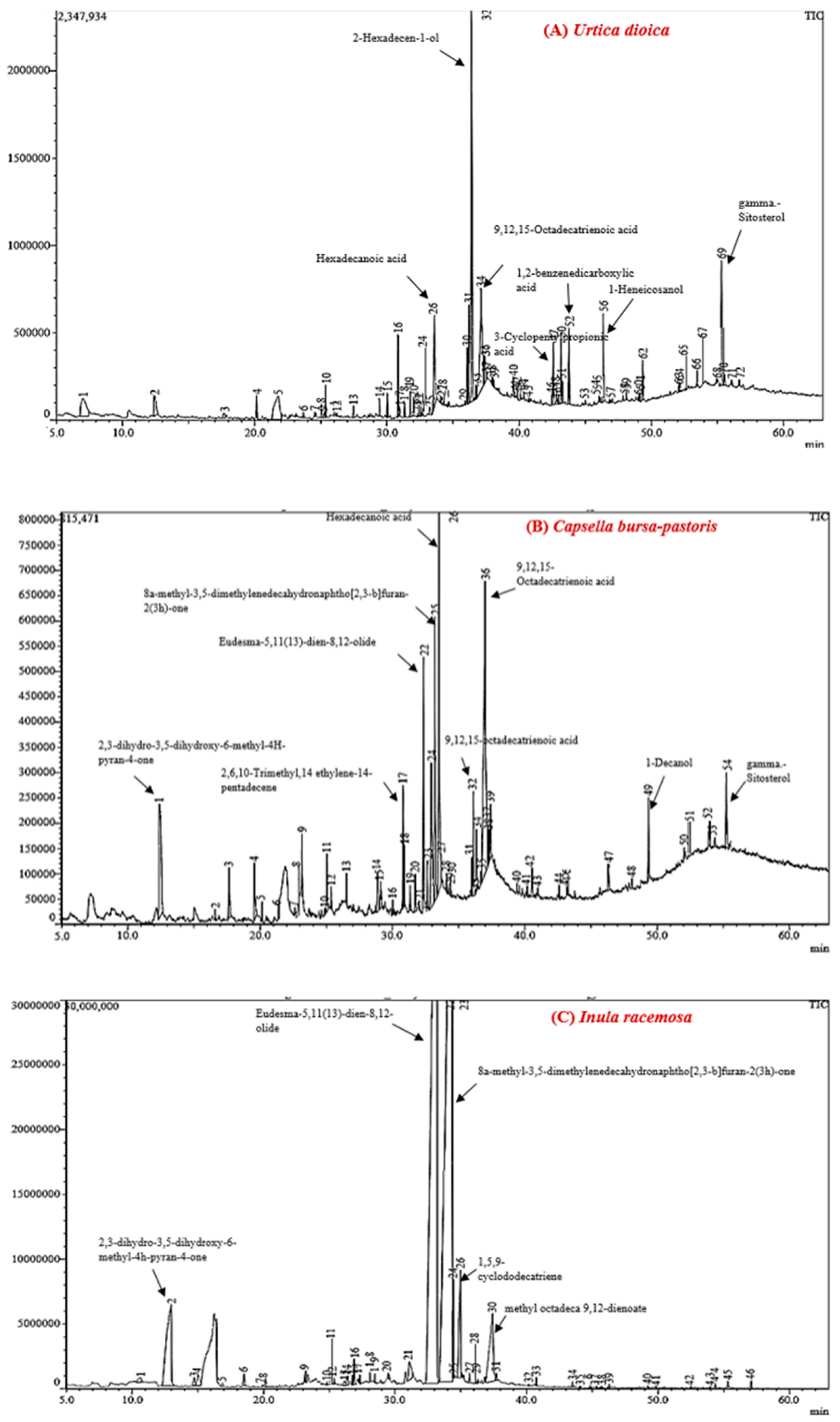
GC–MS chromatograms demonstrated various peaks of volatile organic phytochemicals in all extracts of UD, CBP and IR. On comparison of the mass spectra (MS) of the phytoconstituents with the Wiley library, the ten major phytocompounds were characterized and identified in UD, eleven compounds were detected in CBP, and three phytoconstituents were present in the ethanolic extract of IR, as demonstrated in Figure 3.

3.2.1. Compound Profiling of Urtica dioica

GC–MS chromatograms recorded the presence of a total of 72 peaks in the ethanolic fraction of UD collected from Kashmir (Figure 3a), illustrating 100% of the composition of UD. Prominent phytochemicals on the basis of % peak area, retention time and bioactivity were 9,12,15-octadecatrienoic acid with a % peak area of 17.03%, 2-hexadecen-1-ol (13.05%), 3-cyclopentylpropionic acid (2.30%), 1-Heneicosanol (5.58%), 1-dodecanol (1.51%), beta-tocopherol (1.19%), gamma-sitosterol (6.16%), hexadecanoic acid (6.93%), 1,3-hexadecatrienal (1.78%), 1,2,3-propanetriol (5.03%) and traces of some other compounds, such as catechin, fucosterol, sesquiterpene lactones, squalene, beta-amyrin, and tricosane were evidenced in the UD. These major bioactive compounds present in the UD extract provide scientific authentication that this extract has various therapeutic benefits, such as antioxidant activity, antimicrobial potential, anticancer and hepatoprotective efficacy [17].

3.2.2. Compound Profiling of Capsella bursa-pastoris

GC–MS analysis revealed a total of 54 peaks for compounds in the ethanolic extract of CBP cultivated in Kashmir. (Figure 3b), representing 100% of the CBP composition. The most dominant bioactive substances on the basis of value % peak area and retention time were α-linolenic acid (27.76%), palmitic acid (14.6%), gamma-sitosterol (2.32%), 8A-methyl-3,5-dimethylene (8.13%), 1-decanol (2.00%), 2-pentadecanone (1.19%), proline (1.91%), 2-methoxy-4-vinyl phenol (1.73%), cyclopentane (1.41%), eudesma (5.70%) and phytol (1.26). Additionally, some traces of stearic acid, beta-tocopherol and docosane were found. Bioactive metabolites found in CBP belong to terpenes, phenols, alkane classes and fatty acids. The omega-3 fatty acids were found to be the major bioactive compounds of Capsella bursa-pastoris. These fatty acids have been associated with a plethora of health benefits, such as cardiovascular, neuroprotective, and anti-inflammatory effects [44].

3.2.3. Compound Profiling of Inula racemosa

GC–MS analysis revealed 57 peaks for compounds in the ethanolic extract of IR harvested in hilly regions of Kashmir (Figure 3c), illustrating 100% of the IR composition. The most prominent bioactive substances on the basis of % peak area were Eudesma-5,11(13)-dien-8,12-olide (41.51%), 8a-methyl-3,5-dimethylenedecahydronaphtho [2,3-b]furan-2(3 h)-one (44.16%) and 2,3-dihydro-3,5-dihydroxy-6-methyl-4H-pyran-4-one (4.66%). Additionally, some traces of 1, 5, 9-cyclododecatriene and octadecanoic acid were found. Eudesma-5, 11 (13)-dien-8, 12-olide is a type of sesquiterpene lactone, and these phytoconstituents have various ethnopharmacological activities, such as combating breast cancer, hepatocarcinoma, leukemia, gastric malignancy and bladder cancer [45].

3.3. In Vivo Analysis of Acetaminophen-Induced Renal Toxicity

3.3.1. Assessment of Change in the Body Weight of the Experimental Animals

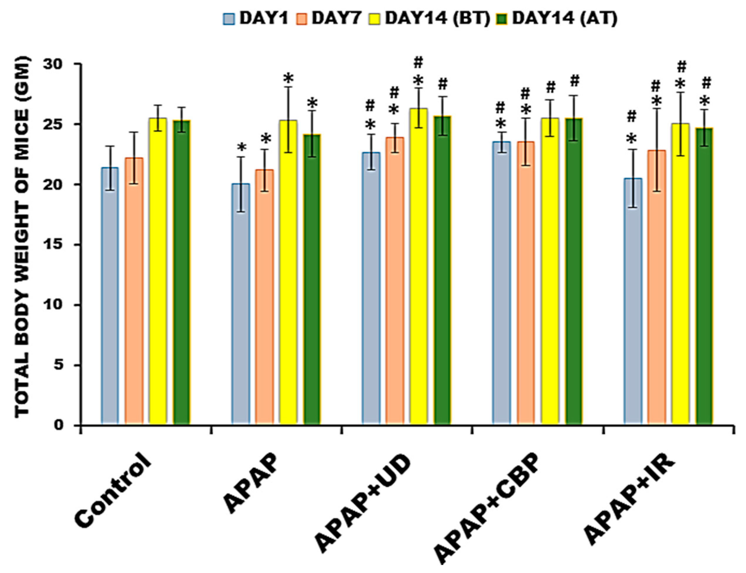
The change in the body weight of all the animals was monitored on the first day of the experiment (Day 1), seventh day (Day 7) and fourteenth day (Day 14) before acetaminophen treatment (BT) and after treatment (AT) to analyze the weight change in the mice due to acetaminophen intoxication. All the experimental groups showed significant weight gain during the first 13 days of the protocol. On day 14, treatment with acetaminophen resulted in a decline in the weight of mice in the APAP group, while the acetaminophen-intoxicated groups pretreated with UD, CBP and IR did not demonstrate any significant weight loss compared to the control group (Figure 4).

3.3.2. Histopathological Observations of Mouse Kidneys

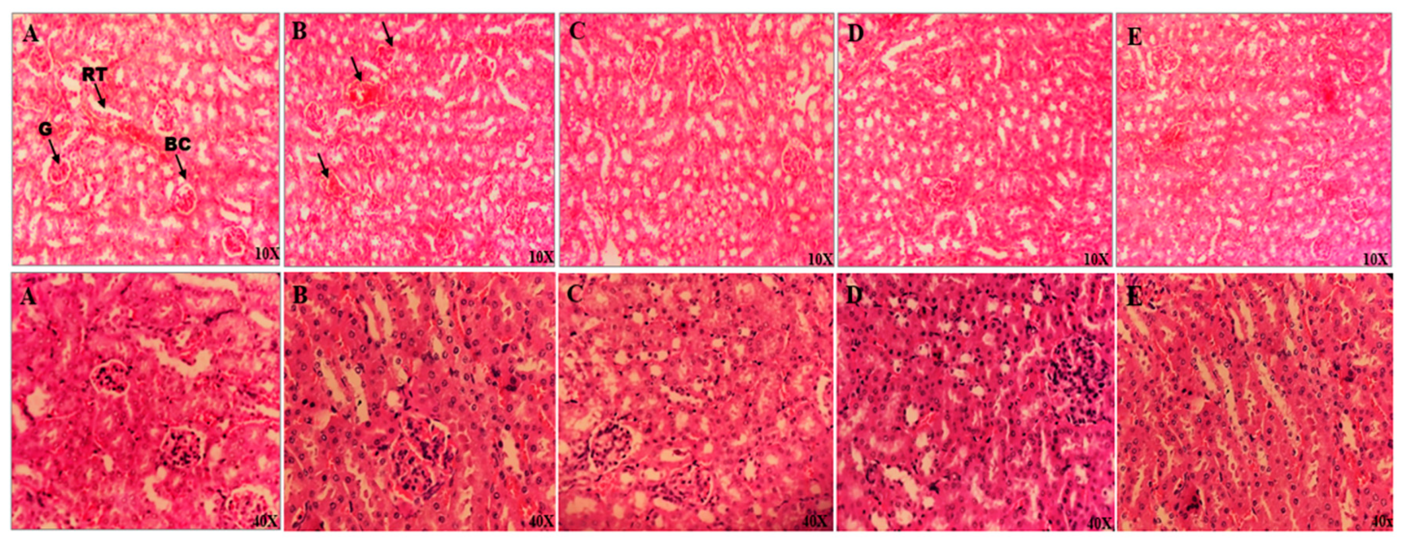
Histopathology observations of renal tissues of mice at 10× and 40× magnification are illustrated in Figure 5.
The photomicrograph of the control group showed a glomerulus surrounded by Bowman’s capsule, normal renal parenchyma and normal tubules (normal architecture). Renal sections from 600 mg/kg acetaminophen-treated mice showed massive fatty changes, destruction of the cortical region, and decreased tubular diameter. The kidney sections of mice treated with 600 mg/kg acetaminophen and 300 mg/kg BW UD showed almost normal nephric tissue with very few apoptotic cells, improved glomerular architecture and almost normal renal architecture. The renal sections of mice treated with 600 mg/kg acetaminophen and 300 mg/kg BW CBP extract showed fewer inflammatory cells around the glomerulus and some traces of necrosis. The kidney sections of mice treated with 600 mg/kg acetaminophen and 300 mg/kg BW IR extract showed minimal recovery.
The histological observation photomicrographs of kidney tissue in the control group demonstrated proper morphology of the parenchyma of the kidneys, with no pathological findings. APAP administration, on the other hand, caused significant kidney damage, as evidenced by the shedding of necrotic tubular cells and necrotic cellular debris found in the intercellular spaces of both the proximal and distal convoluted tubules. In the medullary area, vacuolar degradation of the renal mucosa of the proximal tubule and hyaline casts were observed. In addition, there was significant compaction, edema, and infiltration of inflammatory cells in the interstitial tissue. The portion of the APAP-intoxicated kidney showed extensive disintegration, hematoma, and edema with lymphocyte infiltration. Remarkably, administration of UD, CBP and IR attenuated these pathological alterations, showing the extract’s capacity to repair APAP-induced cell damage. This ameliorative behavior was evident by the normal cellular architecture of APAP-intoxicated groups pretreated with herbal fractions. UD and CBP seem to be more ameliorative than IR, as demonstrated by the minimal recovery of mice from group V (IR) from nephrotoxicity caused by APAP overdose compared to Group III (UD) and Group IV (CBP).

3.4. Degenerated Protein Concentrations in the Mice Exposed to Acetaminophen Overdose

Bradford reagent consists of a dye, namely, Coomassie brilliant Blue (CBB) G-250, which has an absorbance of 465 nm in the unbound state. This assay is based on the formation of complexes between (CBB) G-250 and the protein samples in solution. When the protein in homogenate binds to the dye, the color of the solution changes from brown to blue, and there is a shift in the absorbance from 465 to 495 nm. The binding takes only 5 min. The blue product remained stable for one hour. The concentration of unknown protein can be determined by plotting its absorbance value on the standard curve [46].
Degradation of the protein is associated with cellular damage that could be directly correlated with the toxicity of the compound. To analyze the effect of the herbal fractions on the protein concentration of the mice intoxicated with APAP overdose, a Bradford protein estimation assay was performed. The group of mice exposed to 600 mg/kg APAP exhibited a significant reduction (p < 0.05) in the total protein content of their tissues. The total protein concentration in the kidney tissue was reduced in the APAP group (6.62 ± 0.012 mg/g BSA) compared to the control group (18.58 ± 0.016 mg/g BSA). Among the three medicinal plant pretreated groups along with APAP, APAP pretreated with UD (11.82 ± 0.012 mg/g BSA) exhibited the highest protein content, followed by APAP + CBP (10.65 ± 0.07 mg/g BSA) and APAP + IR (8.02 ± 0.013 mg/g BSA) (Figure 6).

4. Discussion

Although acetaminophen (APAP), also known as paracetamol, has a decent safety profile in prescribed dosages, overdose remains the most common cause of liver injury and even death in many regions of the world [47]. APAP overdose causes hepatotoxic and nephrotoxic impacts through a complicated chain of events. In most cases, acetaminophen-induced nephrotoxicity emerges after hepatotoxicity, although the presence of renal tubular damage and abrupt kidney failure, even in the absence of liver injury, ought not to be overlooked [48].
Although APAP-induced hepatic necrosis has been thoroughly investigated, the extrahepatic symptoms of APAP poisoning are not well reported in the literature, and renal impairment affects 1–2% of acetaminophen overdose patients [49]. Although additional pathways, such as the involvement of prostaglandin synthetase and N-deacetylase enzymes, have been identified, the pathophysiology of acetaminophen-induced nephropathy has been linked to cytochrome P-450 mixed-function oxidase isoenzymes found in the kidney [50]. Although glutathione (GSH) is a crucial facet in the removal of acetaminophen and its metabolites, its conjugates have been related to the creation of nephrotoxic compounds. In most cases, acetaminophen-induced kidney failure emerges after hepatic injury, but it can be differentiated from the hepatorenal syndrome, which can exacerbate severe liver damage [51]. The toxicity mechanism requires glutathione loss, increased reactive oxygen species production, and higher oxidative stress. Many inflammatory markers, such as specific proinflammatory cytokines, are released in conjunction with these critical events, and these facilitators might modulate toxicity. Free radicals can trigger lipid peroxidation and are involved in drug toxicity [52].
Many animal studies have shown a strong positive relationship between oxidative stress and renal toxicity. Acetaminophen nephrotoxicity is caused by the adverse effects of its highly reactive metabolite, N-acetyl-para-amino-benzoquinone imine (NAPQI), which arylate proteins (especially glutamine synthetase and selenium-binding protein) in the S3 portion of the proximal convoluted tubule, causing nephric apoptosis [53]. These drug-induced nephrotoxicity symptoms are frequently linked with significant increases in serum creatinine, blood urea nitrogen, and acute tubular necrosis. Thus, biochemical markers, such as serum creatinine, blood urea, glomerular filtration rate, enzyme urea, and 2-microglobulin urine excretion, have been employed to study drug-induced nephrotoxicity in animals and humans [54].
Herbal medications, unlike chemically synthesized drugs, have a complex chemical structure. As a consequence, the methods used to identify ‘herbal medicine’ are largely intended to obtain a unique fingerprint of specific plants that demonstrates the presence of quality-defining bioactive chemical components [55]. Medicinal plants are the best remedies used for combating nephrotoxicity caused by drug overdose. They are cost-friendly, easily available and have no mutagenic effect [56]. In the current study, three plants, Urtica dioica (UD), Capsella bursa-pastoris (CBP) and Inula racemosa (IR), were used to attenuate acetaminophen-induced nephrotoxicity in male Swiss albino mice. Earlier investigations evaluated that these medicinal plants have greater health benefits, such as antioxidant activity, anticancer properties and neuroprotective effects [29,32,57]. However, various studies have proven the hepatoprotective effects of these extracts against various toxins, including carbon tetrachloride and cisplastin. [58,59,60]. However, to the best of our comprehension, there is no literature that could explain the nephroprotective effects of UD, CBP and IR against acetaminophen-induced nephrotoxicity.
Therefore, in the present study, to check the volatile organic compound profile of the selected medicinal plants, the GC–MS analytical technique was carried out. This analysis demonstrated that the most abundant compounds in UD were 9, 12, 15-octadecatrienoic acid, 2-hexadecen-1-ol, 3-cyclopentylpropionic acid, 1-Heneicosanol, 1-dodecanol, beta-tocopherol, gamma-sitosterol, hexadecanoic acid, 1, 3-hexadecatrienal, and 1, 2, 3-propanetriol. However, in the UD extract, two important pentacyclic triterpenoids, alpha- and beta-amyrin, were detected in minimal amounts. In the case of CBP, the major compounds found were α-linolenic acid, palmitic acid, gamma-sitosterol, 8A-methyl-3, 1-decanol, 2-pentadecanone, proline, 2-methoxy-4-vinyl phenol, cyclopentane, eudesma and phytol. Additionally, there were some traces of stearic acid, beta-tocopherol and docosane, while the two major compounds present in IR were sesquiterpene lactones, eudesma-5,11(13)-dien-8,12-olide, 8A-methyl-3,5-dimethylene (lvasperine) and 2,3-dihydro-3,5-dihydroxy-6-methyl-4H-pyran-4-one. Additionally, some traces of 1, 5, 9-cyclododecatriene and octadecanoic acid were found. It was previously studied that octadecanoic acid has an anti-arthritic, anti-inflammatory, antiandrogenic, hepatoprotective, hypocholesterolemic, 5-alpha-reductase suppressor, nematicide, anti-coronary, antihistaminic, and antiacne activities [61]. Volatile compound profiles in plants have been evaluated to possess various therapeutic effects, such as anti-inflammatory, anti-obesity, anti-cancer and antioxidant activity in vitro and in vivo [62]. Previous evaluations also demonstrated the presence of pentacyclic triterpenoids and fatty acids as the main metabolites in UD [63]. The quantity of the compounds obtained was significantly different from that in our study, which could be due to the difference in the polarity of solvents, as they used the nonpolar solvent hexane. No standard study was found to evaluate the presence of phytochemicals in CBP through GC–MS analytical techniques. In the case of IR, the two major compounds present were sesquiterpene lactones (alantolactone and isoalantolactone). Rathore et al. (2022) reported that these two lactones are considered to be the major sesquiterpene lactones present in rhizomes of IR [64]. Some other compounds were also present in the IR extract but in a minimum amount, such as retinol, tridemorph, and sitosterol, as illustrated in Figure 4. The bioactive substances in the above-mentioned extracts prepared from UD, CBP, and IR are a significant finding for these metabolites’ biological properties (antiradical and antioxidants) and, thus, for their promising applications in pharmacology, such as anticancer, antidiabetic, hepatoprotective, neuroprotective and nephroprotective activities [22,65,66]. The small discrepancy in the phytochemicals present in the herbal fractions in our study compared to previous studies is due to the difference between agroclimatic and seasonal variations, as well as methods used for determining their biological activities, which have a greater impact on the phytochemical composition of herbs.
To be applied as antioxidants in food systems, it is very urgent to take into consideration the optimum technological and processing factors, such as temperature and pH, affecting the stability [33]. The ROS/RNS scavenging capacity of all the herbal fractions was highly influenced by the pH of the system. All aqueous extracts were subjected to different pH systems (4, 7, and 9) to simulate acidic, neutral, and alkaline pH values. The outcomes illustrated the significant influence of pH on the antioxidant activity of herbal extracts. Most of the extracts were stable at pH 4. Hong et al., 2020 suggested that in acidic conditions of aqueous extracts, phenolic content and antioxidant activity increase compared to a neutral aqueous system [67].
Drug-induced nephrotoxicity was established in the current investigation by a one-time intraperitoneal injection of acetaminophen on the 14th day of the experiment. The toxicity was characterized by a significant (p < 0.05) decrease in total protein content and tubule nephritis histological characteristics in the acetaminophen group (negative control) compared to untreated (group I) rats. However, pretreatment with daily, graded oral dosages of UD, CBP, and IR (groups III, IV, and V, respectively) for 14 days reduced these alterations in a dose-related manner. All of the extracts substantially (p < 0.001) reduced the acute toxicity of APAP overdose, as evidenced by higher protein levels in kidney tissue homogenates, keeping their values within the normal range compared to the APAP-treated mice (group II). The examination of the significant weight change in the mice was equally remarkable. The weight of the mice normally increased among all the groups until day 14. Slight but significant weight loss was observed in the acetaminophen-treated group (negative control) compared to the control group. The groups pretreated with herbal extracts showed normal weight changes compared with the control group. The reason for the weight loss could be lethargy and reduced food consumption in the APAP group, which could also be associated with the intraperitoneal injection procedure of acetaminophen. Thus, the anorexic state of the nephrotoxic mice could account for the significant, as well as progressive weight losses recorded for the acetaminophen-intoxicated mice [68].
The histological analysis of kidney tissue in the control group demonstrated proper morphology of the parenchyma of the kidneys, with no pathological findings. APAP administration, on the other hand, caused significant kidney damage, as evidenced by the shedding of necrotic tubular cells and necrotic cellular debris found in the intercellular spaces of both the proximal and distal convoluted tubules. In the medullary area, vacuolar degradation of the renal mucosa of the proximal tubule and hyaline casts were observed. In addition, there was significant compaction, infiltration of inflammatory cells, and edema in the interstitial tissue. The portion of the APAP-intoxicated kidney showed extensive disintegration, hematoma, and edema with lymphocyte infiltration. Remarkably, administration of UD, CBP and IR attenuated these pathological alterations, showing the extract’s capacity to repair APAP-induced cell damage. This ameliorative behavior was evident by the normal cellular architecture of APAP-intoxicated groups pretreated with herbal fractions. UD and CBP seemed to be more ameliorative than IR, which was demonstrated by the minimal recovery of mice from group V (IR) from nephrotoxicity caused by APAP. Previous evaluations showed that the ethanolic extract of U. dioica exhibited significant nephroprotective activity against gentamicin-induced nephrotoxicity in rabbits [69]. It was also documented that Capsella bursa-pastoris has anti-inflammatory properties, as it inhibits the production of IL-6 and TNF-α in the inflammatory development process, and Inula racemosa has adrenaline beta-blocking efficacy [70,71].
Total protein content analysis was performed on excised kidney homogenates from all groups of mice to better understand the disparaging consequences of APAP, which elevated cellular free radical levels and enabled proteins to undergo posttranslational modifications (oxidative). Compared to the control group, mice treated with 600 mg/kg, APAP had a statistically significant drop (p < 0.001) in the total protein concentration of renal tissues. The APAP-treated group with pretreatment of UD had higher protein contents, followed by CBP and IR. This conclusion is consistent with a prior study that revealed that APAP caused organisms to have lower protein content [72]. Earlier studies also claimed that medicinal plants have efficacy in reducing the degradation of cellular proteins and have anti-lipid peroxidative effects, as was proven by our study [73].
Several studies have documented and validated the excellent safety profile of phytoconstituents, which appears to align with our findings. More study in this area, however, is needed. Furthermore, prior phytochemical studies on selected extracts reported the separation and structural characterization of polyphenols (e.g., quercetin and rutin), fatty acids, lignins, and alkaloids [54]. Phytonutrients, specifically quercetin derived from various nephroprotective traditional medicines, have been shown to reduce xenobiotic-induced nephrotoxicity in experimental animals due to their antioxidant and antiradical potency [29,57]. Besides other bioactive components found in the above-mentioned extracts, this biological property could be responsible for the extract’s nephroprotective action [74,75]. The extract could potentially restore renal glutathione levels [76]. Although the precise nephroprotective mechanism(s) of UD, CBP and IR were not examined in this study, this could be a subject of future research.

5. Conclusions

The findings of this investigation suggest that the therapeutic delivery of UD, CBP, and IR provides nephroprotection against the effects of acetaminophen overdose via renal alterations. Pretreatment of these plant extracts significantly alleviated the nephrotoxicity in mice, which was evidenced by biochemical and histopathological analysis. In conclusion, UD, CBP, and IR are virtuous nephroprotective therapeutic agents against drug overdose-induced nephrotoxicity. More prospective research and randomized clinical trials are needed to better understand these extracts’ ameliorative mechanism(s) for considering nephrotoxicity and lowering APAP-related mortality. In the future, Urtica dioica, Capsella bursa-pastoris, and Inula racemosa could be used to develop a novel medication for treating APAP-triggered nephrotoxicity.

Author Contributions

Conceptualization, M.P.S. and S.Y.; methodology, M.P.S. and S.Y.; software, M.P.S. and S.Y.; validation, M.P.S. and S.Y.; formal analysis, M.P.S., S.Y. and S.K.S.; resources, M.P.S.; writing—original draft preparation, S.Y., S.S. (Shabnam Shabir), M.M.M., S.S. (Shailesh Srivastav), M.P.S., Z.M.M., Z.B., M.M.J., M.S.M., Y.F.J., E.V. and S.K.S.; supervision, M.P.S. All authors have read and agreed to the published version of the manuscript.

Funding

This research received no external funding.

Institutional Review Board Statement

The work was approved by the Institute Animal Ethics Committee (IAEC) India under protocol no. LPU/IAEC/2022/01.

Informed Consent Statement

Not applicable.

Data Availability Statement

Data are contained within the article.

Acknowledgments

We acknowledge Lovely Professional University for the infrastructure and the chemicals required for the research. S.K.S. acknowledges the Indian Scientific Education and Technology Foundation, Lucknow.

Conflicts of Interest

The authors declare no conflict of interest.

References

  1. Mahi-Birjand, M.; Yaghoubi, S.; Abdollahpour-Alitappeh, M.; Keshtkaran, Z.; Bagheri, N.; Pirouzi, A.; Karimzadeh, I. Protective effects of pharmacological agents against aminoglycoside-induced nephrotoxicity: A systematic review. Expert Opin. Drug Saf. 2020, 19, 167–186. [Google Scholar] [CrossRef] [PubMed]
  2. Rabani, M.R.; Azarmehr, N.; Moslemi, Z.; Sadeghi, H.; Amini-Khoei, H.; Doustimotlagh, A.H. Protective effects of hydroalcoholic extract of Stachys pilifera on paracetamol-induced nephrotoxicity in female rats. Res. Pharm. Sci. 2021, 16, 643. [Google Scholar]
  3. Ansari, S.; Azarmehr, N.; Barmoudeh, Z.; Moslemi, Z.; Ghahremani, H.; Mirzaei, A.; Doustimotlagh, A.H. Evaluation of the protective potential of hydroalcoholic extract of Thymus daenensis on acetaminophen-induced nephrotoxicity in rats. Heliyon 2020, 6, e03898. [Google Scholar] [CrossRef] [PubMed]
  4. Ghosh, J.; Das, J.; Manna, P.; Sil, P.C. Acetaminophen induced renal injury via oxidative stress and TNF-α production: Therapeutic potential of arjunolic acid. Toxicology 2010, 268, 8–18. [Google Scholar] [CrossRef] [PubMed]
  5. Dua, T.K.; Palai, S.; Roy, A.; Paul, P. Protective effect of probiotics against acetaminophen induced nephrotoxicity. Res. Pharm. Sci. 2022, 49, 8139–8143. [Google Scholar] [CrossRef]
  6. Hussain, Z.; Khan, J.A.; Arshad, A.; Asif, P.; Rashid, H.; Arshad, M.I. Protective effects of Cinnamomum zeylanicum L. (Darchini) in acetaminophen-induced oxidative stress, hepatotoxicity and nephrotoxicity in mouse model. Biomed. Pharm. 2019, 109, 2285–2292. [Google Scholar] [CrossRef]
  7. Dallak, M.; Dawood, A.F.; Haidara, M.A.; Abdel Kader, D.H.; Eid, R.A.; Kamar, S.S.; Eldeen, A.M.S.; Al-Ani, B. Suppression of glomerular damage and apoptosis and biomarkers of acute kidney injury induced by acetaminophen toxicity using a combination of resveratrol and quercetin. Drug Chem. Toxicol. 2022, 45, 1–7. [Google Scholar] [CrossRef]
  8. Akakpo, J.Y.; Ramachandran, A.; Curry, S.C.; Rumack, B.H.; Jaeschke, H. Comparing N-acetylcysteine and 4-methylpyrazole as antidotes for acetaminophen overdose. Arch. Toxicol. 2022, 96, 453–465. [Google Scholar] [CrossRef]
  9. Najafizadeh, A.; Kaeidi, A.; Rahmani, M.; Hakimizadeh, E.; Hassanshahi, J. The protective effect of carvacrol on acetaminophen-induced renal damage in male rats. Mol. Biol. Rep. 2022, 49, 1763–1771. [Google Scholar] [CrossRef]
  10. Krishnan, V.; Loganathan, C.; Thayumanavan, P. Green synthesized selenium nanoparticles using Spermacoce hispida as carrier of s-allyl glutathione: To accomplish hepatoprotective and nephroprotective activity against acetaminophen toxicity. Artif. Cells Nanomed. Biotechnol. 2019, 47, 56–63. [Google Scholar] [CrossRef] [Green Version]
  11. Dhouibi, R.; Affes, H.; Salem, M.B.; Charfi, S.; Marekchi, R.; Hammami, S.; Ksouda, K. Protective effect of Urtica dioica in induced neurobehavioral changes, nephrotoxicity and hepatotoxicity after chronic exposure to potassium bromate in rats. Environ. Pollut. 2021, 287, 117657. [Google Scholar] [CrossRef] [PubMed]
  12. Guil-Guerrero, J.L.; Rebolloso-Fuentes, M.M.; Isasa, M.T. Fatty acids and carotenoids from Stinging Nettle (Urtica dioica L.). J. Food Compos. Anal. 2003, 16, 111–119. [Google Scholar] [CrossRef]
  13. Zeipiņa, S.; Alsiņa, I.; Lepse, L.; Dūma, M. Antioxidant activity in nettle (Urtica dioica L.) and garden orache (Atriplex hortensis L.) leaves during vegetation period. Chem. Technol. 2015, 66, 29–33. [Google Scholar] [CrossRef]
  14. Vajić, U.J.; Grujić-Milanović, J.; Živković, J.; Šavikin, K.; Gođevac, D.; Miloradović, Z.; Mihailović-Stanojević, N. Optimization of extraction of stinging nettle leaf phenolic compounds using response surface methodology. Ind. Crops Prod. 2015, 74, 912–917. [Google Scholar] [CrossRef]
  15. Kregiel, D.; Pawlikowska, E.; Antolak, H. Urtica spp.: Ordinary plants with extraordinary properties. Molecules 2018, 23, 1664. [Google Scholar] [CrossRef] [Green Version]
  16. Di Sotto, A.; Mazzanti, G.; Savickiene, N.; Staršelskytė, R.; Baksenskaite, V.; Di Giacomo, S.; Vitalone, A. Antimutagenic and antioxidant activity of a protein fraction from aerial parts of Urtica dioica. Pharm. Biol. 2015, 53, 935–938. [Google Scholar] [CrossRef] [Green Version]
  17. Gutowska, I.; Jakubczyk, K.; Dec, K.; Baranowska-Bosiacka, I.; Drozd, A.; Janda, K.; Chlubek, D. Effect of the extract from nettle (Urtica dioica L.) fruit cluster on the synthesis of pro-inflammatory agents in hepatocytes treated with fluoride. Fluoride 2014, 47, 109–118. [Google Scholar]
  18. Das, R.; Mitra, S.; Tareq, A.M.; Emran, T.B.; Hossain, M.J.; Alqahtani, A.M.; Simal-Gandara, J. Medicinal plants used against hepatic disorders in Bangladesh: A comprehensive review. J. Ethnopharmacol. 2022, 282, 114588. [Google Scholar] [CrossRef]
  19. Razak, S.; Afsar, T.; Al-Disi, D.; Almajwal, A.; Arshad, M.; Alyousef, A.A.; Chowdary, R.A. GCMS fingerprinting, in vitro pharmacological activities, and in vivo anti-inflammatory and hepatoprotective effect of selected edible herbs from Kashmir valley. J. King Saud. Univ. Sci. 2020, 32, 2868–2879. [Google Scholar] [CrossRef]
  20. Namazi, F.; Shomali, T.; Taghikhani, P.; Nazifi, S. Protective effect of Urtica dioica leaf hydro alcoholic extract against experimentally induced atherosclerosis in rats. Avicenna J. Phytomed. 2018, 8, 254. [Google Scholar]
  21. Al-Snafi, A.E. The chemical constituents and pharmacological effects of Capsella bursa-pastoris—A review. Int. J. Pharmacol. Toxicol. 2015, 5, 76–81. [Google Scholar]
  22. Ma, Q.; Guo, Y.; Wei, R.; Sang, Z.; Liu, W.; Gao, L.; Liu, T. Flavonoids from Capsella bursa-pastoris and their hepatoprotective activities in vitro. Rev. Bras. Farmacogn. 2016, 26, 710–713. [Google Scholar] [CrossRef] [Green Version]
  23. Mohan, S.; Gupta, D. Phytochemical analysis and differential in vitro cytotoxicity assessment of root extracts of Inula racemosa. Biomed. Pharm. 2017, 89, 781–795. [Google Scholar] [CrossRef] [PubMed]
  24. Lateef, R.; Bhat, K.A.; Chandra, S.; Banday, J.A. Antioxidant Activity of Chloroform Extract of Inula racemosa from Kashmir Himalayas. Chem. J. 2018, 1, 179–185. [Google Scholar]
  25. Rao, K.S.; Mishra, S.H. Hepatoprotective activity of Inuta racemosa root. Fitoterapia 1997, 68, 510–514. [Google Scholar]
  26. Basak, P.; Sadhukhan, P.; Sarkar, P.; Sil, P.C. Perspectives of the Nrf-2 signaling pathway in cancer progression and therapy. Toxicol. Rep. 2017, 4, 306–318. [Google Scholar] [CrossRef]
  27. Bai, Z.; Wang, Z. Genistein protects against doxorubicin-induced cardiotoxicity through Nrf-2/HO-1 signaling in mice model. Environ. Toxicol. 2019, 34, 645–651. [Google Scholar] [CrossRef]
  28. James, S.A.; Soltis, P.S.; Belbin, L.; Chapman, A.D.; Nelson, G.; Paul, D.L.; Collins, M. Herbarium data: Global biodiversity and societal botanical needs for novel research. Appl. Plant Sci. 2018, 6, e1024. [Google Scholar] [CrossRef]
  29. Shabir, S.; Yousuf, S.; Singh, S.K.; Vamanu, E.; Singh, M.P. Ethnopharmacological Effects of Urtica dioica, Matricaria chamomilla, and Murraya koenigii on Rotenone-Exposed D. melanogaster: An Attenuation of Cellular, Biochemical, and Organismal Markers. Antioxidants 2022, 11, 1623. [Google Scholar] [CrossRef]
  30. Mensor, L.L.; Menezes, F.S.; Leitão, G.G.; Reis, A.S.; Santos TC, D.; Coube, C.S.; Leitão, S.G. Screening of Brazilian plant extracts for antioxidant activity by the use of DPPH free radical method. Phytother. Res. 2001, 15, 127–130. [Google Scholar] [CrossRef]
  31. Re, R.; Pellegrini, N.; Proteggente, A.; Pannala, A.; Yang, M.; Rice-Evans, C. Antioxidant activity applying an improved ABTS radical cation decolorization assay. Free Radic. Biol. Med. 1999, 26, 1231–1237. [Google Scholar] [CrossRef] [PubMed]
  32. Yousuf, S.; Shabir, S.; Kauts, S.; Minocha, T.; Obaid, A.A.; Khan, A.A.; Singh, M.P. Appraisal of the Antioxidant Activity, Polyphenolic Content, and Characterization of Selected Himalayan Herbs: Anti-Proliferative Potential in HepG2 Cells. Molecules 2022, 27, 8629. [Google Scholar] [CrossRef] [PubMed]
  33. Roy, L.G.; Urooj, A. Antioxidant potency, pH and heat stability of selected plant extracts. J. Food Biochem. 2013, 37, 336–342. [Google Scholar] [CrossRef]
  34. Yusoff NA, H.; Rukayadi, Y.; Abas, F.; Khatib, A.; Hassan, M. Antimicrobial stability of Cosmos caudatus extract at varies pH and temperature, and compounds identification for application as food sanitizer. Food Res. 2021, 5, 83–91. [Google Scholar] [CrossRef]
  35. Chakraborty, P.; Roy, S.S.; Sk, U.H.; Bhattacharya, S. Amelioration of cisplatin-induced nephrotoxicity in mice by oral administration of diphenylmethyl selenocyanate. Free Radic. Res. 2011, 45, 177–187. [Google Scholar] [CrossRef]
  36. Shertzer, H.G.; Schneider, S.N.; Kendig, E.L.; Clegg, D.J.; D’Alessio, D.A.; Genter, M.B. Acetaminophen normalizes glucose homeostasis in mouse models for diabetes. Biochem. Pharmacol. 2008, 75, 1402–1410. [Google Scholar] [CrossRef]
  37. Singh, M.P.; Kim, K.Y.; Kim, H.Y. Methionine sulfoxide reductase A deficiency exacerbates acute liver injury induced by acetaminophen. Biochem. Biophys. Res. Commun. 2017, 484, 189–194. [Google Scholar] [CrossRef]
  38. Johnson, S.J.; Hick, P.M.; Robinson, A.P.; Rimmer, A.E.; Tweedie, A.; Becker, J.A. The impact of pooling samples on surveillance sensitivity for the megalocytivirus Infectious spleen and kidney necrosis virus. Transbound. Emerg. Dis. 2019, 66, 2318–2328. [Google Scholar] [CrossRef]
  39. Chowdhury, S.; Ghosh, S.; Das, A.K.; Sil, P.C. Ferulic acid protects hyperglycemia-induced kidney damage by regulating oxidative insult, inflammation and autophagy. Front. Pharmacol. 2019, 10, 27. [Google Scholar] [CrossRef] [Green Version]
  40. Subedi, P.; Schneider, M.; Philipp, J.; Azimzadeh, O.; Metzger, F.; Moertl, S.; Tapio, S. Comparison of methods to isolate proteins from extracellular vesicles for mass spectrometry-based proteomic analyses. Analyt. Biochem. 2019, 584, 113390. [Google Scholar] [CrossRef]
  41. Sherovski, P.; Stojković, G.; Ristovska, N. Development, validation and application of first derivative spectroscopy ratio method for estimation of Bradford assay. Analyt. Biochem. 2018, 558, 35–40. [Google Scholar] [CrossRef] [PubMed]
  42. Singh, M.P.; Shabir, S.; Deopa, A.S.; Raina, S.R.; Bantun, F.; Jalal, N.A.; Abdel-Razik, N.E.; Jamous, Y.F.; Alhumaidi, M.S.; Altammar, K.A.; et al. Synthesis of Green Engineered Silver Nanoparticles through Urtica dioica: An Inhibition of Microbes and Alleviation of Cellular and Organismal Toxicity in Drosophila melanogaster. Antibiotics 2022, 11, 1690. [Google Scholar] [CrossRef] [PubMed]
  43. Abdulhafiz, F.; Mohammed, A.; Kayat, F.; Bhaskar, M.; Hamzah, Z.; Podapati, S.K.; Reddy, L.V. Xanthine oxidase inhibitory activity, chemical composition, antioxidant properties and GC–MS Analysis of Keladi Candik (Alocasia longiloba Miq). Molecules 2020, 25, 2658. [Google Scholar] [CrossRef] [PubMed]
  44. Nagao, K.; Yanagita, T. Conjugated fatty acids in food and their health benefits. J. Biosci. Bioeng. 2005, 100, 152–157. [Google Scholar] [CrossRef] [PubMed]
  45. Vijayalakshmi, M.; Umamaheswari, A.; Agalya, B.; Jegasubramaniam, S.N.; Prabu, S.L. Foresight on Phytoconstituents and Associated Pharmacological Activities of Traditional Medicinal Plant: Saussurea costus (Falc.) Lipschitz. Curr. Pharmacol. Rep. 2022, 8, 281–289. [Google Scholar] [CrossRef]
  46. Singh, M.P.; Reddy, M.K.; Mathur, N.; Saxena, D.K.; Chowdhuri, D.K. Induction of hsp70, hsp60, hsp83 and hsp26 and oxidative stress markers in benzene, toluene and xylene exposed Drosophila melanogaster: Role of ROS generation. Toxicol. App. Pharmacol. 2009, 235, 226–243. [Google Scholar] [CrossRef]
  47. Paul, I.M.; Walson, P.D. Acetaminophen and ibuprofen in the treatment of pediatric fever: A narrative review. Curr. Med. Res. Opin. 2021, 37, 1363–1375. [Google Scholar] [CrossRef]
  48. Akakpo, J.Y.; Ramachandran, A.; Orhan, H.; Curry, S.C.; Rumack, B.H.; Jaeschke, H. 4-methylpyrazole protects against acetaminophen-induced acute kidney injury. Toxicol. Appl. Pharmacol. 2020, 409, 115317. [Google Scholar] [CrossRef]
  49. Mazer, M.; Perrone, J. Acetaminophen-induced nephrotoxicity: Pathophysiology, clinical manifestations, and management. J. Med. Toxicol. 2008, 4, 2–6. [Google Scholar] [CrossRef] [Green Version]
  50. Eraky, S.M.; El-Magd NF, A. Omega-3 fatty acids protect against acetaminophen-induced hepatic and renal toxicity in rats through HO-1-Nrf2-BACH1 pathway. Arch. Biochem. 2020, 687, 108387. [Google Scholar] [CrossRef]
  51. Seok, P.R.; Kim, J.H.; Kwon, H.R.; Heo, J.S.; Choi, J.R.; Shin, J.H. Protective effects of Gastrodia elata Blume on acetaminophen-induced liver and kidney toxicity in rats. Food Sci. Biotech. 2018, 27, 1445–1454. [Google Scholar] [CrossRef] [PubMed]
  52. Wu, H.M.; Lee, S.G.; Oh, C.S.; Kim, S.G. Hypergravity Load Modulates Acetaminophen Nephrotoxicity via Endoplasmic Reticulum Stress in Association with Hepatic microRNA-122 Expression. Int. J. Mol. Sci. 2021, 22, 4901. [Google Scholar] [CrossRef] [PubMed]
  53. Çoban, F.K.; İnce, S.; Demirel, H.H.; İslam, İ.; Aytuğ, H. Acetaminophen-Induced Nephrotoxicity: Suppression of Apoptosis and Endoplasmic Reticulum Stress Using Boric Acid. Biol. Trace Elem. Res. 2022, 201, 242–249. [Google Scholar] [CrossRef] [PubMed]
  54. Lee, M.T.; Lin, W.C.; Yu, B.; Lee, T.T. Antioxidant capacity of phytochemicals and their potential effects on oxidative status in animals—A review. Asian-Australas J. Anim. Sci. 2017, 30, 299. [Google Scholar] [CrossRef]
  55. Cabrera, C.; Artacho, R.; Giménez, R. Beneficial effects of green tea—A review. J. Am. Coll. Nutr. 2006, 25, 79–99. [Google Scholar] [CrossRef]
  56. Yousuf, S.; Shabir, S.; Singh, M.P. Protection against drug-induced liver injuries through nutraceuticals via amelioration of Nrf-2 signaling. J. Am. Nutr. Assoc. 2022, 1–21. [Google Scholar] [CrossRef]
  57. Caglar, H.G.; Selek Şahabettin Koktasoglu, F.; Koyuncu, I.; Demirel, M.; Sarikaya, A.; Meydan, S. Effect of Camellia sinensis, Hypericum perforatum and Urtica dioica on kidney and liver injury induced by carbon tetrachloride in rats. Cell Mol. Biol. 2019, 65, 79–86. [Google Scholar] [CrossRef]
  58. Dar, M.A.; Mir, R.H.; Mohi-ud-din, R.; Mir, P.A.; Masoodi, M.H.; Akbar, S.; Sawhney, G. Capsella bursa-pastoris (L.) Medic: An Insight into its Pharmacology, Expository Traditional Uses and Extensive Phytochemistry. Curr. Tradit. Med. 2021, 7, 168–179. [Google Scholar] [CrossRef]
  59. Seth, R.; Devi, A.; Sharma, B.; Masand, M.; Singh, G.; Pal, P.; Sharma, R.K. An Integrative Transcriptional Network Revealed Spatial Molecular Interplay Underlying Alantolactone and Inulin Biosynthesis in Inula racemosa Hook f. Int. J. Mol. Sci. 2022, 23, 11213. [Google Scholar] [CrossRef]
  60. Olivia, N.U.; Goodness, U.C.; Obinna, O.M. Phytochemical profiling and GC-MS analysis of aqueous methanol fraction of Hibiscus asper leaves. Future J. Pharm. Sci. 2021, 7, 59. [Google Scholar] [CrossRef]
  61. Gu, I.; Howard, L.; Lee, S.O. Volatiles in Berries: Biosynthesis, Composition, Bioavailability, and Health Benefits. Appl. Sci. 2022, 12, 10238. [Google Scholar] [CrossRef]
  62. Grauso, L.; Emrick, S.; Bonanomi, G.; Lanzotti, V. Metabolomics of the alimurgic plants Taraxacum officinale, Papaver rhoeas and Urtica dioica by combined NMR and GC–MS analysis. Phytochem. Anal. 2019, 30, 535–546. [Google Scholar] [CrossRef]
  63. Farag, M.A.; Weigend, M.; Luebert, F.; Brokamp, G.; Wessjohann, L.A. Phytochemical, phylogenetic, and anti-inflammatory evaluation of 43 Urtica accessions (stinging nettle) based on UPLC–Q-TOF-MS metabolomic profiles. Phytochemistry 2013, 96, 170–183. [Google Scholar] [CrossRef]
  64. Rathore, S.; Raj, Y.; Debnath, P.; Kumar, M.; Kumar, R. Ethnopharmacology, phytochemistry, agrotechnology, and conservation of Inula racemosa Hook f.–A critically endangered medicinal plant of the western Himalaya. J. Ethnopharmacol. 2022, 283, 114613. [Google Scholar] [CrossRef]
  65. Garcìa, L.M.; Ceccanti, C.; Negro, C.; De Bellis, L.; Incrocci, L.; Pardossi, A.; Guidi, L. Effect of drying methods on phenolic compounds and antioxidant activity of Urtica dioica L. Leaves. Horticulturae 2021, 7, 10. [Google Scholar] [CrossRef]
  66. Apaydin Yildirim, B.; Aydin, T.; Kordali, S.; Yildirim, S.; Cakir, A.; Yildirim, F. Antihemorrhoidal activity of organic acids of Capsella bursa-pastoris on croton oil-induced hemorrhoid in rats. J. Food Biochem. 2020, 44, e13343. [Google Scholar] [CrossRef] [PubMed]
  67. Hong, S.; Pangloli, P.; Perumal, R.; Cox, S.; Noronha, L.E.; Dia, V.P.; Smolensky, D. A comparative study on phenolic content, antioxidant activity and anti-inflammatory capacity of aqueous and ethanolic extracts of sorghum in lipopolysaccharide-induced RAW 264.7 macrophages. Antioxidants 2020, 9, 1297. [Google Scholar] [CrossRef]
  68. Adeneye, A.A.; Benebo, A.S. Protective effect of the aqueous leaf and seed extract of Phyllanthus amarus on gentamicin and acetaminophen-induced nephrotoxic rats. J. Ethnopharmacol. 2008, 118, 318–323. [Google Scholar] [CrossRef]
  69. Grauso, L.; de Falco, B.; Lanzotti, V.; Motti, R. Stinging nettle, Urtica dioica L.: Botanical, phytochemical and pharmacological overview. Phytochem. Rev. 2020, 19, 1341–1377. [Google Scholar] [CrossRef]
  70. Cha, J.M.; Suh, W.S.; Lee, T.H.; Subedi, L.; Kim, S.Y.; Lee, K.R. Phenolic glycosides from Capsella bursa-pastoris (L.) Medik and their anti-inflammatory activity. Molecules 2019, 22, 1023. [Google Scholar] [CrossRef] [Green Version]
  71. Kalachaveedu, M.; Raghavan, D.; Telapolu, S.; Kuruvilla, S.; Kedike, B. Phytoestrogenic effect of Inula racemosa Hook f–A cardioprotective root drug in traditional medicine. J. Ethnopharmacol. 2018, 210, 408–416. [Google Scholar] [CrossRef] [PubMed]
  72. Rjeibi, I.; Feriani, A.; Saad, A.B.; Ncib, S.; Sdayria, J.; Hfaiedh, N.; Allagui, M.S. Lycium europaeum Linn as a source of polysaccharide with in vitro antioxidant activities and in vivo anti-inflammatory and hepato-nephroprotective potentials. J. Ethnopharmacol. 2018, 225, 116–127. [Google Scholar] [CrossRef] [PubMed]
  73. Abdeen, A.; Abdelkader, A.; Abdo, M.; Wareth, G.; Aboubakr, M.; Aleya, L.; Abdel-Daim, M. Protective effect of cinnamon against acetaminophen-mediated cellular damage and apoptosis in renal tissue. Environ. Sci. Pollut. Res. 2019, 26, 240–249. [Google Scholar] [CrossRef] [PubMed]
  74. Parameshappa, B.; Ali Basha, M.S.; Sen, S.; Chakraborty, R.; Kumar, G.V.; Sagar, G.V.; Lakshmi AV, S.M. Acetaminophen-induced nephrotoxicity in rats: Protective role of Cardiospermum halicacabum. Pharm. Biol. 2012, 50, 247–253. [Google Scholar] [CrossRef]
  75. Epure, A.; Pârvu, A.E.; Vlase, L.; Benedec, D.; Hanganu, D.; Gheldiu, A.M.; Oniga, I. Phytochemical profile, antioxidant, cardioprotective and nephroprotective activity of Romanian chicory extract. Plants 2020, 10, 64. [Google Scholar] [CrossRef]
  76. Marin, M.; Drăgotoiu, D.; Nicolae, C.G.; Diniţă, G. Research on the influence of the oregano oil use over the productive performances and quality of duck meat. AgroLife Sci. J. 2015, 4, 48–51. [Google Scholar]
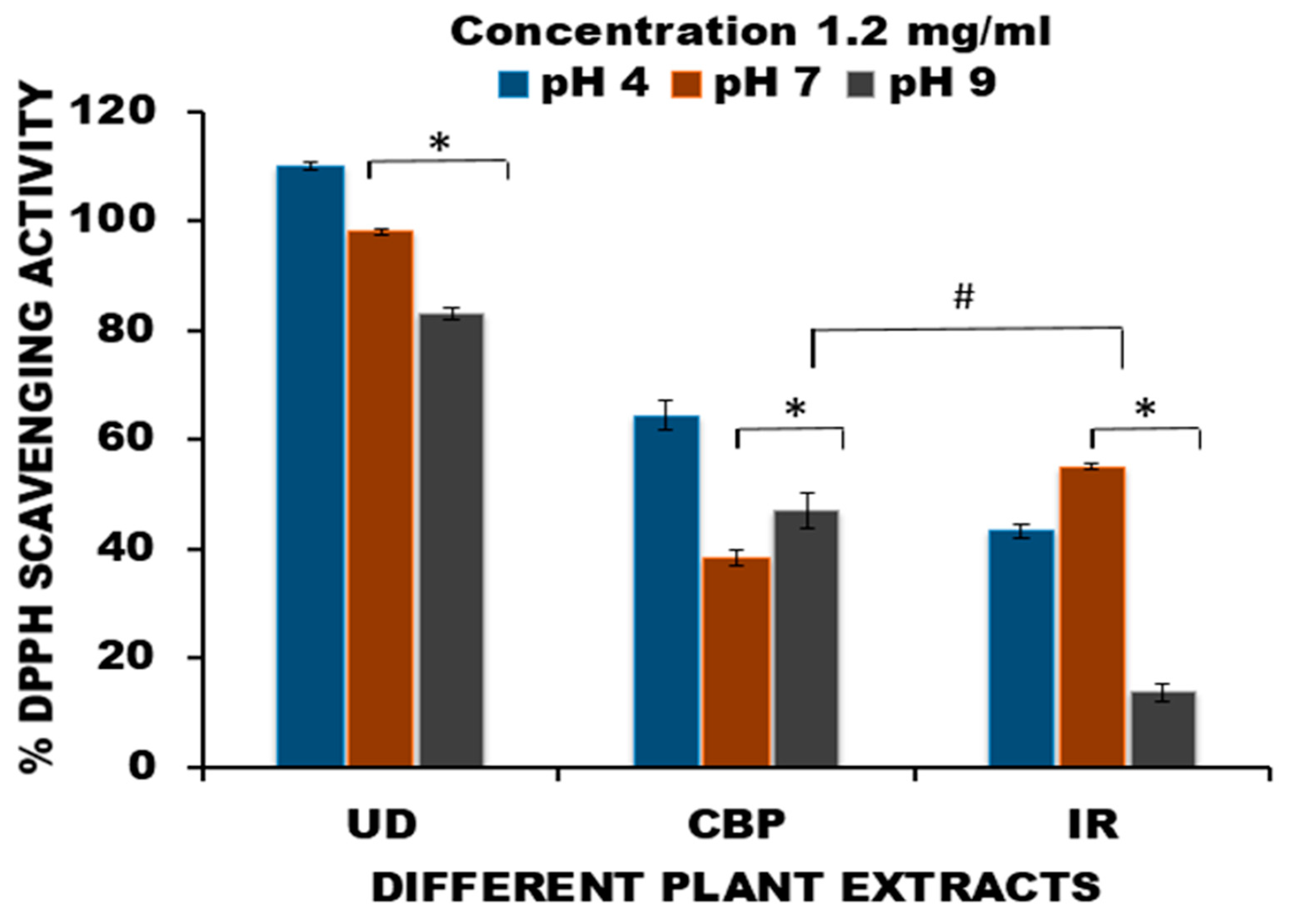
Figure 1. Effect of different pH values (4, 7 and 9) on the DPPH radical scavenging activity of aqueous extracts of Urtica dioica (UD), Capsella bursa-pastoris (CBP) and Inula racemosa (IR) at a concentration of 1.2 mg/mL. Data are represented as the mean ± SD for n = 3. * p < 0.05 (intragroup) and # p < 0.05 (intergroup) compared with pH 4 of the respective groups.
Applsci 13 03925 g001
Figure 2. Dose-effect plots of aqueous fractions of (a) Urtica dioica (UD), (b) Capsella bursa-pastoris (CBP), and (c) Inula racemosa (IR) at three different pH values (pH 4, pH 7, and pH 9) by DPPH radical scavenging assay. All the medicinal plants (UD, CBP, and IR) exhibited a dosage-dependent comportment. With the escalation in the dose, the % DPPH radical scavenging efficacy increased significantly. Data are presented as the mean ± SD for (n = 3). p < 0.05 was considered statistically significant.
Applsci 13 03925 g002
Figure 3. GC–MS chromatograms of the volatile fractions of herbal extracts of (A) leaves of Urtica dioica (UD), (B) whole plants of Capsella bursa-pastoris (CBP), and (C) rhizomes of Inula racemosa (IR).
Applsci 13 03925 g003
Figure 4. Effect of acetaminophen intoxication and pretreatment of medicinal plants Urtica dioica (UD), Capsella bursa-pastoris (CBP) and Inula racemosa (IR) along with acetaminophen treatment on body weight (gm) in mice. The control group is the untreated group fed only a standard pellet diet. The acetaminophen alone group is represented by APAP and APAP + UD, and APAP + CBP and APAP + IR represent groups of pretreated herbs intoxicated with acetaminophen overdose (600 mg/kg) on day 14. On day 14, body weight was monitored before APAP treatment (BT) and after treatment (AT). Significance is ascribed as * p < 0.05 vs. control. # = significance at p < 0.05 compared with APAP. Data represent the mean ± SD (n = 6).
Applsci 13 03925 g004
Figure 5. Sections of the Kidneys of APAP-treated Mice showing the Glomerulus (G) and Renal tubules (RT) and Bowman’s capsule (BC) with hematoxylin-eosin staining at 10× and 40× (AE) magnification (objective) (scale bar = 100 μm and 50 μm, respectively). (A) Control group; (B) APAP, 600 mg/kg BW; (C) APAP-intoxicated mice pretreated with UD: APAP (600 mg/kg BW) + UD (300 mg/kg BW); (D) APAP-intoxicated mice pretreated with CBP: APAP (600 mg/kg BW) + CBP (300 mg/kg BW); (E) APAP-intoxicated mice pretreated with IR: APAP (600 mg/kg BW) + IR (300 mg/kg BW).
Applsci 13 03925 g005
Figure 6. Total protein content in kidney tissue homogenate of male Swiss albino mice. Data represent the mean ± SD (n = 6). Significance is ascribed as * p < 0.05, ** p < 0.01, *** p < 0.001 vs. control. # = significance at p < 0.05, ## = significance at p < 0.01 and ### = significance at p < 0.001 compared with APAP. UD = Urtica dioica, CBP= Capsella bursa-pastoris and IR = Inula racemosa. The control group is the untreated normal group; APAP represents 600 mg/kg BW intoxication; UD is APAP-intoxicated mice pretreated with UD: APAP (600 mg/kg BW) and UD (300 mg/kg BW); CBP illustrates APAP-intoxicated mice pretreated with CBP: APAP (600 mg/kg BW) and CBP (300 mg/kg BW); and IR is APAP-intoxicated mice pretreated with IR: APAP (600 mg/kg BW) and IR (300 mg/kg BW).
Applsci 13 03925 g006
Disclaimer/Publisher’s Note: The statements, opinions and data contained in all publications are solely those of the individual author(s) and contributor(s) and not of MDPI and/or the editor(s). MDPI and/or the editor(s) disclaim responsibility for any injury to people or property resulting from any ideas, methods, instructions or products referred to in the content.