Article

Prevalence and Antibiotic Resistance of Enterococcus spp.: A Retrospective Study in Hospitals of Southeast Romania

by
Alina-Viorica Iancu
1,2,†,
Manuela Arbune
3,4,*,†,
Eliza-Andreea Zaharia
1,5,†,
Dana Tutunaru
6,7,†,
Nicoleta-Maricica Maftei
5,6,*,
Lucian-Daniel Peptine
1,5,†,
George Țocu
6,7,† and
Gabriela Gurău
1,5,†
1
Department of Morphological and Functional Sciences, Faculty of Medicine and Pharmacy, “Dunărea de Jos” University, 800008 Galați, Romania
2
Medical Laboratory Department, Clinical Hospital for Infectious Diseases “Sf. Cuvioasa Parascheva”, 800179 Galati, Romania
3
Clinical Medical Department, Faculty of Medicine and Pharmacy, “Dunărea de Jos” University, 800008 Galati, Romania
4
Infectious Diseases Department, Clinical Hospital for Infectious Diseases “Sf. Cuvioasa Parascheva”, 800179 Galati, Romania
5
Medical Laboratory Department, Clinical Hospital of Children Hospital “Sf. Ioan”, 800494 Galati, Romania
6
Department of Pharmaceutical Sciences, Faculty of Medicine and Pharmacy, “Dunărea de Jos” University, 800008 Galati, Romania
7
Medical Laboratory Department, “Sf. Apostol Andrei” Emergency Clinical Hospital, 800578 Galati, Romania
*
Authors to whom correspondence should be addressed.
These authors contributed equally to this work.
Appl. Sci. 2023, 13(6), 3866; https://doi.org/10.3390/app13063866
Submission received: 22 January 2023 / Revised: 13 March 2023 / Accepted: 15 March 2023 / Published: 17 March 2023
(This article belongs to the Special Issue Feature Review Paper in "Applied Microbiology" Section)

Abstract

:
Enterococci cause infections with various localizations, the most common being urinary infections. The purpose of the study was to identify the profile of the antimicrobial resistance of enterococci species (AMRE) isolated from patients hospitalized in three hospitals in Romania. We evaluated AMRE retrospectively (2019–2021) in various biological samples. The microbiological diagnosis was sustained by classical methods of bacteria culture and automatic identification. The sensitivity testing was performed by the Kirby–Bauer method, and the antibiotic minimum inhibitory concentration was tested by the automated Vitek system. We analyzed 86 strains of Enterococcus spp., identifying the following species: 47.7% E. faecalis, 47.7% E. faecium, 3.55% E. gallinarum, and 1% E. hirae. Most of the bacterial strains were isolated from urocultures (38.4%) and hemocultures (32.6%). Overall, the rate of vancomycin resistance was 5.8% for E. faecalis and 15.1%. for E. faecium. The prevalence of multidrug-resistant (MDR) strains was found to be 100% in E. gallinarum, 75.6% in E. faecium, and 21.9% in E. faecalis. The results confirm the high level of AMRE, which creates difficulties with adequate antibiotic prescriptions. The continuous monitoring of AMRE is essential for updating the local diagnostic and treatment protocols for enterococcal infections.

1. Introduction

The increasing incidence of health care-associated infections (HAI) has been attributed mainly to a group of six pathogenic germs, characterized by multidrug resistance (MDR) and virulence and known by the name ESKAPE: Enterococcus faecium, Staphylococcus aureus, Klebsiella pneumoniae, Acinetobacter baumannii, Pseudomonas aeruginosa, and Enterobacter spp. [1]. In the last 20 years, the incidence of enterococcal infections has increased. This pathogen has been reported the second most common pathogen associated with HAI, mainly in catheter-associated urinary infections and central-line associated bloodstream infections, both in Europe and USA [2]. Vancomycin-resistant enterococci (VRE) colonize the gastrointestinal tract and are frequently associated with Clostridioides difficile infection or colonization, implying interactions with the metabolic pathways [3,4,5].
Previous studies have identified several risk factors for VRE colonization, such as advanced age; severe underlying disease; inter-hospital transfer; home health care; prolonged hospitalization; central venous catheterization; hematological malignancies; solid organ allograft; chronic hemodialysis; exposure to antibiotics, multiple antibiotics, or high-risk antibiotics (vancomycin, third-generation cephalosporins, metronidazole); and longer duration of antibiotic exposure [6,7,8,9].
The increase in antibiotic resistance among enterococci, especially to vancomycin, has developed due to the selective pressure of antibiotics exposure, either by genetic mutation or horizontal gene transfer [2,10].
The increasing minimum inhibitory concentration (MIC) and slow tolerance to the β-lactams are genetically coded, but other acquired resistance mechanisms are also used against this antibiotic group, including plasmid production and transfer, antibiotic inactivation by β-lactamase, and point mutations in genes [11]. The MIC for penicillins is usually 2–8 µg/mL for E. faecalis and 8–16 µg/mL for E. faecium and is much higher than the MIC for other related Gram-positive bacteria [12]. Intrinsic resistance to aminoglycosides was attributable to enterococci, due to natural cell wall impermeability, while other resistance mechanisms include the plasmid transfer that codes the enzymes inactivating the antibiotic and the occurrence of point mutations in the ribosome [11].
Acquired resistance to glycopeptides (vancomycin), macrolides, tetracyclines, linezolid, and chloramphenicol exists. Enterococci resistance to lincosamides and streptogramins is acquired by genes coding a new ion pump, antibiotic inactivation by acetylation, or interference with the binding site on the 50S ribosome. Alteration of the ribosomal binding site is also induced by the acquired gene cfr, that is related to linezolid resistance [11,13].
The development of vancomycin resistance is connected to the synthesis of the modified precursors of peptidoglycan that give rise to a cluster of 11 genes that confer VRE. There are described two clusters of genes, van A, B, D, F, I, M and van C, E, G, L, N, depending on the ending sequence of the peptidoglycan precursors [14].
In practice, there are two main phenotypes of glycopeptide resistance. The first one is VanA, with a high level of resistance to vancomycin and a variable level of resistance to teicoplanin, and the second is VanB, with a variable level of resistance, mainly to vancomycin [2]. The reported nosocomial VRE enterococcal isolates range from 19.1 to 25% of the total enterococcal isolates; they are related to increased morbidity and mortality rates in cases of enterococcal bacteremia, with longer hospitalization and increased in-hospital health care costs, compared with vancomycin-susceptible enterococci [15,16,17,18,19,20,21,22,23,24,25]. HAI are mainly caused by two species: E. faecium (85–90%) and E. faecalis (5–10%) [26]. Most VRE are strains of E. faecium. Characteristic of both E. faecium and E. faecalis is the reduced susceptibility to antibiotics that are commonly active for other Gram-positive cocci and the intrinsic resistance to cephalosporins, amynoglicosides, clindamycin, and trimethoprim-sulphamethoxazole [2].
The reports on AMRE are limited in Romania, although it is recognized as a clinical and epidemiological health problem.
The latest surveillance data on antimicrobial consumption in Romania have shown that the most used antibiotics overall are: beta-lactam penicillines (41.92%), other beta-lactam antibiotics (18.32%), macrolides, lincosamides and streptogramines (18.44%), and quinolones (12.98%) [27].
There are no previous studies on AMRE from the southeast of Romania. However, some related data are provided by the joint report of the European Centre for Disease Prevention and Control (ECDC) and the WHO Regional Office for Europe published in 2022; the report contains AMRE data on invasive isolates in Europe in 2020 and includes information from 13 Romanian labs. The most important AMRE problems in Romania are E. faecalis resistance to gentamicin 29% gentamicin and E. faecalis resistance to vancomycin 16.8%, compared with reported data in other European countries, of 4.1–51.6%, respective 0.0–56.6% [28].
The aim of the study was to identify the antibiotic resistance profile of enterococci species isolated in hospitalized patients from Romania.

2. Materials and Methods

2.1. Samples Collection

The study was a retrospective one on antibiotic resistance among Enterococcus spp. strains. The data of this study were collected from January 2019 to December 2021 at the Emergency Clinical Hospital “Sf. Apostol Andrei”, the Clinical Hospital of Children “Sf. Ioan”, and the Clinical Hospital of Infectious Diseases “Sf. Cuvioasa Parascheva”, in Galati, Romania. Samples from patients with an infection caused by Enterocuccus spp. were included. The samples included hemocultures, urine cultures, purulent secretions from wounds or ulcers of the foot, puncture fluids, catheters, and otic and conjunctival secretions. The treatment of the biological samples was carried out according to the routine identification guidelines of clinical bacteriology, briefly described below. The clinical samples were immediately transported to the bacteriology laboratory and were processed within one hour of sampling.

2.2. Identification of Bacteria

Bacterial culture and biochemical identification of the bacterial strains were carried out according to the classical methodology [29]. The hemocultures were incubated in an automated blood culture-monitoring (BacT/ALERT and Bactec FX 40) blood culture system for up to 5 days, using hemoculture vials with a special nutrient medium whose composition favors the development of aerobic, anaerobic, and microaerophilic microorganisms. Subsequently, one drop from each positive bottle was plated on standard bacteriological media: chocolate agar, Columbia blood agar, Levine agar/MacConkey agar, and Sabouraud agar (bioMérieux, Marcy-l’Étoile, France, Oxoid-Thermo Scientific, Waltham, MA, USA). Other biological samples, such as urine, purulent secretions from wounds or ulcers of the foot, puncture fluids, catheters, and otic and conjunctival secretions, were inoculated on complex and selective solid media, using the technique of inoculation in the sector and by exhaustion. All plates were incubated at 37 °C for 18–24 h. Biochemical identification of bacterial isolates was carried out by the Vitek 2 Compact Automated System, using an identification card (ID-GP), according to the manufacturer’s instructions.

2.3. Antimicrobial Susceptibility Testing

Antibiotic susceptibility testing was determined by the Kirby–Bauer diffusimetric method on the Mueller–Hinton standardized medium and the minimum inhibitory concentration method obtained with the Vitek automated system, using a susceptibility card (AST-592) [30,31,32]. Each bacterial strain suspension was prepared from pure cultures of bacteria cultivated on plates. The bacterial cells were suspended in 3 mL of a sodium chloride solution. The suspension used in the VITEK 2C system was adjusted to a 0.5 McFarland standard, using a Densicheck (bioMérieux, Marcy-l’Étoile, France). The results of the antimicrobial susceptibility were interpreted according to the Clinical Laboratory Standard Institute [30,31,32,33]. The reference strains used for quality control in the identification and antibiotic sensitivity testing were S. aureus ATCC 25923; ATCC 29213 (quality control for disk diffusions and MIC, according to CLSI and VITEK 2C technical insert); and Enterococcus casseliflavus ATCC 700327 (quality control for identification, according to VITEK 2C technical insert). Some antibiotics were tested depending on the type of sample (nitrofurantoin and norfloxacin for testing and reporting of urinary tract isolates only), in accordance with the CLSI recommendations. Isolates of Enterococcus spp. were systematically tested with penicillins (penicillin-10 U, ampicillin-10 µg), fluoroquinolones (ciprofloxacin-5 µg, norfloxacin-10 µg), macrolides (erythromycin-15 µg), nitrofurans (nitrofurantoin-300 µg), high-level aminoglycosides (gentamicin-120 µg, streptomycin-300 µg), glycopeptides (vancomycin-30µg), and oxazolidinones (linezolid-30 µg). For the diffusimetric method, Oxoid Antimicrobial Susceptibility Disks, Thermo Scientific, from the USA, were used. In 2019, the samples were tested using the diffusimetric method, and since 2020, the testing has been conducted according to MIC. Antibiogram results were classified as susceptible (S), intermediate (I) or resistant (R). MDR strains were identified according to the European Center for Disease Control (ECDC) [28,34] as being resistant to at least one agent from at least three antimicrobial categories [28]. Depending on the clinical significance and the demands of the clinician, several MDR strains, such as vancomycin, teicoplanin, and linezolid, were additionally tested for backup antibiotics. Glycopeptide resistance is mediated by two phenotypes: VanA, with high levels of vancomycin resistance and a variable level of teicoplanin resistance in three hospitals, and VanB, with a variable level of resistance, which was limited to vancomycin in most cases. Only the acquired antimicrobial resistance was accounted for, not the intrinsic one.

2.4. Exclusion Criteria of Bacterial Isolates

Duplicates, isolates from contaminated samples, and incompletely identified strains of the enterococcal strains were excluded. We considered contaminated biological samples when there were discordant results between the complete urine examination and the uroculture.

2.5. Statistical Analysis

The results were collected from the databases for common monitorization of antibiotic resistance in three hospitals, and were statistically processed by Microsoft XL software. We used descriptive statistics, according to frequency distribution. Pearson’s chi-square test for independence was used to compare the data from the three hospitals, with a level of p < 0.001 considered to be significant.

3. Results

3.1. Prevalence of Enterococcus spp. from Hospital Bacterial Isolates

A total of 165,098 bacterial cultures, with 26,699 isolated bacterial strains (16.15%), were processed between 2019 and 2021, from three hospitals in Galati, Romania.
The number of microbiological samples decreased by 44.1% from 2019 to 2021. We also noticed a reduction in the diversity of the biological samples and bacterial isolates due to the context of the long period of dedication of hospital care to COVID-19 (Figure A1). The ratio between Gram-positive cocci and Gram-negative rods was similar in each of the three years of the study. The frequency of Staphylococcus species reported to be Gram-positive cocci trend to increase, from 67.9% to 80.9% (Table A1).
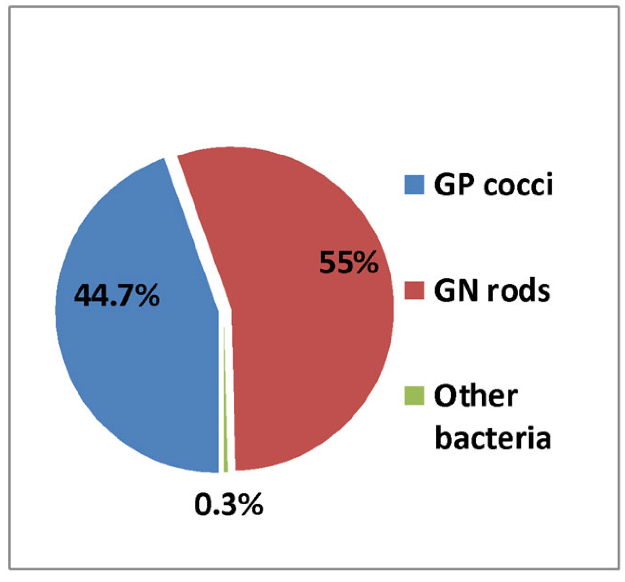
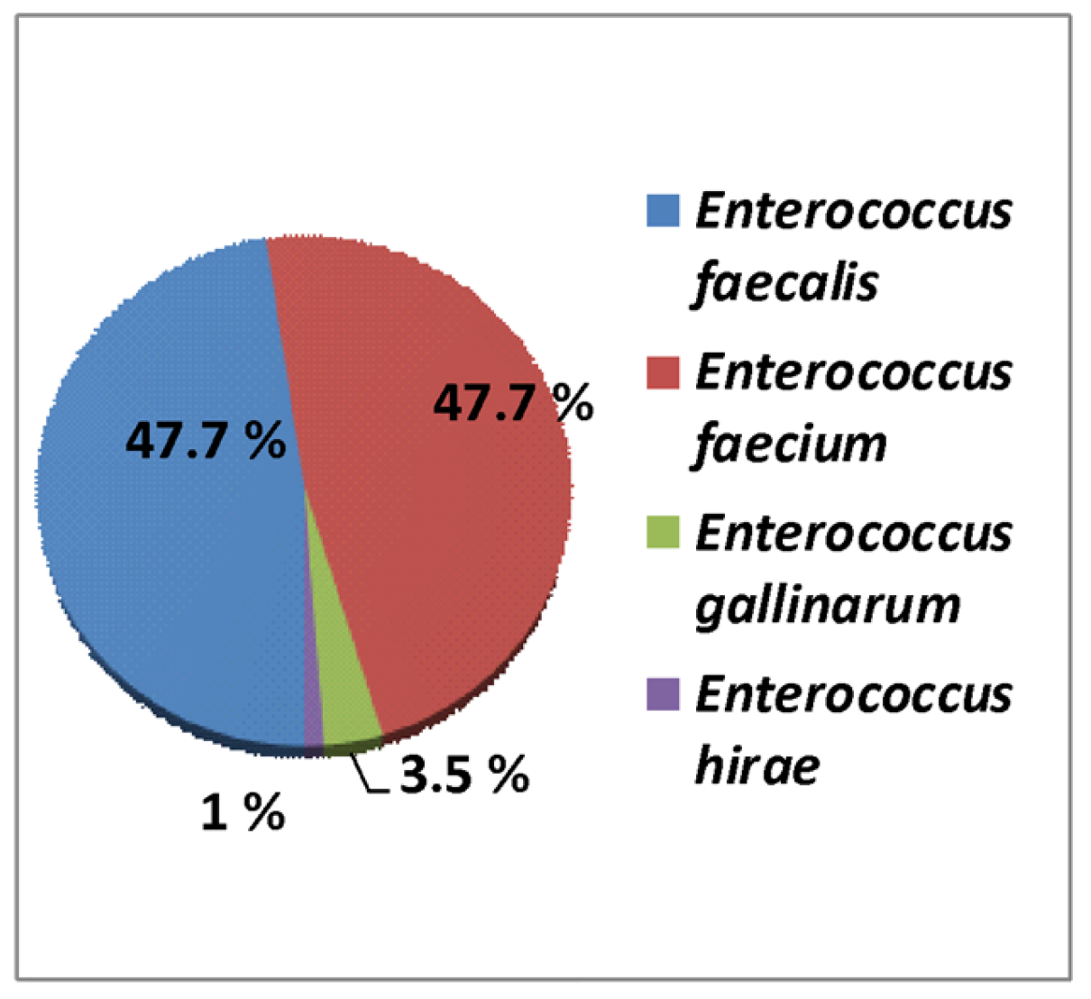
The isolated clinically significant bacterial strains were mainly grouped into 55% Gram-negative bacilli and 44.7% Gram-positive cocci (Figure A2). From the Gram-negative bacilli group, the first place was held by Enterobacteriaceae: E. coli 27.4%, Klebsiella spp. 14.7% (K. pneumoniae, K. oxytoca), Proteus spp. 3.4% (P. mirabilis, P. vulgaris), Enterobacter spp. 1.37% (E. cloacae, E. aerogenes), Salmonella spp. 0.8%, and other Enterobacterales 0.16% (Serratia spp., Citrobacter spp., Morganella spp., Providencia spp.). Non-fermentative Gram-negative bacilli were less frequent, including Pseudomonas spp. 6.03% (P. aeruginosa, P. putida, P. fluorescens, P. stutzeri), and Acinetobacter spp. 1.2% (A. baumannii, A. haemolyticus). Regarding the Gram-positive cocci, the leading strain was Staphylococcus spp. 32.8% (S. aureus, coagulase-negative Staphylococcus), followed by Streptococcus spp. (β- Hemolytic group 5.5%, Streptococcus pneumoniae 5.08%), and Enterococcus spp. 0.86% (Table A1). Among the Gram-positive cocci, after the excluded duplicates, contaminates, and incompletely identified species, we found 86 strains of Enterococcus spp., meaning 0.32%. The distribution of the isolated species was evidenced by 47.7% E. faecalis (41 strains), 47.7% E. faecium (41 strains), 3.55% E. gallinarum (three strains), and 1% E. hirae (one strain) (Figure A3).
The biological samples positive for enterococci consisted of 38.4% (33) urocultures, 32.6% hemocultures (28), 22% purulent secretions of leg ulcers or other skin lesions (19), and 7% others (6), including conjunctival and otic secretions. (Figure 1). The urine isolates were 18 species of E. faecium and 15 strains of E. faecalis. From 28 positive hemocultures, the following species were identified: 13 E. faecium, 11 E. faecalis, 3 E. gallinarum, and 1 E. hirae.

3.2. Antibiotic Resistance of Enterococcus spp.

The E. faecium strains revealed 80.5% ampicillin resistance and a high level of aminoglycosides resistance (HLAR), with 41.1% to gentamicin and 43.8% to streptomycin, respectively, 41.9% ciprofloxacin resistance, 54.7% erythromycin resistance, 24.2% tetracycline resistance, and 10% penicillin resistance. The antimicrobial resistance to E. faecalis was lower than that to E. faecium, especially for ampicillin (7.3%), but the higher resistance to tetracycline (31.8% vs. 24.2%) and penicillin (20% vs. 10%) was uncommon. Ciprofloxacin resistance was 41.9% in either E. faecalis or E. faecium. Both species of enterococci were 98.8% sensitive to linezolid. Testing for nitrofurantoin and norfloxacin was inconsistent and was achieved in 10 strains of E. faecalis originating from urinary infections, with only one nitrofurantoin-resistant strain.
Regarding the glycopeptide resistance, we found the VanA phenotype in 12% of E. faecalis and in 26% of E. faecium species, while the VanB phenotype was less frequent, in 5.8% of E. faecalis and 5% of E. faecium (Figure 2).
The rate of MDR was 75.6% in E. faecium and 21.9% in E. faecalis. There ware only three strains of E. gallinarum, all of them isolated from hemoculture, but they had the remarkable MDR of 100%. Additionally, the three species of E. gallinarum were resistant to ampicillin and ciprofloxacin, whereas two strains expressing the phenotype VanA. E. hirae were also isolated from hemoculture, but conversely, they were sensitive to all the antimicrobial tested agents (Table 1).
The age of the patients hospitalized with enterococcal infections ranged between 0 and 98 years, with an average age of 52.11 ± 30.46 years. There was no significant correlation of age with gender, living area, antibiotic multidrug resistance, or the vancomycin resistance pattern (Table 2).
Considering the isolates from hemocultures, the prevalence of MDR was 75%, though the E. faecalis/faecium isolates preserved sensitivity to vancomycin, teicoplanin, and linezolid.

3.3. Comparative of Enterococcus spp. in Three Hospitals of Southeast Romania

The structure of Emergency County Hospital Galati (EMC) is complex, with various departments, including intensive care and surgical units, which are related to the highest proportion of enterococcal strains (48/83 cases) as well as the most frequent blood samples from invasive infections (26/29) (Table 2).
Lower MDR and VAN-R were found in IDH.
Skin and soft tissue infections are the third cause of enterococcal isolates and were reported in all three hospitals (Table 2).
Significant differences were not found between the three hospitals with regard to the impact of COVID-19 on enterococcal isolates. However, the number of cases increased in ECH during 2021 compared with the pre-pandemic year of 2019, and a lower number of enterococcal isolates were found in the first pandemic year of 2020 in all three hospitals.
The most alarming antimicrobial resistance problems were found in ECH with regard to the MDR strains (34/43 cases) and Van-R strains (16/20 cases), signaling the highest use of antibiotics, the need of control methods for antimicrobial prescriptions, and the risk of spreading the antibiotic resistance to the other local hospitals and the community.

4. Discussion

Enterococci belong to the normal bacterial microbiota of the gastrointestinal tract. Although they are characterized by low virulence, the development of bacteremia, endocarditis, and peritonitis are the consequence of decreased intestinal defense mechanisms [28,35]. They have been of particular concern in recent years due to their ability to spread easily in hospitals among health workers and hospitalized patients and are included among the main microorganisms that cause nosocomial infections [36,37,38]. In the present study, similar percentages of 47.7% E. faecalis (41 strains) and E. faecium (41 strains) were isolated from the clinical samples.
Bacteremia with enterococci is mainly nosocomial, with the community ones signaling the suspicion of endocarditis. In the U.S., three out of four nosocomial bacteremias per 10,000 hospitalizations are caused by enterococci and increase the risk of death [38]. In a recent retrospective study from China, the incidence of enterococcus in the blood infections (BSI) of hospitalized patients was four cases per 10,000 hospitalizations, with the main isolated pathogen being E. faecium (74%) [39,40]. Similar studies suggest that nosocomial BSI with enterococci is increasing and that overall mortality is quite high, ranging from 25% to 50% [41]. Many studies have reported that bacteremia caused by VRE strains leads to higher mortality rates (2.5-fold increase), compared to bacteremia caused by vancomycin-sensitive strains [42,43]. In our study, 32.6% were isolated from hemocultures, with a frequency of 46.4% for E. faecium and 39.2% for E. faecalis.
More common are infections located in the urinary tract (38.4%) and soft tissue infections (22%). According to a similar study, the maximum number of isolates was obtained from urine (46.6%), followed by purulent secretions (19.4%) [44]. These data highlight the prevalence of enterococci in urinary tract infections (UTIs). Many studies have shown the association between biofilm production and urinary catheters in persistent infections [45,46]. In these cases, UTI treatment involves the use of broad-spectrum antibiotics, which are the main cause of the spread of VRE strains.
Another major problem associated with Enterococcus spp. infections, along with the increased incidence rate, is the increased resistance to antimicrobial agents [47,48]. Enterococci have intrinsic resistance to several categories of antibiotics, such as cephalosporins, sulfonamides, and low concentrations of aminoglycosides [28,35]. In enterococcal infections, the success of treatment depends essentially on the ability to acquire new resistance markers through horizontal transfer mediated by plasmids or transposons and through genetic recombination or mutations. Antimicrobial resistance (AMR) is a health threat to millions of people worldwide. The COVID-19 pandemic has exposed the weaknesses of national health systems and the interconnection between countries and continents [28]. The pandemic years had a particular profile in terms of antibiotic resistance through the isolation of a small number of bacteria. In the present study, resistance to ampicillin and gentamicin HL was 7.3% vs. 30.1% for E. faecalis and 80.5% vs. 41.1% for E. faecium. According to EARS-Net data, at the European level, high-level resistance to aminoglycosides (HLAR) in strains of E. faecalis from invasive infections is increasing and was 29% in 2020. In 2020, Romania reported a resistance to gentamicin HL of 43.2%, up from 2019 [2,36]. Enterococci also have a low susceptibility to many β-lactam agents due to their decreasing affinity for penicillins binding proteins that bind penicillin. However, there is typically synergy between aminoglycosides and penicillins or glycopeptides against enterococci without the acquiring of high-level resistance to glycopeptides. Some enterococci have acquired genes conferring high-level resistance to aminoglycosides, causing the loss of any synergistic effect between beta-lactams and aminoglycosides [2].
In our study, the overall prevalence of VRE appears to be lower than the national reports, but the small number of analyzed strains does not support adequate statistical analysis. We obtained 5.8% VRE for E. faecalis and 15.1% for E. faecium. According to the European surveillance data, E. faecium showed a decreasing trend; the VRE was 18.3% in 2019 and 16.8% in 2020, but the data are limited to bloodstream infections. According to the available data, VRE in Romania is still one of the highest in Europe and continues to increase, from 35.7% in 2019 to 39.3% in 2020. AMRE percentages equal to or greater than 50% were found in Bosnia and Herzegovina, Lithuania, North Macedonia, and Serbia [28]. Maintaining the high levels of glycopeptide resistance of E faecium requires the adoption of measures to limit the human transmission of germs and the judicious use of glycopeptides [2]. The rapid spread of VRE has led to the use of new antibiotics such as linezolid and teicoplanin. The resistance to linezolid was 1.16% for both species, and for teicoplanin, 5% was obtained for E. faecalis and 12.5% for E. faecium. The highest prevalence of MDR strains was found in E. gallinarum (100%) and E. faecium (75.6%), followed by E. faecalis (21.9%,), which is in concordance with distribution of species in the other studies [2,36]. The AMRE strains evaluated in this study reflect their prevalence and persistence in the community. The surveillance of MDR strains should be developed by increasing the accuracy of the identification methods used, implementing EUCAST standards, and increasing clinical vigilance for infectious diagnosis and the justified use of antibiotics.
The rapid spread of VRE has led to the requirement for new antibiotics, such as linezolid and teicoplanin. Nevertheless, the local resistance to linezolid and to teicoplanin is low; however, the higher MDR of E. faecium and E. gallinarum are worrying for the clinical practice. If ampicillin remains the first choice for the treatment of community infections with E. faecalis, when it is involved E. faecium and other Enterococcus spp., more data must be analyzed for the first line of therapeutical decision, considering the local antibiotic resistance situation, the source of infection, the localization and severity of infection, and the individual profile of the patient. The surveillance of AMR and MDR strains must be developed by increasing the accuracy of the identification methods used, by implementing the EUCAST standards, and by increasing clinical vigilance for infectious diagnosis and applying the principles of antibiotic stewardship in clinical practice.

Limits of the Study

The small number of enterococcal strains included in the study was related to the pandemic context when the hospitals were crowded with patients with COVID-19 and the recommendations for microbiological investigations were limited. Moreover, the hesitance regarding hospitalization was frequently found in patients with other medical problems, which could influence the reliability of the statistical results (Table A1). The small sample size affects the reliability of the statistical analysis. Other limits are the retrospective type of study and the inconsistent availability of reagents for the Vitek system bacterial identification, due to financial problems.

5. Conclusions

The frequency of Enterococcus spp. isolated from hospitalized patients decreased during the first two years of the COVID-19 pandemic. The nature of the positive biological samples, identified enterococcal species, and antibiotic resistance are different in the hospitals from the same geographical and administrative region of Romania, which is associated with the distinctive profiles of the departments and local medical practice routines. The main resistance challenges are MDR and VRE, mainly in E. faecium isolates from blood stream infections. The development of local antibiotic stewardship programmes needs to consider the AMRE surveillance and the risk of transferring germs from one hospital to another or to the community. Development of the bacterial identification techniques is necessary for the improvement of the etiologic diagnosis of hospitalized infection. The use of molecular investigations and the identification of the resistance genes are required for earlier microbiological diagnosis and appropriate therapeutic decisions.

Author Contributions

Conceptualization, G.G., M.A., D.T. and A.-V.I.; methodology, G.G., A.-V.I. and D.T.; software, E.-A.Z., A.-V.I. and G.Ț.; validation, N.-M.M., E.-A.Z. and M.A.; formal analysis, E.-A.Z., N.-M.M. and L.-D.P.; investigation, L.-D.P., N.-M.M., A.-V.I. and M.A.; resources, G.G., D.T. and A.-V.I.; data curation, E.-A.Z., N.-M.M. and M.A.; writing—original draft preparation, A.-V.I. and D.T.; writing—review and editing, M.A. and G.G.; visualization, M.A., G.G., G.Ț., and N.-M.M.; supervision, G.G., D.T. and M.A. All authors have read and agreed to the published version of the manuscript.

Funding

This research received no external funding.

Institutional Review Board Statement

The study did not require ethical approval. The consent was given as our retrospective study used laboratory management which regularly collected data from the databases.

Informed Consent Statement

Not applicable.

Data Availability Statement

Not applicable.

Acknowledgments

The authors acknowledge the research staff of “Dunarea de Jos” University” Galati for the financial support for the publishing of our study.

Conflicts of Interest

The authors declare no conflict of interest.

Appendix A

Figure A1. Annual evolution of the bacterial strain isolates and number of the biological samples in Galati, Romania (2019–2021).
Applsci 13 03866 g0a1
Figure A2. Distribution of types of microorganisms.
Applsci 13 03866 g0a2
Figure A3. The distribution of Enterococcus spp.
Applsci 13 03866 g0a3
Table A1. Annual Frequency of Group Species.
Genera2019No—2020No—2021Total%
GP CocciNo%No%No%
Staphylococcus spp.370167.9%212877.6%292680.71%875532.8%
Streptococcus spp. β-Hemolytic group81214.9%33012%3258.96%14675.5%
Streptococcus pneumoniae82615.16%2258.2%3078.46%13585.08%
Enterococcus spp.1061.94%572.08%671.84%2300.86%
Total5445100%2740100%3625100%11,81044.24%
GN rods
Escherichia coli332554.6%169146.4%230146.4%731727.4%
Klebsiella spp.132821.8%106829.3%152630.8%392214.7%
Proteus spp.4006.6%2206.03%2785.6%8983.4%
Enterobacter spp.2103.45%802.19%781.57%3681.37%
Salmonella spp.911.5%441.2%741.5%2090.8%
Alte Enterobacterales200.32%90.24%140.28%430.16%
Pseudomonas spp.62410.2%4379.52%54911%16106.03%
Acinetobacter spp.881.4%952.6%1392.8%3221.2%
Total6086100%3644100%4959100%14,68955.1%
Other bacteria6378481890.7%
Total11,5946473863226,699100%
Note: % annual frequency.

References

  1. Rice, L.B. Federal Funding for the Study of Antimicrobial Resistance in Nosocomial Pathogens: No ESKAPE. J. Infect. Dis. 2008, 197, 1079–1081. [Google Scholar] [CrossRef] [PubMed]
  2. García-Solache, M.; Rice, L.B. The Enterococcus: A Model of Adaptability to Its Environment. Clin. Microbiol. Rev. 2019, 32, e00058-18. [Google Scholar] [CrossRef] [PubMed] [Green Version]
  3. Monticelli, J.; Di Bella, S.; Giacobbe, D.R.; Amato, G.; Antonello, R.M.; Barone, E.; Brachelente, G.; Busetti, M.; Carcione, D.; Carretto, E.; et al. Trends in the Incidence and Antibiotic Resistance of Enterococcal Bloodstream Isolates: A 7-Year Retrospective Multicenter Epidemiological Study in Italy. Microb. Drug Resist. 2021, 27, 529–535. [Google Scholar] [CrossRef]
  4. Monticelli, J.; Di Bella, S.; Giacobbe, D.R.; Amato, G.; Antonello, R.M.; Barone, E.; Brachelente, G.; Busetti, M.; Carcione, D.; Carretto, E.; et al. Enterococci enhance Clostridioides difficile pathogenesis. Nature 2022, 611, 780–786. [Google Scholar] [CrossRef]
  5. Stevens, V.W.; Khader, K.; Echevarria, K.; E Nelson, R.; Zhang, Y.; Jones, M.; Timbrook, T.T.; Samore, M.H.; A Rubin, M. Use of Oral Vancomycin for Clostridioides difficile Infection and the Risk of Vancomycin-Resistant Enterococci. Clin. Infect. Dis. 2020, 71, 645–651. [Google Scholar] [CrossRef]
  6. Tokars, J.I.; Satake, S.; Rimland, D.; Carson, L.; Miller, E.R.; Killum, E.; Sinkowitz-Cochran, R.L.; Arduino, M.J.; Tenover, F.C.; Marston, B.; et al. The prevalence of colonization with vancomycin-resistant Enterococcus at a Veterans’ Affairs institution. Infect. Control Hosp. Epidemiol. 1999, 20, 171–175. [Google Scholar] [CrossRef] [PubMed]
  7. Harbarth, S.; Samore, M.H.; Lichtenberg, D.; Carmeli, Y. Prolonged antibiotic prophylaxis after cardiovascular surgery and its effect on surgical site infections and antimicrobial resistance. Circulation 2000, 101, 2916–2921. [Google Scholar] [CrossRef] [Green Version]
  8. DeLisle, S.; Perl, T.M. Vancomycin-resistant enterococci: A road map on how to prevent the emergence and transmission of antimicrobial resistance. Circulation 2003, 123, 504S–518S. [Google Scholar]
  9. Padiglione, A.A.; Wolfe, R.; Grabsch, E.A.; Olden, D.; Pearson, S.H.; Franklin, C.; Spelman, D.; Mayall, B.; Johnson, P.D.R.; Grayson, M.L. Risk factors for new detection of vancomycin-resistant enterococci in acute-care hospitals that employ strict infection control procedures. Antimicrob. Agents Chemother. 2000, 47, 2492–2498. [Google Scholar] [CrossRef] [Green Version]
  10. Edmond, M.B.; Wallace, S.E.; McClish, D.K.; Pfaller, M.A.; Jones, R.N.; Wenzel, R.P. Nosocomial bloodstream infections in United States hospitals: A three-year analysis. Clin. Infect. Dis. 1999, 29, 239–244. [Google Scholar] [CrossRef] [Green Version]
  11. Růžičková, M.; Vítězová, M.; Kushkevych, I. The characterization of Enterococcus genus: Resistance mechanisms and inflammatory bowel disease. Open Med. 2020, 15, 211–224. [Google Scholar] [CrossRef] [Green Version]
  12. Hollenbeck, B.L.; Rice, L.B. Intrinsic and acquired resistance mechanisms in Enterococcus. Virulence 2012, 3, 421–433. [Google Scholar] [CrossRef] [Green Version]
  13. Guffey, A.A.; Loll, P.J. Regulation of Resistance in Vancomycin-Resistant Enterococci: The VanRS Two-Component System. Microorganisms 2021, 9, 2026. [Google Scholar] [CrossRef]
  14. Aqib, A.I.; Alsayeqh, A.F. Vancomycin drug resistance, an emerging threat to animal and public health. Front. Vet. Sci. 2022, 28, 1010728. [Google Scholar] [CrossRef]
  15. Cooper, E.; Paull, A.; O’Reilly, M. Characteristics of a large cluster of vancomycin-resistant enterococci in an Australian hospital. Infect. Control. Hosp. Epidemiol. 2002, 23, 151–153. [Google Scholar] [CrossRef]
  16. Centers for Disease Control and Prevention. National nosocomial infections surveillance (NNIS) system report, data summary from January 1992–April 2000. Am. J. Infect. Control 2000, 28, 429–448. [Google Scholar] [CrossRef]
  17. Murray, B.E. Vancomycin-resistant enterococcal infections. N. Engl. J. Med. 2000, 342, 710–721. [Google Scholar] [CrossRef]
  18. Brinkwirth, S.; Ayobami, O.; Eckmanns, T.; Markwart, R. Hospital-acquired infections caused by enterococci: A systematic review and meta-analysis, WHO European Region, 1 January 2010 to 4 February 2020. Euro. Surveill. 2021, 26, 2001628. [Google Scholar] [CrossRef] [PubMed]
  19. Hemapanpairoa, J.; Changpradub, D.; Santimaleeworagun, W. Clinical Impact of Vancomycin Treatment in Ampicillin-Susceptible Enterococci Bloodstream Infections. Antibiotics 2022, 11, 1698. [Google Scholar] [CrossRef] [PubMed]
  20. Janjusevic, A.; Cirkovic, I.; Minic, R.; Stevanovic, G.; Soldatovic, I.; Mihaljevic, B.; Vidovic, A.; Markovic Denic, L. Predictors of Vancomycin-Resistant Enterococcus spp. Intestinal Carriage among High-Risk Patients in University Hospitals in Serbia. Antibiotics 2022, 11, 1228. [Google Scholar] [CrossRef] [PubMed]
  21. Büchler, A.C.; Ragozzino, S.; Wicki, M.; Spaniol, V.; Jäger, S.; Seth-Smith, H.M.B.; Goldenberger, D.; Hinic, V.; Egli, A.; Frei, R.; et al. Patients exposed to vancomycin-resistant enterococci during in-hospital outbreaks in a low endemic setting: A proposal for risk-based screening. Antimicrob. Resist. Infect. Control 2022, 11, 60. [Google Scholar] [CrossRef]
  22. Beale, J.W.; Durward-Diioia, M. Clinical Considerations in the Approach to Vancomycin-Resistant Enterococci: A Narrative Review. Int. J. Med. Students 2022, 10, 202–209. [Google Scholar] [CrossRef]
  23. Todorić, Z.; Majdandžić, I.; Kregar, T.K.; Herljević, Z.; Ćorić, M.; Lešin, J.; Kuliš, T. Increasing trend in enterococcal bacteraemia and vancomycin resistance in a tertiary care hospital in Croatia, 2017–2021. Infect. Dis. 2022, 55, 9–16. [Google Scholar] [CrossRef] [PubMed]
  24. DiazGranados, C.A.; Zimmer, S.M.; Klein, M.; Jernigan, J.A. Comparison of mortality associated with vancomycin-resistant and vancomycin-susceptible enterococcal bloodstream infections: A meta-analysis. Clin. Infect. Dis. 2005, 41, 327–333. [Google Scholar] [CrossRef] [PubMed] [Green Version]
  25. Correa-Martinez, C.L.; Tönnies, H.; Froböse, N.J.; Mellmann, A.; Kampmeier, S. Transmission of Vancomycin-Resistant Enterococci in the Hospital Setting: Uncovering the Patient-Environment Interplay. Microorganisms 2020, 8, 203. [Google Scholar] [CrossRef] [Green Version]
  26. Ramos, S.; Silva, V.; Dapkevicius, M.d.L.E.; Igrejas, G.; Poeta, P. Enterococci, from Harmless Bacteria to a Pathogen. Microorganisms 2020, 8, 1118. [Google Scholar] [CrossRef]
  27. Zaheer, R.; Cook, S.R.; Barbieri, R.; Goji, N.; Cameron, A.; Petkau, A.; Polo, R.O.; Tymensen, L.; Stamm, C.; Song, J.; et al. Surveillance of Enterococcus spp. reveals distinct species and antimicrobial resistance diversity across a One-Health continuum. Sci. Rep. 2020, 10, 3937. [Google Scholar] [CrossRef] [PubMed] [Green Version]
  28. European Centre for Disease Prevention and Control. Antimicrobial Resistance Surveillance in Europe 2022. Antimicrobial Resistance in the EU/EEA (EARS-Net)—Annual Epidemiological Report for 2020. Available online: https://www.ecdc.europa.eu/en/publications-data/antimicrobial-resistance-eueea-ears-net-annual-epidemiological-report-2020 (accessed on 10 September 2022).
  29. Procop, G.W.; Church, D.L.; Hall, G.S.; Janda, W.M. Koneman’s Color Atlas and Textbook of Diagnostic Microbiology, 7th ed.; Lippincott Williams & Wilkins: Philadelphia, PA, USA, 2017. [Google Scholar]
  30. Clinical and Laboratory Standards Institute. Performance Standards for Antimicrobial Susceptibility Testing: 29th Informational Supplement, CLSI Document M100-S29; Clinical and Laboratory Standards Institute: Wayne, PA, USA, 2019; Available online: https://clsi.org/media/2663/m100ed29_sample.pdf (accessed on 12 January 2019).
  31. Clinical and Laboratory Standards Institute. Performance Standards for Antimicrobial Susceptibility Testing: 30th Informational Supplement, CLSI Document M100-S30; Clinical and Laboratory Standards Institute: Wayne, PA, USA, 2020; Available online: https://clsi.org/media/3481/m100ed30_sample.pdf (accessed on 25 January 2020).
  32. Clinical and Laboratory Standards Institute. Performance Standards for Antimicrobial Susceptibility Testing: 31th Informational Supplement, CLSI Document M100-S31; Clinical and Laboratory Standards Institute: Wayne, PA, USA, 2021; Available online: https://clsi.org/media/z2uhcbmv/m100ed31_sample.pdf (accessed on 8 January 2021).
  33. Gajic, I.; Kabic, J.; Kekic, D.; Jovicevic, M.; Milenkovic, M.; Mitic Culafic, D.; Trudic, A.; Ranin, L.; Opavski, N. Antimicrobial Susceptibility Testing: A Comprehensive Review of Currently Used Methods. Antibiotics 2022, 11, 427. [Google Scholar] [CrossRef] [PubMed]
  34. Magiorakos, A.P.; Srinivasan, A.; Carey, R.B.; Carmeli, Y.; Falagas, M.E.; Giske, C.G.; Harbarth, S.; Hindler, J.F.; Kahlmeter, G.; Olsson-Liljequist, B.; et al. Multidrug-resistant, extensively drugresistant and pandrug-resistant bacteria: An international expert proposal for interim standard definitions for acquired resistance. Clin. Microbiol. Infect. 2012, 18, 268–281. [Google Scholar] [CrossRef] [PubMed] [Green Version]
  35. Popescu, G.A.; Șerban, R.; Niculcea, A.; Centrul National de Supraveghere si Control al Bolilor Transmisibile. CARMIN-ROM 2017 (Consumul de Antibiotice, Rezistența Microbiană și Infecții Asociate Asistenței Medicale în România—2017). Available online: https://www.cnscbt.ro/index.php/analiza-date-supraveghere/infectii-nosocomiale-1/1309-consumul-de-antibiotice-rezistenta-microbiana-si-infectii-asociate-asistentei-medicale-nosocomiale-in-romania-2017/file (accessed on 29 September 2022).
  36. Rogers, L.A.; Strong, K.; Cork, S.C.; McAllister, T.A.; Liljebjelke, K.; Zaheer, R.; Checkley, S.L. The Role of Whole Genome Sequencing in the Surveillance of Antimicrobial Resistant Enterococcus spp.: A Scoping Review. Front. Public Health 2021, 9, 599285. [Google Scholar] [CrossRef] [PubMed]
  37. Arbune, M.; Gurau, G.; Niculet, E.; Iancu, A.V.; Lupasteanu, G.; Fotea, S.; Vasile, M.C.; Tatu, A.L. Prevalence of Antibiotic Resistance of ESKAPE Pathogens Over Five Years in an Infectious Diseases Hospital from South-East of Romania. Infect. Drug Resist. 2021, 14, 2369–2378. [Google Scholar] [CrossRef] [PubMed]
  38. Wisplinghoff, H.; Bischoff, T.; Tallent, S.M.; Seifert, H.; Wenzel, R.P.; Edmond, M.B. Nosocomial bloodstream infections in U.S. hospitals: Analysis of 24,000 cases from a prospective national wide surveillance study. Clin.Infect. Dis. 2004, 39, 309–317. [Google Scholar] [CrossRef] [Green Version]
  39. El-Sokkary, R.; Erdem, H.; Kullar, R.; Pekok, A.U.; Amer, F.; Grgić, S.; Carevic, B.; El-Kholy, A.; Liskova, A.; Özdemir, M.; et al. Self-reported antibiotic stewardship and infection control measures from 57 intensive care units: An international ID-IRI survey. J. Infect. Public Health. 2022, 15, 950–954. [Google Scholar] [CrossRef] [PubMed]
  40. Boccella, M.; Santella, B.; Pagliano, P.; De Filippis, A.; Casolaro, V.; Galdiero, M.; Borrelli, A.; Capunzo, M.; Boccia, G.; Franci, G. Prevalence and Antimicrobial Resistance of Enterococcus Species: A Retrospective Cohort Study in Italy. Antibiotics 2021, 10, 1552. [Google Scholar] [CrossRef]
  41. Zhang, Y.; Du, M.; Chang, Y.; Chen, L.A.; Zhang, Q. Incidence, clinical characteristics, and outcomes of nosocomial Enterococcus spp. bloodstream infections in a tertiary-care hospital in Beijing, China: A four-year retrospective study. Antimicrob. Resist. Infect. Control 2017, 6, 73. [Google Scholar] [CrossRef] [Green Version]
  42. Pinholt, M.; Ostergaard, C.; Arpi, M.; Bruun, N.E.; Schønheyder, H.C.; Gradel, K.O.; Søgaard, M.; Knudsen, J.D. Incidence, clinical characteristics and 30-day mortality of enterococcal bacteraemia in Denmark 2006-2009: A population-based cohort study. Clin. Microbiol. Infect. 2014, 20, 145–151. [Google Scholar] [CrossRef] [Green Version]
  43. Santella, B.; Folliero, V.; Pirofalo, G.M.; Serretiello, E.; Zannella, C.; Moccia, G.; Santoro, E.; Sanna, G.; Motta, O.; De Caro, F.; et al. Sepsis-A Retrospective Cohort Study of Bloodstream Infections. Antibiotics 2020, 9, 851. [Google Scholar] [CrossRef]
  44. O’Driscoll, T.; Crank, C.W. Vancomycin-resistant enterococcal infections: Epidemiology, clinical manifestations, and optimal management. Infect. Drug Resist. 2015, 8, 217–230. [Google Scholar] [CrossRef] [Green Version]
  45. Salem-Bekhit, M.; Moussa, I.; Muharram, M.; Alanazy, F.; Hefni, H. Prevalence and antimicrobial resistance pattern of multidrug resistant enterococci isolated from clinical specimens. Indian J. Med. Microbiol. 2012, 30, 44–51. [Google Scholar] [CrossRef]
  46. Galvan, E.M.; Mateyca, C.; Ielpi, L. Role of interspecies interactions in dual-species biofilms developed in vitro by uropathogens isolated from polymicrobial urinary catheter-associated bacteriuria. Biofouling 2016, 32, 1067–1077. [Google Scholar] [CrossRef] [PubMed]
  47. Akhter, J.; Ahmed, S.; Saleh, A.A.; Anwar, S. Antimicrobial resistance and in vitro biofilm-forming ability of Enterococci spp. isolated from urinary tract infection in a tertiary care hospital in Dhaka. Bangladesh Med. Res. Counc. Bull. 2014, 40, 6–9. [Google Scholar] [CrossRef] [PubMed] [Green Version]
  48. Miftode, I.-L.; Pasare, M.-A.; Miftode, R.-S.; Nastase, E.; Plesca, C.E.; Lunca, C.; Miftode, E.-G.; Timpau, A.-S.; Iancu, L.S.; Dorneanu, O.S. What Doesn’t Kill Them Makes Them Stronger: The Impact of the Resistance Patterns of Urinary Enterobacterales Isolates in Patients from a Tertiary Hospital in Eastern Europe. Antibiotics 2022, 11, 548. [Google Scholar] [CrossRef] [PubMed]
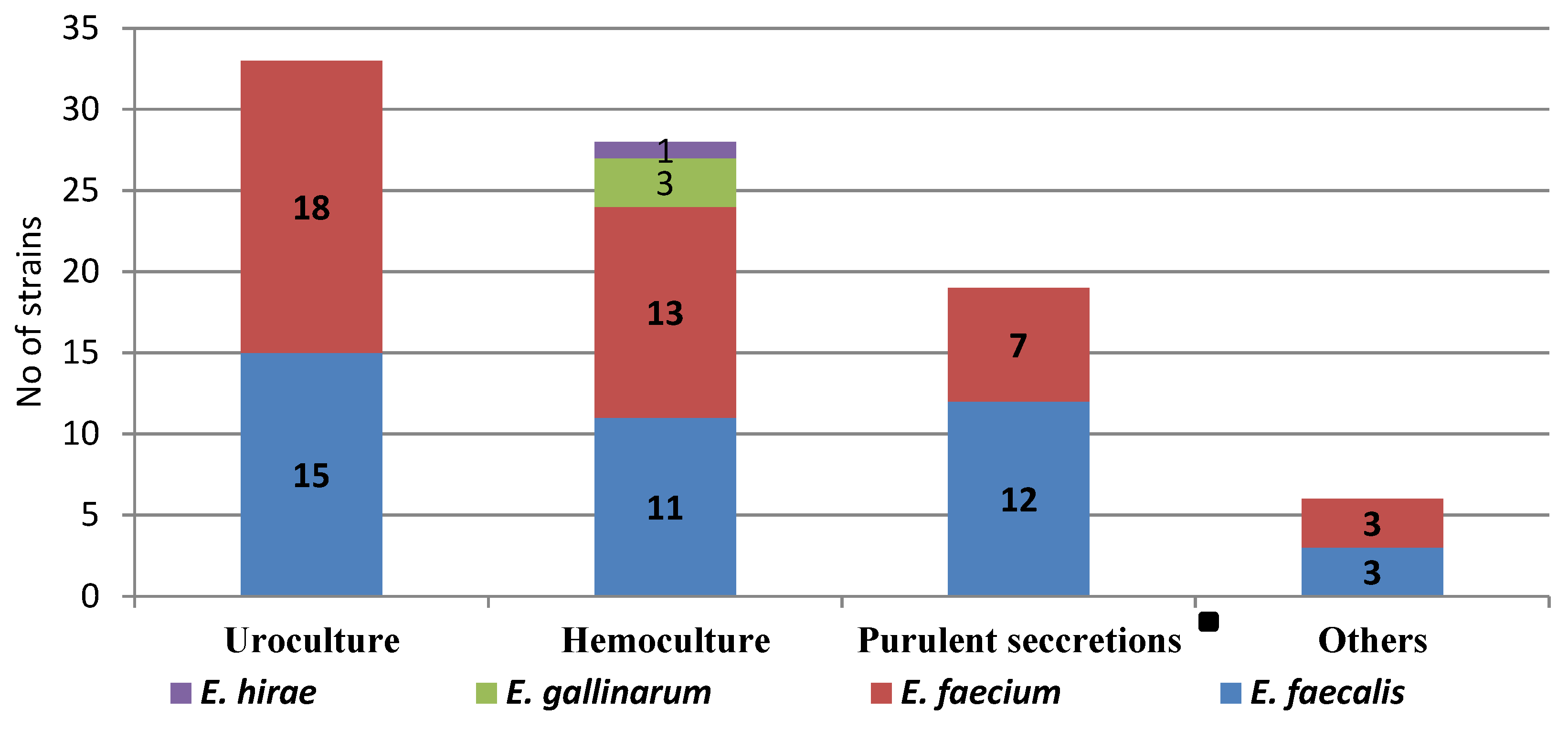
Figure 1. Distribution of Enterococcus spp. isolates according to the type of biologic sample in Galati, Romania.
Applsci 13 03866 g001
Figure 2. The distribution of antibiotic resistance in the strains of E. gallinarum, E. faecium, and E. faecalis and from Galati, Romania.
Applsci 13 03866 g002
Table 1. Antibiotic resistance profile of Enterococcus spp.
Enterococcus  SpeciesN1R (n)1R (%)2R (n)2R (%)MDR (n)MDR (%)
E. faecalis4137.3717.1921.9
E. faecium4124.949.83175.6
E. gallinarum30-0-3100
E. hirae10-0-0-
Total8655.81112.84350
Legend: 1R—resistant to one antimicrobial category; 2R—resistant to two antimicrobial categories; MDR—multidrug-resistant, N—total number, n—partial number.
Table 2. Comparison of Enterococcus spp. in three hospitals of southeast Romania.
nECH
(N1 = 48)
IDH
(N2 = 20)
ECHC
(N3 = 18)
Chi-Square Test
(p)
GenderMale442211110.502
Female422697
Living areaUrban50301460.047
Rural3618612
Year of isolation2019255812<0.001
2020201253
2021413173
COVID-19Positive85300.261
Negative78431718
Biological sampleHemoculture2926210.005
Uroculture33111111
SSTI18864
Other6312
Enterococcus spp.E. faecium41283100.018
E. faecalis4117177
Other4301
MDRYes433427<0.001
No43141811
Van-RYes2016130.031
No66321915
Legend: ECH: Emergency County Hospital; IDH: Infectious Diseases Hospital; ECHC: Emergency County Hospital for Children; SSTI: skin and soft tissue infection.
Disclaimer/Publisher’s Note: The statements, opinions and data contained in all publications are solely those of the individual author(s) and contributor(s) and not of MDPI and/or the editor(s). MDPI and/or the editor(s) disclaim responsibility for any injury to people or property resulting from any ideas, methods, instructions or products referred to in the content.