Article

Challenge Test in Catalan “Mató” Fresh Cheese to Assess the Antimicrobial Activity of Ericaria selaginoides Extracts against Bacillus cereus

1
IRTA, Food Safety and Functionality Program, Finca Camps i Armet s/n, Monells, 17121 Girona, Spain
2
Spanish Institute of Oceanography of the Spanish National Research Council (IEO, CSIC), Oceanographic Centre of Santander, Marine Culture Units “El Bocal”, Seaweeds Centre, Barrio Corbanera s/n, Monte, 39012 Santander, Spain
*
Author to whom correspondence should be addressed.
Appl. Sci. 2023, 13(4), 2207; https://doi.org/10.3390/app13042207
Submission received: 20 January 2023 / Revised: 5 February 2023 / Accepted: 7 February 2023 / Published: 9 February 2023
(This article belongs to the Special Issue Food Safety and Microbiological Hazards)

Abstract

:
Growing consumer demand for high-quality products coupled with minimally processed products and a minor use of synthetic food additives have increased the need to search for new sources of natural antimicrobials to ensure product safety. This study aimed to evaluate the antimicrobial activity of extracts from the brown algae Ericaria selaginoides against Bacillus cereus in typical Catalan fresh cheese (“mató”) by means of challenge testing. Three concentrations of a crude extract and its corresponding two subfractions (non-polar and mid-polar) obtained after purification showed an antimicrobial dose-dependent effect on B. cereus, from inhibition to inactivation. The best results were obtained with higher concentrations of the non-polar subfraction that caused a total inactivation of the inoculated pathogen after 2 or 4 days, followed by the mid-polar that inactivated B. cereus after 2 or 6 days. The results showed an improvement in the antimicrobial effect after purification compared with the effect observed when the crude extract was tested. Moreover, compounds of different chemical natures may be involved in this antimicrobial activity since it remained in both subfractions after purification. The results obtained in this work show the great potential of macroalgae extracts as natural food preservatives against B. cereus in fresh cheese.

Graphical Abstract

1. Introduction

Bacillus cereus is an aerobic facultative, spore-forming Gram-positive bacteria, widely distributed in the natural environment and considered an important food contaminant responsible for food spoilage. It can grow in highly variable conditions, including a broad pH range between 4.5 and 9.5, a minimum water activity (aw) for growth of 0.93, a broad range of temperature from 4 °C (psychrotrophic or psychrotolerant strains) to 48 °C, and NaCl concentrations up to 7% [1]. Furthermore, it is an opportunistic human pathogen producing virulence factors, such as toxins, associated with two forms of food poisoning, depending on the context in which it grows: emetic and diarrheal syndrome. The emetic syndrome, characterized by intense vomiting, is caused by a heat-stable peptide toxin, called cereulide, which is pre-formed in food or ingredients under various conditions, whereas diarrheal is caused by three heat-labile enterotoxins, the tripartite haemolysin BL (Hbl), the non-haemolytic enterotoxin (Nhe), and the single protein cytotoxin K (CytK), produced in the small intestine after contaminated food is consumed [2,3]. However, a human dose–response relationship has not been described for either the emetic or diarrheal toxin produced by B. cereus [1].
Current consumer demands for complex and mildly processed food with limited refrigerated shelf-life as ready-to-eat (RTE) products are leading to an increase on the B. cereus outbreaks all over the world, being associated with concentrations higher than 105 cfu/g in implicated foods [4]. B. cereus is responsible for a large number of cases of food poisoning, resulting in 155 outbreaks, 1636 cases, 44 hospitalizations, and 7 deaths reported by EFSA in 2019 [5]. Due to its ubiquity, it can contaminate a great variety of food products, such as pasta, rice, soups, sauces, meat, or dairy products, and most cases are attributed to incorrectly preserved food, including temperatures as high as 14 °C or long-term storage of the food at room temperature [6,7]. Moreover, its ability to produce endospores provides great resistance to heat, radiation, chemicals, and desiccation, allowing this bacteria to cope with adverse conditions for prolonged periods of time [8]. In this sense, it represents a concern in the dairy industry due to spores’ resistance to pasteurization and standard sanitation processes, being found in pasteurized milk and milk-derived products, such as milk powder and infant formulas [9,10]. B. cereus is also considered one of the main spoilage microorganisms in milk and dairy products. Its growth at levels over 5 log cfu/g is considered hazardous for the consumer and is also responsible for the spoilage [11,12]. Among dairy products, fresh cheese constitutes a very suitable food matrix for the growth of this pathogen because of its physicochemical characteristics (pH 5.8–6.8, aw 0.94–0.99, 0.1–1.2% NaCl). Thus, a fresh cheese highly consumed in Catalan regions of Spain, called “mató”, was selected as a food model in this study since its pH (6.78), aw (0.99), and NaCl (0.1%) values make it suitable for the growth of B. cereus. In addition, evidence has placed cheese products as a great reservoir of this pathogen since it has been widely described in a variety of cheeses, i.e., with a count range of 2–6 log cfu/g in buffalo mozzarella cheese [13], 5.5–5.7 log cfu/g in artisanal cheeses, such as Requeson, Cotija, or Adobero from Mexico [14], or 1–3.1 log cfu/g in Ricotta cheese [15].The main focus of the contamination of raw milk in the dairy chain is farms during milking via soil-contaminated udders and teats and the milking equipment, although additional contamination can occur in dairy plants [16]. Furthermore, its presence in food may result in recalls of contaminated products due to the unsafe status of the food, producing significant food waste and economic losses in the milk and dairy industry, which represents a dynamic global business sector, with the EU cheese market being the largest in the world [17]. In this context, its control is of great importance since, to date, there is no food safety criteria for B. cereus in Europe defining the acceptability of a foodstuff in terms of its microbiological safety; rather, there is a process of hygienic criterion established instead (Commission Regulation (EC) No 2073/2005) [18] for presumptive B. cereus in dried infant formulae and dried dietary foods for special medical purposes intended for infants below six months of age.
Regarding current preservation strategies, the use of additives from synthetic origin to improve the shelf-life of food products has been traditionally extended. However, new trends in consumer demand towards the use of natural and sustainable preservatives are gaining momentum, encouraging the search for novel sources of compounds [19] that could also represent different complementary biopreservation strategies to those currently used [20].
Marine ecosystems harbor a high diversity of organisms with unique composition. Thus, macroalgae have been traditionally consumed over centuries as food, mainly in Asian countries, due to their high nutritional value, and have also been used as gelling and thickening agents in the cosmeceutical and agri-food sector [21]. Over the past few years, they have become increasingly popular since they represent an underexploited source of chemical structures different from those described in terrestrial organisms. They have developed complex defense mechanisms to cope with harsh environmental conditions, resulting in a great variety of novel compounds [22]. In this sense, interest in the scientific community has been focused on brown macroalgae (Phaeophyceae) because of their content in chemical structures typical from this algal group, such as phlorotannins (polyphenols), fucoxanthin (carotenoid), or fucoidans (sulphated polysaccharides) [23]. They have been reported to show a variety of bioactivities, such as antioxidant, anticancer, antifungal, anti-inflammatory [24,25], as well as great antimicrobial potential, which make macroalgae a great source of compounds with potential as an alternative for natural food preservatives [26]. In this study, Ericaria selaginoides, a brown macroalgae commonly found in the North Atlantic coasts of Spain, was selected according to its antimicrobial efficacy previously observed in vitro against B. cereus [27]. Currently, although lots of studies have described the high in vitro antimicrobial potential of these marine organisms against foodborne pathogens and spoilage bacteria, few studies assessing this potential in food matrices have been performed so far [28] aiming to assess the functionality of macroalgae in food products.
The aim of this work was to evaluate the antimicrobial potential of E. selaginoides extracts as a control strategy against B. cereus artificially inoculated in “mató”, a Catalan fresh cheese chosen as a food matrix. To the best of our knowledge, it is the first time that the behavior of a foodborne pathogen, B. cereus, is evaluated under exposure to macroalgal extracts in a dairy matrix, such as fresh cheese, thus, helping to extend the current knowledge and establish a basis to make further applications of macroalgal extracts or their bioactive compounds feasible in the food industry as natural food preservatives.

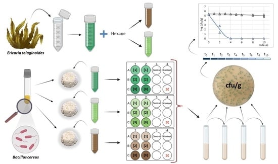
2. Materials and Methods

2.1. Macroalgae Collection

Ericaria selaginoides (Linnaeus) Molinary & Guiryi, 2020 [29] (Ochrophyta, Phaeophyceae, Fucales, Sargassaceae) (formerly Carpodesmia tamariscifolia (Hudson) Orellana & Sansón, 2019 [30], and Cystoseira tamariscifolia (Hudson) Papenfuss, 1950) were collected on rocky substrata in the lower intertidal and upper subtidal zones (<1 m depth) in March 2020 in Comillas (43°23′ N/4°17′ W; Cantabria region, Spain). The proper identification of the specimens of this species was performed by an experienced researcher in the field based on morphological characteristics using specialized taxonomic keys [31,32].
Apical and median parts of the thallus of adult specimens were selected and washed with sterile seawater to remove residual sand, salt, and particles attached to their surface. Likewise, remaining epiphytes and epizooties were carefully picked out. Then, the selected fresh and clean fronds were wrapped in sterile cloths moistened with seawater and kept under darkness, humid atmosphere (>84%), and cool conditions with ice packs (<15 °C) into expanded polystyrene (EPS) boxes to keep algae alive and healthy until transport to our laboratory at IRTA (Monells, Girona, Spain). Samples were kept at −80 °C until processing. In the laboratory, algal biomass was grinded and stored at −80 °C protected from light and vacuum packaged in polyethylene terephthalate and metallic/polyethylene (PET + MET/PE) bags (oxygen permeability of 1.5 cm3/m2/24 h and water vapor permeability of 1 g/m2/24 h) (Sistemvac, Estudi Graf S.A, Girona, Spain) until extract preparation.

2.2. Macroalgae Extract Processing

2.2.1. Preparation of Crude Extracts

Crude algae extract was prepared using a mid-polarity extraction medium composed of a mixture of hexane–isopropanol–water (10:80:10) according to Rubiño et al. (2022a) [33]. Food-grade solvents were selected according to current European legislation (Directive 2009/32/EC) [34] about the safe use of some organic solvents for food purposes. Pooled supernatants (5000 rpm, 10 min, 4 °C; Eppendorf, Germany) of three consecutive extractions performed at room temperature for 60 min each were evaporated to dryness under vacuum conditions at room temperature (Thermo Scientific Savant SpeedVac SPD120 Vacuum; Thermo Fisher Scientific, Waltham, MA, USA). The dried extract was collected, weighted, and kept frozen at −20 °C until further analysis.

2.2.2. Two-Phase Extraction Procedure

Crude extract was subjected to further liquid–liquid purification. It was transferred into a separating funnel and hexane (VWR Chemicals, Radnor, PA, USA) was added in a proportion 1:1. Mixture was shaken for 3 min and kept aside for layer separation. After clear phase separation, the top (non-polar) and bottom (mid-polar) subfractions were collected. The non-polar subfraction was evaporated to dryness at room temperature under vacuum conditions and the mid-polar subfraction was lyophilized (LyoMicron Freeze dryer, Coolvacuum Technologies, Barcelona, Spain). The dried subfractions were collected, weighted, and kept frozen at −20 °C until further analysis.

2.2.3. Preparation of Extracts for Application in Fresh Cheese

Prior to assay, the antimicrobial activity in the fresh cheese model, crude extract, and non-polar subfraction was resuspended in a medium composed of a mixture of water–glycerol–tween 80 (80:10:10) and the mid-polar subfraction was dissolved under sterile conditions in water.
MBC values obtained previously for crude extract through in vitro microdilution plate assays performed according to the recommended CLSI criteria with modifications [27,35] were taken as a reference to establish the tested concentrations, corresponding to 2×, 4×, and 10× the MBC values. The selected concentrations for subfractions were calculated according to the proportion they represent in the crude extract: 70% and 30% for non-polar and mid-polar subfractions, respectively. Thus, extracts were tested in three different concentrations: 14, 28, and 70 mg/g for crude extract; 5, 10, and 20 mg/g for the non-polar subfraction and 10, 25, and 50 mg/g for the mid-polar subfraction.

2.3. Challenge Test—Experimental Design

2.3.1. Bacterial Strains and Culture Conditions

B. cereus LMG 12335, used as a target bacteria, was a strain isolated from food that belongs to the Laboratory of Microbiology of Gent (LMG) as part of the Belgian Coordinated Collections of Microorganisms (BCCM).
Stocks of B. cereus were kept at −80 °C in 20% glycerol as cryoprotectant. Two consecutive cultures of B. cereus strain were made in BHI medium (Brain Heart Infusion broth, Oxoid Thermo Fisher Scientific, Waltham, MA, USA) both incubated at 30 °C over 18 h to reach the stationary growth phase (i.e., final counts of 107–108 cfu/mL).

2.3.2. Laboratory-Scale Fresh Cheese Model

Pasteurized cow’s milk fresh cheese, typical for Catalonia, called “mató”, commercially available at local retailers, was selected as a food matrix and used at the early stage of its shelf-life. The labelled cheese composition was 10% fat (including 6.9% saturated fat), 8.3% protein, 3.1% carbohydrates (being 1.3% free sugars), 0.1% NaCl, 250 mg/100 g calcium (31% Nutrient Reference Values (NRV)), and 165 mg/100 g phosphorus (23% NRV).
The antimicrobial effect of E. selaginoides against B. cereus was tested in two independent trials performed on two different days using two batches of fresh cheese and newly dissolved crude extract and subfractions. Cheeses were prepared in miniaturized format in 12-well plates in a sterile environment into a biosafety cabinet (Scalaf Mars Pro, Labogene, Allerød, Denmark). Three grams of fresh cheese was weighed in each individual well and each concentration of extract was added (0.45 v/w) in two wells (duplicate) into the food matrix. Afterwards, each well was inoculated (1% v/w) with 104–105 cfu/g of B. cereus and properly mixed with a sterile stick until complete homogenization. One well without B. cereus inoculum or extracts was also included to verify the hygienic conditions of handling and storage.
Once performed, the plates were covered and sealed in polyamide/polyethylene (PA/PE) bags (oxygen permeability of 50 cm3/m2/24 h and water vapor permeability of 2.8 g/m2/24 h) (Sistemvac, Estudi Graf S.A, Girona, Spain). Sealed 12-well plates were stored for 10 days at 8 °C. The temperature was chosen to assess a worst-case scenario, with a slight abuse of the refrigeration temperature corresponding to a reasonably foreseeable condition at the consumer stage [36,37]. Temperature was constantly recorded by a real-time wireless LABGUARD® system (BioMerieux, Marcy l’Etoile, France).

2.4. Analytical Determinations

A representative sample of the fresh cheese model was used to test variations in pH value (Crison Instruments, Barcelona, Spain) and water activity (aw) via an Aqualab® system (Ferrer Lab, Lleida, Spain). The hygienic initial microbiological conditions of miniaturized cheeses were controlled by Enterobacteriaceae counts (TEMPO, Biomerieux, Marcy-l’Étoile, France). Total aerobic counts were also measured initially and at subsequent sampling times.
B. cereus enumeration was performed after 2 h of the inoculation in the fresh cheese model (t0) and almost every day in control lots (t1, t2, t3, t4, t5, t6, t8, t10), whereas samples with macroalgae extracts were sampled every 2 days (t2, t4, t6, t8, t10) during refrigerated storage at 8 °C.
Two grams of cheese sample was diluted 1:10 in 0.1% Bacto Peptone physiological saline solution (Difco Laboratories, Detroit, MI, United States) with 0.85% NaCl and mixed thoroughly. Appropriate ten-fold serial dilutions were made in saline solution and then spread onto Brilliance Bacillus cereus agar (Oxoid, Thermo Fisher Scientific Inc., Waltham, MA, USA). Plate counting was performed after incubation at 30 °C after 24 h, with the detection limit set at 4 cfu/g (0.6 log cfu/g).

2.5. Statistical Analysis of Data

B. cereus counts were log transformed and statistical analyses were performed using the JMP 16.0.1 statistical software from SAS Institute Inc. (Cary, NC, USA). The effect of extracts and concentration at initial and final time of log counts was studied through one-way analysis of variance (ANOVA), followed by post hoc multiple comparison pairwise Tukey–Kramer test.

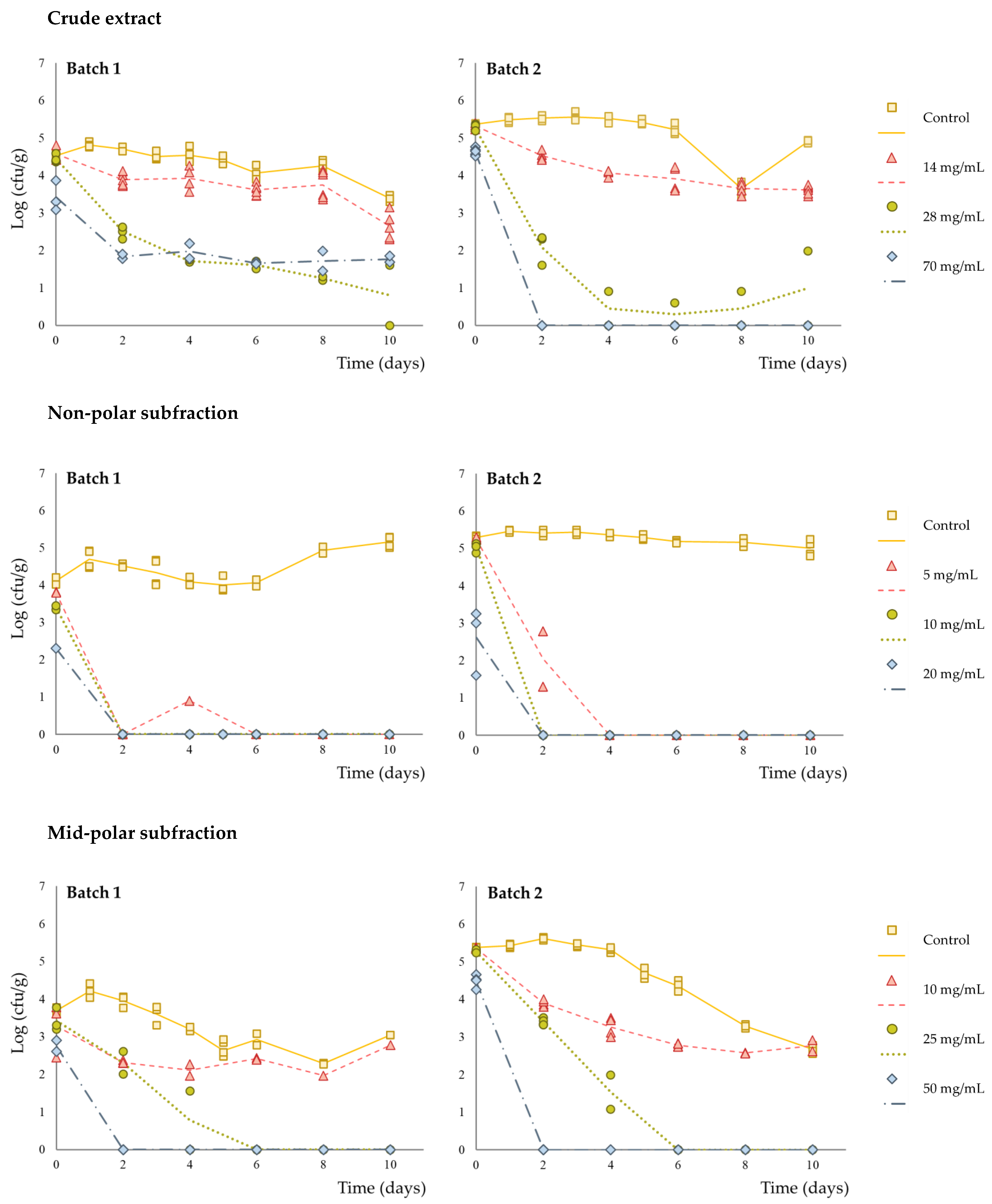
3. Results and Discussion

In this study, a “mató” fresh cheese was selected, since the pH and aw values, together with its low content of NaCl (0.1%) and the absence of other antimicrobial preservatives, make this cheese suitable for B. cereus growth. In this sense, the composition and volume of the medium used to dissolve the crude extract and non-polar subfraction were carefully selected to have a minimal effect on the aw (0.989 ± 0.002) and pH (6.76 ± 0.02) values of “mató” cheese under experimental conditions. B. cereus vegetative cells were inoculated in “mató” cheese in order to simulate natural germination of spores that survive milk pasteurization, post-contamination after milk pasteurization, or during cheese manufacturing.
Total aerobic microorganisms and Enterobacteriaceae counts were under the limit of detection in control samples (<10 cfu/g and <100 cfu/g, respectively) and no colonies were counted in control samples without pathogen inoculation nor addition of extract during the storage time, indicating the microbiological hygienic quality of the product and proper handling conditions during the experiment.
Counts of the control samples were kept at the initial level throughout storage since the selected B. cereus strain was a mesophile and, therefore, not able to grow under refrigerated conditions at 8 °C. However, counts for control of mid-polar subfraction diminished significatively (p < 0.05) at the end of the storage time, accounting for a 1.76 log reduction.
Compared to control cheeses, B. cereus counts decreased to a higher extent in fresh cheeses with E. selaginoides crude extract and the respective subfractions over the storage time (Table 1), although the inactivation rate and the magnitude of the count reduction were dependent on the extract and the concentration applied. A significative immediate bactericidal effect (p < 0.05), registered after two hours of inoculation (t0) compared with initial counts in controls, was only observed at the highest concentration of the non-polar subfraction, with a reduction of 2.24 ± 0.61 log.
In the presence of the crude extract, the total inactivation observed accounted for almost a 2 log reduction (1.81 ± 0.11 log) with the lowest concentration (14 mg/g), and the effect was similar with the intermediate (28 mg/g) and highest (70 mg/g) concentrations where the log reduction doubled, reaching 4 log. However, the effect in the presence of the highest concentration was variable between batches, whereas counts diminished quickly under the limit of detection by plate counting (<0.6 log cfu/g) after 2 days in batch 2, with an almost 3 log reduction (to 2.76 log cfu/g) observed in batch 1 and residual counts of 1.77 log remained in fresh cheese after 10 days of storage (Figure 1).
Antimicrobial activity, mainly with inhibitory effect, for these compounds and/or macroalgae extracts from brown macroalgae has been widely reported by many authors in in vitro assays [23]. The antimicrobial potential of E. selaginoides extracts to inhibit various foodborne pathogens and spoilage bacteria, including Bacillus subtilis, Escherichia coli, Listeria monocytogenes, Pseudomonas aeruginosa, and Staphylococcus aureus, has been supported by several authors [27,38,39,40,41,42]. Despite B. cereus not having been extensively used as a target strain in antimicrobial assays so far in other studies, the in vitro antimicrobial activity of extracts from E. selaginoides collected from the Atlantic coast of Spain against this foodborne pathogen was described previously by Salvador et al. (2007) [39] and Rubiño et al. (2022b) [27].
Nevertheless, most of the research developed in this sense has been evaluated mainly through in vitro assays. Studies assessing the antimicrobial potential of algal extracts in food matrices are currently limited and they are mostly focused on their effect on total viable counts over time. In addition, the reported effects differed depending on the nature of the food model. Whereas in meat products such as pork patties with added extracts from Laminaria digitata and Ulva spp., no effect was observed on the microbial population [43,44], total viable counts decreased when extracts from Myagropsis myagroides, Ecklonia cava, or Sargassum sagamianum were added to bakery products, including bread and muffins [45,46,47]. There is no available information about the application in cheese of macroalgae extracts for this purpose. However, the addition of Ascophyllum nodosum and Fucus vesiculosus extracts in other dairy products, such as raw whole cow’s milk and yogurt, did not affect the studied microorganisms, including total plate counts, total coliforms, yeasts or molds in the milk, and lactic acid bacteria in the yogurt [48,49].
The composition of macroalgae is highly dependent on a wide range of factors, intrinsic (i.e., life stage) and extrinsic (i.e., environmental and experimental conditions), determining, therefore, the nature of the extracted chemical structures. The composition of crude extracts used in this study was previously described in Rubiño et al. (2022b) [27], being a mixture of compounds with bioactive potential, including 0.73% of lipophilic pigments (such as chlorophyll a and fucoxanthin), 7.97% of polyphenols, 3.57% of polysaccharides, and other uncharacterized compounds.
Phlorotannins constitute a wide and diverse family of compounds restricted to brown seaweeds (Phaeophyceae), which are derived from the polymerization of phloroglucinol units, with diverse molecular weights and different levels of complexity. The growth inhibition and bactericidal effects against Gram+ and Gram− bacteria reported in several in vitro studies are proposed to occur by suppressing oxidative phosphorylation, the permeability alteration in microbial cell membrane, and the interaction with target microbial proteins [50,51]. Thus, the interaction of the phenolic aromatic ring and hydroxyl groups of various phlorotannins with bacterial enzymes and proteins can induce cell lysis [52]. Likewise, the active mechanism remains unclear, the available information about different existing structures in brown seaweeds is still limited, and only low-molecular-weight phlorotannins (2–8 phloroglucinol units) have been described in the literature. In vitro antimicrobial activity against B. cereus of phlorotannin-purified extracts from Cystoseira tamariscifolia (currently Ericaria selaginoides (Linnaeus) Molinary & Guiryi, 2020) was described by Lopes et al. (2012) [53]. In addition, the occurrence of five different phenolic structures isolated from the brown algae Ecklonia kurome with antimicrobial activity against B. cereus was described by Nagayama et al. (2002) [54]. Among them, dieckol and 8,8′-bieckol exerted the best results (0.54 μmol/mL), followed by phlorofucofuroeckol A (0.66 μmol/mL), eckol (1.08 μmol/mL), and phloroglucinol (>6.35 μmol/mL).
Other authors have also described an effect against B. cereus when purified fucoxanthin and fucoidan were tested. Deyab et al. (2013) [55] tested concentrations between 10 and 100 μg/mL of fucoxanthin obtained from Turbinaria triquatra via the disk-diffusion method, demonstrating a dose-dependent effect as the size of the inhibition zones increased with the concentration, with values from 1.8 ± 0.1 to 6.5 ± 0.3 mm. Ethanolic and acetone:methanol extracts performed with other brown algae species, such as Saccharina japonica and Sargassum horneri, with fucoxanthin content between 0.41 and 0.48 mg/g as well as 0.71 and 0.77 mg/g, respectively, showed an effect against this target strain, being higher with acetone:methanol extracts [56]. Regarding fucoidans, Rajeshkumar (2016) [57] also described an enhancement in the inhibition of Bacillus sp. when encapsulated purified fucoidan from Padina tetrastromatica was tested together with a batch of different antibiotics.
Research on the action mechanisms of the potential antimicrobial compounds from macroalgae is still scarce, although evidence of interactions at the cell membrane level causing cell disruption has been described for polyphenolic compounds, fucoidans, and fucoxanthin [23,58]. In addition to the interactions of these compounds with the bacterial cell wall, interactions between their functional groups could have occurred. Thus, potential interactions between these compounds together with their polarity differences were assumed to be responsible of the difficulties found in solubilizing the components of the crude extract in a polar dissolution medium. Hence, a specific medium was designed to solubilize the mid-polarity crude extract and fractioning was performed to minimize this issue, which could also explain the improvement in the antimicrobial effect observed with both subfractions discussed below.
Although references evaluating the antimicrobial activity of these compounds after purification are limited so far, the evidence described above, together with the fact that there is an occurrence of compounds of this nature in E. selaginoides crude extract used in this study [27], lead one to consider that the observed antimicrobial effect could not be attributed to a single compound but to an additive, synergic, and/or antagonistic action of compounds of different chemical natures.
On the other hand, in the presence of the non-polar subfraction, counts of B. cereus were under the limit of detection (<0.6 log cfu/g) after 2 days in fresh cheese samples with the intermediate and highest concentrations (10 and 20 mg/g), and in 2–4 days in cheese with the lowest concentration (5 mg/g). At the end of the storage time, compared to the control, all the concentrations tested had achieved almost 5 log reduction (4.70 ± 0.83 log).
The behavior of B. cereus in fresh cheese with the lowest concentration of mid-polar subfraction (10 mg/g) accounted for almost 2 log reduction (1.76 ± 1.20 log). At the intermediate concentration applied (25 mg/g), B. cereus counts diminished below the detection limit after 6 days of storage, while, with the highest concentration (50 mg/g), counts were under the limit of detection after 2 days (p < 0.05). Thus, 4.53 ± 1.19 log reduction was observed after 10 days of storage with the intermediate and highest concentrations when compared to control.
Among all the assayed extracts, the non-polar subfraction was the most efficacious to reduce the counts of B. cereus on the fresh cheese samples, followed by the mid-polar subfraction and the crude extract. The non-polar subfraction showed a higher antimicrobial effect against B. cereus, even at the lowest concentration assayed compared with the lowest concentration of the other two extracts (crude and mid-polar subfraction). Results also showed an increase in the antimicrobial effect after separation of the extract in two subfractions. Whereas with crude extract, a total inactivation was observed at the highest concentration only in batch 2, with the mid-polar subfraction, it was observed in the presence of the intermediate and highest concentration in 2–6 days and with the non-polar subfraction at all the concentrations tested in even less time (2–4 days). Moreover, the fact that the antimicrobial activity was maintained in both subfractions may indicate that compounds of different chemical natures are involved in the observed effect.

4. Conclusions

An antimicrobial effect against B. cereus was observed when crude extract from E. selaginoides and the subsequent purified subfractions were added to Catalan “mató” fresh cheese. The best results when the crude extract was added were observed at the highest concentration. The obtention of two purified subfractions proved an increased effect, being more remarkable with the non-polar subfraction, since B. cereus was under the limit of detection after 2-4 days. The presence of different bioactive chemical compounds occurring in E. selaginoides extract, as well as the remaining activity in both subfractions, leads one to consider that more than one single compound of different chemical nature is responsible for the antimicrobial activity observed in the crude extract.
Results obtained in this work proved that the macroalgae extract from E. selaginoides has potential as a natural preservative in the cheese-making industry against B. cereus, establishing a good basis for further research and the development of potential macroalgae in the food industry. The inclusion of this extract can lead to a marked quality and safety enhancement in fresh cheese matrices, as well as a successful strategy for the development of novel innovative products and an increase in the commercial value of macroalgae. Further research focused on the purification of extracts and the identification of the active principle/s could allow one to minimize the variability in the antimicrobial effect observed as well as to evaluate the impact on the sensory features of the product in order to ensure consumer acceptance.

Author Contributions

Conceptualization, T.A., S.B.-C. and M.H.; Methodology, S.R., T.A., S.B.-C. and M.H.; Formal analysis, S.R., T.A., S.B.-C. and M.H.; Resources, C.P.; Writing—original draft preparation, S.R.; writing—review and editing, S.R., T.A., C.P., S.B.-C., M.H.; supervision, T.A., S.B.-C. and M.H.; Project administration, M.H.; Funding acquisition, M.H. All authors have read and agreed to the published version of the manuscript.

Funding

This work was supported by the Spanish Ministry of Economy and Competitiveness (INIA Project: RTA2015-00010-C03-01), the CERCA Program (Generalitat de Catalunya), the Consolidated Research Group (2017 SGR 1650, 2021 SGR 00468) and the FPI Ph.D. grant (BES-2017-0027) awarded to Susana Rubiño by the Spanish Ministry of Economy and Competitiveness.

Institutional Review Board Statement

Not applicable.

Informed Consent Statement

Not applicable.

Data Availability Statement

Not applicable.

Acknowledgments

Susana Rubiño acknowledges the FPI Ph.D. grant from the Spanish Ministry of Economy and Competitiveness (BES-2017-0027).

Conflicts of Interest

The authors declare no conflict of interest.

References

  1. Bottone, E.J. Bacillus cereus, a volatile human pathogen. Clin. Microbiol. Rev. 2010, 23, 382–398. [Google Scholar] [CrossRef]
  2. From, C.; Pukall, R.; Schumann, P.; Hormazábal, V.; Granum, P.E. Toxin-producing ability among Bacillus spp. outside the Bacillus cereus group. Appl. Environ. Microbiol. 2005, 71, 1178–1183. [Google Scholar] [CrossRef] [PubMed]
  3. Dietrich, R.; Jessberger, N.; Ehling-Schulz, M.; Märtlbauer, E.; Granum, P.E. The food poisoning toxins of Bacillus cereus. Toxins 2021, 13, 98. [Google Scholar] [CrossRef] [PubMed]
  4. Rodrigo, D.; Rosell, C.M.; Martinez, A. Risk of Bacillus cereus in relation to rice and derivatives. Foods 2021, 10, 302. [Google Scholar] [CrossRef] [PubMed]
  5. EFSA. The European Union One Health 2019 Zoonoses Report. EFSA J. 2021, 19, e06406. [Google Scholar] [CrossRef]
  6. Choi, W.; Kim, S.S. Outbreaks, germination, and inactivation of Bacillus cereus in food products: A review. J. Food Prot. 2020, 83, 1480–1487. [Google Scholar] [CrossRef]
  7. Griffiths, M.W.; Schraft, H. Bacillus cereus food poisoning. In Foodborne Diseases: Third Edition; Dodd, C.E.R., Aldsworth, T., Stein, R.A., Cliver, D.O., Riemann, H.P., Eds.; Academic Press: Cambridge, MA, USA, 2017; pp. 395–405. [Google Scholar] [CrossRef]
  8. Nicholson, W.L.; Munakata, N.; Horneck, G.; Melosh, H.J.; Setlow, P. Resistance of Bacillus endospores to extreme terrestrial and extraterrestrial environments. Microbiol. Mol. Biol. Rev. 2000, 64, 548–572. [Google Scholar] [CrossRef]
  9. Becker, H.; Schaller, G.; von Wiese, W.; Terplan, G. Bacillus cereus in infant foods and dried milk products. Int. J. Food Microbiol. 1994, 23, 1–15. [Google Scholar] [CrossRef]
  10. Di Pinto, A.; Bonerba, E.; Bozzo, G.; Ceci, E.; Terio, V.; Tantillo, G. Occurrence of potentially enterotoxigenic Bacillus cereus in infant milk powder. Eur. Food Res. Technol. 2013, 237, 275–279. [Google Scholar] [CrossRef]
  11. Lücking, G.; Stoeckel, M.; Atamer, Z.; Hinrichs, J.; Ehling-Schulz, M. Characterization of aerobic spore-forming bacteria associated with industrial dairy processing environments and product spoilage. Int. J. Food Microbiol. 2013, 166, 270–279. [Google Scholar] [CrossRef]
  12. Food and Drug Administration (FDA). Bacillus cereus and Other Bacillus spp. In Bad Bug Book: Foodborne Pathogenic Microorganisms and Natural Toxins Handbook; International Medical Publishing: London, UK, 2012; pp. 92–95. [Google Scholar]
  13. Montone, A.M.I.; Capuano, F.; Mancusi, A.; Di Maro, O.; Peruzy, M.F.; Proroga, Y.T.R.; Cristiano, D. Exposure to Bacillus cereus in water buffalo mozzarella cheese. Foods 2020, 9, 1899. [Google Scholar] [CrossRef]
  14. Adame-Gómez, R.; Muñoz-Barrios, S.; Castro-Alarcón, N.; Leyva-Vázquez, M.A.; Toribio-Jiménez, J.; Ramírez-Peralta, A. Prevalence of the strains of Bacillus cereus group in artisanal Mexican cheese. Foodborne Pathog. Dis. 2020, 17, 8–14. [Google Scholar] [CrossRef] [PubMed]
  15. Cosentino, S.; Mulargia, A.F.; Pisano, B.; Tuveri, P.; Palmas, F. Incidence and biochemical characteristics of Bacillus flora in Sardinian dairy products. Int. J. Food Microbiol. 1997, 38, 235–238. [Google Scholar] [CrossRef] [PubMed]
  16. Ehling-Schulz, M.; Messelhäusser, U.; Granum, P.E. Bacillus cereus in milk and dairy production. In Rapid Detection, Characterization, and Enumeration of Foodborne Pathogens; Hoorfar, J., Ed.; ASM Press: Washington DC, USA, 2011. [Google Scholar] [CrossRef]
  17. Statista Research Department. Global per Capita Consumption of Cheese 2020 by Country. 2021. Available online: https://www.statista.com/statistics/527195/consumption-of-cheese-per-capita-worldwide-country/ (accessed on 26 July 2022).
  18. European Commission. Commission Regulation (EC) No 2073/2005 of 15 November 2005 on microbiological criteria for foodstuffs. Off. J. Eur. Union 2005, 50, 1–26. [Google Scholar]
  19. Bondi, M.; Lauková, A.; De Niederhausern, S.; Messi, P.; Papadopoulou, C. Natural preservatives to improve food quality and safety. J. Food Qual. 2017, 2017, 090932. [Google Scholar] [CrossRef]
  20. Shi, C.; Maktabdar, M. Lactic Acid Bacteria as biopreservation against spoilage molds in dairy products–A review. Front. Microbiol. 2022, 12, 819684. [Google Scholar] [CrossRef] [PubMed]
  21. Kilinç, B.; Cirik, S.; Turan, G.; Tekogul, H.; Koru, E. Seaweeds for food and industrial applications. Chem. Age India 2013, 33, 475–482. [Google Scholar]
  22. Barbosa, M.; Valentão, P.; Andrade, P.B. Bioactive compounds from macroalgae in the new millennium: Implications for neurodegenerative diseases. Mar. Drugs 2014, 12, 4934–4972. [Google Scholar] [CrossRef]
  23. Silva, A.; Silva, S.A.; Carpena, M.; Garcia-Oliveira, P.; Gullón, P.; Barroso, M.F.; Prieto, M.A.; Simal-Gandara, J. Macroalgae as a source of valuable antimicrobial compounds: Extraction and applications. Antibiotics 2020, 9, 642. [Google Scholar] [CrossRef]
  24. Biris-Dorhoi, E.S.; Michiu, D.; Pop, C.R.; Rotar, A.M.; Tofana, M.; Pop, O.L.; Socaci, S.A.; Farcas, A.C. Macroalgae—A sustainable source of chemical compounds with biological activities. Nutrients 2020, 12, 3085. [Google Scholar] [CrossRef]
  25. Martínez, M.A.; Ares, I.; Martínez, M.; Lopez-Torres, B.; Maximiliano, J.E.; Rodríguez, J.L.; Martínez-Larrañaga, M.R.; Anadón, A.; Peteiro, C.; Rubiño, S.; et al. Brown marine algae Gongolaria baccata extract protects Caco-2 cells from oxidative stress induced by tert-butyl hydroperoxide. Food Chem. Toxicol. 2021, 156, 112460. [Google Scholar] [CrossRef] [PubMed]
  26. Pérez, M.J.; Falqué, E.; Domínguez, H. Antimicrobial action of compounds from marine seaweed. Mar. Drugs 2016, 14, 52. [Google Scholar] [CrossRef] [PubMed]
  27. Rubiño, S.; Peteiro, C.; Aymerich, T.; Hortós, M. Brown macroalgae (Phaeophyceae): A valuable reservoir of antimicrobial compounds in North coasts of Spain. Mar. Drugs 2022, 20, 775. [Google Scholar] [CrossRef]
  28. Cabral, E.M.; Oliveira, M.; Modala, J.R.M.; Curtin, J.; Tiwari, B.K.; García-Vaquero, M. Antimicrobials from seaweeds for food applications. Mar. Drugs 2021, 19, 211. [Google Scholar] [CrossRef] [PubMed]
  29. Molinari-Novoa, E.A.; Guiry, M.D. Reinstatement of the genera Gongolaria Boehmer and Ericaria Stackhouse (Sargassaceae, Phaeophyceae). Notulae Algarum 2020, 172, 1–10. [Google Scholar]
  30. Orellana, S.; Hernandez, M.; Sanson, M. Diversity of Cystoseira sensu lato (Fucales, Phaeophyceae) in the eastern Atlantic and Mediterranean based on morphological and DNA evidence, including Carpodesmia gen. emend. and Treptacantha gen. emend. Eur. J. Phycol. 2019, 54, 447–465. [Google Scholar] [CrossRef]
  31. Fletcher, R.L. Seaweeds of the British Isles; Fucophyceae (Phaeophyceae) Part 1.; British Museum (Natural History): London, UK, 1987; Volume 3. [Google Scholar]
  32. Gómez Garreta, A.; Barceló Martí, M.C.; Gallardo García, T.; Pérez-Ruzafa, I.M.; Ribera Sigual, M.A.; Rull Lluch, J. Flora Phycológica Ibérica. 1. Fucales; Universidad de Murcia, Servicio de Publicaciones: Murcia, Spain, 2000. [Google Scholar]
  33. Rubiño, S.; Peteiro, C.; Aymerich, T.; Hortós, M. Major lipophilic pigments in Atlantic seaweeds as valuable food ingredients: Analysis and assessment of quantification methods. Food Res. Int. 2022, 159, 111609. [Google Scholar] [CrossRef]
  34. European Parliament; Council of the European Union. Directive 2009/32/EC of the European Parliament and of the Council of 23 April 2009 on the approximation of the laws of the Member States on extraction solvents used in the production of foodstuffs and food ingredients. Off. J. Eur. Union 2009, 141, 3–11. [Google Scholar]
  35. CLSI Standard M07; Methods for Dilution Antimicrobial Susceptibility Test for Bacteria that Grow Aerobically. Approved standard, 11th ed.; Clinical and Laboratory Standards Institute: Wayne, PA, USA, 2018.
  36. Jofré, A.; Latorre-Moratalla, M.L.; Garriga, M.; Bover-Cid, S. Domestic refrigerator temperatures in Spain: Assessment of its impact on the safety and shelf-life of cooked meat products. Food Res. Int. 2019, 126, 108578. [Google Scholar] [CrossRef]
  37. EURL Lm. Technical Guidance Document on Challenge Tests and Durability Studies for Assessing Shelf-Life of Ready-to-Eat Foods Related to Listeria monocytogenes; Version 4 of 1 July 2021; Anses – Food Safety Laboratory: Maisons-Alfort, France, 2021. [Google Scholar]
  38. Abourriche, A.; Charrouf, M.; Berrada, M.; Bennamara, A.; Chaib, N.; Francisco, C. Antimicrobial activities and cytotoxicity of the brown alga Cystoseira tamariscifolia. Fitoterapia 1999, 70, 611–614. [Google Scholar] [CrossRef]
  39. Salvador, N.; Garreta, A.G.; Lavelli, L.; Ribera, M.A. Antimicrobial activity of Iberian macroalgae. Sci. Mar. 2007, 71, 101–114. [Google Scholar] [CrossRef]
  40. Karima, S.; Fatiha, B. Study of the antimicrobial activity of four Algerian marin algae species. In Microbes in Applied Research; World Scientific: Singapore, 2012; pp. 578–581. [Google Scholar]
  41. Ainane, T.; Abourriche, A.; Kabbaj, M.; Elkouali, M.; Bennamara, A.; Charrouf, M.; Lemrani, M. Biological activities of extracts from seaweed Cystoseira tamariscifolia: Antibacterial activity, antileishmanial activity and cytotoxicity. J. Chem. Pharm. Res. 2014, 6, 607–611. [Google Scholar]
  42. De La Fuente, G.; Pinteus, S.; Silva, J.; Alves, C.; Pedrosa, R. Antioxidant and antimicrobial potential of six fucoids from the Mediterranean Sea and the Atlantic. Ocean. J. Sci. Food. Agric. 2022, 102, 5568–5575. [Google Scholar] [CrossRef]
  43. Moroney, N.C.; O’Grady, M.N.; O’Doherty, J.V.; Kerry, J.P. Effect of a brown seaweed (Laminaria digitata) extract containing laminarin and fucoidan on the quality and shelf-life of fresh and cooked minced pork patties. Meat Sci. 2013, 94, 304–311. [Google Scholar] [CrossRef] [PubMed]
  44. Lorenzo, J.M.; Sineiro, J.; Amado, I.R.; Franco, D. Influence of natural extracts on the shelf life of modified atmosphere-packaged pork patties. Meat Sci. 2014, 96, 526–534. [Google Scholar] [CrossRef] [PubMed]
  45. Lee, C.-J.; Choi, J.-S.; Song, E.-J.; Lee, S.-Y.; Kim, K.-B.-W.-R.; Kim, S.-J.; Yoon, S.-Y.; Lee, S.-J.; Park, N.-B.; Jung, J.-Y. Effect of Myagropsis myagroides extracts on shelf-life and quality of bread. Korean J. Food Sci. Technol. 2010, 42, 50–55. [Google Scholar]
  46. Jung, K.-I.; Choi, Y.-J.; Cho, E.-K. Effect of Ecklonia cava hot water extracts on shelf-life and quality of muffin. J. Korean Soc. Food Sci. Nutr. 2010, 39, 1672–1677. [Google Scholar] [CrossRef]
  47. Kim, M.-J.; Kim, K.-B.-W.-R.; Lee, C.-J.; Kwak, J.-H.; Kim, D.-H.; SunWoo, C.; Jung, S.-A.; Kang, J.-Y.; Kim, H.-J.; Choi, J.-S. Effect of Sargassum sagamianum extract on shelf-life and improved quality of morning bread. Korean J. Food Sci. Technol. 2011, 43, 723–728. [Google Scholar] [CrossRef]
  48. O’Sullivan, A.M.; O’Callaghan, Y.C.; O’Grady, M.N.; Waldron, D.S.; Smyth, T.J.; O’Brien, N.M.; Kerry, J.P. An examination of the potential of seaweed extracts as functional ingredients in milk. Int. J. Dairy Technol. 2014, 67, 182–193. [Google Scholar] [CrossRef]
  49. O’Sullivan, A.M.; O’Grady, M.N.; O’Callaghan, Y.C.; Smyth, T.J.; O’Brien, N.M.; Kerry, J.P. Seaweed extracts as potential functional ingredients in yogurt. Innov. Food Sci. Emerg. Technol. 2016, 37, 293–299. [Google Scholar] [CrossRef]
  50. Bhowmick, S.; Mazumdar, A.; Moulick, A.; Adam, V. Algal metabolites: An inevitable substitute for antibiotics. Biotechnol. Adv. 2020, 43, 107571. [Google Scholar] [CrossRef] [PubMed]
  51. Shannon, E.; Abu-Ghannam, N. Antibacterial derivatives of marine algae: An overview of pharmacological mechanisms and applications. Mar. Drugs 2016, 14, 81. [Google Scholar] [CrossRef] [PubMed]
  52. Shrestha, S.; Zhang, W.; Smid, S.D. Phlorotannins: A review on biosynthesis, chemistry and bioactivity. Food Biosci. 2021, 39, 100832. [Google Scholar] [CrossRef]
  53. Lopes, G.; Sousa, C.; Silva, L.R.; Pinto, E.; Andrade, P.B.; Bernardo, J.; Mouga, T.; Valentão, P. Can phlorotannins purified extracts constitute a novel pharmacological alternative for microbial infections with associated inflammatory conditions? PLoS ONE 2012, 7, e31145. [Google Scholar] [CrossRef] [PubMed]
  54. Nagayama, K.; Iwamura, Y.; Shibata, T.; Hirayama, I.; Nakamura, T. Bactericidal activity of phlorotannins from the brown alga Ecklonia kurome. J. Antimicrob. Chemother. 2002, 50, 889–893. [Google Scholar] [CrossRef]
  55. Deyab, M.A.; Abou-Dobara, M.I. Antibacterial activity of some marine algal extracts against most nosocomial bacterial infections. Egypt. J. Exp. Biol. 2013, 9, 281–286. [Google Scholar]
  56. Sivagnanam, S.P.; Yin, S.; Choi, J.H.; Park, Y.B.; Woo, H.C.; Chun, B.S. Biological properties of fucoxanthin in oil recovered from two brown seaweeds using supercritical CO2 extraction. Mar. Drugs 2015, 13, 3422–3442. [Google Scholar] [CrossRef]
  57. Rajeshkumar, S. Phytochemical constituents of fucoidan (Padina tetrastromatica) and its assisted AgNPs for enhanced antibacterial activity. IET Nanobiotechnol. 2017, 11, 292–299. [Google Scholar] [CrossRef]
  58. Khan, F.; Jeong, G.J.; Khan, M.S.A.; Tabassum, N.; Kim, Y.M. Seaweed-Derived phlorotannins: A review of multiple biological roles and action mechanisms. Mar. Drugs 2022, 20, 384. [Google Scholar] [CrossRef]
Figure 1. Behavior of B. cereus inoculated in fresh cheeses stored at 8 °C under E. selaginoides extract exposure at the three concentrations for the two independent batches performed. Symbols represent the experimental observed data and lines the mean of observed data.
Applsci 13 02207 g001
Table 1. Log reduction in B. cereus (log10; mean ± standard deviation) in fresh cheese inoculated with three different concentrations of crude extract and its two corresponding subfractions.
Crude extract
14 mg/g28 mg/g70 mg/g
Initial log reduction a<0.5<0.50.92 ± 0.27
Inactivation potential b1.81 ± 0.114.05 ± 0.454.06 ± 1.84
Non-polar subfraction
5 mg/g10 mg/g20 mg/g
Initial log reduction a<0.5<0.52.24 ± 0.61
Inactivation potential b4.70 ± 0.834.70 ± 0.834.70 ± 0.83
Mid-polar subfraction
10 mg/g25 mg/g50 mg/g
Initial log reduction a<0.5<0.50.91 ± 0.04
Inactivation potential b1.76 ± 1.204.53 ± 1.194.53 ± 1.19
a Immediate bactericidal effect after 2 h of inoculation. b Total log reduction after 10 days storage at 8 °C.
Disclaimer/Publisher’s Note: The statements, opinions and data contained in all publications are solely those of the individual author(s) and contributor(s) and not of MDPI and/or the editor(s). MDPI and/or the editor(s) disclaim responsibility for any injury to people or property resulting from any ideas, methods, instructions or products referred to in the content.

