Article

Effects of Cheonwangbosim-dan in a Mouse Model of Chronic Obstructive Pulmonary Disease: Anti-Inflammatory and Anti-Fibrotic Therapy

1
Department of Veterinary Pathology, College of Veterinary Medicine, Chungnam National University, Daejeon 34134, Republic of Korea
2
Herbal Medicine Research Division, Korea Institute of Oriental Medicine, Daejeon 34054, Republic of Korea
*
Author to whom correspondence should be addressed.
These authors equally contributed to this work.
Appl. Sci. 2023, 13(3), 1829; https://doi.org/10.3390/app13031829
Submission received: 19 December 2022 / Revised: 20 January 2023 / Accepted: 28 January 2023 / Published: 31 January 2023
(This article belongs to the Special Issue Bioactive Compounds from Natural Products - Volume II)

Abstract

:
Chronic obstructive pulmonary disease (COPD) is a lung illness, marked by dyspnea, coughing, and sputum production. Cheonwangbosim-dan (CBD) is a traditional East Asian medicine, consisting of a combination of 15 medicinal herbs, which is frequently used to treat arterial/auricular flutter, neuroses, cardiac-malfunction-induced diseases, and insomnia. The present study evaluated the therapeutic effect of CBD (100 or 200 mg/kg) on COPD using a mouse model of COPD induced by cigarette smoke (CS) and lipopolysaccharide (LPS). The increase in inflammatory cell numbers caused by exposure to CS and LPS was significantly reduced by CBD administration. In addition, CBD therapy reduced interleukin-6 (IL-6) and tumor necrosis factor-α (TNF-α) in bronchoalveolar lavage fluid (BALF). In lung tissue, CBD not only reduced the levels of IL-1β (CBD 100: p < 0.001 and CBD 200: p < 0.001), IL-6 (CBD 100: p < 0.001 and CBD 200: p < 0.001), TNF-α (CBD 100: p = 0.005 and CBD 200: p = 0.014), and monocyte chemoattractant protein-1 (MCP-1; CBD 100: p = 0.018 and CBD 200: p = 0.003), but also decreased the expression of α-smooth muscle actin (α-SMA; CBD 100: p < 0.001 and CBD 200: p < 0.001), transforming growth factor-β (TGF-β; CBD 100: p < 0.001 and CBD 200: p < 0.001), matrix metallopeptidase-7 (MMP-7; CBD 100: p = 0.019 and CBD 200: p < 0.001), MMP-9 (CBD 100: p = 0.015 and CBD 200: p = 0.013), and tissue inhibitor of metalloproteinase-1 (TIMP-1; CBD 100: p = 0.035 and CBD 200: p = 0.013) compared with the COPD group. CBD was also found to suppress the phosphorylation of nuclear factor kappa B (NF-κB), extracellular signal-regulated kinase 1/2 (ERK1/2), and p38 mitogen-activated protein kinases (p38 MAPK). Taken together, these findings showed that CBD can attenuate respiratory inflammation and airway remodeling induced by exposure to CS and LPS, suggesting that CBD has probable preventive and therapeutic applications in patients with COPD.

1. Introduction

Airflow blockage and recurrent respiratory symptoms are two characteristics of chronic obstructive pulmonary disease (COPD) [1]. Despite being both preventable and treatable, COPD is one of the leading factors in morbidity and death around the globe [2]. Exposure to cigarette smoke is the main risk factor for COPD, although other variables, including exposure to air pollution and biomass combustion, have also been associated with the disease [1]. It is anticipated that COPD will remain a significant burden on both patients and healthcare professionals for the foreseeable future. Therefore, it is crucial to determine whether innovative anti-inflammatory drugs may stop or delay the decrease in lung function that is a hallmark of COPD, because it is a chronic inflammatory condition. Chronic inflammation can cause airway remodeling and constriction, which then contribute to a typical decrease in airflow and symptoms of cough, shortness of breath, and increased sputum production seen in COPD [3]. Most of the pathways upstream of COPD genes and targets are involved in immunological control [4]. For instance, higher levels of TNF-α, as well as higher phosphorylation levels of p38 MAPK and ERK1/2, have been noted in people with COPD; moreover, inhibitors of p38 MAPK and TNF-α have been found to exhibit inhibitory effects on the inflammatory responses of COPD [5,6]. The final etiology of COPD may, thus, be significantly influenced by the molecular mechanisms of inflammation. As a result, complementary and alternative therapies for the treatment of COPD are gaining popularity. Plant-derived compounds have demonstrated immunomodulatory and anti-inflammatory characteristics by inhibiting the manufacture and function of cytokines, chemokines, and adhesion molecules [7]. Plant-derived agents could be utilized alone as anti-cytokine activators or in combination with existing known anti-inflammatory medications, thereby reducing potential costs and side effects while increasing effectiveness. Several herbal medicines have been used in the treatment of patients with COPD because of unsatisfactory treatment results and the side effects of existing drugs [8].
A herbal cure under the name Cheonwangbosim-dan in Korea, Tennohosintan in Japan, and Tian Wang Bu Xin Dan in China has been used for centuries in East Asia. Cheonwangbosim-dan (CBD) contains 15 medicinal ingredients: Acorus gramineus, Angelica gigas, Asparagus cochinchinensis, Coptis japonica, Liriope platyphylla, Panax ginseng, Polygala tenuifolia, Platycodon grandiflorum, Poria cocos, Rehmannia glutinosa, Salvia miltiorrhiza, Scrophularia buergeriana, Schisandra chinensis Thuja orientalis, and Ziziphus jujuba [9]. CBD has been used to treat conditions, such as heart dysfunction, neuroses, sleeplessness, and arterial or auricular flutter [10]. Numerous biological effects of CBD have been observed, including antihypertensive, vasorelaxant, and neurological effects [11]. Additionally, antioxidant [12] and antidepressant [13] effects of CBD, as well as effects on the central nervous system and cardiovascular system [10], have been found.
According to research, the main environmental risk factors for COPD include cigarette smoking, biofuel exposure, industrial dust, and indoor and outdoor air pollution [14]. Numerous toxic components in cigarette smoke (CS) stimulate the respiratory system. Long-term smoking has an impact on the alveolar septa, alters the structure of the air duct walls, and results in interstitial fibrosis [15,16,17]. Smoking also causes obstructive bronchiolitis and increases mucous gland secretions, which exacerbate the advancement of lung tissue diseases [18,19]. Lipopolysaccharide (LPS) is an endotoxin produced by Gram-negative bacteria that increases the production of proinflammatory cytokines and induces an influx of inflammatory cells, such as lymphocytes, neutrophils, and macrophages [20,21]. Repeated exposure to LPS has been shown to cause pathological alterations comparable to those seen in COPD patients, such as chronic neutrophilia in the bronchi [22] and goblet cell hyperplasia in the airways [23]. The present study evaluated the effects of CBD in COPD induced by CS and LPS in a mouse model, and attempted to determine whether CBD can prevent the inflammatory processes and airway remodeling characteristic of lungs in this model of COPD.

2. Materials and Methods

2.1. Animals

At the age of six weeks, 40 male Balb/c mice were bought from ORIENT BIO Inc. (Seongnam, Republic of Korea). Before the research began, the animals were isolated and acclimated. The mice were kept in specialized pathogen-free environments and given regular rodent food and filtered water at their discretion. All animal experiments were approved by the Animal Experimental Ethics Committee of Chungnam National University (Daejeon, Republic of Korea). The 40 animals were put into five groups of eight mice each using randomization. Four groups of mice were exposed to CS and treated with intranasal installation of LPS (COPD group), with one group each treated with 5 mg/kg roflumilast (RO group), 100 mg/kg CBD (CBD 100 group), and 200 mg/kg CBD (CBD 200 group). The fifth group was treated with intratracheal instillation of normal saline (control group). By smoking 3R4F research cigarettes, cigarette smoke (CS) was produced (Tobacco and Health Research Institute, Lexington, KY, USA). In an exposure chamber, eight cigarettes’ worth of CS was given to the mice for 60 min every day for eight weeks. Mice were exposed to LPS (10 µg) by nasal instillation under anesthesia during weeks 1, 3, 5, and 7. CBD was prepared and quantitated by the Korea Institute of Oriental Medicine, as described in [24]. Mice were administered roflumilast (5 mg/kg) or CBD (100 or 200 mg/kg) by oral lavage for 4 weeks 1 h before exposure to CS. Beginning 36 h after the final challenge with CS, the mice were fasted for 12 h and sacrificed.

2.2. Analysis of Inflammatory Cells in the Lungs

Mice were sacrificed by CO2 inhalation in a CO2 chamber. A syringe catheter was used to intubate the trachea after it had been surgically exposed. The lungs were progressively infused with Dulbecco’s phosphate-buffered saline (DPBS), and subsequently removed using a cannula placed into the trachea. Cells in the bronchoalveolar lavage fluid (BALF) were pelleted by centrifugation at 500× g for 10 min at 4 °C. Cells were resuspended in cold DPBS, and a hemocytometer was used to calculate the differential and total cell counts in BALF. For differential cell counts, slides made by cytocentrifugation (Hanil Scientific Inc., Gimpo, Republic of Korea) and marked with Diff-Quik Stain reagents (IMEB Inc., San Marcos, CA, USA) were used.

2.3. BALF IL-6 and TNF-α Measurements

Using ELISA kits (R&D Systems, Minneapolis, MN, USA), we assessed the concentrations of the proinflammatory cytokines IL-6 and TNF-α in BALF in accordance with the manufacturer’s instructions. To summarize, samples were put on test plates at a volume of 50 µL/well and incubated at room temperature for 2 h. The kit’s wash solution was used to clean the plates before IL-6 and TNF-α conjugates were applied to each well and incubated for 2 h. Double measurements were used to determine the relative cytokine levels. The optical density value was compared to a standard curve to determine the concentration, and the dimensions of the results were in pg/mL. The assay sensitivity ranges were 0.8–50 pg/mL for TNF-α and 7.8–500 pg/mL for IL-6. The absorbance of each well was determined at 450 nm with a microplate reader (Infinite 200pro, TECAN, Männedorf, Switzerland).

2.4. Histopathology of the Lungs

Using a microtome, lungs were sectioned into 4 µm-thick slices after being preserved in formalin and paraffin. The sections underwent xylene deparaffinization, a declining ethanol series, and hematoxylin and eosin staining (H&E). Following dehydration in an increasing ethanol series, the sections were cleaned with xylene, mounted in DPX mounting media, and examined under a microscope. Peribronchiolar and perivascular inflammation was scored using a 0–3 scale, as described previously [25]. Five randomly selected airway segments from each mouse’s lungs were examined, and an average score was determined.

2.5. RNA Extraction and Real-Time PCR

Total RNA was extracted from lung tissue and reverse-transcribed to cDNA using a ReverTra Ace™ qPCR RT Kit (Toyobo, Tokyo, Japan). Using SYBR Green PCR Master Mix (Applied Biosystems, Waltham, MA, USA) and previously mentioned primers [26], triplicate samples were subjected to quantitative real-time PCR on an Applied Biosystems 7500 Real-Time PCR System (Life Technologies, Carlsbad, CA, USA). The utilized PCR primers are listed in Table 1. The Ct values acquired from different samples were determined using the 2−ΔΔCt method, with the relative expression of each gene standardized to that of the endogenous control, GAPDH.

2.6. Western Blot Analysis

A protease inhibitor cocktail containing RIPA lysis buffer (Cell Signaling Technology, Danvers, MA, USA) was used to homogenize lung tissue. According to the manufacturer’s recommendations, each sample’s protein concentration was measured using bicinchoninic acid assays (Thermo Fisher Scientific, Waltham, MA, USA). By using 8% SDS-polyacrylamide gel electrophoresis, identical amounts of total cellular protein were separated and then transferred to polyvinylidene fluoride (PVDF) membranes. The appropriate primary antibody was then incubated on the membranes at 4 °C overnight after the blocking solution (5% BSA) had been applied. The following primary antibodies and dilutions were used: anti-β-actin (1:5000 dilution; Sigma-Aldrich, St. Louis, MO, USA), anti-phospho-Erk1/2 (1:1000 dilution; Cell Signaling Technology), anti-Erk1/2 (1:1000 dilution; Cell Signaling Technology), anti-phospho-NF-κB (1:1000 dilution; Cell Signaling Technology), anti-NF-κB (1:1000 dilution; Cell Signaling Technology), anti-phospho-p-38 (1:1000 dilution; Cell Signaling Technology), and anti-p-38 (1:1000 dilution; Cell Signaling Technology). After being rinsed three times with Tween 20-containing PBS (PBST), the blots were incubated with a secondary antibody conjugated with horseradish peroxidase (HRP) (Bio-Rad Laboratories, Hercules, CA, USA) for 2 h at room temperature. The blots were washed and then developed with an enhanced chemiluminescence kit (Thermo Fisher Scientific). The EzWestLumi plus system (ATTO Corporation, Tokyo, Japan) was used to detect protein expression. Protein expression levels were then measured using CSAnalyzer 4 and adjusted to levels of β-actin.

2.7. Statistical Evaluation

The data were presented as mean ± SEM, with results of multiple groups compared using one-way ANOVA. A p value of 0.05 or below was used to determine statistical significance for all analyses, which were conducted using the GraphPad prism 6 program (GraphPad, San Diego, CA, USA).

3. Results

3.1. Lung Histological Analysis

In comparison to those in the control group (Figure 1(Aa)), mice subjected to CS and LPS (COPD group) had exudative alterations in the intraluminal regions of the big and small bronchi, as well as a significant infiltration of inflammatory cells (Figure 1(Ab),B; COPD: p < 0.001). However, RO (Figure 1(Ac)), CBD 100 (Figure 1(Ad)) or CBD 200 (Figure 1(Ae)) therapy reduced lung inflammation (Figure 1B; RO: p = 0.001, CBD 100: p = 0.004, CBD 200: p = 0.001).

3.2. Therapeutic Effect of CBD

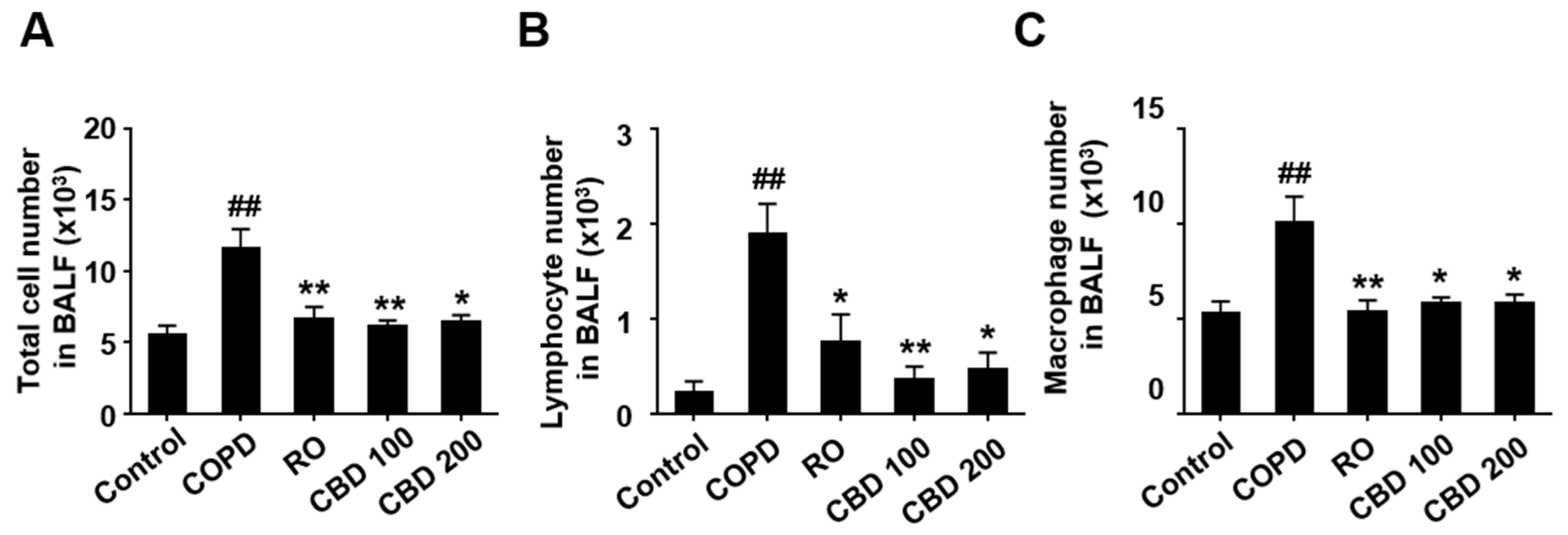
In comparison to mice treated with saline alone (control group), mice treated with CS plus LPS (COPD group) had a considerably larger total number of cells in their BALF (p < 0.001). In addition, CS- and LPS-treated mice (COPD group) had a much larger inflow of lymphocytes (p < 0.001) and macrophages (p = 0.003) than mice in the control group. Treatment with RO (total cell: p = 0.004, lymphocyte: p = 0.017, and macrophage: p = 0.002), CBD 100 (total cell: p = 0.004, lymphocyte: p = 0.002, and macrophage: p = 0.013), or CBD 200 (total cell: p = 0.020, lymphocyte: p = 0.026, and macrophage: p = 0.036) led to an attenuation of these effects of CS and LPS (Figure 2).

3.3. Lung Acute Inflammation and CBD

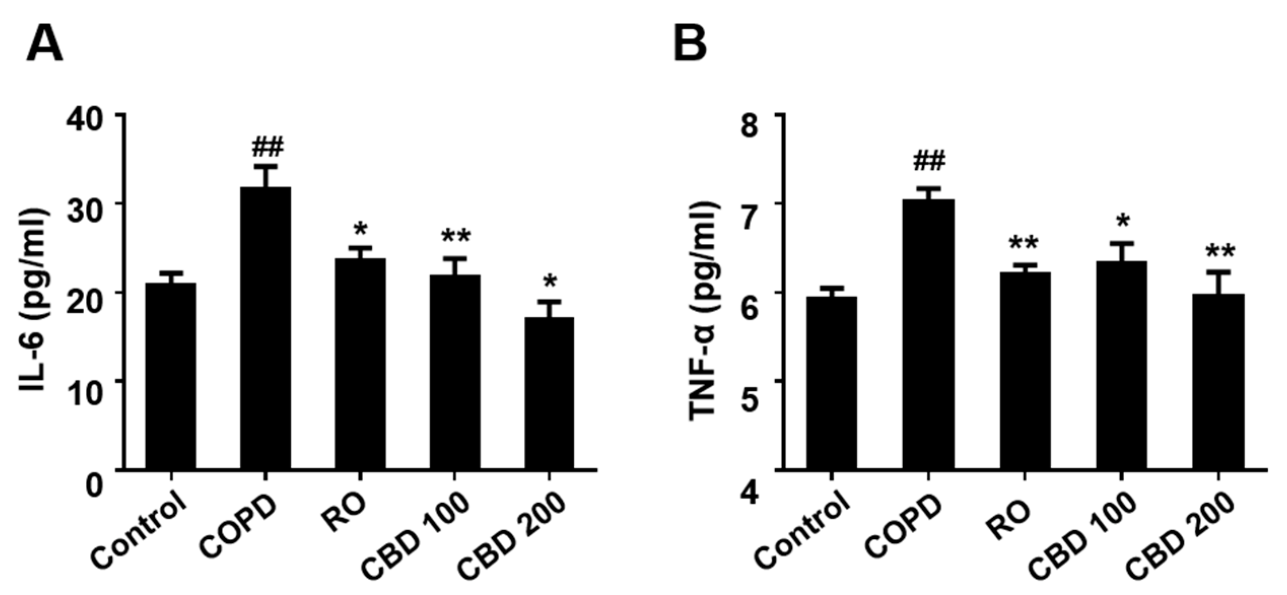
Compared to mice treated with saline (control group), animals treated with CS and LPS (COPD group) had increased levels of IL-6 (p = 0.003) and TNF-α (p < 0.001) in their BALF. Treatment with RO (IL-6: p = 0.039 and TNF-α: p = 0.008), CBD 100 (IL-6: p = 0.008 and TNF-α: p = 0.034), or CBD 200 (IL-6: p < 0.001 and TNF-α: p = 0.002) greatly lowered these elevations (Figure 3).

3.4. Effect of CBD on Gene Expression Associated with Inflammation

Mice treated with CS and LPS (COPD group) had considerably greater mRNA levels of IL-1β (p < 0.001), IL-6 (p = 0.023), TNF-α (p = 0.006), and MCP-1 (p = 0.001) than untreated mice (control group) (Figure 4). However, therapy with RO (IL-1β: p = 0.002, IL-6: p < 0.001, TNF-α: p = 0.010, and MCP-1: p = 0.010), CBD 100 (IL-1β: p < 0.001, IL-6: p < 0.001, TNF-α: p = 0.005, and MCP-1: p = 0.018), or CBD 200 (IL-1β: p < 0.001, IL-6: p < 0.001, TNF-α: p = 0.014, and MCP-1: p = 0.003) greatly reduced these increases (Figure 4).

3.5. Effect of CBD on Gene Expression Associated with Pulmonary Fibrosis

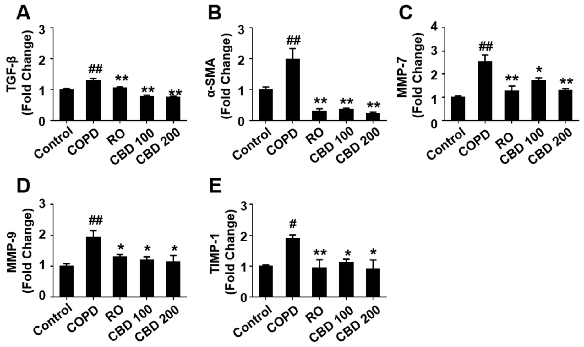
RT-PCR analysis of the pulmonary expression of TGF-β (p < 0.001), α-SMA (p = 0.009), MMP-7 (p < 0.001), MMP-9 (p < 0.001), and TIMP-1 (p = 0.011) mRNAs showed that exposure to CS and LPS (COPD group) induced significant increases in the expression of these genes in lung tissues, compared with saline-exposed mice (control group) (Figure 5). Treatment with RO (TGF-β: p = 0.009, α-SMA: p < 0.001, MMP-7: p < 0.001, MMP-9: p = 0.023, and TIMP-1: p = 0.007), CBD 100 (TGF-β: p < 0.001, α-SMA: p < 0.001, MMP-7: p = 0.019, MMP-9: p = 0.015, and TIMP-1: p = 0.035), or CBD 200 (TGF-β: p < 0.001, α-SMA: p < 0.001, MMP-7: p < 0.001, MMP-9: p = 0.013, and TIMP-1: p = 0.013), however, significantly attenuated the COPD-induced increases in these mRNA levels (Figure 5).

3.6. Effect of CBD on Phosphorylation Levels of NF-kB and MAPKs

To evaluate whether CBD has therapeutic effects on COPD through the MAPKs and NF-κB signaling pathways, the levels of both the phosphorylated and non-phosphorylated forms of proteins belonging to these pathways were measured in lung tissues. The phosphorylation levels of ERK1/2 (p = 0.009), p38 MAPK (p < 0.001), and NF-κB (p = 0.004) were higher in the COPD group than in the control group, but were significantly lower in mice treated with RO (ERK1/2: p = 0.009, p38 MAPK: p < 0.001, and NF-κB: p = 0.014), CBD 100 (ERK1/2: p = 0.006, p38 MAPK: p = 0.001, and NF-κB: p = 0.012), or CBD 200 (ERK1/2: p = 0.004, p38 MAPK: p < 0.001, and NF-κB: p = 0.021) than in the COPD group (Figure 6).

4. Discussion

The present study demonstrated that the BALF and lung parenchyma of mice experienced inflammatory cell infiltration as a result of exposure to CS and LPS. The influx of lymphocytes and macrophages was accompanied by increased concentrations of IL-6 and TNF-α. Treatment with CBD, however, attenuated these effects of CS and LPS. CBD was found to exert its anti-inflammatory effects in this mouse model of COPD by preventing the phosphorylation and activation of NF-κB and MAPKs, by reducing the production of the proinflammatory mediators IL-1β, IL-6, TNF-α, and MCP-1, and by attenuating the expression of the fibrosis inducers TGF-β, α-SMA, MMP-7, MMP-9, and TIMP-1.
COPD is predominantly caused by cigarette smoking and is defined by pulmonary inflammation, including increases in cells of both the innate and adaptive immune systems. LPS present in CS is a substance that forms the cell membrane of Gram-negative bacteria and causes pulmonary and systemic inflammation in humans [27]. The present study established a COPD model in mice, induced by exposure to CS for 8 weeks and exposure every other week to intranasal LPS. The pathological changes resulting from exposure to CS and LPS were comparable to the clinical aspects observed in patients with COPD [28]. CBD administered by oral gavage to these mice for 4 weeks, 1 h before exposure to CS, was found to be effective in reducing the manifestations of COPD in this model.
The ability of CBD to ameliorate pathological changes in the lungs of CS- and LPS-exposed mice suggests that CBD may be a potent anti-inflammatory agent that inhibits the accumulation of inflammatory cells caused by CS and LPS exposure. The present study demonstrated that mice exposed to CS and LPS had higher amounts of IL-6 and TNF-α in their BALF and lung tissue, whereas CBD-treated mice had much lower levels of these cytokines. Increased BALF IL-6, IL-8, and TNF-α levels in COPD patients are used as indicators of the systemic inflammatory response, and their levels are associated with the severity of the disease [29]. The finding that CBD downregulated the production of proinflammatory cytokines during lung inflammation suggests that CBD therapy may affect downstream processes, such as the inflow of inflammatory cells and the amounts of mediators that exacerbate the inflammatory response.
Additionally, the expression of α-SMA, TGF-β, MMPs, and TIMPs was significantly decreased by CBD. Large levels of TGF-β are created during the onset and maintenance of pulmonary fibrosis, which can lead to additional nearby fibrosis. MCP-1, a recognized inducer of TGF-β, maintains the fibrotic process through the direct control of TGF-β and possibly the ensuing accumulation of myofibroblasts as well [30]. MCP-1 regulation of inflammation may be crucial in the fibrotic response. One of the most important indicators of fibrosis is the protein α-SMA, which TGF-β upregulates [31]. TGF-β enhanced α-SMA expression, along with a decrease in E-cadherin expression, in a dose- and time-dependent manner [32]. These changes result in the secretion of excess collagen-I, leading to pulmonary fibrosis [33]. The expression of α-SMA was shown to be involved in interleukin-1β (IL-1β)-regulated inflammation in rat lung myofibroblasts [34]. MMP-9 and TIMP-1 are also involved in the pathophysiology of COPD, as they degrade the extracellular matrix (ECM) [35]. MMP-9, which is secreted by macrophages originating from neutrophils and from circulating monocytes, is in charge of tissue remodeling and healing through the destruction of basement membrane collagen-IV [36]. Increased MMP-9 activity may be essential in the development of airway fibrosis and emphysema by encouraging the breakdown of alveolar wall basement membranes [37]. TIMP-1 binds to the precursors and active form of MMP-9 to reduce its activity [38]. In COPD patients, higher levels of MMP-9 with higher enzymatic activity are produced as a result of elevated MMP-9 and TIMP-1 expression as well as increased alveolar macrophages. Asymmetry in the levels of MMP-9 and TIMP-1 may cause COPD by generating abnormal ECM breakdown or an accumulation of ECM proteins in the pulmonary alveoli and small airway walls [39]. According to histopathological evidence, CS- and LPS-induced inflammation and fibrosis were alleviated by CBD treatment. Taken together, these findings imply that CBD may be sufficient to stop the structural changes that are typical of COPD, such as pulmonary emphysema and widespread lung inflammation.
Inflammation is controlled at the molecular level by intracellular signaling pathways, such as MAPK and NF-κB [40]. Activation of MAPK and/or NF-κB was found to correlate with the production of proinflammatory mediators. As demonstrated here, mice exposed to CS and LPS had significantly higher levels of IL-1β, IL-6, and TNF-α in their BALF and lung tissue, and this increase in cytokines appears to depend on the NF-κB pathway. Both IL-1β and TNF-α promoted the NF-κB-DNA binding complex, and the combined action of the two cytokines on human pulmonary microvascular endothelial cells increased the magnitude of this effect [41]. The MAPK is also a pathway through which various proinflammatory cytokines, such as IL-1β and TNF-α, serve as key effectors in numerous cellular responses [42,43]. To identify the molecular mechanisms responsible for CBD-induced suppression of inflammatory responses, the ability of CBD to alter MAPK phosphorylation and NF-κB activation in COPD-induced mice was tested. Treatment with CS and LPS markedly increased phosphorylation levels of ERK and p38 MAPK, with both being suppressed by CBD. In addition, CBD reduced nuclear expression of NF-κB and blocked its phosphorylation in the lung tissue of mice exposed to CS and LPS, indicating the crucial roles of MAPKs and NF-κB pathways in CBD-induced anti-inflammatory activity.
CBD contains approximately 15 herbs. The ratios of the components required to create CBD are described very differently in each study in the literature. Rehmannia, the main herb in the blend, makes up between 8% and 32% of the total weight [44]. Iridoids, triterpene glycosides (saponins), and steroid glycosides are the three types of terpene series glycosides found in most herbs. Although distinct terpene glycosides have different properties, the general effects are sedative, antipyretic, and anti-inflammatory [45]. The anti-inflammatory action of the CBD formula is thought to have a significant impact on improving conditions induced by CS and LPS.

5. Conclusions

In summary, our study determined that CBD containing glycosides of the terpene series prevented COPD induced by LPS and CS in mice by lowering inflammatory response and airway remodeling. The capacity of CBD to inhibit the MAPKs and NF-κB pathways may be responsible for these effects. Patients with COPD may benefit from using CBD as a therapy.

Author Contributions

Conceptualization, H.-J.K. and M.-Y.L.; Methodology, E.-J.H.; Formal analysis, J.H.K., E.B.B. and E.-J.H.; Investigation, J.H.K. and E.B.B.; Writing and original draft, H.-J.K. and J.H.K.; Writing, review and editing, E.J., H.-J.K. and J.H.K.; Supervision, H.-J.K.; Project administration, H.-J.K. and M.-Y.L.; Funding acquisition, H.-J.K. and M.-Y.L. All authors have read and agreed to the published version of the manuscript.

Funding

This research was supported by grants from the Korea Institute of Oriental Medicine (grant numbers KSN2013220 and KSN2021220).

Institutional Review Board Statement

The animal study protocol was approved by the Animal Experimental Ethics Committee of Chungnam National University (CNU-01141 and 19 December 2018).

Data Availability Statement

The data that support the findings of this study are available from the corresponding author upon reasonable request.

Conflicts of Interest

The authors declare that they have no conflicts of interest.

References

  1. Global Strategy for Diagnosis, Management, and Prevention of COPD. Available online: https://goldcopd.org/2022-gold-reports/ (accessed on 1 February 2022).
  2. Rabe, K.F.; Watz, H. Chronic obstructive pulmonary disease. Lancet 2017, 389, 1931–1940. [Google Scholar] [CrossRef] [PubMed]
  3. Vestbo, J.; Hurd, S.S.; Agustí, A.G.; Jones, P.W.; Vogelmeier, C.; Anzueto, A.; Barnes, P.J.; Fabbri, L.M.; Martinez, F.J.; Nishimura, M. Global strategy for the diagnosis, management, and prevention of chronic obstructive pulmonary disease: GOLD executive summary. Am. J. Respir. Crit. Care Med. 2013, 187, 347–365. [Google Scholar] [CrossRef] [PubMed]
  4. Li, J.; Zhao, P.; Tian, Y.; Li, K.; Zhang, L.; Guan, Q.; Mei, X.; Qin, Y. The anti-inflammatory effect of a combination of five compounds from five Chinese herbal medicines used in the treatment of COPD. Front. Pharmacol. 2021, 12, 709702. [Google Scholar] [CrossRef] [PubMed]
  5. Lemire, B.B.; Debigaré, R.; Dubé, A.; Thériault, M.-E.; Côté, C.H.; Maltais, F. MAPK signaling in the quadriceps of patients with chronic obstructive pulmonary disease. J. Appl. Physiol. 2012, 113, 159–166. [Google Scholar] [CrossRef] [PubMed] [Green Version]
  6. Zhang, J.; Yang, C.; Deng, F. MicroRNA-378 inhibits the development of smoking-induced COPD by targeting TNF-α. Eur Rev Med. Pharmacol. Sci. 2019, 23, 9009–9016. [Google Scholar]
  7. Calixto, J.B.; Campos, M.M.; Otuki, M.F.; Santos, A.R. Anti-inflammatory compounds of plant origin. Part II. Modulation of pro-inflammatory cytokines, chemokines and adhesion molecules. Planta Med. 2004, 70, 93–103. [Google Scholar]
  8. Guo, R.; Pittler, M.; Ernst, E. Herbal medicines for the treatment of COPD: A systematic review. Eur. Respir. J. 2006, 28, 330–338. [Google Scholar] [CrossRef] [Green Version]
  9. Kim, O.-S.; Kim, Y.; Yoo, S.-R.; Lee, M.-Y.; Shin, H.-K.; Jeong, S.-J. Cheonwangbosimdan, a traditional herbal formula, inhibits inflammatory responses through inactivation of NF-κB and induction of heme oxygenase-1 in raw264 7 murine macrophages. Int. J. Clin. Exp. Med. 2016, 9, 1692–1699. [Google Scholar]
  10. Kim, N.-J.; Kong, Y.-Y.; Chang, S.-W. Studies on the Efficacy of Combined Preparation of Crude Drugs (XXXVII)-The effects of Chunwangboshimdan on the central nervous system and cardio-vascular system. Korean J. Pharmacogn. 1988, 19, 208–215. [Google Scholar]
  11. Kim, B.; Jo, C.; Choi, H.-Y.; Lee, K. Vasorelaxant and hypotensive effects of Cheonwangbosimdan in SD and SHR rats. Evid. Based Complement. Alternat. Med. 2018, 2018, 6128604. [Google Scholar] [CrossRef] [Green Version]
  12. Gim, G.T.; Kim, H.-M.; Kim, J.; Kim, J.; Whang, W.-W.; Cho, S.-H. Antioxidant effect of tianwang buxin pills a traditional Chinese medicine formula: Double-blind, randomized controlled trial. Am. J. Chin. Med. 2009, 37, 227–239. [Google Scholar] [CrossRef]
  13. Park, J.-H.; Bae, C.-w.; Jun, H.-S.; Hong, S.-Y.; Park, S.-D. Antidepressant effect of chunwangboshimdan and its influence on monoamines. Herb. Formula Sci. 2004, 12, 77–93. [Google Scholar]
  14. Zhu, B.; Wang, Y.; Ming, J.; Chen, W.; Zhang, L. Disease burden of COPD in China: A systematic review. Int. J. Chron. Obstruct. Pulmon. Dis. 2018, 13, 1353. [Google Scholar] [CrossRef] [Green Version]
  15. Hou, W.; Hu, S.; Li, C.; Ma, H.; Wang, Q.; Meng, G.; Guo, T.; Zhang, J. Cigarette smoke induced lung barrier dysfunction, EMT, and tissue remodeling: A possible link between COPD and lung cancer. BioMed Res. Int. 2019, 2019, 2025636. [Google Scholar] [CrossRef]
  16. Leopold, P.L.; O’Mahony, M.J.; Lian, X.J.; Tilley, A.E.; Harvey, B.-G.; Crystal, R.G. Smoking is associated with shortened airway cilia. PLoS ONE 2009, 4, e8157. [Google Scholar] [CrossRef]
  17. Zong, D.; Liu, X.; Li, J.; Ouyang, R.; Chen, P. The role of cigarette smoke-induced epigenetic alterations in inflammation. Epigenetics Chromatin 2019, 12, 1–25. [Google Scholar] [CrossRef] [Green Version]
  18. Maestrelli, P.; Saetta, M.; Mapp, C.E.; Fabbri, L.M. Remodeling in response to infection and injury: Airway inflammation and hypersecretion of mucus in smoking subjects with chronic obstructive pulmonary disease. Am. J. Respir. Crit. Care Med 2001, 164, S76–S80. [Google Scholar] [CrossRef]
  19. van Eeden, S.F.; Hogg, J.C. Immune-modulation in chronic obstructive pulmonary disease: Current concepts and future strategies. Respiration 2020, 99, 550–565. [Google Scholar] [CrossRef]
  20. Gharaee-Kermani, M.; Ullenbruch, M.; Phan, S.H. Animal models of pulmonary fibrosis. In Fibrosis Research; Springer: Berlin, Germany, 2005; pp. 251–259. [Google Scholar]
  21. Hsueh, W.; Sun, X.; Rioja, L.; Gonzalez-Crussi, F. The role of the complement system in shock and tissue injury induced by tumour necrosis factor and endotoxin. Immunology 1990, 70, 309. [Google Scholar]
  22. Barnes, P.J. Mechanisms in COPD: Differences from asthma. Chest 2000, 117, 10S–14S. [Google Scholar] [CrossRef]
  23. Rogers, D. Mucus pathophysiology in COPD: Differences to asthma, and pharmacotherapy. Monaldi. Arch. Chest. Dis. 2000, 55, 324–332. [Google Scholar] [PubMed]
  24. Park, H.-S.; Jeong, H.-Y.; Kim, Y.-S.; Seo, C.-S.; Ha, H.; Kwon, H.-J. Anti-microbial and anti-inflammatory effects of Cheonwangbosim-dan against Helicobacter pylori-induced gastritis. J. Vet. Sci. 2020, 21, e39. [Google Scholar] [CrossRef]
  25. Wijerathne, C.U.; Seo, C.-S.; Song, J.-W.; Park, H.-S.; Moon, O.-S.; Won, Y.-S.; Kwon, H.-J.; Son, H.-Y. Isoimperatorin attenuates airway inflammation and mucus hypersecretion in an ovalbumin-induced murine model of asthma. Int. Immunopharmacol. 2017, 49, 67–76. [Google Scholar] [CrossRef] [PubMed]
  26. Rho, J.; Seo, C.-S.; Hong, E.-J.; Baek, E.B.; Jung, E.; Park, S.; Lee, M.-Y.; Kwun, H.-J. Yijin-Tang Attenuates Cigarette Smoke and Lipopolysaccharide-Induced Chronic Obstructive Pulmonary Disease in Mice. Evid. Based Complement Alternat. Med. 2022, 2022, 7902920. [Google Scholar] [CrossRef] [PubMed]
  27. Hasday, J.D.; Bascom, R.; Costa, J.J.; Fitzgerald, T.; Dubin, W. Bacterial endotoxin is an active component of cigarette smoke. Chest 1999, 115, 829–835. [Google Scholar] [CrossRef]
  28. Chung, K. Cytokines in chronic obstructive pulmonary disease. Eur. Respir. J. 2001, 18, 50s–59s. [Google Scholar] [CrossRef]
  29. El-Shimy, W.S.; El-Dib, A.S.; Nagy, H.M.; Sabry, W. A study of IL-6, IL-8, and TNF-α as inflammatory markers in COPD patients. Egyp. J. Bronchol. 2014, 8, 91–99. [Google Scholar] [CrossRef]
  30. Ferreira, A.M.; Takagawa, S.; Fresco, R.; Zhu, X.; Varga, J.; DiPietro, L.A. Diminished induction of skin fibrosis in mice with MCP-1 deficiency. J. Investig. Dermatol. 2006, 126, 1900–1908. [Google Scholar] [CrossRef] [Green Version]
  31. Hu, B.; Wu, Z.; Phan, S.H. Smad3 mediates transforming growth factor-β–induced α-smooth muscle actin expression. Am. J. Respir. Cell Mol. Biol. 2003, 29, 397–404. [Google Scholar] [CrossRef] [Green Version]
  32. Ding, H.; Chen, J.; Qin, J.; Chen, R.; Yi, Z. TGF-β-induced α-SMA expression is mediated by C/EBPβ acetylation in human alveolar epithelial cells. Mol. Med. 2021, 27, 1–12. [Google Scholar]
  33. Ma, W.-h.; Li, M.; Ma, H.-f.; Li, W.; Liu, L.; Yin, Y.; Zhou, X.-m.; Hou, G. Protective effects of GHK-Cu in bleomycin-induced pulmonary fibrosis via anti-oxidative stress and anti-inflammation pathways. Life Sci. 2020, 241, 117139. [Google Scholar] [CrossRef]
  34. Hu, B.; Wu, Z.; Jin, H.; Hashimoto, N.; Liu, T.; Phan, S.H. CCAAT/enhancer-binding protein β isoforms and the regulation of α-smooth muscle actin gene expression by IL-1β. J. Immunol. 2004, 173, 4661–4668. [Google Scholar] [CrossRef] [Green Version]
  35. Li, Y.; Lu, Y.; Zhao, Z.; Wang, J.; Li, J.; Wang, W.; Li, S.; Song, L. Relationships of MMP-9 and TIMP-1 proteins with chronic obstructive pulmonary disease risk: A systematic review and meta-analysis. J. Res. Med. Sci. 2016, 21, 12. [Google Scholar] [CrossRef]
  36. Brajer, B.; Batura-Gabryel, H.; Nowicka, A.; Kuznar-Kaminska, B.; Szczepanik, A. Concentration of matrix metalloproteinase-9 in serum of patients with chronic obstructive pulmonary disease and a degree of airway obstruction and disease progression. J. Physiol. Pharmacol. 2008, 59, 145–152. [Google Scholar]
  37. Lindberg, A.; Larsson, L.-G.; Muellerova, H.; Rönmark, E.; Lundbäck, B. Up-to-date on mortality in COPD-report from the OLIN COPD study. BMC Pulm. Med. 2012, 12, 1–7. [Google Scholar] [CrossRef] [Green Version]
  38. Zhuang, Y.; Qian, Z.; Huang, L. Elevated expression levels of matrix metalloproteinase-9 in placental villi and tissue inhibitor of metalloproteinase-2 in decidua are associated with prolonged bleeding after mifepristone-misoprostol medical abortion. Fertil. Steril. 2014, 101, 166–171.e162. [Google Scholar] [CrossRef]
  39. Tency, I.; Verstraelen, H.; Kroes, I.; Holtappels, G.; Verhasselt, B.; Vaneechoutte, M.; Verhelst, R.; Temmerman, M. Imbalances between matrix metalloproteinases (MMPs) and tissue inhibitor of metalloproteinases (TIMPs) in maternal serum during preterm labor. PLoS ONE 2012, 7, e49042. [Google Scholar] [CrossRef] [Green Version]
  40. Saklatvala, J.; Nagase, H.; Salvesen, G.; Dean, J.; Clark, A. Control of the expression of inflammatory response genes. In Proceedings of the Biochemical Society Symposia; Portland Press: London, UK, 2003; pp. 95–106. [Google Scholar]
  41. Said, F.A.; Werts, C.; Elalamy, I.; Couetil, J.P.; Jacquemin, C.; Hatmi, M. TNF-α, inefficient by itself, potentiates IL-1β-induced PGHS-2 expression in human pulmonary microvascular endothelial cells: Requirement of NF-κB and p38 MAPK pathways. Br. J. Pharmacol. 2002, 136, 1005–1014. [Google Scholar] [CrossRef]
  42. Kyriakis, J.M.; Avruch, J. Protein kinase cascades activated by stress and inflammatory cytokines. Bioessays 1996, 18, 567–577. [Google Scholar] [CrossRef] [PubMed]
  43. Wang, Y.-Z.; Zhang, P.; Rice, A.B.; Bonner, J.C. Regulation of interleukin-1β-induced platelet-derived growth factor receptor-α expression in rat pulmonary myofibroblasts by p38 mitogen-activated protein kinase. J. Biol. Chem. 2000, 275, 22550–22557. [Google Scholar] [CrossRef]
  44. Bingshan, H.; Yuxia, W. Thousand Formulas and Thousand Herbs of Traditional Chinese Medicine; Heilongjiang Education Press: Harbin, China, 1993; Volume 1. [Google Scholar]
  45. Cai, J.; Chao, G.; Chen, D. State Administration of Traditional Chinese Medicine: Advanced Textbook on Traditional Chinese Medicine and Pharmacology; New World Press: Beijing, China, 1997; Volume IV. [Google Scholar]
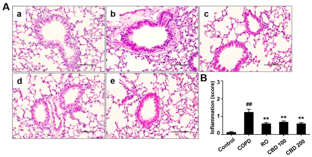
Figure 1. Effects of CBD on lung tissue histological damage. (A) H&E staining. (B) The degree of inflammation. (a). Control: mice instilled intratracheally with normal saline; (b). COPD: mice exposed to CS and LPS; (c). RO: mice exposed to 10 mg/kg roflumilast plus CS and LPS; (d). CBD 100: mice exposed to 100 mg/kg CBD plus CS and LPS; (e). CBD 200: mice exposed to 200 mg/kg CBD plus CS and LPS. Results are presented as means ± SEMs. ## p < 0.01 compared with the control group; ** p < 0.01 compared with the COPD group. Scale bar 100 μm.
Applsci 13 01829 g001
Figure 2. Effects of CBD on inflammatory cell recruitment to COPD-induced mice BALF. (A) Number of total cells. (B) Number of lymphocytes. (C) Number of macrophages. Control: mice instilled intratracheally with normal saline; COPD: mice exposed to CS and LPS; RO: mice exposed to 10 mg/kg roflumilast plus CS and LPS; CBD 100: mice exposed to 100 mg/kg CBD plus CS and LPS; CBD 200: mice exposed to 200 mg/kg CBD plus CS and LPS. Results are presented as means ± SEMs. ## p < 0.01 compared with the control group; * p < 0.05, ** p < 0.01 compared with the COPD group.
Applsci 13 01829 g002
Figure 3. Effects of CBD on concentrations of proinflammatory cytokines in BALF. Individual samples were analyzed using ELISA. (A) IL-6. (B) TNF-α. Control: mice instilled intratracheally with normal saline; COPD: mice exposed to CS and LPS; RO: mice exposed to 10 mg/kg roflumilast plus CS and LPS; CBD 100: mice exposed to 100 mg/kg CBD plus CS and LPS; CBD 200: mice exposed to 200 mg/kg CBD plus CS and LPS. Results are presented as means ± SEMs. ## p < 0.01 compared with the control group; * p < 0.05, ** p < 0.01 compared with the COPD group.
Applsci 13 01829 g003
Figure 4. Analysis of mRNA levels of inflammatory cytokines in lung tissues. Expression of (A) IL-1β, (B) IL-6, (C) TNF-α, and (D) MCP-1 mRNAs was measured by RT-PCR. Control: mice instilled intratracheally with normal saline; COPD: mice exposed to CS and LPS; RO: mice exposed to 10 mg/kg roflumilast plus CS and LPS; CBD 100: mice exposed to 100 mg/kg CBD plus CS and LPS; CBD 200: mice exposed to 200 mg/kg CBD plus CS and LPS. Results are presented as means ± SEMs. # p < 0.05, ## p < 0.01 compared with the control group; * p < 0.05, ** p < 0.01 compared with the COPD group.
Applsci 13 01829 g004
Figure 5. Effects of CBD on levels of mRNAs encoding pulmonary-fibrosis-related genes in lung tissues. Levels of (A) TGF-β, (B) α-SMA, (C) MMP-7, (D) MMP-9, and (E) TIMP-1 mRNAs were measured by RT-PCR. Control: mice instilled intratracheally with normal saline; COPD: mice exposed to CS and LPS; RO: mice exposed to 10 mg/kg roflumilast plus CS and LPS; CBD 100: mice exposed to 100 mg/kg CBD plus CS and LPS; CBD 200: mice exposed to 200 mg/kg CBD plus CS and LPS. Results are presented as means ± SEMs. # p < 0.05, ## p < 0.01 compared with the control group; * p < 0.05, ** p < 0.01 compared with the COPD group.
Applsci 13 01829 g005
Figure 6. NF-kB and MAPK activity in lung tissues resulting from CBD effects. Western blotting showing the levels of phosphorylation of ERK, p38 MAPK, and NF-κB in lung tissues. Control: mice instilled intratracheally with normal saline; COPD: mice exposed to CS and LPS; RO: mice exposed to 10 mg/kg roflumilast plus CS and LPS; CBD 100: mice exposed to 100 mg/kg CBD plus CS and LPS; CBD 200: mice exposed to 200 mg/kg CBD plus CS and LPS. Results are presented as means ± SEMs. ## p < 0.01 compared with the control group; * p < 0.05, ** p < 0.01 compared with the COPD group.
Applsci 13 01829 g006
Table 1. Primers used to amplify target genes.
Gene IDGenePrimer
NM_007392α-SMAForward: 5′-TGCTGACAGAGGCACCACTGAA-3′
Reverse: 5′-CAGTTGTACGTCCAGAGGCATAG-3′
NM_031512IL-1βForward: 5′-AGGACCCAAGCACCTTCTTT-3′
Reverse: 5′-AGACAGCACGAGGCATTTT-3′
NM_012589IL-6Forward: 5′-TAGTCCTTCCTACCCCAACT-3′
Reverse: 5′-TTGGTCCTTAGCCACTCCTT-3′
NM_001278601TNF-αForward: 5′- CATGAGCACAGAAAGCATGA -3′
Reverse: 5′- AAGCAGGAATGAGAAGAGGC -3′
NM_011333MCP-1Forward: 5′-GCATCCACGTGTTGGCTCA-3′
Reverse: 5′-CTCCAGCCTACTTCATTGGGATC-3′
NM_013599MMP-9Forward: 5′-GTTTTTGATGCTATTGCTGA-3′
Reverse: 5′- ACCCAACTTATCCAGATCC-3′
NM_010810MMP-7Forward: 5′- AGGTGTGGAGTGCCAGATGTTG -3′
Reverse: 5′- CCACTACGATCCGAGGTAAGTC -3′
NM_011577.2TGF-βForward: 5-TTGCTTCAGCTCCACAGAGA-3′
Reverse: 5-TGGTTGTAGAGGGCAAGGAC-3′
NM_017008.3GAPDHForward: 5-ACAGCAACAGGGTGGTGGAC-3′
Reverse: 5-TTTGAGGGTGCAGCGAACTT-3′
Disclaimer/Publisher’s Note: The statements, opinions and data contained in all publications are solely those of the individual author(s) and contributor(s) and not of MDPI and/or the editor(s). MDPI and/or the editor(s) disclaim responsibility for any injury to people or property resulting from any ideas, methods, instructions or products referred to in the content.