
“A Woman Clothed with the Sun”: The Diagnostic Study and Testing of Enzyme-Based Green Products for the Restoration of an Early 17th Century Wall Painting in the Palazzo Gallo in Bagnaia (Italy)


Abstract
:Featured Application
Abstract
1. Introduction
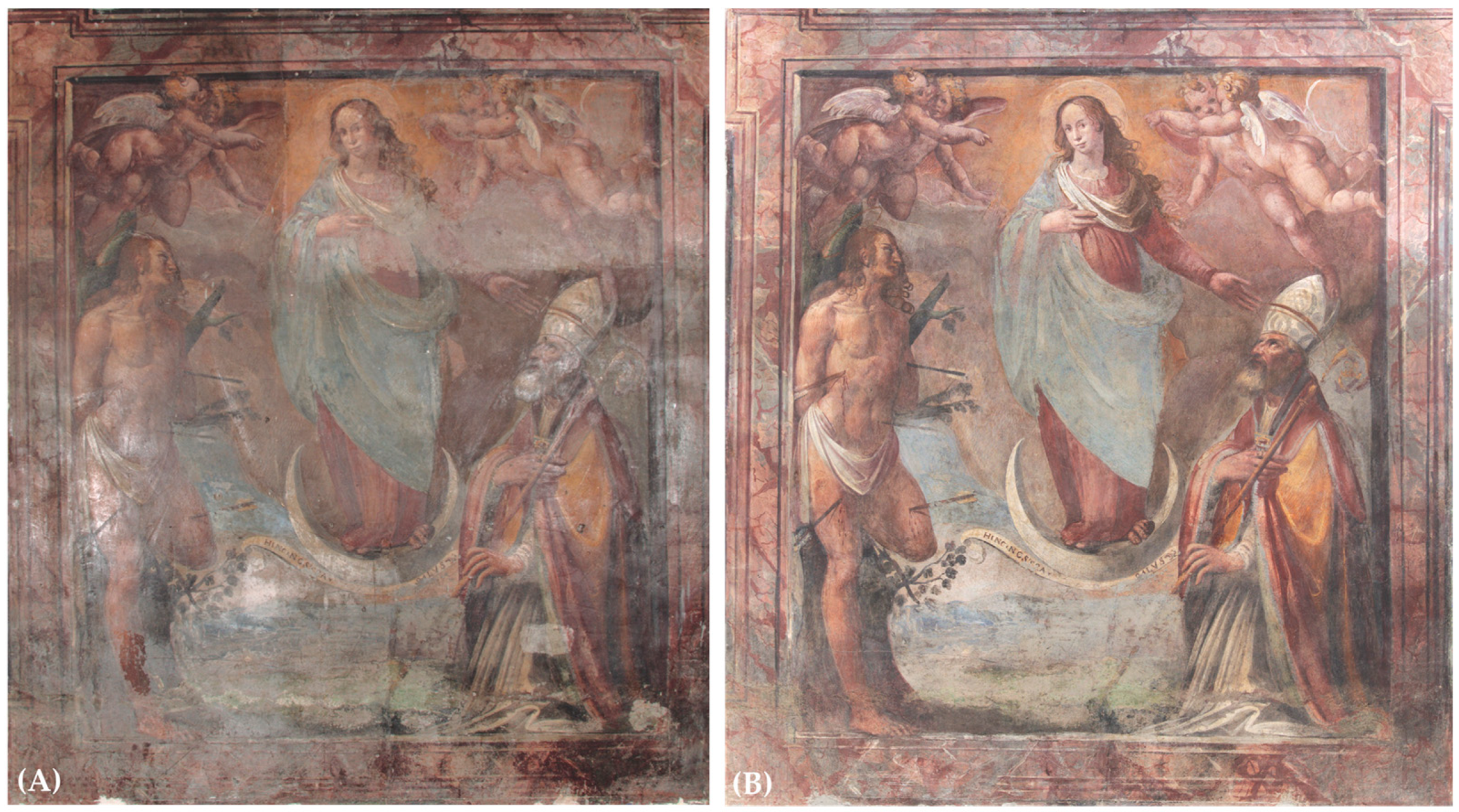
1.1. The Case Study
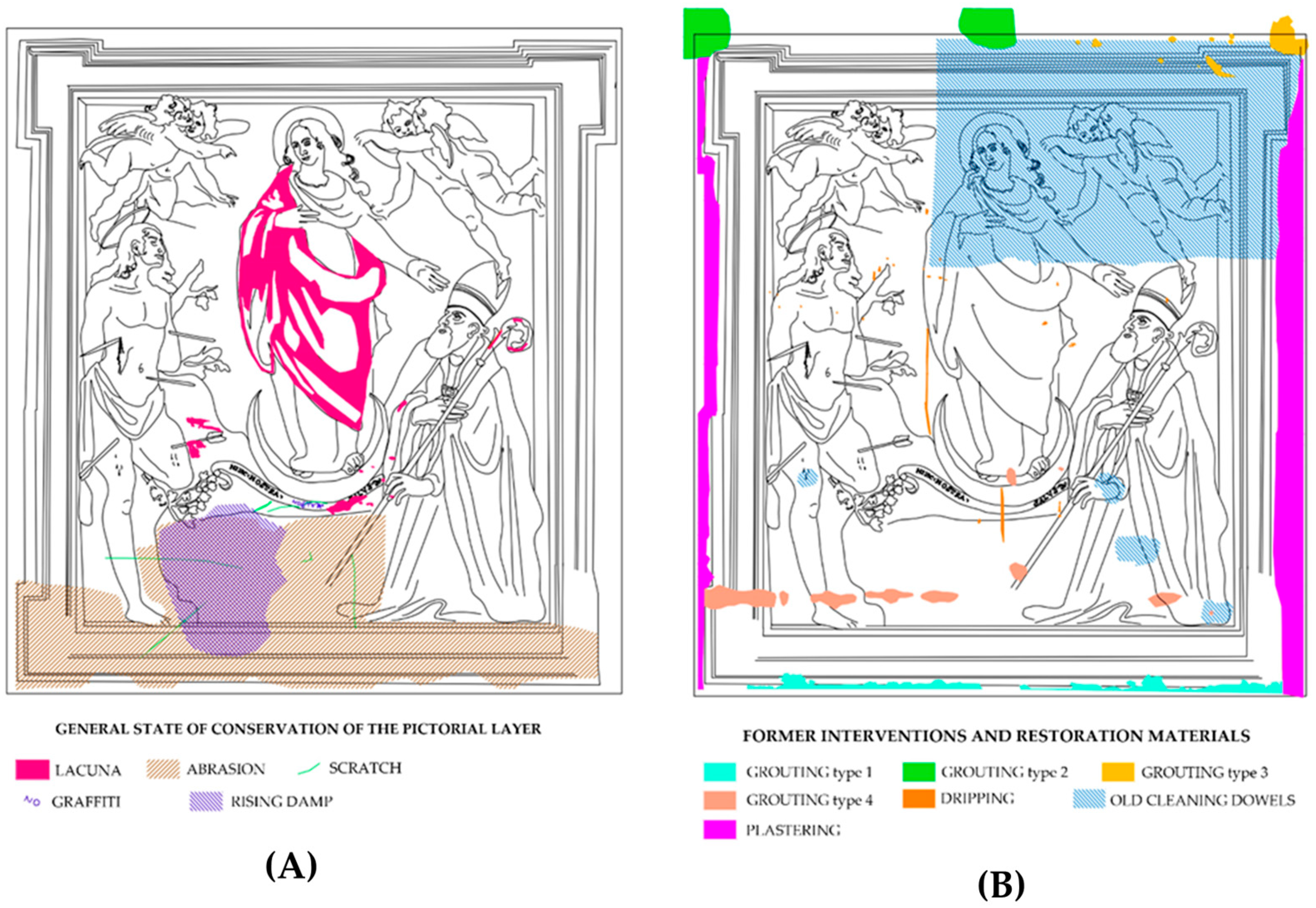
1.2. The Conservation History of the Wall Painting
1.3. The Aims of the Present Research
2. Materials and Methods
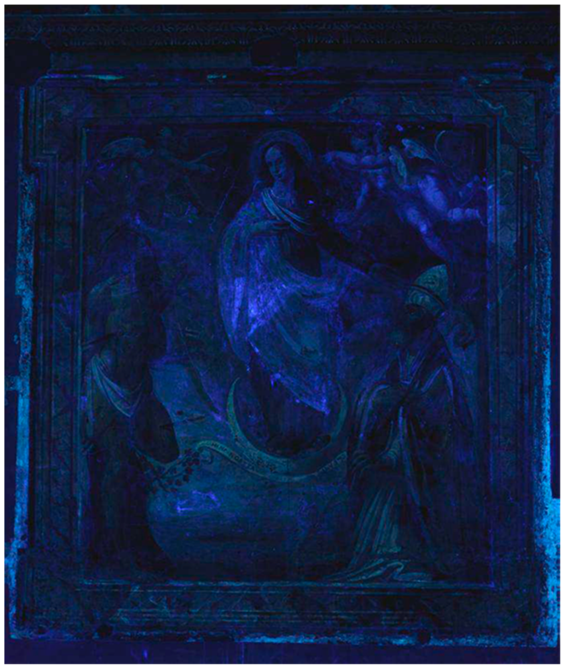
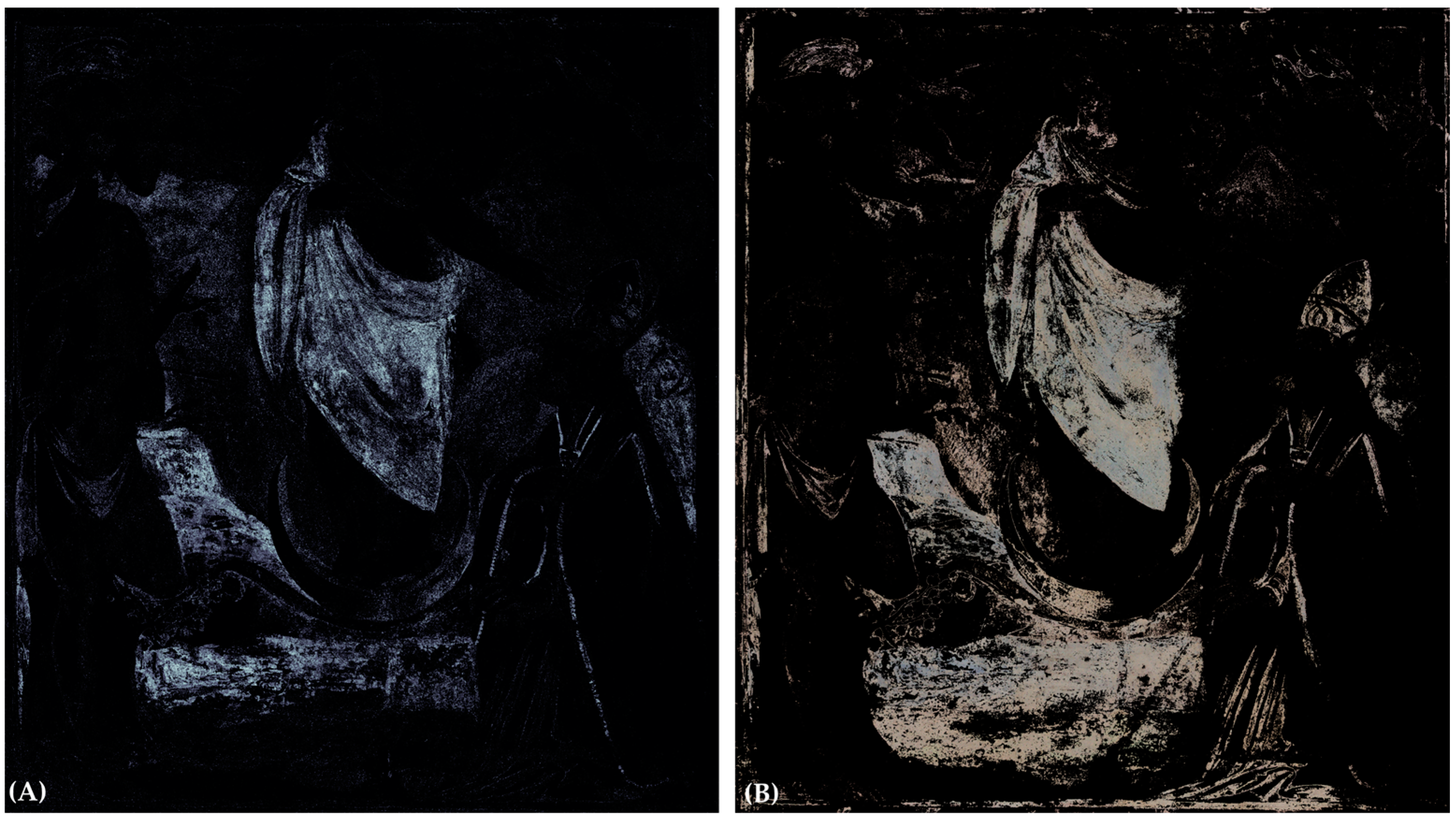
2.1. Hypercolorimetric Multispectral Imaging (HMI) and Ultraviolet Fluorescence Photography (UVF)
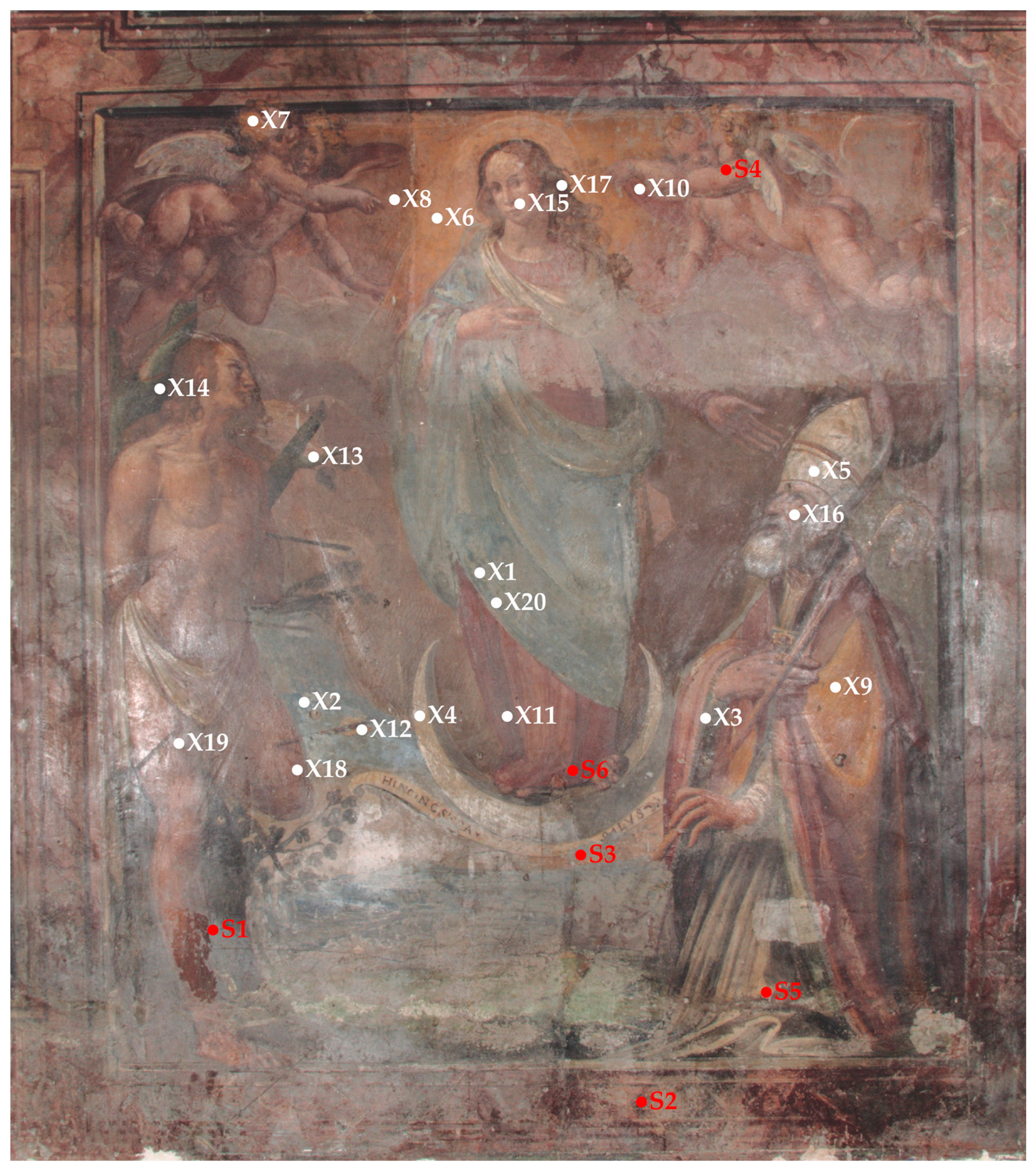
2.2. X-ray Fluorescence Spectroscopy (XRF)
2.3. Laboratory Analysis of Micro-Samples
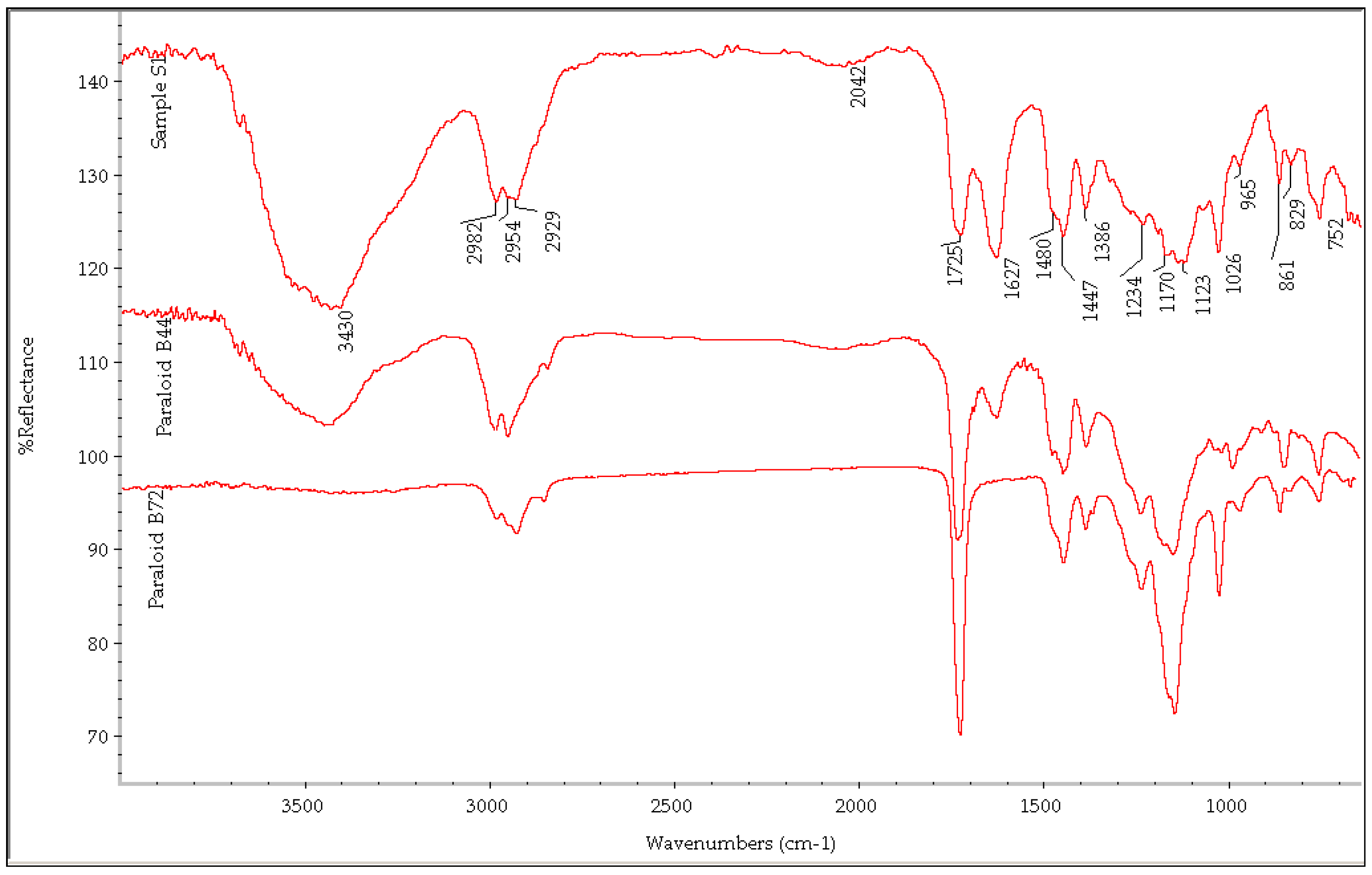
2.3.1. Fourier Transform Infrared Spectroscopy (FTIR)
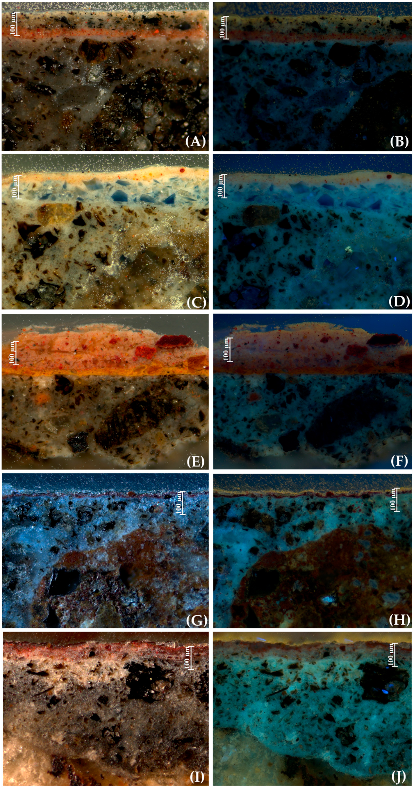
2.3.2. Microstratigraphic Analysis of the Cross-Sections
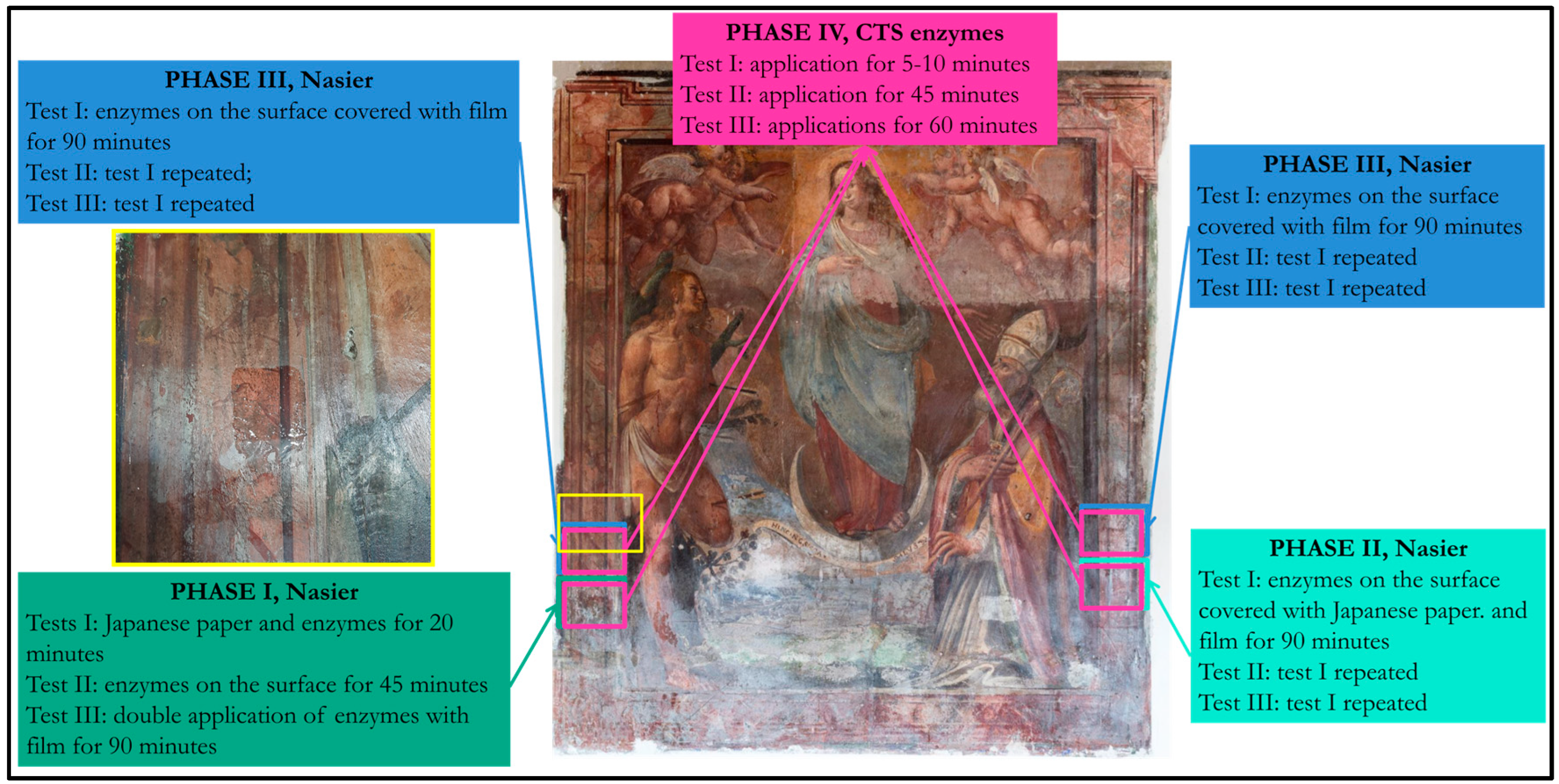
2.4. Testing of Enzyme-Based Cleaning Products
3. Results
3.1. HMI and UVF Results
3.2. XRF Data
3.3. Results of the Analysis of the Micro-Samples
3.3.1. FTIR Data
3.3.2. Result of Microstratigraphic Analysis
3.4. Results of the Cleaning Tests
4. Discussion
Author Contributions
Funding
Institutional Review Board Statement
Informed Consent Statement
Data Availability Statement
Acknowledgments
Conflicts of Interest
References
- Serone, G. Le mura medievali di Bagnaia. Analisi diacronica di un manufatto architettonico. In Il Tesoro Delle Città. Strenna dell’Associazione Storia Delle Città; Edizioni Kappa: Roma, Italy, 2007; pp. 473–491. [Google Scholar]
- Pezzuto, L. (Ed.) La Roma di Raffaele Riario tra XV e XVI secolo. In Cultura Antiquaria e Quartieri Decorativi; Fascicolo 1; Horti Hesperidum: Rome, Italy, 2017. [Google Scholar]
- Vettraino, R.; Valentini, V.; Pogliani, P.; Ricci, M.; Laureti, S.; Calvelli, S.; Zito, R.; Lanteri, L.; Pelosi, C. Multi-Technique Approach by Traditional and Innovative Methodologies to Support the Restoration of a Wall Painting from the 16th Century at Palazzo Gallo in Bagnaia, Viterbo, Central Italy. Buildings 2023, 13, 783. [Google Scholar] [CrossRef]
- Ricci, S. Bagnaia e Villa Lante. La Fortuna Riflessa di un Centro Minore del Lazio; Edizioni Saverio Ricci: San Gemini, Italy, 2017. [Google Scholar]
- Hibbard, H. Guido Reni’s Painting of the Immaculate Conception. Metrop. Mus. Art Bull. 1969, 28, 19–32. [Google Scholar] [CrossRef]
- Morello, G.; Francia, V.; Fusco, R. (Eds.) Una Donna Vestita di Sole. L’Immacolata Concezione Nelle Opere dei Grandi Maestri, 1st ed.; Motta: Milano, Italy, 2005; p. 238. [Google Scholar]
- Röttgen, H. (Ed.) Il Cavalier d’Arpino; De Luca: Roma, Italy, 1973; pp. 39, 78–79, 110–111, 163. [Google Scholar]
- Mancini, C.; Profili, S. Il giovane Marzio Ganassini firma gli affreschi della Cappella Baglioni di Sipicciano. Bibl. E Soc. 2019, 72, 42–51. [Google Scholar]
- Pintus, V.; Schreiner, M. Characterization and identification of acrylic binding media: Influence of UV light on the ageing process. Anal. Bioanal. Chem. 2011, 399, 2961–2976. [Google Scholar] [CrossRef] [PubMed]
- Wendelbo, O.; Fosse, B. Protein “Surgey” A Restoring Procedure Applied on Paper. Restaurator 1970, 1, 245–248. [Google Scholar]
- Segal, J.; Cooper, D. The use of enzymes to release adhesives. Pap. Conserv. 1977, 2, 47–50. [Google Scholar] [CrossRef]
- Cooper, D.; Segal, J. The use of enzyme in partially non aqueous media. In Conservation of Library and Archive Materials and the Graphic Arts, 1st ed.; Butterworth: London, UK, 1980; pp. 25–30. [Google Scholar]
- Bellucci, R.; Cremonesi, P.; Pignagnoli, G. A note on the use of enzymes in conservation. A preliminary report on the removal of aged acrylic resin coatings with Lipase. Stud. Conserv. 1999, 44, 278–281. [Google Scholar]
- Beutel, S.; Klein, K.; Knobbe, G.; Königfeld, P.; Petersen, K.; Ulber, R.; Scheper, T. Controlled enzymatic removal of damaging casein layers on medieval wall paintings. Biotechnol. Bioeng. 2002, 80, 13–21. [Google Scholar] [CrossRef]
- Ranalli, G.; Alfano, G.; Belli, C.; Lustrato, G.; Colombini, M.P.; Bonaduce, I.; Zanardini, E.; Abbruscato, P.; Cappitelli, F.; Sorlini, C. Biotechnology applied to cultural heritage: Biorestoration of frescoes using viable bacterial cells and enzymes. J. Appl. Microbiol. 2005, 98, 73–83. [Google Scholar] [CrossRef]
- Ahmed, H.E.; Kolisis, F.N. An investigation into the removal of starch paste adhesives from historical textiles by using the enzyme α-amylase. J. Cult. Herit. 2011, 12, 169–179. [Google Scholar] [CrossRef]
- Pereira, C.; Busani, T.; Branco, L.C.; Joosten, I.; Sandu, I.C.A. Nondestructive characterization and enzyme cleaning of painted surfaces: Assessment from the macro to nano level. Microsc. Microanal. 2013, 19, 1632–1644. [Google Scholar] [CrossRef] [PubMed]
- Palla, F. Blue-biotechnology and Biocleaning of historic-artistic artifacts. Conserv. Sci. Cult. Herit. 2016, 16, 185–196. [Google Scholar]
- Jeszeová, L.; Bauerová-Hlinková, V.; Baráth, P.; Puškárová, A.; Bučková, M.; Kraková, L.; Pangallo, D. Biochemical and proteomic characterization of the extracellular enzymatic preparate of Exiguobacterium undae, suitable for efficient animal glue removal. Appl. Microbiol. Biotechnol. 2018, 102, 6525–6536. [Google Scholar] [CrossRef]
- Cremonesi, P.; Casoli, A. Enzymes as tools for conservation of works of art. J. Cult. Herit. 2021, 50, 73–87. [Google Scholar] [CrossRef]
- Ricci, M.; Laureti, S.; Malekmohammadi, H.; Sfarra, S.; Lanteri, L.; Colantonio, C.; Calabrò, G.; Pelosi, C. Surface and Interface Investigation of a 15th Century Wall Painting Using Multispectral Imaging and Pulse-Compression Infrared Thermography. Coatings 2021, 11, 546. [Google Scholar] [CrossRef]
- Groppi, F.; Vigliotti, D.; Lanteri, L.; Agresti, G.; Casoli, A.; Laureti, S.; Ricci, M.; Pelosi, C. Advanced documentation methodologies combined with multi-analytical approach for the preservation and restoration of 18th century architectural decorative elements at Palazzo Nuzzi in Orte (Central Italy). Int. J. Conserv. Sci. 2021, 12, 921–934. [Google Scholar]
- Bonizzoni, L.; Caglio, S.; Galli, A.; Lanteri, L.; Pelosi, C. Materials and Technique: The First Look at Saturnino Gatti. Appl. Sci. 2023, 13, 6842. [Google Scholar] [CrossRef]
- Annarilli, S.; Casoli, A.; Colantonio, C.; Lanteri, L.; Marseglia, A.; Pelosi, C.; Sottile, S. A Multi-Instrument Analysis of the Late 16th Canvas Painting, “Coronation of the Virgin with the Saints Ambrose and Jerome”, Attributed to the Tuscany-Umbria Area to Support the Possibility of Bio-Cleaning Using a Bacteria-Based System. Heritage 2022, 5, 2904–2921. [Google Scholar] [CrossRef]
- Colantonio, C.; Clivet, L.; Laval, E.; Coquinot, Y.; Maury, C.; Melis, M.; Boust, C. Integration of multispectral imaging, XRF mapping and Raman analysis for noninvasive study of illustrated manuscripts: The case study of fifteenth century Humay meets the Princess Humayun Persian masterpiece from Louvre Museum. Eur. Phys. J. Plus 2021, 136, 958. [Google Scholar] [CrossRef]
- Lanteri, L.; Pelosi, C. 2D and 3D ultraviolet fluorescence applications on cultural heritage paintings and objects through a low-cost approach for diagnostics and documentation. In Proceedings of the SPIE 11784, Optics for Arts, Architecture, and Archaeology VIII, Munich, Germany, 8 July 2021; pp. 1–10. [Google Scholar]
- Derrick, M.R.; Stulik, D.C.; Landry, J.M. In Spectroscopy in Conservation Science, 1st ed.; Getty Conservation Institute: Los Angeles, CA, USA, 1999. [Google Scholar]
- Miliani, C.; Rosi, F.; Daveri, A.; Brunetti, B.G. Reflection infrared spectroscopy for the non-invasive in situ study of artists’ pigments. Appl. Phys. A 2012, 106, 295–307. [Google Scholar] [CrossRef]
- Walker, J.; Hodgkins, R.; Berrie, B. On the Surface: Reflectance FTIR Spectroscopy in Cultural Heritage Research. Microsc. Microanal. 2021, 27, 2800–2804. [Google Scholar] [CrossRef]
- Vahur, S.; Teearu, A.; Peets, P.; Joosu, L.; Leito, I. ATR-FT-IR spectral collection of conservation materials in the extended region of 4000–80 cm−1. Anal. Bioanal. Chem. 2016, 408, 3373–3379. [Google Scholar] [CrossRef] [PubMed]
- IRUG. Infrared and Raman User Group. Available online: http://www.irug.org/search-spectral-database (accessed on 18 September 2023).
- Database of ATR-FT-IR Spectra of Various Materials. Available online: https://spectra.chem.ut.ee/ (accessed on 18 September 2023).
- Moon, T.; Schilling, M.R.; Thirkettle, S. A Note on the Use of False-Color Infrared Photography in Conservation. Stud. Conserv. 1992, 37, 42–52. [Google Scholar] [CrossRef]
- Cosentino, A. Identification of pigments by multispectral imaging a flow chart method. Herit. Sci. 2014, 2, 8. [Google Scholar] [CrossRef]
- Mühlethaler, B.; Thissen, J. Smalt, in Artists’ Pigments: A Handbook of Their History and Characteristics; Roy, A., Ed.; National Gallery of Art: Washington, DC, USA, 1993; Volume 2, pp. 113–130. [Google Scholar]
- Gratuze, B.; Pactat, I.; Schibille, N. Changes in the signature of cobalt colorants in Late Antique and Early Islamic glass production. Minerals 2018, 8, 225. [Google Scholar] [CrossRef]
- Colomban, P.; Kırmızı, B.; Simsek Franci, G. Cobalt and Associated Impurities in Blue (and Green) Glass, Glaze and Enamel: Relationships between Raw Materials, Processing, Composition, Phases and International Trade. Minerals 2021, 11, 633. [Google Scholar] [CrossRef]
- Vinçotte, A.; Beauvoit, E.; Boyard, N.; Guilminot, E. Effect of solvent on PARALOID® B72 and B44 acrylic resins used as adhesives in conservation. Herit. Sci. 2019, 7, 42. [Google Scholar] [CrossRef]
- Chiantore, O.; Lazzari, M. Characterization of acrylic resins. Int. J. Polym. Anal. Charac. 1996, 2, 395–408. [Google Scholar] [CrossRef]
- Casini, A.; Chelazzi, D.; Baglioni, P. Advanced methodologies for the cleaning of works of art. Sci. China Technol. Sci. 2023, 66, 2162–2182. [Google Scholar] [CrossRef]
- Macchia, A.; Zaratti, C.; Biribicchi, C.; Colasanti, I.A.; Barbaccia, F.I.; Favero, G. Evaluation of Green Solvents’ Applicability for Chromatic Reintegration of Polychrome Artworks. Heritage 2023, 6, 3353–3364. [Google Scholar] [CrossRef]


| Condition of The Surface | T < 12–15 °C | 12–15 °C < T < 25 °C | T > 25 °C |
|---|---|---|---|
| Well-preserved surface | 90 min | 45 min | 45 min + PVC pellicula |
| Not well-preserved surface | 15 min or higher | 15 min or higher | 15 min or higher + PVC pellicula |
| Point | Color | Detected Elements | Hypothesized Pigments |
|---|---|---|---|
| X1 | Blue (clear) | As, Sr, Fe, Ca, Rb, Co, Bi, Zr, Pb | Smalt blue + lead white |
| X2 | Blue (clear) | As, Fe, Sr, Co, Ca, Bi, Zr, Ni, Rb, Pb | Smalt blue + lead white |
| X3 | Blue (clear) | Fe, As, Sr, Ca, Co, Rb, Bi, Ni, Pb, Zr | Smalt blue + earth pigments + lead white |
| X4 | White | Ca, Sr, Fe, As, Rb, Zr, Pb | Calcium carbonate + lead white |
| X5 | White | Ca, Sr, Rb, Zr, Fe, Pb | Calcium carbonate + lead white |
| X6 | Yellow | Fe, Ca, Sr, Rb, Zr, Pb | Calcium carbonate + yellow ochre |
| X7 | Yellow | Fe, Ca, Sr, Rb, Zr, Pb | Calcium carbonate + yellow ochre |
| X8 | Yellow | Fe, Ca, Sr, Rb, Pb | Calcium carbonate + yellow ochre |
| X9 | Yellow | Fe, Sr, Ca, Rb, Pb, As | Yellow ochre |
| X10 | Red | Fe, Ca, Sr, Hg, Rb, Pb | Red earth + cinnabar |
| X11 | Red | Fe, Hg, Ca, Sr, Rb, As, Pb | Red earth + cinnabar |
| X12 | White | Ca, As, Sr, Pb, Bi, Fe, Zr, Co, Rb, Ni | Smalt blue + lead white + earth pigments |
| X13 | Green | Fe, Sr, Ca, As, Pb, Rb, Zr, Co, Bi, Ni | Earth pigment + lead white + smalt blue |
| X14 | Green | Fe, Sr, Ca, Rb, Zr, Pb | Earth pigment + lead white |
| X15 | Pink (clear) | Ca, Fe, Sr, Rb, Pb | Calcium carbonate + earth pigments + lead white |
| X16 | Pink | Ca, Fe, Sr, Hg, Rb, Pb | Earth pigments + cinnabar + lead white |
| X17 | Brown | Fe, Ca, Sr, Rb, Hg, Mn, Pb | Red earth + cinnabar + umber |
| X18 | Red | Ca, Sr, Fe, Rb, Zr, Pb, Mn | Red earth + cinnabar + umber |
| X19 | Red | Fe, Sr, Ca, Zr, Rb, Pb | Red earth + lead white or minimum |
| X20 | Blue (clear) | Sr, Fe, Ca, Zr, Rb, As, Co, Pb, Ni, Bi | Smalt blue + lead white |
| Test Number | Enzyme Mixture | Application Modality | Visible Result |
|---|---|---|---|
| PHASE I–TEST I | Nasier L02 | By brush, on the surface for 20 min, inserting a sheet of Japanese paper between the surface and the enzyme mixture | No result |
| PHASE I–TEST II | Nasier L02 | By brush, directly on the surface for 45 min without paper | No result |
| PHASE I-TEST III | Nasier L02 | By brush, on the surface for 90 min, covering the enzymatic gel with transparent film | Partial removal is visible |
| PHASE II–TEST I | Nasier L02 | By brush, directly on the surface and covered with Japanese paper and transparent film for 90 min | No result, the mixture is too fluid and drips onto the wall |
| PHASE II–TEST II | Nasier L02 | The same as test I | The same result as test I |
| PHASE II–TEST III | Nasier L02 | The same as test I | The same result as test I |
| PHASE III–TEST I | Naser L02 modified | By brush, on the surface for 90 min, covering the enzymatic gel with transparent film—first application | No result |
| PHASE III–TEST II | Nasier L02 modified | By brush, on the surface for 90 min, covering the enzymatic gel with transparent film—second application | Partial removal of the surface material |
| PHASE III–TEST III | Nasier L02 modified | By brush, on the surface for 90 min, covering the enzymatic gel with transparent film—third application | The removal of the surface material is more evident than in Test II, second application |
| PHASE IV–TEST I | CTS product | By brush, for 5 min | No result |
| PHASE IV–TEST II | CTS product | By brush, for 10 min | No result |
| PHASE IV–TEST III | CTS product | By brush, for 45 min | Partial removal of the surface material |
| PHASE IV–TEST IV | CTS product | By brush, for 60 min | Partial removal of the surface material |
Disclaimer/Publisher’s Note: The statements, opinions and data contained in all publications are solely those of the individual author(s) and contributor(s) and not of MDPI and/or the editor(s). MDPI and/or the editor(s) disclaim responsibility for any injury to people or property resulting from any ideas, methods, instructions or products referred to in the content. |
© 2023 by the authors. Licensee MDPI, Basel, Switzerland. This article is an open access article distributed under the terms and conditions of the Creative Commons Attribution (CC BY) license (https://creativecommons.org/licenses/by/4.0/).
Share and Cite
Colantonio, C.; Lanteri, L.; Bocci, R.; Valentini, V.; Pelosi, C. “A Woman Clothed with the Sun”: The Diagnostic Study and Testing of Enzyme-Based Green Products for the Restoration of an Early 17th Century Wall Painting in the Palazzo Gallo in Bagnaia (Italy). Appl. Sci. 2023, 13, 12884. https://doi.org/10.3390/app132312884
Colantonio C, Lanteri L, Bocci R, Valentini V, Pelosi C. “A Woman Clothed with the Sun”: The Diagnostic Study and Testing of Enzyme-Based Green Products for the Restoration of an Early 17th Century Wall Painting in the Palazzo Gallo in Bagnaia (Italy). Applied Sciences. 2023; 13(23):12884. https://doi.org/10.3390/app132312884
Chicago/Turabian StyleColantonio, Claudia, Luca Lanteri, Ramona Bocci, Valeria Valentini, and Claudia Pelosi. 2023. "“A Woman Clothed with the Sun”: The Diagnostic Study and Testing of Enzyme-Based Green Products for the Restoration of an Early 17th Century Wall Painting in the Palazzo Gallo in Bagnaia (Italy)" Applied Sciences 13, no. 23: 12884. https://doi.org/10.3390/app132312884
APA StyleColantonio, C., Lanteri, L., Bocci, R., Valentini, V., & Pelosi, C. (2023). “A Woman Clothed with the Sun”: The Diagnostic Study and Testing of Enzyme-Based Green Products for the Restoration of an Early 17th Century Wall Painting in the Palazzo Gallo in Bagnaia (Italy). Applied Sciences, 13(23), 12884. https://doi.org/10.3390/app132312884